HAL Id: hal-03025636 https://hal.archives-ouvertes.fr/hal-03025636 Submitted on 26 Nov 2020 HAL is a multi-disciplinary open access archive for the deposit and dissemination of sci- entific research documents, whether they are pub- lished or not. The documents may come from teaching and research institutions in France or abroad, or from public or private research centers. L’archive ouverte pluridisciplinaire HAL, est destinée au dépôt et à la diffusion de documents scientifiques de niveau recherche, publiés ou non, émanant des établissements d’enseignement et de recherche français ou étrangers, des laboratoires publics ou privés. Proteotranscriptomic Insights into the Venom Composition of the Wolf Spider Lycosa tarantula Dominique Koua, Rosanna Mary, Anicet Ebou, Celia Barrachina, Khadija El Koulali, Guillaume Cazals, Pierre Charnet, Sebastien Dutertre To cite this version: Dominique Koua, Rosanna Mary, Anicet Ebou, Celia Barrachina, Khadija El Koulali, et al.. Pro- teotranscriptomic Insights into the Venom Composition of the Wolf Spider Lycosa tarantula. Toxins, MDPI, 2020, 12 (8), 10.3390/toxins12080501. hal-03025636
Transcript
HAL Id: hal-03025636https://hal.archives-ouvertes.fr/hal-03025636
Submitted on 26 Nov 2020
HAL is a multi-disciplinary open accessarchive for the deposit and dissemination of sci-entific research documents, whether they are pub-lished or not. The documents may come fromteaching and research institutions in France orabroad, or from public or private research centers.
L’archive ouverte pluridisciplinaire HAL, estdestinée au dépôt et à la diffusion de documentsscientifiques de niveau recherche, publiés ou non,émanant des établissements d’enseignement et derecherche français ou étrangers, des laboratoirespublics ou privés.
Proteotranscriptomic Insights into the VenomComposition of the Wolf Spider Lycosa tarantula
To cite this version:Dominique Koua, Rosanna Mary, Anicet Ebou, Celia Barrachina, Khadija El Koulali, et al.. Pro-teotranscriptomic Insights into the Venom Composition of the Wolf Spider Lycosa tarantula. Toxins,MDPI, 2020, 12 (8), �10.3390/toxins12080501�. �hal-03025636�
Received: 9 June 2020; Accepted: 2 August 2020; Published: 5 August 2020�����������������
Abstract: Spider venoms represent an original source of novel compounds with therapeutic andagrochemical potential. Whereas most of the research efforts have focused on large mygalomorphspiders, araneomorph spiders are equally promising but require more sensitive and sophisticatedapproaches given their limited size and reduced venom yield. Belonging to the latter group, the genusLycosa (“wolf spiders”) contains many species widely distributed throughout the world. These spidersare ambush predators that do not build webs but instead rely strongly on their venom for preycapture. Lycosa tarantula is one of the largest species of wolf spider, but its venom composition isunknown. Using a combination of RNA sequencing of the venom glands and venom proteomics,we provide the first overview of the peptides and proteins produced by this iconic Mediterraneanspider. Beside the typical small disulfide rich neurotoxins, several families of proteins were alsoidentified, including cysteine-rich secretory proteins (CRISP) and Hyaluronidases. Proteomic analysisof the electrically stimulated venom validated 30 of these transcriptomic sequences, including nineputative neurotoxins and eight venom proteins. Interestingly, LC-MS venom profiles of manualversus electric stimulation, as well as female versus male, showed some marked differences in massdistribution. Finally, we also present some preliminary data on the biological activity of L. tarantulacrude venom.
Key Contribution: We provide here the first in-depth analysis of the venom composition ofL. tarantula using an integrated proteotranscriptomic approach. Through bioinformatics, venomgland transcriptome coupled to LC-MS/MS proteomics of the electrically-stimulated venom revealsnovel venom peptides and proteins useful to better understand the biology of L. tarantula and for themining of novel pharmacological compounds of interest.
1. Introduction
Animal venoms consist of a complex mixture of bioactives, including small molecules, peptides,and proteins [1–3]. These natural libraries of compounds have evolved to target specific ion channelsand receptors, and they are now actively being mined to discover new pharmacological probes but also
potential drug and eco-friendly agrochemical candidates [4]. Among the venomous arthropods, spidersrepresent one of the most speciose invertebrate group, with more than 48,000 species described todate [1]. Spiders can be found in very diverse environments, having adapted to nearly all ground levelniches and up to high in the canopy as well as under water [5]. One of the reasons for the evolutionarysuccess of spiders comes from their ability to produce complex venom to protect themselves frompredators (defense), and to facilitate prey capture (predation) [6,7]. Besides a sister clade known as theMesothelae, spiders are broadly divided into the mygalomorphs (“ancient spiders”), the araneomorphs(“modern spiders”), the latter containing the vast majority of described species, (with approximately39,000 species or >90% of all spiders).
Only a few spider species can be lethal to a fully grown adult human being, including the“terrible trio” made of the black widows (Latrodectus sp.), the wandering banana spider of SouthAmericas (Phoneutria nigriventer), and the infamous Australian funnel-web spiders (Atrax robustus andHadronyche sp.) [8]. Yet, any “black hairy spider” will almost instantaneously trigger uncontrollablefear in many people. As a result of their deadly potential, the venom of spiders has been the focus ofmany studies early on, notably to investigate the mode of action of the medically important venomcomponents [8]. Remarkably, in the case of the Australian funnel-web spiders, it is the male that isparticularly dangerous [9]. One explanation for this sexual dimorphism is that males need to leave thesafety of their burrow to go find a female for mating, exposing themselves to predators and requiring“defensive toxins” rather than predatory toxins. These sex-driven venom intraspecific variations havesince been demonstrated for other spider groups, including the ctenids and tetragnathids [10,11].Other venom variations can be attributed to the method of collection [12]. Largely employed, electricstimulation of the venom glands produces higher yields but may differ from naturally producedvenom. Therefore, caution is required when interpreting the results of electrically stimulated venom,since proteins and other components released from damaged secretory cells can alter the composition.
Whereas many araneomorph spiders have evolved web-building skills to facilitate prey capture,some remain active hunters and rely on speed and fast acting venom to subdue their prey. From thislatter group, the wolf spiders (Lycosidae) are widely distributed across the globe, with more than2300 species described [13]. Lycosa tarantula is one of the largest representatives of the Lycosidaefamily in the Mediterranean basin [14], reaching a body length of 30 mm. It lives in burrows up to40 cm deep, which end with a “turret” made of twigs, plant debris and small pebbles agglomeratedwith silk (Figure 1). From this burrow, the spider will ambush passing prey. It is found in southernEurope, including France, but also Italy, where this legendary spider was wrongly held responsiblefor the “tarantism”. A bite from this “tarantula” was supposed to cause a state of lethargy that maylead to death, and affected victims had to dance the “tarantella” if they were to survive [15]. It waslater suggested that the true culprit was likely the local black widow (Latrodectus tredecimguttatus).Remarkably, although locally common in Mediterranean regions and relatively large, the venom ofL. tarantula is still mostly unknown. Despite being considered harmless to humans, early experimentsfrom naturalist Jean-Henri Fabre demonstrated that its venom can have deleterious effects on smallvertebrates [16]. Indeed, a young sparrow and a mole bitten by an adult L. tarantula would both succumbto the effect of its venom in less than 48 h, suggesting that this species should be handled carefully.
Accordingly, the venom of other large Lycosidae, such as L. singoriensis, was shown to affect thephysiology of vertebrates, including the contraction of a frog’s heart and the rat vas deferens [17].However, the class of toxins responsible for these biological effects are unknown, and the full toxinrepertoire produced by a Lycosa spider remains unclear. Although the venom gland transcriptomes ofL. singoriensis [18] and L. vittata [19] have been obtained via traditional Sanger sequencing, the highthroughput next generation sequencing technologies have only been applied to a single species todate, namely Pardosa pseudoannulata [20]. Yet, combination of venom gland transcriptomics and venomproteomic analysis has not been reported for any Lycosidae. Thus, we provide here our in-depthanalysis of the venom composition of L. tarantula using an integrated proteotranscriptomic approach.Bioinformatic-based identification of putative toxin-like and protein sequences coupled to LC-MS/MS
Toxins 2020, 12, 501 3 of 18
proteomic analysis of the electrically stimulated venom provide an important resource for a betterunderstanding of the biology of L. tarantula and for the mining of novel pharmacological compoundsof interest.Toxins 2020, 12, x FOR PEER REVIEW 3 of 19
2.1. Major Venom Peptide and Protein Families Retrieved from L. tarantula Venom Gland Transcriptome
To gain an unprecedented insight into the venom repertoire of L. tarantula, high throughputsequencing technology was employed to decipher the venom gland transcriptome. From the 28,793,065raw Illumina reads obtained, a total of 389,316 contigs were assembled using Trinity version 2.1.1.All Contigs were annotated using an improved version of the previously published Ekenda HiddenMarkov Models (HMM) library and the hmmcompete program [21]. Identified hits were classified intotoxins family using Ekenda Hidden Markov Models (HMM) and the hmmcompete program. Thisclassification is supported by an exhaustive set of 219 new profile hidden Markov models (HMMs)able to attribute a given peptide to its precise peptide type, family, and group [21]. Sequences werethen annotated based on two consecutive BLASTs, one using a spider specific database and the secondusing the whole UniprotKB database (version of 2019_03). Signal Peptide and propeptide werepredicted using respectively SignalP version 5.0 [22] and SpiderP tool available on the Arachnoserverwebserver [23]. This automatic procedure takes advantage of the ability of HMMs to detect distantlyrelated sequences and allowed to retrieve and to annotate a total of 18 putative sequences from 10structural families including both venom proteins and neurotoxin-like typical peptides (includingcysteine-rich, neurotoxin-like and linear peptides; Figure 2).
Toxins 2020, 12, 501 4 of 18
Toxins 2020, 12, x FOR PEER REVIEW 4 of 19
hidden Markov models (HMMs) able to attribute a given peptide to its precise peptide type, family,
and group [21]. Sequences were then annotated based on two consecutive BLASTs, one using a spider
specific database and the second using the whole UniprotKB database (version of 2019_03). Signal
Peptide and propeptide were predicted using respectively SignalP version 5.0 [22] and SpiderP tool
available on the Arachnoserver webserver [23]. This automatic procedure takes advantage of the
ability of HMMs to detect distantly related sequences and allowed to retrieve and to annotate a total
of 18 putative sequences from 10 structural families including both venom proteins and neurotoxin-
like typical peptides (including cysteine-rich, neurotoxin-like and linear peptides; Figure 2).
Figure 2. Representation of the 10 families of toxins (SN) and venom proteins (VP) from the venom
gland transcriptome of L. tarantula.
The sequences were deposited to GenBank (accession numbers: MT725462-MT725492). Among
the 10 distinct structural families of venom related sequences, we found four families of spider
neurotoxins (SN) and six families of venom proteins (VP). Classification as SN or VP were directly
provided by the HMM-based annotation system used indicating sequence homology with spider
neurotoxins and venom protein respectively [21] (Table 1). Although classified into venom proteins,
VP-11 and VP-12 families contain short and disulfide rich peptides. Therefore, we included these two
families in the next section on the description of disulfide rich peptide toxins.
Table 1. Classification of putative neurotoxins and venom proteins.
Family Name Family Description Number of Contigs
SN_04 Omega-agatoxin superfamily 1
SN_19 Spider toxin CsTx superfamily 4
SN_33 CsTx-26 1
SN_29 Omega-lycotoxin family 1
VP_11 Spider Whey Acidic Protein (WAP) family 2
VP_12 Venom Kunitz-type family 1
VP_05 Hyaluronidase 2
Figure 2. Representation of the 10 families of toxins (SN) and venom proteins (VP) from the venomgland transcriptome of L. tarantula.
The sequences were deposited to GenBank (accession numbers: MT725462-MT725492). Amongthe 10 distinct structural families of venom related sequences, we found four families of spiderneurotoxins (SN) and six families of venom proteins (VP). Classification as SN or VP were directlyprovided by the HMM-based annotation system used indicating sequence homology with spiderneurotoxins and venom protein respectively [21] (Table 1). Although classified into venom proteins,VP-11 and VP-12 families contain short and disulfide rich peptides. Therefore, we included these twofamilies in the next section on the description of disulfide rich peptide toxins.
Table 1. Classification of putative neurotoxins and venom proteins.
Family Name Family Description Number of Contigs
SN_04 Omega-agatoxin superfamily 1SN_19 Spider toxin CsTx superfamily 4SN_33 CsTx-26 1SN_29 Omega-lycotoxin family 1VP_11 Spider Whey Acidic Protein (WAP) family 2VP_12 Venom Kunitz-type family 1VP_05 Hyaluronidase 2VP_07 Angiotensin-converting enzyme 1VP_10 Venom Serine protease 1VP_13 Cysteine rich secretory protein 4
2.1.1. Disulfide Rich Peptide Toxins
In this section, the disulfide rich peptide toxins found in the transcriptome of Lycosa tarantulahave been classified and named according to the nomenclature proposed by King et al. [24] The bestmatching sequence from BLAST search against UniprotKB database was aligned with each of theretrieved L. tarantula sequence for comparison.
Family SN_04
One sequence (U1-lycotoxin Lt04a) exhibited high identity (93%) with U2-lycotoxin-Ls1b fromthe related Lycosa singoriensis (Figure 3). Both signal peptides (1–18) were identical, whereasthe propeptide contained five substitutions that were mostly conservative (e.g., E to D or Vto L). The predicted mature toxin (cleavage after residue 42) contained eight cysteine residues
Toxins 2020, 12, 501 5 of 18
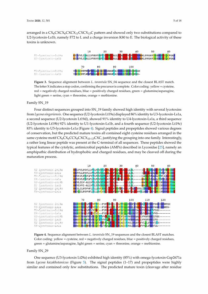
arranged in a CX6CXCX6CXCX11CXCX13C pattern and showed only two substitutions compared toU2-lycotoxin-Ls1b, namely F72 to L and a charge inversion K80 to E. The biological activity of thesetoxins is unknown.
Toxins 2020, 12, x FOR PEER REVIEW 5 of 19
VP_07 Angiotensin-converting enzyme 1
VP_10 Venom Serine protease 1
VP_13 Cysteine rich secretory protein 4
2.1.1. Disulfide Rich Peptide Toxins
In this section, the disulfide rich peptide toxins found in the transcriptome of Lycosa tarantula
have been classified and named according to the nomenclature proposed by King et al. [24] The best
matching sequence from BLAST search against UniprotKB database was aligned with each of the
retrieved L. tarantula sequence for comparison.
Family SN_04
One sequence (U1-lycotoxin Lt04a) exhibited high identity (93%) with U2-lycotoxin-Ls1b from
the related Lycosa singoriensis (Figure 3). Both signal peptides (1–18) were identical, whereas the
propeptide contained five substitutions that were mostly conservative (e.g., E to D or V to L). The
predicted mature toxin (cleavage after residue 42) contained eight cysteine residues arranged in a
CX6CXCX6CXCX11CXCX13C pattern and showed only two substitutions compared to U2-lycotoxin-
Ls1b, namely F72 to L and a charge inversion K80 to E. The biological activity of these toxins is
unknown.
Figure 3. Sequence alignment between L. tarantula SN_04 sequence and the closest BLAST match. The
letter X indicates a stop codon, confirming the precursor is complete. Color coding: yellow=cysteine,
Four distinct sequences grouped into SN_19 family showed high identity with several lycotoxins
from Lycosa singoriensis. One sequence (U2-lycotoxin Lt19a) displayed 86% identity to U3-lycotoxin-
Ls1a, a second sequence (U2-lycotoxin Lt19d), showed 91% identity to U4-lycotoxin-Ls1a, a third
sequence (U2-lycotoxin Lt19b) 92% identity to U1-lycotoxin-Ls1b, and a fourth sequence (U2-
lycotoxin Lt19c) 83% identity to U5-lycotoxin-Ls1a (Figure 4). Signal peptides and propeptides
showed various degrees of conservation, but the predicted mature toxins all contained eight cysteine
residues arranged in the same cysteine motif CX6CX6CCX8CXCX10–13CXC, justifying the grouping into
one family. Interestingly, a rather long linear peptide was present at the C-terminal of all sequences.
These peptides showed the typical features of the cytolytic, antimicrobial peptides (AMPs) described
in Lycosidae [25], namely an amphipathic distribution of hydrophobic and charged residues, and
may be cleaved off during the maturation process.
Figure 3. Sequence alignment between L. tarantula SN_04 sequence and the closest BLAST match.The letter X indicates a stop codon, confirming the precursor is complete. Color coding: yellow = cysteine,red = negatively charged residues, blue = positively charged residues, green = glutamine/asparagine,light green = serine, cyan = threonine, orange = methionine.
Family SN_19
Four distinct sequences grouped into SN_19 family showed high identity with several lycotoxinsfrom Lycosa singoriensis. One sequence (U2-lycotoxin Lt19a) displayed 86% identity to U3-lycotoxin-Ls1a,a second sequence (U2-lycotoxin Lt19d), showed 91% identity to U4-lycotoxin-Ls1a, a third sequence(U2-lycotoxin Lt19b) 92% identity to U1-lycotoxin-Ls1b, and a fourth sequence (U2-lycotoxin Lt19c)83% identity to U5-lycotoxin-Ls1a (Figure 4). Signal peptides and propeptides showed various degreesof conservation, but the predicted mature toxins all contained eight cysteine residues arranged in thesame cysteine motif CX6CX6CCX8CXCX10–13CXC, justifying the grouping into one family. Interestingly,a rather long linear peptide was present at the C-terminal of all sequences. These peptides showed thetypical features of the cytolytic, antimicrobial peptides (AMPs) described in Lycosidae [25], namely anamphipathic distribution of hydrophobic and charged residues, and may be cleaved off during thematuration process.
Toxins 2020, 12, x FOR PEER REVIEW 6 of 19
Figure 4. Sequence alignment between L. tarantula SN_19 sequences and the closest BLAST matches.
Color coding: yellow=cysteine, red=negatively charged residues, blue= positively charged residues,
One sequence (U4-lycotoxin Lt33a) exhibited high identity (71%) with toxin 26 (accession
MH754609.1) from the American wandering spider (Cupiennius salei; Figure 6). Despite both species
being more distantly related, the signal peptides (1–25) were remarkably similar. After a relatively
short propeptide, the predicted mature toxin (cleavage after residue 37) contained eight cysteine
residues arranged in a CX6CX4CCX4CXCX6CXC pattern.
Figure 4. Sequence alignment between L. tarantula SN_19 sequences and the closest BLAST matches.Color coding: yellow = cysteine, red = negatively charged residues, blue = positively charged residues,green = glutamine/asparagine, light green = serine, cyan = threonine, orange = methionine.
Family SN_29
One sequence (U3-lycotoxin Lt29a) exhibited high identity (85%) with omega-lycotoxin-Gsp2671afrom Lycosa kazakhstanicus (Figure 5). The signal peptides (1–17) and propeptides were highlysimilar and contained only few substitutions. The predicted mature toxin (cleavage after residue
Toxins 2020, 12, 501 6 of 18
40) contained eight cysteine residues arranged in a CX6CX6CCX4CXCX15CXC pattern and displayedseven substitutions compared to omega-lycotoxin-Gsp2671a. Omega-lycotoxin-Gsp2671a was shownto specifically modulate P-type Ca2+ channels [26].
Toxins 2020, 12, x FOR PEER REVIEW 6 of 19
Figure 4. Sequence alignment between L. tarantula SN_19 sequences and the closest BLAST matches.
Color coding: yellow=cysteine, red=negatively charged residues, blue= positively charged residues,
One sequence (U4-lycotoxin Lt33a) exhibited high identity (71%) with toxin 26 (accession
MH754609.1) from the American wandering spider (Cupiennius salei; Figure 6). Despite both species
being more distantly related, the signal peptides (1–25) were remarkably similar. After a relatively
short propeptide, the predicted mature toxin (cleavage after residue 37) contained eight cysteine
residues arranged in a CX6CX4CCX4CXCX6CXC pattern.
Figure 5. Sequence alignment between L. tarantula SN_29 sequence and the closest BLAST match.Color coding: yellow = cysteine, red = negatively charged residues, blue = positively charged residues,green = glutamine/asparagine, light green = serine, cyan = threonine, orange = methionine.
Family SN_33
One sequence (U4-lycotoxin Lt33a) exhibited high identity (71%) with toxin 26 (accessionMH754609.1) from the American wandering spider (Cupiennius salei; Figure 6). Despite both speciesbeing more distantly related, the signal peptides (1–25) were remarkably similar. After a relatively shortpropeptide, the predicted mature toxin (cleavage after residue 37) contained eight cysteine residuesarranged in a CX6CX4CCX4CXCX6CXC pattern.
Toxins 2020, 12, x FOR PEER REVIEW 7 of 19
Figure 6. Sequence alignment between L. tarantula SN_33 sequence and the closest BLAST match.
Color coding: yellow=cysteine, red=negatively charged residues, blue= positively charged residues,
Figure 6. Sequence alignment between L. tarantula SN_33 sequence and the closest BLAST match.Color coding: yellow = cysteine, red = negatively charged residues, blue = positively charged residues,green = glutamine/asparagine, light green = serine, cyan = threonine, orange = methionine.
Family VP_12
One sequence (U6-lycotoxin Lt12a) showed only limited identity (45%) to Kunitz-typekappaPI-theraphotoxin-Hs1e from the Chinese bird spider (Haplopelma schmidti; Figure 7). Despite alow sequence identity for the signal and propeptide regions, the six cysteine residues of the maturetoxin were conserved and arranged in a CX8CX13CX7CX12CX3C motif.
Toxins 2020, 12, x FOR PEER REVIEW 7 of 19
Figure 6. Sequence alignment between L. tarantula SN_33 sequence and the closest BLAST match.
Color coding: yellow=cysteine, red=negatively charged residues, blue= positively charged residues,
Figure 7. Sequence alignment between L. tarantula VP_12 sequence and the closest BLAST match.Color coding: yellow = cysteine, red = negatively charged residues, blue = positively charged residues,green = glutamine/asparagine, light green = serine, cyan = threonine, orange = methionine.
Family VP_11
Two incomplete sequences (U6-lycotoxin Lt11a and U6-lycotoxin Lt11b) exhibited highidentity with U15-lycotoxin-Ls1a (81%) and with U20-lycotoxin-Ls1c (83%) from Lycosa singoriensis
Toxins 2020, 12, 501 7 of 18
(Figure 8). Both signal peptides (1–20) were moderately conserved and no propeptide waspredicted. The mature toxins (cleavage after residue 20) contained 10 cysteine residues arranged in aCX7CX8CCX4CX5CCX3CX3 CX17C pattern.
Toxins 2020, 12, x FOR PEER REVIEW 7 of 19
Figure 6. Sequence alignment between L. tarantula SN_33 sequence and the closest BLAST match.
Color coding: yellow=cysteine, red=negatively charged residues, blue= positively charged residues,
Figure 8. Sequence alignment between L. tarantula VP_11 sequences and the closest BLAST matches.Color coding: yellow = cysteine, red = negatively charged residues, blue = positively charged residues,green = glutamine/asparagine, light green = serine, cyan = threonine, orange = methionine.
2.1.2. Venom Proteins
In addition to the classic disulfide rich peptides, several sequences retrieved from L. tarantula venomgland transcriptome showed similarity with known venom proteins. These include hyaluronidase,angiotensin-converting enzyme, venom serine protease, and cysteine rich secretory protein (Table 1).The two partial hyaluronidase sequences identified showed 80–81% sequence identity to Cupienniussalei hyaluronidase. One complete sequence matched (52%) a putative angiotensin converting enzymeprecursor from Carcinus maenas. One partial sequence showed high sequence identity (97%) to aputative processing quadruplet motif (PQM) protease precursor from L. hispanica, a sister speciesto L. tarantula. Finally, four partial sequences showed similarity to cysteine rich secretory proteins.From these, three sequences displayed high sequence identity to L. singoriensis’ venom allergen 5proteins (81–91%), and one sequence shows moderate homology (62%) to a cysteine rich secretoryprotein 1 isoform a1 from Cupiennius salei.
2.2. Mass Spectrometry Analyses of L. tarantula Venom
To gain further insights into the venom composition of L. tarantula, mass spectrometry analyseswere carried out, including comparative LC-MS of the electrically stimulated venom (Figure 9)and the manually collected venom from male and female specimens (Figure 10). In addition, a fullproteomic (LC-MS/MS) analysis was performed on the electrically stimulated venom in order to testfor the presence of some transcriptome-annotated venom peptides and proteins and validate theirmature sequences.
2.2.1. LC-MS of the Electrically Stimulated Venom
The venom from several specimens of L. tarantula was collected via electrostimulation andpooled. Approximatively 600 µg of venom was analyzed by LC-MS over 80 min. The overall total ioncurrent (TIC) profile showed the highest complexity between 5 and 25 min (corresponding to 5–25%acetonitrile), where most of the ions were detected (Figure 9). The calculated monoisotopic massesfor the dominant ions in each peak are reported on Figure 9, and the distribution shows a majority ofmasses <3 kDa, and then between 5 to 9 kDa (Figure 11). Interestingly, the top five most intense peakscorrespond to small molecular weight compounds, between 1500–2500 Da (2260.16 Da, 1660.96 Da,2274.2 Da, 2213.34 Da, and 2001.12 Da).
Toxins 2020, 12, 501 8 of 18Toxins 2020, 12, x FOR PEER REVIEW 9 of 19
Figure 9. Total ion current trace of electrically stimulated (ES) L. tarantula venom. Major monoisotopic
masses determined for each peak are indicated.
2.2.2. LC-MS of Manually Stimulated Female vs. Male Venom
Figure 9. Total ion current trace of electrically stimulated (ES) L. tarantula venom. Major monoisotopicmasses determined for each peak are indicated.
2.2.2. LC-MS of Manually Stimulated Female vs. Male Venom
Although more convenient and producing higher yields, electrostimulation can damage secretorycells, resulting in the collected venom being “contaminated” with unwanted cellular proteins. Therefore,in an attempt to collect venom reflecting a more natural composition, we used a manual stimulation,where the spiders are aggravated and induced to bite into a plastic tubing. The resulting defensivevenom droplets recovered from both male and female specimens were analyzed using LC-MS. The moststriking difference with the electrically stimulated venom profile lies in the reduced complexity,especially in the early eluting compounds (no distinguishable peaks <10 min). Whereas the overallfemale vs. male profiles show obvious similarities in terms of complexity and peak intensities,the underlying differences appear more evident when considering the calculated masses and theiroverlap. Indeed, although the mass distribution showed a similar pattern, more than 50% of the massesdetected in female venom were unique and not found in the male venom (Figure 11). Interestingly,in both venoms, one of the most intense ions corresponded to a mass of 1908.14 Da (together with1803.16 Da), which appeared remarkably absent from the electrically stimulated venom.
2.2.3. Proteomic Analysis of the Electrically Stimulated Venom
With the aim of identifying a maximum of the peptides and proteins present in the venom ofL. tarantula, shotgun proteomics on a high-resolution mass spectrometer was performed on the morecomplex electrically stimulated venom. After reduction, alkylation, and trypsin digestion of the venomsample, the resulting peptides were fragmented, leading to the acquisition of 15,224 MS and 89,834MS/MS scans, and further analyzed using PEAKS software (Bioinformatics solutions, Waterloo, ON,Canada). The search database was composed of our translated transcriptome, and a false discovery rateof 1% was applied. The results were filtrated in PEAKS Studio using stringent parameters, includingpeptide −10lgP ≥ 24.6, protein −10lgP ≥ 20, proteins unique peptides ≥2, and de novo average localconfidence (ALC) score ≥80%. Under these conditions, 30 proteins were identified (Table S1). Amongthe validated sequences, the short neurotoxin-like peptides are well represented, with nine out the10 sequences retrieved from the venom gland transcriptome that are validated. Overall, all disulfiderich peptide families were confirmed, except for the family VP_12 (Kunitz-type U5-lycotoxin-Lt12a).Next, the venom proteins were also well represented in the venom, with eight sequences validated,
Toxins 2020, 12, 501 9 of 18
including all four CRISP (Venom allergen 5), two hyaluronidases, a putative PQM protease, and aputative angiotensin converting enzyme. Finally, some ubiquitous cellular proteins were identified,namely several heat shock proteins, cytochrome, elongation factor, arginine kinase, glyceraldehyde-3phosphate dehydrogenase, actin as well as several sequences producing no significant match toknown proteins.
Toxins 2020, 12, x FOR PEER REVIEW 10 of 19
Figure 10. Total ion current traces of female and male L. tarantula venom. Representative traces of a
single specimen (female, top panel and male, bottom panel). Major masses calculated for each peak
are indicated.
Although more convenient and producing higher yields, electrostimulation can damage
secretory cells, resulting in the collected venom being “contaminated” with unwanted cellular
proteins. Therefore, in an attempt to collect venom reflecting a more natural composition, we used a
manual stimulation, where the spiders are aggravated and induced to bite into a plastic tubing. The
Figure 10. Total ion current traces of female and male L. tarantula venom. Representative traces of asingle specimen (female, top panel and male, bottom panel). Major masses calculated for each peakare indicated.
Toxins 2020, 12, 501 10 of 18
Toxins 2020, 12, x FOR PEER REVIEW 11 of 19
resulting defensive venom droplets recovered from both male and female specimens were analyzed
using LC-MS. The most striking difference with the electrically stimulated venom profile lies in the
reduced complexity, especially in the early eluting compounds (no distinguishable peaks <10 min).
Whereas the overall female vs. male profiles show obvious similarities in terms of complexity and
peak intensities, the underlying differences appear more evident when considering the calculated
masses and their overlap. Indeed, although the mass distribution showed a similar pattern, more
than 50% of the masses detected in female venom were unique and not found in the male venom
(Figure 11). Interestingly, in both venoms, one of the most intense ions corresponded to a mass of
1908.14 Da (together with 1803.16 Da), which appeared remarkably absent from the electrically
stimulated venom.
Figure 11. Venn diagram and mass distribution of electrically stimulated (ES), female (F), and male
(M) L. tarantula venom. (Left) panel shows the overlap of masses between venom samples. (Right)
panels show the mass distributions for each venom sample.
2.2.3. Proteomic Analysis of the Electrically Stimulated Venom
With the aim of identifying a maximum of the peptides and proteins present in the venom of L.
tarantula, shotgun proteomics on a high-resolution mass spectrometer was performed on the more
complex electrically stimulated venom. After reduction, alkylation, and trypsin digestion of the
venom sample, the resulting peptides were fragmented, leading to the acquisition of 15,224 MS and
89,834 MS/MS scans, and further analyzed using PEAKS software (Bioinformatics solutions,
Waterloo, ON, Canada). The search database was composed of our translated transcriptome, and a
false discovery rate of 1% was applied. The results were filtrated in PEAKS Studio using stringent
parameters, including peptide −10lgP ≥ 24.6, protein −10lgP ≥ 20, proteins unique peptides ≥2, and de
novo average local confidence (ALC) score ≥80%. Under these conditions, 30 proteins were identified
(Table S1). Among the validated sequences, the short neurotoxin-like peptides are well represented,
with nine out the 10 sequences retrieved from the venom gland transcriptome that are validated.
Overall, all disulfide rich peptide families were confirmed, except for the family VP_12 (Kunitz-type
U5-lycotoxin-Lt12a). Next, the venom proteins were also well represented in the venom, with eight
sequences validated, including all four CRISP (Venom allergen 5), two hyaluronidases, a putative
PQM protease, and a putative angiotensin converting enzyme. Finally, some ubiquitous cellular
Figure 11. Venn diagram and mass distribution of electrically stimulated (ES), female (F), and male (M)L. tarantula venom. (Left) panel shows the overlap of masses between venom samples. (Right) panelsshow the mass distributions for each venom sample.
2.3. Electrophysiology Assay of Crude L. tarantula Venom
The biological activity of crude (electrically stimulated) Lycosa tarantula venom was investigatedusing a two-electrode voltage clamp method on honeybee CaV4 (DSC1) expressed in Xenopus laevisoocytes. Upon application of 0.01 mg/mL diluted venom, no significant effect was observed, but at0.1 mg/mL, the increase in the leak current was so strong that the oocyte could not be properly clampedanymore, and thus value of the holding potential and the depolarization could not be maintained,preventing the adequate measurement of the Ca2+ current (Figure 12A). Suspecting that the venomstrongly permeabilizes the oocyte membrane, the venom was also tested without depolarizationon CaV4–injected (n = 6) and non-injected (n = 7) oocytes. Indeed, application of 25 µL of venom(1 mg/mL) produced a similar increase in holding current, indicating that this effect is independent of theexpression of CaV4 (Figure 12B). However, in some oocytes (n = 2) a notable difference between injectedand non-injected oocytes appeared upon washing of the venom. Whereas these two non-injectedoocytes “recovered” from the leak (holding current amplitude back to smaller values), the other oocytes(six injected with honeybee CaV4 and five non-injected) were unable to recover. This behavior preventsa clear detection of any CaV4 channel blocker within the venom. Further deconvolution of L. tarantulavenom will clearly be necessary to determine if it contains specific CaV4 blockers.
Toxins 2020, 12, 501 11 of 18
Toxins 2020, 12, x FOR PEER REVIEW 12 of 19
proteins were identified, namely several heat shock proteins, cytochrome, elongation factor, arginine
kinase, glyceraldehyde-3 phosphate dehydrogenase, actin as well as several sequences producing no
significant match to known proteins.
2.3. Electrophysiology Assay of Crude L. tarantula Venom
The biological activity of crude (electrically stimulated) Lycosa tarantula venom was investigated
using a two-electrode voltage clamp method on honeybee CaV4 (DSC1) expressed in Xenopus laevis
oocytes. Upon application of 0.01 mg/mL diluted venom, no significant effect was observed, but at
0.1 mg/mL, the increase in the leak current was so strong that the oocyte could not be properly
clamped anymore, and thus value of the holding potential and the depolarization could not be
maintained, preventing the adequate measurement of the Ca2+ current (Figure 12A). Suspecting that
the venom strongly permeabilizes the oocyte membrane, the venom was also tested without
depolarization on CaV4–injected (n = 6) and non-injected (n = 7) oocytes. Indeed, application of 25 µl
of venom (1 mg/mL) produced a similar increase in holding current, indicating that this effect is
independent of the expression of CaV4 (Figure 12B). However, in some oocytes (n = 2) a notable
difference between injected and non-injected oocytes appeared upon washing of the venom. Whereas
these two non-injected oocytes “recovered” from the leak (holding current amplitude back to smaller
values), the other oocytes (six injected with honeybee CaV4 and five non-injected) were unable to
recover. This behavior prevents a clear detection of any CaV4 channel blocker within the venom.
Further deconvolution of L. tarantula venom will clearly be necessary to determine if it contains
specific CaV4 blockers.
Figure 12. Biological effect of L. tarantula venom on Apis mellifera Cav4 channel expressed in oocytes.
A) Left panel shows the effect of L. tarantula venom on oocytes expressing the CaV4 Ca2+ channel. Note
the lack of effect at the 0.01 mg/mL dilution, and the strong increase in the holding current at the 0.1
mg/mL dilution. Middle panel represents the time course of the Ca2+ current and holding current
amplitudes measured during the protocol shown in the left panel. The right panel shows the averaged
effect on the peak Ca2+ current amplitude measured at the steady state or just before the wash. B)
Effect of L. tarantula venom on CaV4-expressing (left) or non-injected oocytes (right) recorded on the
holding current without any channel stimulation (constant holding potential of −100 Mv). Top left
panel shows the holding current recorded continuously without depolarization in the Bant10
solution. The perfusion was stopped at the vertical arrowhead, a puff of 25 µ l of Bant10 was applied
Figure 12. Biological effect of L. tarantula venom on Apis mellifera Cav4 channel expressed in oocytes.(A) Left panel shows the effect of L. tarantula venom on oocytes expressing the CaV4 Ca2+ channel.Note the lack of effect at the 0.01 mg/mL dilution, and the strong increase in the holding current at the0.1 mg/mL dilution. Middle panel represents the time course of the Ca2+ current and holding currentamplitudes measured during the protocol shown in the left panel. The right panel shows the averagedeffect on the peak Ca2+ current amplitude measured at the steady state or just before the wash. (B)Effect of L. tarantula venom on CaV4-expressing (left) or non-injected oocytes (right) recorded on theholding current without any channel stimulation (constant holding potential of −100 Mv). Top leftpanel shows the holding current recorded continuously without depolarization in the Bant10 solution.The perfusion was stopped at the vertical arrowhead, a puff of 25 µL of Bant10 was applied in therecording chamber, without any effect, and the perfusion was started again at the horizontal arrow.Bottom left panel displays the same protocol applied but using 25 µL of the L. tarantula venom at1 mg/mL instead of Bant10 solution. Note the big increase in the holding current and the lack ofreversibility during the wash. Right panel shows the same protocol with a puff of L. tarantula venom at1 mg/mL but applied to non-injected oocytes, with a similar increase in the holding current, indicatingthat this effect of the venom on the oocyte is independent of the expression of CaV4.
3. Discussion
Spider venoms consist of complex mixtures of biologically active compounds that are for the mostpart gene encoded polypeptides and proteins. Therefore, combining venom gland transcriptomics withvenom proteomics is a powerful method to accelerate the identification of full precursors and maturetoxins for a better understanding of spider biology, venom-ecology relationships, and for the mining ofuseful pharmaceutical and agrochemical molecules. In this work, we used such proteotranscriptomicsstrategy to provide the first insights into the venom of one of the largest Lycosid spiders found inthe Mediterranean region, Lycosa tarantula. Automated bioinformatics analyses followed by manualvalidation of the venom gland transcriptome revealed 18 distinct venom-related sequences classifiedinto 10 structural families. The disulfide rich neurotoxin-like peptides comprised 10 sequences from sixfamilies, whereas the venom proteins were grouped into four distinct classes. Besides these sequences,proteomics investigations also revealed the presence of common cellular proteins, confirming thatelectrically stimulated venom includes contaminants. Indeed, the manually stimulated venom fromboth male and female specimens showed a less complex LC-MS profile and a different mass distributioncompared to electrically stimulated venom. Interestingly, more than 50% of the masses detected in
Toxins 2020, 12, 501 12 of 18
female venom were unique and not found in the male’s venom, suggesting that some intraspecificvariations may be due to sex. Such intraspecific variations between male and female has already beenreported in several species of spiders [27–29].
Whereas the biological activity of the neurotoxin-like peptides remains to be elucidated, ourpreliminary investigation of the crude venom on honeybee CaV4 ion channel indicated the possiblepresence of selective blockers. However, further deconvolution of the crude venom will be necessaryto uncover the peptides responsible for this activity, since the cytolytic activity present in the venomprevented accurate electrical measurement. Indeed, application of the crude venom to injected andnon-injected oocytes induced a strong leak current, consistent with the cytolytic activity describedfor several other Lycosidae venoms. The molecular entities responsible for this cytolytic activity areknown as antimicrobial peptides (AMPs), which are usually small, highly positively charged linearpeptides adopting an amphipathic secondary structure in lipid membrane. Several such AMPs havebeen isolated and sequenced from Lycosidae venom [25]. Often, only the mature peptide sequencesare available, not the full precursors, raising the question about the molecular origin of these AMPs.Interestingly, in the recently published high throughput sequencing of the venom gland of the LycosidaePardosa pseudoannulata, there is no mention of AMPs. However, a closer inspection of the reportedsequences reveals that family A resembles the “inhibitory cysteine knot (ICK) + α-helix” modulartoxin described from a Zodariidae spider, Lachesana tarabaevi. In these modular toxins, the C-terminalfragment synthesized separately was shown to possess membrane-binding activity consistent with acytolytic effect [30]. These AMPs are often major components of the venom in Lycosidae, as seen withLyeTx I, a peptide isolated from Lycosa erythrognatha [31]. In our transcriptome, family SN_19 alsodisplays the same architecture, with a N-terminal ICK motif and a C-terminal AMP-like sequence, andLC-MS of the venom shows a major contribution of peptides in the 1500–2500 Da range. Interestingly,the C-terminal peptide (QQPKSHKIAEKIVDKAKTVI) of U2-lycotoxin Lt19a has a mass (2260.32 Da)that corresponds to the major peak present in the venom (see Figure 9). The C-terminal peptides ofthe other SN_19 family sequences are also in the same mass range of 2000–2500 Da. Further work,including HPLC fractionation and purification steps, will be necessary to confirm this hypothesis.
Compared to the transcriptomes of other Lycosidae, such as Lycosa singoriensis or Lycosa vittata,our Lycosa tarantula transcriptome revealed a similar number of structural families, but fewer paralogsfor each family [18,19]. However, it should be noted that in these studies, many of the reportedparalogs were actually often single substitution sequence variants, and several of these substitutionswere located in the propeptide, therefore producing identical mature toxin. We suspect that theassembly step of our Illumina reads eliminated the majority of these minor substitution variantsthat were otherwise picked up by the traditional Sanger sequencing technology used in thesestudies. However, we cannot exclude that additional neurotoxin-like sequences were missed. Forinstance, interrogation of the PEAKS “de novo” peptides that did not match any sequence fromour transcriptome revealed a number of fragments that show high similarity to known neurotoxins,such as YPESGEGELCTCQQPK (75% U3-lycotoxin-Ls1h, Lycosa singoriensis), CTPLLHDCSHDR(92% U4-lycotoxin-Ls1b, Lycosa singoriensis), GCGFLDFNYPGDGR (93% Venom allergen 5, Lycosasingoriensis), and CCWPWSCVCWSQTLS (87% Omega-lycotoxin-Gsp2671e, Alopecosa marikovskyi).These unmatched yet high quality proteomic sequences may arise from the different specimens usedfor venom gland transcriptomics and venom proteomics.
In summary, we have reported here the first proteotranscriptomics analysis of Lycosa tarantulavenom, including 18 distinct sequences of short neurotoxin-like peptides and venom proteins from10 structural families. Future works should focus on the synthesis and pharmacological characterizationof some of the neurotoxin-like peptides, as well as the cytolytic activity of some C-terminal fragments.Our data contribute to a treasure trove for the mining of useful pharmacological compounds.
Toxins 2020, 12, 501 13 of 18
4. Materials and Methods
4.1. Spiders, Venom Collection, and Venom Gland Dissection
Twelve specimens, including two mature males and 10 adult females of Lycosa tarantula werecollected in the scrublands around Montpellier, France. These spiders (except males, caught wanderingin open areas) were lured out of their burrow using a small stick wiggling around the entrance andcaught into plastic jars. Specimens were then individually isolated in small boxes and maintainedin the laboratory at room temperature. They were watered twice a week and fed once a week withcommercially available mealworms.
To collect the venom from these specimens, two methods were used. First, a “manual stimulation”was applied similar to that described by Liu et al. [17], where each spider was presented with a piece ofsoft tubing (0.5 cm in diameter) and aggravated with tweezers to trigger a bite. Venom drops depositedon the tube were recovered using a pipette and diluted in distilled water. Secondly, electrostimulationwas carried out on several specimens (n > 7) using an electric venom extractor based on the Arduino®
Mega 2560 board, specifically designed for the extraction of venom from arthropods and other smallsize animals [32].
Spiders were not fed for at least a week prior to the milking session. Specimens were anesthetizedbefore milking (with 5% CO2). Chelicerae were stimulated by electrical impulses (3 to 7 V andapproximately 0.5 to 2 A) discharged in a 2 s “working time” and 2 s of “rest time” steps. Releasedvenom was collected from the tip of the fangs using a pipette and transferred to a 1.5 mL microcentrifugetube containing approximately 20 µL of distilled water. Protein concentration of the venom sampleswere assessed using a nanophotometer N60 (Implen GmbH, München, Germany). Venom collectedfrom individual spiders was pooled, freeze-dried and stored at −20 ◦C for subsequent use (proteomiccharacterization and electrophysiology).
To obtain the amount of mRNA required for the transcriptome sequencing, venom glands of sixanesthetized adult female spiders were dissected on ice and placed in a 1.5 mL microcentrifuge tubecontaining 500 µL of lysis buffer. Next, the mRNA was extracted using a commercial kit (MagneticmRNA isolation kit, Biolabs) following the manufacturer’s instructions. After extraction, the mRNAconcentration was measured using a nanophotometer N60 (Implen GmbH, München, Germany).
4.2. Library prepaRation and Illumina Sequencing
RNA-Seq libraries were constructed with the Truseq stranded mRNA sample preparation (lowthroughput protocol) kit from Illumina (San Diego, CA, USA). Depending on the samples, 100 or 200 ngof mRNA was used for the construction of the libraries. Next, the mRNA was fragmented into smallpieces using divalent cations under elevated temperature. The cleaved RNA fragments were copiedinto first strand cDNA using SuperScript II reverse transcriptase, Actinomycin D and random hexamerprimers. The second strand cDNA was synthesized by replacing deoxythymidine triphosphate (dTTP)with deoxyuridine triphosphate (dUTP). These cDNA fragments have the addition of a single ‘A’ baseand subsequent ligation of the adapter. The products are then purified and enriched with 15 cycles ofPCR. The final cDNA libraries were validated with a Fragment Analyzer (Agilent Santa Clara, CA,USA) and quantified with a KAPA qPCR kit (Kapa Biosystems, Wilmington, MA, USA).
The transcriptome of L. tarantula was sequenced as part of a larger project comprising 15 othervenom gland transcriptomes. On three sequencing lanes of V2 flowcells, the 16 libraries were pooledin equal proportions, denatured with NaOH and diluted to 18 pM before clustering. Cluster formation,primer hybridization and single-end read, 125 cycles sequencing were performed on cBot and HiSeq2500(Illumina, San Diego, CA, USA) respectively.
Image analysis and base calling were performed using the HiSeq Control Software v.2.2.68(Illumina, San Diego, CA, USA) and Real-Time Analysis component v.1.18.66.3 (Illumina, San Diego,CA, USA). Demultiplexing was performed using Illumina’s conversion software (bcl2fastq 2.18).The quality of the data was assessed using FastQC from the Babraham Institute v.0.11.5 and the
Toxins 2020, 12, 501 14 of 18
Illumina software SAV (Sequencing Analysis Viewer) v. 2.1.8 (Illumina, San Diego, CA, USA). Potentialcontaminants were investigated with the FastQ Screen software from the Babraham Institute v.0.9.5.
4.3. Bioinformatics Sequence Analysis
Data issued from the sequencing platform were trimmed using the Trinity trimmomatic toolwith default parameters. Reads were assembled using the Trinity software (version 2.1.1) [33].Obtained contigs were translated in-silico into their six reading frames and annotated using thefollowing procedure. An in-house database composed of all spider toxins from Arachnoserver,UniprotKB/SwissProt and Venomzone were created using makeblastdb of BLAST+ package afterredundancy removal using CD-HIT [34,35] at the threshold of 1.00.
All Contigs were searched using an improved version of the previously published Ekenda HiddenMarkov Models (HMM) library and the hmmcompete program [21].
All Contigs were submitted to a first BLAST step against this database to provide anannotated subset of the transcriptome. Annotated contigs were again BLASTed against the wholeUniprotKB/SwissProt database to confirm the exactitude of obtained hits and remove false positive hits(BlastP against UniProtKB with e-threshold = 0.0001; matrix BLOSUM-62, non-filtering and gapped;UniProtKB/SwissProt 2019_03).
Spider toxin-related sequences were identified and classified into toxins family using EkendaHidden Markov Models (HMM) and the hmmcompete program. Signal Peptide and propeptide werepredicted using respectively SignalP version 5.0 [22] and SpiderP [23] directly from the Arachnoserverweb server at http://www.arachnoserver.org/spiderP.html. A final manual validation step wasperformed: multiple sequence alignments using MAFFT Version 7 [36], variant identification, andcleavage site validation. All peptide hits as well as their corresponding contigs sequences were furtheranalyzed at nucleotide level to detect eventual mutations. Nucleotide sequence variants that obviouslyresulted from sequencing errors, assembly errors or frame shifts were excluded.
4.4. Proteomics
4.4.1. Liquid Chromatography Coupled Mass Spectrometry (LC-MS)
RP-UPLC was operated on an Acquity H-Class ultrahigh performance liquid chromatography(UPLC) system (Waters, Corp., Milford, MA, United States) fitted with a UV detector (diode arraydetector) under the control of Waters MassLynx software (version 4.1). Separation of the L. tarantulavenom (~600 µg) was achieved using a Kinetex C18 100 Å column (2.1 × 150 mm, 3 µm) fitted with apre-column. Elution was carried out using a gradient of 0–80% B (0.1% formic acid in acetonitrile) in80 min. Samples eluting from the UPLC were introduced into the mass spectrometer at a flow rateof 500 µL/min. Acquisitions were carried out over the range 50 Da to 1800 Da m/z every 0.1 s on aSynapt-G2-S high-definition MS system (Waters, Corp., Milford, MA, United States). To obtain themolecular masses of the venom components eluting between 0 and 40 min, each peak from the totalion current (TIC) chromatogram was analyzed with Waters Mass Lynx software (version 4.1) (Waters,Milford, MA, USA).
4.4.2. Shotgun Proteomics (LC-MS/MS)
Prior to shotgun proteomics, venom protein extracts were denatured, reduced, and alkylated.Briefly, each sample (~50 µg) was dissolved in 89 µL of triethylammonium bicarbonate (TEABC)100 mM. One microliter of dithiothreitol (DTT) 1 M was added and incubation was performed for30 min at 60 ◦C. A volume of 10 µL of iodoacetamide (IAA) 0.5 M was added (incubation for 30 min inthe dark). Enzymatic digestion was performed by addition of 2 µg trypsin (Gold, Promega, Madison,WI, USA) in TEABC 100 mM and incubation overnight at 30 ◦C. After completing the digestionstep, peptides were purified and concentrated using OMIX Tips C18 reverse-phase resin (Agilent
Technologies Inc., Santa Clara, CA, USA) according to the manufacturer’s specifications. Peptides weredehydrated in a vacuum centrifuge.
Samples were then subjected to nano-flow liquid chromatography coupled to tandem massspectrometry (NanoLC-MS/MS). Samples were resuspended in 20 µL formic acid (0.1%, buffer A) and1 µL was loaded onto an analytical 25 cm reversed-phase column (75 mm inner diameter, AcclaimPepmap 100® C18, Thermo Fisher Scientific) and separated with an Ultimate 3000 RSLC system(Thermo Fisher Scientific, Waltham, MA, USA) coupled to a Q Exactive HF-X (Thermo Fisher Scientific,Waltham, MA, USA) via a nano-electrospray source, using a 123 min gradient of 6% to 40% of bufferB (80% ACN, 0.1% formic acid) and a flow rate of 300 nL/min. MS/MS analyses were performedin a data-dependent mode. Full scans (375–1500 m/z) were acquired in the Orbitrap mass analyzer(Thermo Fisher Scientific, Waltham, MA, USA) with a 60,000 resolution at 200 m/z. For the full scans,3 × 106 ions were accumulated within a maximum injection time of 60 ms and detected in the Orbitrapmass analyzer. The twelve most intense ions with charge states ≥2 were sequentially isolated to atarget value of 1 × 10⁵ with a maximum injection time of 45 ms and fragmented by higher-energycollisional dissociation (HCD) in the collision cell (normalized collision energy of 28%) and detected inthe Orbitrap mass analyzer at 30,000 resolution.
4.4.3. Bioinformatic Integration of Proteomic and Transcriptomic Data
PEAKS Studio 8.5 software (Bioinformatics solutions, Waterloo, ON, Canada) was used to matchMS/MS spectra obtained from proteomic analysis of L. tarantula venom. MS spectra were elucidatedbased on a personalized database resulting from assembled contigs translated into their six readingframes. Carbamidomethylation was set as fixed modification, while oxidation (M) was set as variablemodifications, with maximum missed cleavages at 3 for trypsin digestion. Parent mass and fragmentmass error tolerance were set at 5 ppm and 0.015 Da respectively. False discovery rate (FDR) of 1%and unique peptide ≥2 were used for filtering out inaccurate proteins. A −10lgP > 120 was used toestimate whether the detected proteins was identified by enough reliable peptides MS/MS spectra.In order to identify more relevant sequences, the Spider algorithm from PEAKS Studio software wasused to find additional mutations or to correct the sequences. This algorithm corrects the sequencesstored in transcriptomic database with de novo sequences based on MS/MS spectra, which allowed toidentify post-translational modifications (PTMs) and mutations. Minimum ion intensity for mutationand PTMs was set to 5%, and ALC score ≥ 90 for de novo sequences leading to low precursor masserror in order to identify reliable PTM’s and potential mutations.
4.5. Electrophysiology
Ovaries were surgically removed from Xenopus laevis female, anesthetized using a 0.2% MS222solution (Sigma Saint-Louis, MO, USA). After a first mechanical dissociation and extensive washingusing the OR-2 solution (containing in mM: NaCl, 100; MgCl2, 2; KCl, 2; HEPES, 10), oocytes wereisolated by approximately 2 h enzymatic dissociation using 1 mg/mL collagenase IA (Sigma Saint-Louis,MO, USA) dissolved in OR-2. Oocytes were then washed several times with OR-2 and selected in thesurvival medium (containing in mM: NaCl, 96; MgCl2, 2; KCl, 2; CaCl2, 1.8; HEPES, 10; pyruvic acid,2.5; gentamycin, 50 ~µg/mL; neutralized at pH 7.2 using NaOH).
Oocytes injection was performed in the equatorial region by employing a home-made pneumaticinjectory. Xenopus oocytes were microinjected with RNA corresponding to the AmCaV4 channel(1 µg/µL) of domestic honeybee, Apis mellifera. About 40 oocytes were injected with 1 µL of solution,and these injected oocytes were incubated at 18 ◦C in OR-2 solution for at least 24 h for 2–7 days at19 ◦C under gentle agitation before recording. The survival medium was renewed daily.
Whole cell Ba2+ currents were recorded under two electrode voltage-clamp by employing theGeneClamp 500 amplifier (Axon Inst., Burlingame, CA, USA). Current and voltage electrodes werefilled with a solution containing: KCl 3M; KOH. The bath-clamp head stage was connected to the bathusing two agar bridges filled with 2% agar in 3M KCl, and the extracellular solution (physiological
Toxins 2020, 12, 501 16 of 18
solution) was BANT10 (BaOH: 10 mM, TEAOH 20%: 12 mL, NMDG: 30 mM, CsOH: 2 mM, HEPES:10 mM, pH = 7.2 with methane sulfonate). Injection of BAPTA (in mM: BAPTA-free acid (SigmaSaint-Louis, MO, USA), 100; CsOH, 10; HEPES, 10; pH 7.2) into oocytes was performed using athird microelectrode (in order to eliminate any Ca2+-activated Cl current). Under these conditionsuncontaminated Ba2+ currents can be recorded. Ba2+ currents were elicited by series of depolarizingsteps of 400 ms duration from a holding potential of −100 mV, to 10 mV every 10 s. Voltage-protocol andionic currents were generated and recorded using the Clampex software (pClamp, ver 7.0, Axon Inst)(Molecular Devices, San Jose, CA, USA). Venom solution to be tested were prepared just prior to theexperiment by adding the desired concentration in the BANT10 physiological solution. The differentconcentrations of venom (0.01, 0.1, and 1 µM) were then applied manually in a static bath using apipette delivering a dose from about 20 µL.
The effect of administered venom was measured when steady state was reached (after about 1to 2 min, i.e., 6–12 depolarizations) as a percentage of inhibition of the peak Ba2+ current amplituderecorded during a depolarizing pulse ranging of −100 to 10 mV. Data are presented as means ± S.E.M.from at least three oocytes.
Supplementary Materials: The following are available online at http://www.mdpi.com/2072-6651/12/8/501/s1,Table S1: Proteomics results.
Author Contributions: Conceptualization: S.D.; data curation: D.K. and S.D.; funding acquisition: S.D.;investigation: R.M., P.C., C.B., K.E.K., and G.C.; methodology: S.D., P.C., C.B., K.E.K., G.C., and D.K.; software:A.E. and D.K.; supervision: P.C. and S.D.; visualization: D.K., and A.E.; writing—original draft: D.K., R.M., A.E.,and S.D.; writing—review and editing: D.K., A.E., and S.D. All authors have read and agreed to the publishedversion of the manuscript.
Funding: This research was funded by a grant of the French National Research Agency (ANR-16-CE34-0002to S.D.).
Acknowledgments: The authors acknowledge access to the cluster MUSE for computation time, and the FunctionalProteomics Platform of Montpellier, where high resolution mass spectrometry experiments were carried out. MGXacknowledges financial support from France Génomique National infrastructure, funded as part of “Investissementd’avenir” program managed French National Research Agency (ANR-10-INBS-09).
Conflicts of Interest: The authors declare no conflict of interest.
5. Dos Santos, L.D.; Dias, N.B.; Roberto, J.; Pinto, A.S.; Palma, M.S. Brown recluse spider venom: Proteomicanalysis and proposal of a putative mechanism of action. Protein Pept. Lett. 2009, 16, 933–943. [CrossRef]
7. King, G.F. The wonderful world of spiders: Preface to the special Toxicon issue on spider venoms. Toxicon2004, 43, 471–475. [CrossRef]
8. Vetter, R.S.; Isbister, G.K. Medical aspects of spider bites. Annu. Rev. Entomol. 2008, 53, 409–429. [CrossRef]9. Atkinson, R.K.; Walker, P. The effects of season of collection, feeding, maturation and gender on the potency
of funnel-web spider (Atrax Infensus) Venom. Aust. J. Exp. Biol. Med. Sci. 1985, 63, 555–561. [CrossRef]10. Binford, G.J. An analysis of geographic and intersexual chemical variation in venoms of the spider Tegenaria
11. Herzig, V.; John Ward, R.; Ferreira dos Santos, W. Intersexual variations in the venom of the Brazilian ‘armed’spider Phoneutria nigriventer (Keyserling, 1891). Toxicon 2002, 40, 1399–1406. [CrossRef]
12. Tobassum, S.; Tahir, H.M.; Zahid, M.T.; Gardner, Q.A.; Ahsan, M.M. Effect of milking method, diet, andtemperature on venom production in scorpions. J. Insect Sci. 2018, 18. [CrossRef] [PubMed]
13. Piacentini, L.N.; Ramírez, M.J. Hunting the wolf: A molecular phylogeny of the wolf spiders (Araneae,Lycosidae). Mol. Phylogenet. Evol. 2019, 136, 227–240. [CrossRef] [PubMed]
14. Planas, E.; Fernández-Montraveta, C.; Ribera, C. Molecular systematics of the wolf spider genus Lycosa(Araneae: Lycosidae) in the Western Mediterranean Basin. Mol. Phylogenet. Evol. 2013, 67, 414–428.[CrossRef]
15. Corral-Corral, I. Tarantism in Spain in the eighteen century: Latrodectism and suggestion. Rev. Neurol. 2016,63, 370–379.
16. Jean-Henri, F. Souvenirs Entomologiques, 10th ed.; Editions Delagrave: Paris, France, 1925.17. Liu, Z.; Qian, W.; Li, J.; Zhang, Y.; Liang, S. Biochemical and pharmacological study of venom of the wolf
spider Lycosa singoriensis. J. Venom. Anim. Toxins Trop. Dis. 2009, 15, 79–92. [CrossRef]18. Zhang, Y.; Chen, J.; Tang, X.; Wang, F.; Jiang, L.; Xiong, X.; Wang, M.; Rong, M.; Liu, Z.; Liang, S.
Transcriptome analysis of the venom glands of the Chinese wolf spider Lycosa singoriensis. Zoology 2010, 113,10–18. [CrossRef]
19. Zhang, F.; Liu, C.; Tan, H.; Wang, H.; Jiang, Y.; Liang, S.; Zhang, F.; Liu, Z. A survey of the venom of thespider Lycosa vittata by biochemical, pharmacological and transcriptomic analyses. Toxicon 2015, 107, 335–343.[CrossRef]
20. Huang, L.; Wang, Z.; Yu, N.; Li, J.; Liu, Z. Toxin diversity revealed by the venom gland transcriptome ofPardosa pseudoannulata, a natural enemy of several insect pests. Comp. Biochem. Physiol. Part D GenomicsProteomics 2018, 28, 172–182. [CrossRef]
21. Koua, D.; Kuhn-Nentwig, L. Spider neurotoxins, short linear cationic peptides and venom protein classificationimproved by an automated competition between exhaustive profile HMM classifiers. Toxins 2017, 9, 245.[CrossRef]
22. Almagro Armenteros, J.J.; Tsirigos, K.D.; Sønderby, C.K.; Petersen, T.N.; Winther, O.; Brunak, S.; vonHeijne, G.; Nielsen, H. SignalP 5.0 improves signal peptide predictions using deep neural networks. Nat.Biotechnol. 2019, 37, 420–423. [CrossRef] [PubMed]
23. Pineda, S.S.; Chaumeil, P.-A.; Kunert, A.; Kaas, Q.; Thang, M.W.C.; Le, L.; Nuhn, M.; Herzig, V.; Saez, N.J.;Cristofori-Armstrong, B.; et al. ArachnoServer 3.0: An online resource for automated discovery, analysis andannotation of spider toxins. Bioinformatics 2018, 34, 1074–1076. [CrossRef] [PubMed]
24. King, G.F.; Gentz, M.C.; Escoubas, P.; Nicholson, G.M. A rational nomenclature for naming peptide toxinsfrom spiders and other venomous animals. Toxicon 2008, 52, 264–276. [CrossRef] [PubMed]
25. Melo-Braga, M.N.; Almeida, F.D.M.; Dos Santos, D.M.; de Avelar Júnior, J.T.; Dos Reis, P.V.M.; de Lima, M.E.Antimicrobial peptides from lycosidae (Sundevall, 1833) Spiders. Curr. Protein Pept. Sci. 2020. [CrossRef]
27. Binford, G.J.; Gillespie, R.G.; Maddison, W.P. Sexual dimorphism in venom chemistry in Tetragnatha spidersis not easily explained by adult niche differences. Toxicon 2016, 114, 45–52. [CrossRef]
28. Herzig, V.; Hodgson, W.C. Intersexual variations in the pharmacological properties of Coremiocnemis tropix(Araneae, Theraphosidae) spider venom. Toxicon 2009, 53, 196–205. [CrossRef]
29. Rash, L.D.; King, R.G.; Hodgson, W.C. Sex differences in the pharmacological activity of venom from thewhite-tailed spider (Lampona cylindrata). Toxicon 2000, 38, 1111–1127. [CrossRef]
31. Santos, D.M.; Verly, R.M.; Piló-Veloso, D.; de Maria, M.; de Carvalho, M.A.R.; Cisalpino, P.S.; Soares, B.M.;Diniz, C.G.; Farias, L.M.; Moreira, D.F.F.; et al. LyeTx I, a potent antimicrobial peptide from the venom of thespider Lycosa erythrognatha. Amino Acids 2010, 39, 135–144. [CrossRef]
32. Besson, T.; Debayle, D.; Diochot, S.; Salinas, M.; Lingueglia, E. Low cost venom extractor based on Arduino®
board for electrical venom extraction from arthropods and other small animals. Toxicon 2016, 118, 156–161.[CrossRef] [PubMed]
33. Grabherr, M.G.; Haas, B.J.; Yassour, M.; Levin, J.Z.; Thompson, D.A.; Amit, I.; Adiconis, X.; Fan, L.;Raychowdhury, R.; Zeng, Q.; et al. Full-length transcriptome assembly from RNA-Seq data without areference genome. Nat. Biotechnol. 2011, 29, 644–652. [CrossRef] [PubMed]
34. Li, W.; Godzik, A. Cd-hit: A fast program for clustering and comparing large sets of protein or nucleotidesequences. Bioinformatics 2006, 22, 1658–1659. [CrossRef] [PubMed]
35. Fu, L.; Niu, B.; Zhu, Z.; Wu, S.; Li, W. CD-HIT: Accelerated for clustering the next-generation sequencingdata. Bioinformatics 2012, 28, 3150–3152. [CrossRef] [PubMed]