4 Psychobiological Stress and Preterm Birth Curt A. Sandman 1 , Elysia P. Davis 1,2 and Laura M. Glynn 1,3 1 Department of Psychiatry and Human Behavior University of California, Irvine, 2 Department of Pediatrics University of California, Irvine, 3 Crean School of Health and Life Sciences Chapman University, USA 1. Introduction Despite current research progress, preterm birth (delivery before 37 weeks gestation) remains a significant problem in maternal-child health because of its high prevalence rate and association with severe adverse health consequences (March of Dimes, 2009; Goldenberg et al., 2000; Sibai et al., 2005; Ventura et al., 2000). Globally, an estimated 13 million babies are born preterm each year representing a 9.6% prevalence of preterm birth (March of Dimes, 2009). The incidence and consequences of preterm birth (PTB) are particularly high and harsh in Africa and Asia where over 11 million (85%) of all preterm births occur. Preterm birth is the leading cause of infant morbidity and mortality. About one million deaths in the first month of life (or 28 percent of total newborn deaths) are attributable to preterm birth. In the United States (US) preterm birth occurs in 10-15% of all pregnancies and the rate has increased by 35% in the past 25 years (March of Dimes, 2009; Institute of Medicine, 2006). There is a significantly higher rate of preterm birth among African-American women (17.8%) compared to Caucasian women (8.8%) (Institute of Medicine, 2006). In the US, preterm births are associated with 75% of perinatal mortality (Adams & Barfield, 2008; Nathanielsz, 1995; Novy et al., 1995). Long term follow-up indicates that between one-third to one-fifth of preterm children have moderate to severe sensory handicaps by age two (including cerebral palsy, mental retardation, epilepsy, blindness or deafness) (Escobar et al., 1991; Kramer, 2009; Kuban & Leviton, 1994). Because of this the economic consequences of PTB are of similar magnitude as smoking, alcohol abuse and AIDS (Novy et al., 1995). An Institute of Medicine (2006) report estimated the cost of PTB to be $26.2 billion in 2005 with daily NICU costs exceeding $3,500 per infant, and it is not unusual for costs to top $1 million for a prolonged stay (Catlin, 2006). Despite the magnitude of this problem, the etiology of preterm birth remains poorly understood. The precise mechanisms by which human parturition is initiated spontaneously, either at term or preterm, are not well understood (Kramer et al., 2009). It is established that microbial colonization and inflammation in the maternal genital tract is one cause of preterm birth (Gibbs et al., 1992) and account for the majority of preterm births between 21 and 24 weeks. As gestation progresses to 33 weeks, however, the incidence of preterm birth due to infection drops below 10%. Thus, in the large majority of preterm births there is no known etiological agent. While the exact causes of preterm labor are not known, they may include behavioral, environmental, biological and psychosocial factors, medical conditions and genetics. As described above, there are striking racial-ethnic and socioeconomic www.intechopen.com
Transcript
4
Psychobiological Stress and Preterm Birth
Curt A. Sandman1, Elysia P. Davis1,2 and Laura M. Glynn1,3 1Department of Psychiatry and Human Behavior University of California, Irvine,
2Department of Pediatrics University of California, Irvine, 3Crean School of Health and Life Sciences Chapman University,
USA
1. Introduction
Despite current research progress, preterm birth (delivery before 37 weeks gestation) remains a significant problem in maternal-child health because of its high prevalence rate and association with severe adverse health consequences (March of Dimes, 2009; Goldenberg et al., 2000; Sibai et al., 2005; Ventura et al., 2000). Globally, an estimated 13 million babies are born preterm each year representing a 9.6% prevalence of preterm birth (March of Dimes, 2009). The incidence and consequences of preterm birth (PTB) are particularly high and harsh in Africa and Asia where over 11 million (85%) of all preterm births occur. Preterm birth is the leading cause of infant morbidity and mortality. About one million deaths in the first month of life (or 28 percent of total newborn deaths) are attributable to preterm birth. In the United States (US) preterm birth occurs in 10-15% of all pregnancies and the rate has increased by 35% in the past 25 years (March of Dimes, 2009; Institute of Medicine, 2006). There is a significantly higher rate of preterm birth among African-American women (17.8%) compared to Caucasian women (8.8%) (Institute of Medicine, 2006). In the US, preterm births are associated with 75% of perinatal mortality (Adams & Barfield, 2008; Nathanielsz, 1995; Novy et al., 1995). Long term follow-up indicates that between one-third to one-fifth of preterm children have moderate to severe sensory handicaps by age two (including cerebral palsy, mental retardation, epilepsy, blindness or deafness) (Escobar et al., 1991; Kramer, 2009; Kuban & Leviton, 1994). Because of this the economic consequences of PTB are of similar magnitude as smoking, alcohol abuse and AIDS (Novy et al., 1995). An Institute of Medicine (2006) report estimated the cost of PTB to be $26.2 billion in 2005 with daily NICU costs exceeding $3,500 per infant, and it is not unusual for costs to top $1 million for a prolonged stay (Catlin, 2006). Despite the magnitude of this problem, the etiology of preterm birth remains poorly understood. The precise mechanisms by which human parturition is initiated spontaneously, either at term or preterm, are not well understood (Kramer et al., 2009). It is established that microbial colonization and inflammation in the maternal genital tract is one cause of preterm birth (Gibbs et al., 1992) and account for the majority of preterm births between 21 and 24 weeks. As gestation progresses to 33 weeks, however, the incidence of preterm birth due to infection drops below 10%. Thus, in the large majority of preterm births there is no known etiological agent. While the exact causes of preterm labor are not known, they may include behavioral, environmental, biological and psychosocial factors, medical conditions and genetics. As described above, there are striking racial-ethnic and socioeconomic
www.intechopen.com
Preterm Birth - Mother and Child
96
differences in preterm birth rates that are largely unexplained (Kramer & Hogue, 2009; Institute of Medicine, 2006). At the physiological level, it is clear that many factors are involved in the onset of labor, including hormonal metabolism and structural changes to the uterus and myometrium (Petraglia et al., 1996) but the effects and interactions among these factors are not fully understood (Challis, 1994). Research from our group (Wadhwa et al., 2004; Sandman et al., 2006; Sandman et al., 1995, 1999b; Wadhwa et al., 1996; Wadhwa et al., 2004; Wadhwa et al., 1998) and many others (Braastad, 1998; Lindsay & Nieman, 2005; Makrigiannakis et al., 2007; Neumann et al., 1998) indicates that a primary pathway of the effects of stress on the human fetus is the HPA stress axis. This review will focus on the role that stress plays in determining preterm birth.
2. Stress: Definitions
In physics, stress historically was defined as the degree of distortion in a malleable metal when it is subjected to an external load. A similar concept of systemic stress was introduced to the life sciences by Hans Selye in the 1930s. He defined stress as "the non-specific response of the body to any demand for change" (Selye, 1936). In connecting stress to disease states, Selye emphasized the non-specificity of stressful events--it could be heat, cold, exercise, bacterial infection, and a host of other agents (Selye, 1959). Selye refined and broadened his initial definition over the years by adding to the concept the idea that stress included an inadequate physiological response to any demand that resulted in "wear and tear on the body" (Selye, 1956). He recognized that individuals adapted to, and developed defenses against, stress (Selye, 1955). His General Adaptation Syndrome was characterized by an alarm reaction or shock phase, a stage of resistance and finally, if defenses fail, an exhaustion stage placing the organism at risk for ill health.
2.1 Endocrine stress system
Systemic stress activates the expression of the master stress hypothalamic (H) hormone, corticotrophic releasing hormone (CRH), which stimulates the cascade of events preparing the organism for “fight or flight”. CRH, a 41-amino acid neuropeptide, is synthesized primarily in the paraventricular nucleus of the hypothalamus and has a major role in regulating pituitary (P)-adrenal (A) function and the physiological response to stress (Chrousos, 1992; Vale et al., 1981). CRH stimulates the synthesis of a bioinactive 31K dalton prohormone, proopiomelanocortin (POMC) in the pituitary which is converted by enzymes into adrenocorticotrophic hormone, ACTH and other active peptides. ACTH enters the blood stream and elicits secretion of glucocorticoids (cortisol in humans) from the adrenal gland. There is negative feedback between the adrenal gland and both the hypothalamus and pituitary gland that shuts down the stress response under normal conditions. In addition, cortisol crosses the blood–brain barrier and activates specific receptors in limbic brain structures and in the cortex. The limbic structures, especially the hippocampus, prefrontal cortex (PFC) and amygdala have both excitatory and inhibitory connections with the HPA axis (Avishai-Eliner et al., 2002). The amygdala activates the HPA axis. Stimulation of the amygdala promotes synthesis and release of CRH from the hypothalamus and begins the sequence of events which ultimately results in corticosteroid biosynthesis and secretion in the adrenal gland. In contrast to the
amygdala, the hippocampus is involved in terminating the HPA axis responses to stress. Hippocampal lesions are associated with basal hypersecretion of glucocorticoids (Knigge &
www.intechopen.com
Psychobiological Stress and Preterm Birth
97
Hays, 1963), enhanced basal CRH mRNA expression (Herman et al., 1995; Herman et al., 1989), increased ACTH secretion in paraventricular nucleus (PVN) and prolonged corticosterone and ACTH release following exposure to a variety of stressors (Herman et al., 1998; Nettles et al., 2000). Like the hippocampus, the prefrontal cortex also plays an important role in negative feedback regulation of the HPA axis (Meaney et al., 1996). Studies in rats (Bagley & Moghaddam, 1997; Feldman & Conforti, 1985; Moghaddam, 1993) and humans (Murros et al., 1993; Shimizu et al., 1997) show that the PFC is a significant target for the negative-feedback actions of circulating corticosteroids. Direct implants of corticosterone into the medial prefrontal region decrease stress-induced ACTH and corticosterone secretion following acute or repeated restraint (Akana et al., 2001; Diorio et al., 1993). Administration of CRH enhances CRF1 RNA expression throughout the medial prefrontal cortex. There is evidence that the CRH peptide interacts with CRH neurons in the PFC to inhibit the hypothalamic–pituitary adrenal axis via indirect pathways reducing CRH release from the PVN (Brunson et al., 2002).
2.2 Psychological stress
In our work we adopt an umbrella concept (Lazarus, 1966, 1968) to characterize prenatal stress. This view includes both stress exposures and responses under the same framework of prenatal stress. The overarching concept is divided into stressors (environmental exposures) and responses. Exposures include new, intense or rapidly changing conditions, or conversely, absence of expected stimulation, fatigue, boredom and even misperceptions. Responses include biological, emotional, cognitive, and behavioral reactions. A further theoretical component is cognitive appraisals of stress, which operate as a critical mediator between stressors and responses in human research (Lazarus & Folkman, 1984).
3. Stress and pregnancy
3.1 Endocrine stress system during pregnancy
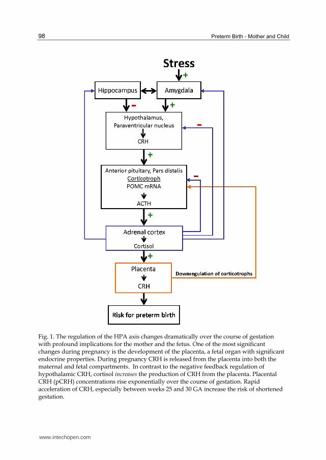
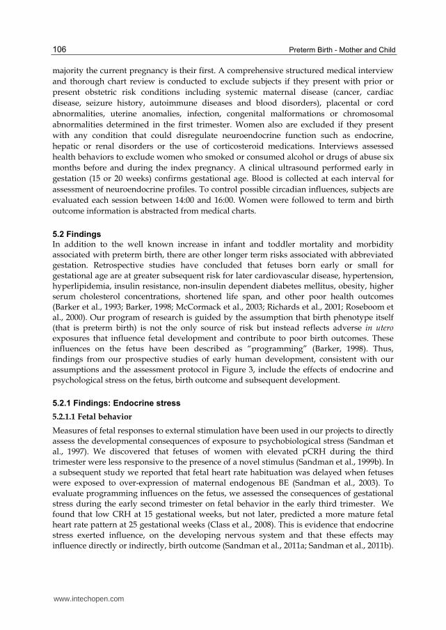
The endocrine stress or "fight or flight" system is profoundly altered during human pregnancy. The pituitary gland doubles in size and the output of pituitary peptides increases severalfold as gestation progresses. But it is the growth and development of a new organ, the placenta, in primates that is primarily responsible for the profound changes in the stress circuit (Figure 1). CRH immunoreactivity in the plasma of non-pregnant women is very low or undetectable. The human placenta and amniotic membrane expresses the genes for the major stress hormones, CRH (hCRHmRNA) and POMC by the seventh week of gestation. All of the HPA and placental stress hormones increase as pregnancy advances, but the exponential increase in placental CRH in maternal plasma is especially dramatic, reaching levels observed only in the hypothalamic portal system during physiological stress (Lowry, 1993). The levels of hCRHmRNA increase more than 20-fold in the five weeks preceding delivery (Frim et al., 1988) resulting in a significant elevation in maternal CRH plasma concentrations during the second half of pregnancy. Levels rise exponentially as pregnancy advances, peaking during labor, and falling to very low or undetectable levels within 24 hours after delivery (Campbell et al., 1987; Chan et al., 1993; Goland et al., 1992; Sasaki et al., 1987; Wolfe et al., 1988). Placental CRH is identical to hypothalamic CRH in structure, immunoreactivity and bioactivity (Petraglia et al., 1989; Sasaki et al., 1988). However, in contrast to the inhibitory influence on the promoter region of the CRH gene in the hypothalamus,
www.intechopen.com
Preterm Birth - Mother and Child
98
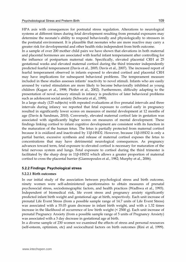
Fig. 1. The regulation of the HPA axis changes dramatically over the course of gestation with profound implications for the mother and the fetus. One of the most significant changes during pregnancy is the development of the placenta, a fetal organ with significant endocrine properties. During pregnancy CRH is released from the placenta into both the maternal and fetal compartments. In contrast to the negative feedback regulation of hypothalamic CRH, cortisol increases the production of CRH from the placenta. Placental CRH (pCRH) concentrations rise exponentially over the course of gestation. Rapid acceleration of CRH, especially between weeks 25 and 30 GA increase the risk of shortened gestation.
www.intechopen.com
Psychobiological Stress and Preterm Birth
99
maternal stress signals (cortisol) from the adrenal glands activate the promoter region in the placenta and stimulate the expression of hCRHmRNA establishing a positive feedback loop that allows for the simultaneous increase of CRH, ACTH and cortisol over the course of gestation. The difference in behavior of the CRH gene in the placenta and hypothalamus is due to the expression of different transcription factors, co-activators and co-repressors in these two tissues (King et al., 2002). The increase of CRH especially over the latter part of human gestation plays a fundamental role in the organization of the fetal nervous system (Sandman et al., 1999b), influencing the timing of the onset of spontaneous labor and delivery (McLean et al., 1995; Sandman et al., 2006; Smith et al., 2002; Smith & Nicholson, 2007; Tyson et al., 2009) and in maternal adaptation during pregnancy, including dampening psychological stress (Glynn & Sandman, in press).
3.2 Alterations in stress responding during pregnancy
The dramatic maternal endocrine alterations that accompany pregnancy have implications not only for the maintenance of gestation, successful parturition and optimal fetal/infant/child development, but also have ramifications for the maternal brain and behavior. HPA axis, blood pressure, heart rate and catecholamine responses to stress are dampened as pregnancy progresses (de Weerth & Buitelaar, 2005). These changes in physiological responding are mirrored by changes in psychological responding. Exposures to stress are found to be less distressing when they occur later in pregnancy compared to when they occur early in pregnancy or in the non-pregnant state (Glynn et al., 2008; Glynn et al., 2004). The changes in stress responding as gestation advances may be adaptive and promote survival (Glynn et al., 2008). Specifically, down-regulated psychological and physiological maternal stress responding provides protection for mother and fetus from the effects of adversity as pregnancy progresses toward term. For instance, stress experienced early in gestation, but not later, is associated with preterm birth (Glynn et al., 2004; Lederman et al., 2004). Moreover, women who fail to show the expected decrease in generalized stress and anxiety or dampening in the cortisol awakening response during pregnancy are at increased risk for preterm delivery (Buss et al., 2009b; Glynn et al., 2008).
4. Risk for preterm birth
Because each developing organism plays an active role in its own construction, the embryo and the fetus must acquire information about the environment that guide its development. The human placenta is both a sensory and effector organ that incorporates and transduces information from its maternal host environment into the fetal developmental program. The fetal/placental unit’s early detection of stress signals from the maternal environment “informs” the fetus that there may be a threat to survival. If the nature of the environment is perceived to be stressful or hostile, it may promote developmental trajectories that ensure survival. Compelling evidence from the desert-dwelling Western spadefoot toad illustrates this conserved function (Boorse & Denver, 2002; Denver, 1997, 1999; Seasholtz et al., 2002). This toad lays its eggs in pools of desert rainwater. Tadpoles exposed to rapidly evaporating pools, accelerate their metamorphosis to escape imminent peril. This highly adaptive function allows the tadpole to reach maturity before the life-sustaining environment desiccates. If stress hormones are blocked during environmental desiccation, then the rate of development is arrested and the tadpole’s survival is compromised. Survival under these
www.intechopen.com
Preterm Birth - Mother and Child
100
circumstances, however, is associated with long-term costs. Tadpoles that survived by accelerating their development were smaller than normal at emergence as toads and had reduced capacity to forage for food and to mate (Denver, 1997; John-Adler & Morin, 1990; Newman, 1989; Smith, 1987).
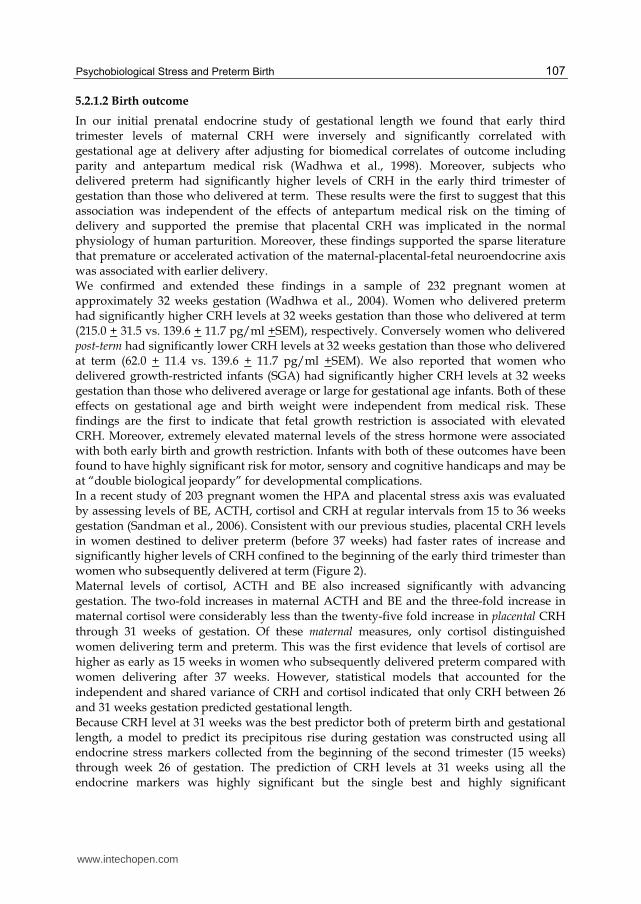
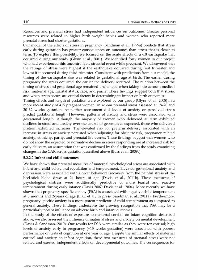
Fig. 2. The BLUE panel shows the normal activity of the HPA axis during pregnancy. One of
the most significant changes during pregnancy is the development of the placenta, a fetal
organ with significant endocrine properties. CRH is released from the placenta into both the
maternal and fetal compartments. In contrast to the negative feedback regulation of
hypothalamic CRH, cortisol increases the production of CRH from the placenta. Placental
CRH (pCRH) concentrations rise exponentially over the course of gestation. The RED panel
illustrates (by the thicker lines) that the normal changes are exaggerated under conditions of
high stress that results in accelerated release of CRH and increasing the risk for preterm
fold within 12 hours in response to the administration of synthetic glucocorticoids such as
betamethasone (Korebrits et al., 1998; Marinoni et al., 1998). Further, placental CRH
concentrations remain elevated for at least one week after a single course of treatment. These finding are consistent with the report of decreased gestational length among women
administered corticosteroids during their first trimester (Gur et al., 2004) probably by
stimulating synthesis and release of placental CRH. The placental detection of stress or adversity may prime or advance the "placental clock"
by activating the promoter region of the CRH gene and increase the placental synthesis of
CRH. The rapid increase in circulating CRH begins the cascade of events influencing
myometria (Tyson et al., 2009) and in extreme cases, precipitating preterm birth. Placental
CRH has been shown to increase the placental production of estrogens and to inhibit the
synthesis of progesterone (Yang et al., 2006; You et al., 2006). Placental CRH is
additionally released into the fetal compartment where it stimulates the fetal adrenal
gland to release dehydroepiandrosterone sulfate (DHEAS) an obligate precursor for
placental estriol production (Smith et al., 1998). Alterations in the production of
progesterone and estriol may be one pathway by which placental CRH regulates the
www.intechopen.com
Preterm Birth - Mother and Child
102
timing of delivery (Smith et al., 2009). Data indicate that it is the trajectory of placental
CRH production over gestation, rather than the absolute hormone concentration that best
predicts preterm delivery, suggesting that target cells are highly responsive to relative
changes in placental CRH concentrations. The effects of HPA and placental axis hormones
on gestational length are modulated by the activities of binding proteins and enzymes.
For example, concurrent with increases in circulating levels of placental CRH, a CRH-
binding protein (CRH-BP) is produced in the liver and also in the trophoblast and
intrauterine tissues during pregnancy, and binds to circulating CRH, reducing its
biological action (Orth & Mount, 1987; Petraglia, et al., 1996; Petraglia et al., 1993). In
contrast to the exponentially increasing levels of circulating CRH over the course of
gestation, CRH-BP levels, which are constant in the first, second, and early third trimester
and are not significantly different from non-pregnant levels, fall by approximately 30% as
birth approaches (Linton et al., 1993). The net effect of these changes in levels of CRH and
CRH-BP is a sharp increase in the availability of free and bioactive CRH during this last
part of gestation. There is some evidence that women who deliver preterm have lower
levels of CRH-BP (Hobel et al., 1999). Maternal plasma cortisol binding globulin (CBG)
levels also change across pregnancy. CBG is stimulated by estrogen and these levels
increase progressively with advancing gestation until the end of gestation when there is a
significant decline in CBG leading to an increase in bioactive cortisol (Ho et al., 2007). The
activity of placental 11β-HSD2 (which oxidizes cortisol into its inactive form, cortisone)
(Sun et al., 1999) increases as gestation progresses before falling precipitously near term.
Both the decrease in CBG and the decrease in activity of placental 11β-HSD2 increase
fetal exposure to maternal cortisol ensuring maturation of the fetal lungs, CNS and other
organ systems in full term births (Ma et al., 2003; Murphy & Clifton, 2003).
The association between maternal plasma concentrations of CRH and preterm
labor/delivery has been examined in many published studies (Markovic et al., 2007). During
pregnancy, maternal stress threatens the fetal nervous system (Coe et al., 2003; Insel et al.,
1990; Poland et al., 1999; Sanchez et al., 1993; Sandman et al., 2003; Sandman et al., 1999a;
Sandman et al., 1999b; Weinstock, 1996) and shortens the length of gestation (Campbell et
al., 1987; McLean et al., 1995; Wadhwa et al., 2004; Wadhwa et al., 1998; Wadhwa et al., 1993;
Warren et al., 1992; Wolfe et al., 1988). The general findings are that plasma CRH
concentrations of women in preterm labor are significantly higher than those of gestational-
age matched controls and the rate of change of CRH over gestation is accelerated in women
destined to deliver early. Studies measuring CRH at a single point during gestation produce
equivocal findings because there are wide individual differences that can only be assessed
with longitudinal designs (Sibai et al., 2005) and because it is the trajectory of placental CRH
production over gestation that best predicts preterm birth (Smith et al., 2009). The most
convincing early support for the role of CRH in the timing of human delivery was
demonstrated by McLean et al (1995). In a prospective, longitudinal study, CRH levels were
assessed between one and four times from 16-20 weeks gestation to term. Plasma CRH
levels at 18-20 weeks gestation were significantly higher in women delivering preterm
(N=24) than at term (N=308), and were significantly lower in women delivering post-term
(N=29). These findings demonstrated that patterns of plasma CRH are associated with the
timing of delivery, both early and late, and may be established as early as the beginning of
the second trimester of gestation.
www.intechopen.com
Psychobiological Stress and Preterm Birth
103
4.2 Psychological risk
Early work examining psychological risk was plagued by methodological issues rendering a definitive answer about the role of psychological stress in preterm birth difficult to assess. An early careful review by Savitz & Pastore (1999) of 20 of the more rigorous studies, stated that it was difficult to draw conclusions due to methodological limitations. Study designs until that time had been a mix of retrospective and case-control studies, with relatively few prospective studies and approximately half of the studies reviewed found associations of stress and length of gestation. However, more recent work with more methodologically sound approaches, including reports from our own group (Campos et al., 2008; Dominguez et al., 2008; Glynn et al., 2008; Glynn et al., 2001; Hilmert et al., 2008; Rini et al., 1999; Wadhwa et al., 1993) have consistently demonstrated significant associations between prenatal stress and adverse birth outcomes. In a recent comprehensive review, Dunkel Schetter and Glynn (2011) concluded that prenatal stress represents a significant risk for preterm birth or shortened gestation. However the findings also indicated that stress was not a unitary construct and some characterizations of stress were stronger predictors of birth outcome than others. For instance, of the fourteen studies of exposure to stressful life events during pregnancy, eight found a significant influence on the risk for shortened gestation, and another form of episodic stress, catastrophes, also were consistently identified as risk factors. High levels of chronic forms of stress (chronic strain, perceived racism, and neighborhood or community stressors) showed consistent links with decreased gestational length. Inconsistent and very modest effects are detected with instruments targeting appraised or perceived stress (all twelve studies used Cohen’s Perceived Stress Scale [Cohen et al., 1983], and only four found an association). Similarly, depression is not a reliable predictor of shortened gestation. Perhaps the most compelling pattern was the finding that anxiety related to pregnancy outcomes, mostly reflecting fears, concerns and beliefs, significantly shortened gestation in ten of eleven studies. Viewed collectively, the evidence is quite clear that stress conceived of as a general multidimensional concept contributes to the etiology of preterm birth. There are at least three emerging themes, however, that dictate future directions and refinement of models examining the role of stress in preterm birth. First, some conceptions or dimensions of stress now are emerging as more potent predictors than others, suggesting a need for further theoretical specificity. By far the most consistent results are found for pregnancy-related stress and anxiety, and a close second is major life events. Also notable are chronic strains, catastrophes, community stressors, and racism, but these literatures still are in the early stages. Second, the predictable changes in maternal stress responding (both psychological and physiological) represent a critical moderating variable (Glynn, 2010). Normative changes in stress responding have important implications both for the impact of exposures to stress and also for interpreting the relations between stress, measured at different points in gestation, and adverse outcomes. It is worth noting that from a methodological standpoint, it is difficult if not impossible, to understand timing without prospective, longitudinal study designs. Last, an additional moderating variable worth considering relates to ethnic and cultural differences. Some stress concepts do not generalize well across ethnic, cultural and foreign populations and some dimensions of stress apply only to specific groups. For example, lifetime exposures to racism are predictive of restricted fetal growth, but only among African American women (Dominquez et al., 2008). Similarly, anxiety in pregnancy has been shown to characterize Latina women in particular, especially
www.intechopen.com
Preterm Birth - Mother and Child
104
new and unacculturated immigrants from countries with poor medical care (Rini et al., 1999; Zambrana et al., 1997). Adding further complexity to this issue is the fact that the relevance of biological mediators and the physiological pathways to preterm birth may differ depending on race/ethnicity. For example, the threshold of CRH exposure that is associated with preterm birth is lower among African American women (Holzman et al., 2001), and others have shown that elevated prenatal cortisol is more likely to be associated with an accelerated CRH trajectory in African American and Latina women than among white women (Glynn et al., 2007). Even more recently it was shown that among Latinas, perceived discrimination is associated with elevated prenatal cortisol trajectories, which predict reduced fetal growth, but this is not the case for non-Hispanic white women (Glynn, 2011). These findings highlight the importance of developing population-specific concepts and measures of stress. In addition, stress concepts and measures that generalize well over ethnic, cultural, and international populations are needed. The emergence of pregnancy-specific validated measures of stress and anxiety represents a promising avenue to achieve this goal (Huizink et al., 2004; Lobel et al., 2008). In recent years, many more studies have been published on this topic, often with
prospective designs, large sample sizes, and appropriate controls. Recent reviews concur
that the evidence regarding stress as a significant independent risk factor for spontaneous
preterm labor and delivery is now clearer (Beydoun & Saftlas, 2008; Institute of Medicine,
2006). Beydoun and Saflas (2008) report that nine of eleven studies between 2000 and 2006
found significant effects of prenatal maternal stress on length of gestation or risk of preterm
labor or birth, although not all studies adjusted for appropriate control variables.
5. Our approach
Our research team has been exploring the effects of stress and specifically the HPA axis on developmental processes for over 30 years (Glynn & Sandman, in press; Sandman & Davis, 2010; Sandman et al., 2011a; Sandman et al., 2011b). The initial studies were among the first to describe the long lasting (perhaps permanent and programming) effects of neonatal exposure to ACTH on the brain and behavior of rats (Beckwith et al., 1977; Champney et al., 1976; Sandman & O'Halloran, 1986). In another comprehensive project, we discovered that in utero exposure of rats to high levels of beta endorphin (BE) delayed developmental milestones, permanently altered pain threshold, exploration, and both active and passive avoidance responding (Sandman & Kastin, 1981). We found that fetal exposure to BE increased the expression of opioids (Moldow et al., 1981) and down-regulated dopamine (D2) receptors (Sandman & Yessaian, 1986) in the brains of these animals as adults. During the past 15+ years our group has been examining the effects of stress and activation of the HPA/placental axis on birth outcomes and on the human fetus. As described below, our findings contribute to the growing acceptance that maternal stress is a risk factor for adverse outcomes. Our studies also have made significant contributions to understanding the mechanisms of the effects of stress on gestational length. Other findings were the first to indicate that very high levels of CRH are associated with both preterm delivery and infants who are small for gestational age. Moreover, we discovered that very low levels of CRH were associated with post-term birth adding strong support to the suggestion that CRH primed a “placental clock” controlling the timing of delivery. We published evidence that a maternal stress message early in pregnancy may prime a subsequent fetal/placental CRH
www.intechopen.com
Psychobiological Stress and Preterm Birth
105
response and increase the risk for preterm birth and that racial/ethnic differences may exist in this priming process (Glynn et al., 2007). Other studies from our project were the first to show that elevated levels of CRH in the maternal circulation influences the human fetal nervous system and fetal exposure to elevated levels of CRH persist into infancy and childhood.
5.1 Assessment
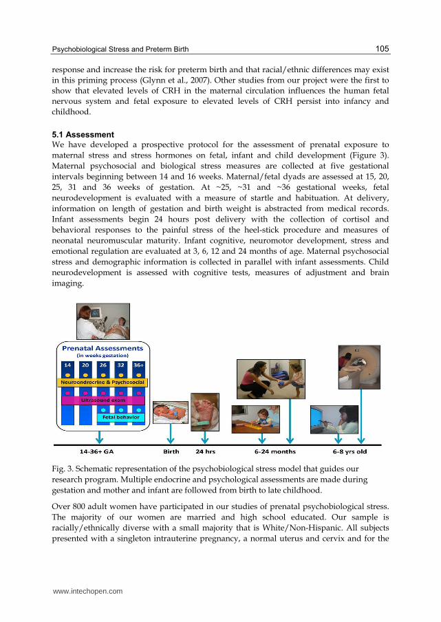
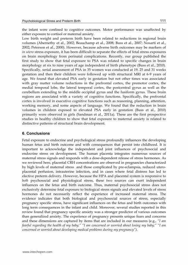
We have developed a prospective protocol for the assessment of prenatal exposure to
maternal stress and stress hormones on fetal, infant and child development (Figure 3).
Maternal psychosocial and biological stress measures are collected at five gestational
intervals beginning between 14 and 16 weeks. Maternal/fetal dyads are assessed at 15, 20,
25, 31 and 36 weeks of gestation. At ~25, ~31 and ~36 gestational weeks, fetal
neurodevelopment is evaluated with a measure of startle and habituation. At delivery,
information on length of gestation and birth weight is abstracted from medical records.
Infant assessments begin 24 hours post delivery with the collection of cortisol and
behavioral responses to the painful stress of the heel-stick procedure and measures of
neonatal neuromuscular maturity. Infant cognitive, neuromotor development, stress and
emotional regulation are evaluated at 3, 6, 12 and 24 months of age. Maternal psychosocial
stress and demographic information is collected in parallel with infant assessments. Child
neurodevelopment is assessed with cognitive tests, measures of adjustment and brain
imaging.
Fig. 3. Schematic representation of the psychobiological stress model that guides our
research program. Multiple endocrine and psychological assessments are made during
gestation and mother and infant are followed from birth to late childhood.
Over 800 adult women have participated in our studies of prenatal psychobiological stress.
The majority of our women are married and high school educated. Our sample is
racially/ethnically diverse with a small majority that is White/Non-Hispanic. All subjects
presented with a singleton intrauterine pregnancy, a normal uterus and cervix and for the
www.intechopen.com
Preterm Birth - Mother and Child
106
majority the current pregnancy is their first. A comprehensive structured medical interview
and thorough chart review is conducted to exclude subjects if they present with prior or
present obstetric risk conditions including systemic maternal disease (cancer, cardiac
disease, seizure history, autoimmune diseases and blood disorders), placental or cord
abnormalities, uterine anomalies, infection, congenital malformations or chromosomal
abnormalities determined in the first trimester. Women also are excluded if they present
with any condition that could disregulate neuroendocrine function such as endocrine,
hepatic or renal disorders or the use of corticosteroid medications. Interviews assessed
health behaviors to exclude women who smoked or consumed alcohol or drugs of abuse six
months before and during the index pregnancy. A clinical ultrasound performed early in
gestation (15 or 20 weeks) confirms gestational age. Blood is collected at each interval for
assessment of neuroendocrine profiles. To control possible circadian influences, subjects are
evaluated each session between 14:00 and 16:00. Women were followed to term and birth
outcome information is abstracted from medical charts.
5.2 Findings
In addition to the well known increase in infant and toddler mortality and morbidity associated with preterm birth, there are other longer term risks associated with abbreviated gestation. Retrospective studies have concluded that fetuses born early or small for gestational age are at greater subsequent risk for later cardiovascular disease, hypertension, hyperlipidemia, insulin resistance, non-insulin dependent diabetes mellitus, obesity, higher serum cholesterol concentrations, shortened life span, and other poor health outcomes (Barker et al., 1993; Barker, 1998; McCormack et al., 2003; Richards et al., 2001; Roseboom et al., 2000). Our program of research is guided by the assumption that birth phenotype itself (that is preterm birth) is not the only source of risk but instead reflects adverse in utero exposures that influence fetal development and contribute to poor birth outcomes. These influences on the fetus have been described as “programming” (Barker, 1998). Thus, findings from our prospective studies of early human development, consistent with our assumptions and the assessment protocol in Figure 3, include the effects of endocrine and psychological stress on the fetus, birth outcome and subsequent development.
5.2.1 Findings: Endocrine stress
5.2.1.1 Fetal behavior
Measures of fetal responses to external stimulation have been used in our projects to directly assess the developmental consequences of exposure to psychobiological stress (Sandman et al., 1997). We discovered that fetuses of women with elevated pCRH during the third trimester were less responsive to the presence of a novel stimulus (Sandman et al., 1999b). In a subsequent study we reported that fetal heart rate habituation was delayed when fetuses were exposed to over-expression of maternal endogenous BE (Sandman et al., 2003). To evaluate programming influences on the fetus, we assessed the consequences of gestational stress during the early second trimester on fetal behavior in the early third trimester. We found that low CRH at 15 gestational weeks, but not later, predicted a more mature fetal heart rate pattern at 25 gestational weeks (Class et al., 2008). This is evidence that endocrine stress exerted influence, on the developing nervous system and that these effects may influence directly or indirectly, birth outcome (Sandman et al., 2011a; Sandman et al., 2011b).
www.intechopen.com
Psychobiological Stress and Preterm Birth
107
5.2.1.2 Birth outcome
In our initial prenatal endocrine study of gestational length we found that early third trimester levels of maternal CRH were inversely and significantly correlated with gestational age at delivery after adjusting for biomedical correlates of outcome including parity and antepartum medical risk (Wadhwa et al., 1998). Moreover, subjects who delivered preterm had significantly higher levels of CRH in the early third trimester of gestation than those who delivered at term. These results were the first to suggest that this association was independent of the effects of antepartum medical risk on the timing of delivery and supported the premise that placental CRH was implicated in the normal physiology of human parturition. Moreover, these findings supported the sparse literature that premature or accelerated activation of the maternal-placental-fetal neuroendocrine axis was associated with earlier delivery. We confirmed and extended these findings in a sample of 232 pregnant women at approximately 32 weeks gestation (Wadhwa et al., 2004). Women who delivered preterm had significantly higher CRH levels at 32 weeks gestation than those who delivered at term (215.0 + 31.5 vs. 139.6 + 11.7 pg/ml +SEM), respectively. Conversely women who delivered post-term had significantly lower CRH levels at 32 weeks gestation than those who delivered at term (62.0 + 11.4 vs. 139.6 + 11.7 pg/ml +SEM). We also reported that women who delivered growth-restricted infants (SGA) had significantly higher CRH levels at 32 weeks gestation than those who delivered average or large for gestational age infants. Both of these effects on gestational age and birth weight were independent from medical risk. These findings are the first to indicate that fetal growth restriction is associated with elevated CRH. Moreover, extremely elevated maternal levels of the stress hormone were associated with both early birth and growth restriction. Infants with both of these outcomes have been found to have highly significant risk for motor, sensory and cognitive handicaps and may be at “double biological jeopardy” for developmental complications. In a recent study of 203 pregnant women the HPA and placental stress axis was evaluated by assessing levels of BE, ACTH, cortisol and CRH at regular intervals from 15 to 36 weeks gestation (Sandman et al., 2006). Consistent with our previous studies, placental CRH levels in women destined to deliver preterm (before 37 weeks) had faster rates of increase and significantly higher levels of CRH confined to the beginning of the early third trimester than women who subsequently delivered at term (Figure 2). Maternal levels of cortisol, ACTH and BE also increased significantly with advancing gestation. The two-fold increases in maternal ACTH and BE and the three-fold increase in maternal cortisol were considerably less than the twenty-five fold increase in placental CRH through 31 weeks of gestation. Of these maternal measures, only cortisol distinguished women delivering term and preterm. This was the first evidence that levels of cortisol are higher as early as 15 weeks in women who subsequently delivered preterm compared with women delivering after 37 weeks. However, statistical models that accounted for the independent and shared variance of CRH and cortisol indicated that only CRH between 26 and 31 weeks gestation predicted gestational length. Because CRH level at 31 weeks was the best predictor both of preterm birth and gestational length, a model to predict its precipitous rise during gestation was constructed using all endocrine stress markers collected from the beginning of the second trimester (15 weeks) through week 26 of gestation. The prediction of CRH levels at 31 weeks using all the endocrine markers was highly significant but the single best and highly significant
www.intechopen.com
Preterm Birth - Mother and Child
108
independent predictor of third trimester CRH was cortisol at 15 weeks gestation. Our longitudinal study of human pregnancy provided evidence that the earliest, and perhaps critical, period for the effects of CRH on gestational length was the interval between weeks 26 and 31. The rate of increase during this interval is faster and the level of CRH at 31 weeks is higher in women destined to deliver preterm. Serial sampling of maternal plasma provides no support for the possibility that CRH earlier in pregnancy influenced gestational length. New findings from this study indicated that a plausible stress-related endocrine signal, elevated cortisol from the mother very early in pregnancy, predicts the precocious rise in CRH leading to an abbreviated gestation. The pattern of findings supports the argument that the effect of elevated cortisol early in pregnancy on gestational length reflects a priming or programming (Barker, 1998; McLean et al., 1995; McLean & Smith, 2001) effect on the eventual fetal/placental CRH response. A recent study further assessed the association of prenatal levels of cortisol with gestational length using a naturally occurring circadian challenge, the cortisol awakening response (CAR) (Buss et al., 2009b). Complete data from a home-based awakening cortisol response were obtained from 51 women early (~17 weeks gestation) and late (~31 weeks gestation). The CAR progressively declined over the course of gestation. A larger CAR in late pregnancy and reduced attenuation of the CAR from early to late gestation were associated with shorter gestational length. Thus, women who exhibited high HPA responsiveness in late gestation and showed reduced dampening of the CAR over gestation were at an increased risk to deliver early.
5.2.1.3 Neonatal and infant outcomes
In a study from our group (Ellman et al., 2008) the New Ballard Maturation Score was used to assess physical and neuromuscular maturation of 158 newborns within 24 hours after birth. Specifically, the neuromuscular and physical characteristics of the newborn were rated and consisted of measures of muscle tone, distinct posture, and angles of resistance in key muscle groups. The results of this study provided evidence that fetal exposure to increases in levels of maternal cortisol at 15 and at 19 weeks gestation and increases in levels of pCRH at 31 weeks gestation were associated with significant decreases in newborn physical and neuromuscular maturation. These effects were observed after adjusting for length of gestation, indicating that fetal exposure to stress hormones programs neonatal neuromuscular maturation independent of gestational age. Prenatal exposure to maternal stress hormones similarly programs the development of the fetal HPA axis with consequences for neonatal functioning. Recently we reported (Davis et al., 2011b) in a sample of 116 mothers and their healthy full term infants assessed at five gestational intervals and at 24 hours after birth, that prenatal maternal cortisol and psychosocial stress each exerted influences on neonatal stress regulation and these influences were dependent upon the gestational period during which the fetus was exposed. Specifically elevated maternal cortisol early in gestation was associated with slower neonatal behavioral recovery from the painful stress of a heel-stick procedure. Elevated maternal cortisol during the second half of gestation was associated with a larger and more prolonged neonatal cortisol response to stress. The data from this study are consistent with evidence that prenatal exposure to synthetic glucocorticoids during the late second and early third trimester is associated with an amplified cortisol response to stress among healthy full term neonates (Davis et al., 2011c). Together, these data provide evidence that gestational exposure to excess glucocorticoids alters the developmental trajectory of the fetal
www.intechopen.com
Psychobiological Stress and Preterm Birth
109
HPA axis with consequences for postnatal stress regulation. Alterations to neurological systems at different times during fetal development resulting from prenatal exposures may determine the neonate’s ability to respond behaviorally and physiologically to stressors in the postnatal environment. It is plausible that neonates who are more reactive may carry a greater risk for developmental and other health risks independent from birth outcome. In a sample of over 200 mother child pairs we have shown that elevations in both maternal and placental hormones are associated with fearful infant temperament after controlling for the influence of postpartum maternal state. Specifically, elevated placental CRH at 25 gestational weeks and elevated maternal cortisol during the third trimester independently predicted fearful temperament (Davis et al., 2005; Davis et al., 2007). The increased report of fearful temperament observed in infants exposed to elevated cortisol and placental CRH may have implications for subsequent behavioral problems. The temperament measure included in these studies assesses infants’ reactivity to novel stimuli. Infants who are easily aroused by varied stimulation are more likely to become behaviorally inhibited as young children (Kagan et al., 1998; Pfeifer et al., 2002). Furthermore, difficulty adapting to the presentation of novel sensory stimuli in infancy is predictive of later behavioral problems such as adolescent social anxiety (Schwartz et al., 1999). In a large study (125 subjects) with repeated evaluations at five prenatal intervals and three intervals during infancy we reported that fetal exposure to cortisol early in pregnancy resulted in significantly lower scores on measures of mental development at 12 months of age (Davis & Sandman, 2010). Conversely, elevated maternal cortisol late in gestation was associated with significantly higher scores on measures of mental development. These findings linking cortisol to infant cognitive development are consistent with its function in the maturation of the human fetus. The fetus is partially protected from maternal cortisol because it is oxidized and inactivated by 11β-HSD2. However, because 11β-HSD2 is only a partial barrier, excessive synthesis and release of maternal cortisol exposes the fetus to concentrations that may have detrimental neurological consequences. As pregnancy advances toward term, fetal exposure to elevated cortisol is necessary for maturation of the fetal nervous system and lungs. Fetal exposure to cortisol during the third trimester is facilitated by the sharp drop in 11β-HSD2 which allows a greater proportion of maternal cortisol to cross the placental barrier (Giannopoulos et al., 1982; Murphy et al., 2006).
5.2.2 Findings: Psychological stress
5.2.2.1 Birth outcomes
In our initial study of the association between psychological stress and birth outcome, ninety women were self-administered questionnaires to obtain measures of prenatal psychosocial stress, sociodemographic factors, and health practices (Wadhwa et al., 1993). Independent of biomedical risk, life event stress and pregnancy anxiety significantly predicted infant birth weight and gestational age at birth, respectively. Each unit increase of prenatal Life Event Stress (from a possible sample range of 14.7 units of Life Event Stress) was associated with a 55.03 gram decrease in infant birth weight, and with a 1.32 times increase in the likelihood of occurrence of low birth weight (< 2500 g). Each unit increase of prenatal Pregnancy Anxiety (from a possible sample range of 5 units of Pregnancy Anxiety) was associated with a 3 day decrease in gestational age at birth. In a diverse sample of 230 women we examined the effects of stress and personal resources (self-esteem, optimism, etc) and sociocultural factors on birth outcomes (Rini et al, 1999).
www.intechopen.com
Preterm Birth - Mother and Child
110
Resources and prenatal stress had independent influences on outcomes. Greater personal resources were related to higher birth weight babies and women who reported more prenatal stress had shorter gestations. Our model of the effects of stress in pregnancy (Sandman et al., 1999a) predicts that stress early during gestation has greater consequences on outcomes than stress that is closer to term. To explore this possibility, we focused on the acute effects of a 6.8 earthquake that occurred during our study (Glynn et al., 2001). We identified forty women in our project who had experienced this uncontrollable stressful event while pregnant. We discovered that the ratings of stress were highest if the earthquake occurred during first trimester and lowest if it occurred during third trimester. Consistent with predictions from our model, the timing of the earthquake also was related to gestational age at birth. The earlier during pregnancy the stress occurred, the earlier the delivery occurred. The relation between the timing of stress and gestational age remained unchanged when taking into account medical risk, maternal age, marital status, race, and parity. These findings suggest both that stress, and when stress occurs are critical factors in determining its impact on birth outcomes. Timing effects and length of gestation were explored by our group (Glynn et al., 2008) in a more recent study of 415 pregnant women in whom prenatal stress assessed at 18–20 and 30–32 weeks gestation. At neither assessment did levels of anxiety or perceived stress predict gestational length. However, patterns of anxiety and stress were associated with gestational length. Although the majority of women who delivered at term exhibited declines in stress and anxiety over the course of gestation as expected, those who delivered preterm exhibited increases. The elevated risk for preterm delivery associated with an increase in stress or anxiety persisted when adjusting for obstetric risk, pregnancy related anxiety, ethnicity, parity, and prenatal life events. These findings suggest that women who do not show the expected or normative decline in stress responding are at increased risk for early delivery, an assumption that was confirmed by the findings from the study examining changes in the CAR across gestation described above (Buss et al., 2009c).
5.2.2.2 Infant and child outcomes
We have shown that prenatal measures of maternal psychological stress are associated with infant and child behavioral regulation and temperament. Elevated gestational anxiety and depression were associated with slower behavioral recovery from the painful stress of the heel-stick blood draw at 24 hours of age (Davis et al., 2011b). These measures of psychological distress were additionally predictive of more fearful and reactive temperament during early infancy (Davis 2007; Davis et al., 2004). More recently we have shown that pregnancy specific anxiety (PSA) is associated with negative child temperament at 3 months and 2-years of age (Blair et al., in press; Sandman et al., 2011a). Furthermore, pregnancy specific anxiety is a more potent predictor of child temperament as compared to general anxiety. These findings underscore the growing recognition that PSA may be a particularly potent influence on adverse birth and infant outcomes. In the study of the effects of exposure to maternal cortisol on infant cognition described above, we also assessed the influence of maternal stress and anxiety on mental development (Davis & Sandman, 2010). Our results for PSA were similar as they were for cortisol; high levels of anxiety early in pregnancy (~15 weeks gestation) were associated with poorest performance on tests of cognition at one year of age. Despite the similar effects of maternal cortisol and anxiety on infant cognition, these two measures of prenatal stress were not related and exerted independent effects on developmental outcomes. The consequences for
www.intechopen.com
Psychobiological Stress and Preterm Birth
111
the infant were confined to cognitive outcomes. Motor performance was unaffected by either exposure to cortisol or maternal anxiety. Low birth weight and preterm birth have been related to reductions in regional brain volumes (Abernethy et al., 2002; Beauchamp et al., 2008; Buss et al., 2007; Nosarti et al., 2002; Peterson et al., 2000). However, because adverse birth outcomes may be markers of in utero stress exposure, it has been difficult to separate the effects of fetal stress exposures on brain morphology from perinatal complications. Recently, our group published the first study to show that fetal exposure to PSA was related to specific changes in brain morphology at six to nine years of age independent of birth phenotype (Buss et al., 2010). Specifically, serial assessment of PSA in 35 women was conducted at 19, 25 and 31 weeks gestation and then their children were followed up with structural MRI at 6-9 years of age. We found that elevated PSA early in gestation but not other times was associated with gray matter volume reductions in the prefrontal cortex, the premotor cortex, the medial temporal lobe, the lateral temporal cortex, the postcentral gyrus as well as the cerebellum extending to the middle occipital gyrus and the fusiform gyrus. These brain regions are associated with a variety of cognitive functions. Specifically, the prefrontal cortex is involved in executive cognitive functions such as reasoning, planning, attention, working memory, and some aspects of language. We found that the reduction in brain volumes in children exposed to elevated PSA early in gestation (Buss et al., 2010) primarily were observed in girls (Sandman et al., 2011a). These are the first prospective studies in healthy children to show that fetal exposure to maternal anxiety is related to distinctive patterns of structural brain development.
6. Conclusions
Fetal exposure to endocrine and psychological stress profoundly influences the developing human fetus and birth outcome and with consequences that persist into childhood. It is important to acknowledge the independent and joint influences of psychosocial and endocrine stress on development. The human placenta integrates numerous sources of maternal stress signals and responds with a dose-dependent release of stress hormones. As we reviewed here, placental CRH concentrations are observed in pregnancies characterized by high levels of maternal stress and those complicated by pre-eclampsia, reduced utero-placental perfusion, intrauterine infection, and in cases where fetal distress has led to elective preterm delivery. However, because the HPA and placental system is responsive to both psychosocial and physiological stress, these two sources can exert independent influences on the fetus and birth outcome. Thus, maternal psychosocial stress does not exclusively determine fetal exposure to biological stress signals and elevated levels of stress hormones do not necessarily reflect the experience of increased maternal stress. The evidence indicates that both biological and psychosocial sources of stress, especially pregnancy specific stress, have significant influences on the fetus and birth outcomes with long term consequences in the infant and child. Moreover, several studies reported in this review found that pregnancy specific anxiety was a stronger predictor of various outcomes than generalized anxiety. The experience of pregnancy presents unique fears and concerns and these dimensions are captured by items that are included in our measures (e.g., “I am fearful regarding the health of my baby;” “I am concerned or worried about losing my baby;” “I am concerned or worried about developing medical problems during my pregnancy”).
www.intechopen.com
Preterm Birth - Mother and Child
112
There are plausible routes of maternal biological stress influencing birth outcome and the fetal nervous system. One possible endocrine route discussed here posits that the placental detection of stress or adversity primes or advances the "placental clock" by activating the promoter region of the CRH gene and increases the placental synthesis of CRH. The rapid increase in circulating CRH begins the cascade of events influencing myometria (Tyson et al., 2009) and may precipitate preterm birth. As such, fetal exposure to pCRH may be a final common pathway for the “programming” effects of some stressors on birth outcome and the developing fetus. The precise mechanism by which the pregnant woman communicates her psychological state of stress or adversity to her fetus and influences birth outcome, however, is less clear. As discussed here fetal exposure to stress hormones alone is not the mechanism of communication. We did not discuss other possible pathways, such as vascular or immunological, or even other endocrine systems, but these are areas that deserve further study. One new area of research that we only briefly acknowledged was sex differences in birth and developmental outcomes. We have reported that (i) female fetuses displayed more mature responses than males at 31 and 36 gestational weeks (Buss et al., 2009a), (ii) delayed neuromotor development associated with fetal exposure to cortisol early in gestation and CRH late in gestation was confined to males (Ellman et al., 2008) and (iii) the reduction in brain volumes in children exposed to elevated PSA early in gestation primarily were observed in girls (Sandman et al., 2011a). These findings are consistent with findings of sex specific trajectories of fetal development and the sexually dimorphic risk of neurological impairment associated with neonatal complications (Kesler et al., 2008). There is evidence that sexually specific patterns are formed very early in development and are reflected in the function and response to stress of the placenta. Clifton (2010) has argued that sexually dimorphic placental sensitivity to signals of adversity (elevated glucocorticoids) results in different patterns of response and in particular in different patterns of growth. Male fetuses, Clifton suggests, do not alter their patterns of development in response to adversity and continue to grow despite reduced resources. Because the male fetus has not adjusted to the initial adversity and has not conserved its resources, it is more susceptible to later stress with increases in morbidity and mortality. In contrast, the female placenta responds or adjusts to an adverse maternal environment in multiple ways resulting in reduced growth. If exposed to stress that reduces nutrients and resources later in gestation, the female fetus has conserved its energy needs which increases the probability of survival. By this mechanism, sexually specific patterns of response to stress may be determined very early in fetal development.
7. Future perspectives
Despite the fact that this area of research is in its embryonic stage, the findings have created a paradigm shift. It is now essential to consider fetal experience (or exposure) and birth outcomes in order to fully understand human development. There are several areas of research that must continue to evolve. First, a more comprehensive understanding of the fetal experience is critical. This review focused on stress-related exposures and primarily endocrine and psychological markers. However there is growing interest in vascular and immune exposures that exert both independent and additive influences on fetal health and developmental outcomes. It is important that the field of fetal neurology/psychology explores the mechanisms of communication of stress and adversity between the host
www.intechopen.com
Psychobiological Stress and Preterm Birth
113
(mother) and the fetus. The precise mechanisms of communication are largely unknown and in some cases the most plausible candidates have been ruled out. New results from our studies indicate that fetuses receive neurodevelopmental benefit from longer gestation even after 37 weeks (Davis et al., 2011a). Neurodevelopment was evaluated with structural magnetic resonance imaging in 100 healthy right-handed 6- to 10-year-old children born between 28 and 41 gestational weeks with a stable neonatal course. We found that longer duration of gestation was associated with region-specific increases in gray matter brain density. Further, the benefit of longer gestation for brain development was present even in children born after 37 weeks. The significant linear association between gestational age at birth and brain development in young children challenges the commonly held assumption of a “non-linear” developmental course that defines fetal maturity as occurring at 37 gestational weeks. Our findings emphasize that there is a benefit for the developing brain of increased gestational length throughout the course of fetal development. In addition to providing new information about the importance of longer gestation beyond 37 gestational weeks for brain development, this finding has implications for medical decisions involving assisted deliveries. The decision about when to deliver a fetus, especially after 37 weeks, rarely involves concerns about the fetal nervous system. The findings reported here suggest that the neurological maturity of the fetus should enter the decision algorithm because even modest increases in gestational length have significant and long-lasting influences on the structure and function of the nervous system.
8. Acknowledgments
The research reported here was supported by awards from the National Institutes of Health: NS-41298, HD-51852 and HD-28413 to CAS, HD-50662 and HD-65823 to EPD and HD-40967 to LMG. The assistance of Cheryl Crippen, Megan Blair, Christina Canino, Natalie Hernandez, Kendra Leak and Christine Cordova is gratefully acknowledged. The authors are especially grateful to the families who participated in these studies.
9. References
Abernethy, L. J., Palaniappan, M., & Cooke, R. W. (2002). Quantitative magnetic resonance imaging of the brain in survivors of very low birth weight. Arch Dis Child Fetal Neonatal Ed, 87(4), 279-283.
Adams, M. M., & Barfield, W. D. (2008). The future of very preterm infants: learning from the past. Jama, 299(12), 1477-1478.
Akana, S. F., Chu, A., Soriano, L., & Dallman, M. F. (2001). Corticosterone exerts site-specific and state-dependent effects in prefrontal cortex and amygdala on regulation of adrenocorticotropic hormone, insulin and fat depots. J Neuroendocrinol, 13(7), 625-637.
Avishai-Eliner, S., Brunson, K. L., Sandman, C. A., & Baram, T. Z. (2002). Stressed-out, or in (utero)? Trends Neurosci, 25(10), 518-524.
Bagley, J., & Moghaddam, B. (1997). Temporal dynamics of glutamate efflux in the prefrontal cortex and in the hippocampus following repeated stress: effects of pretreatment with saline or diazepam. Neuroscience, 77, 65-73.
www.intechopen.com
Preterm Birth - Mother and Child
114
Barker, D. J., Osmond, C., Simmonds, S. J., & Wield, G. A. (1993). The relation of small head circumference and thinness at birth to death from cardiovascular disease in adult life. BMJ, 306(6875), 422-426.
Barker, D. J. P. (1998). Mothers, babies and health in later life. Edinburgh: Churchill Livingston. Beauchamp, M. H., Thompson, D. K., Howard, K., Doyle, L. W., Egan, G. F., Inder, T. E., &
Anderson, P. J. (2008). Preterm infant hippocampal volumes correlate with later working memory deficits. Brain, 131(Pt 11), 2986-2994.
Beckwith, B. E., Sandman, C. A., Hothersall, D., & Kastin, A. J. (1977). Influence of neonatal injections of alpha-MSH on learning, memory and attention in rats. Physiol Behav, 18(1), 63-71.
Beydoun, H., & Saftlas, A. F. (2008). Physical and mental health outcomes of prenatal maternal stress in human and animal studies: a review of recent evidence. Paediatr Perinat Epidemiol, 22(5), 438-466.
Blair, M., Glynn, L. M., Sandman, C. A., & Davis, E. P. (in press). Prenatal maternal anxiety and early childhood temperament. Stress.
Boorse, G. C., & Denver, R. J. (2002). Acceleration of Ambystoma tigrinum metamorphosis by corticotropin-releasing hormone. Journal of Experimental Zoology, 293(1), 94-98.
Braastad, B. O. (1998). Effects of prenatal stress on behaviour of offspring in laboratory and farmed animals. Applied Animal Behaviour Science, 61, 159-180.
Brunson, K. L., Grigoriadis, D. E., Lorang, M. T., & Baram, T. Z. (2002). Corticotropin-releasing hormone (CRH) downregulates the function of its receptor (CRF1) and induces CRF1 expression in hippocampal and cortical regions of the immature rat brain. Exp Neurol, 176(1), 75-86.
Buss, C., Davis, E. P., Class, Q. A., Gierczak, M., Pattillo, C., Glynn, L. M., & Sandman, C. A. (2009a). Maturation of the human fetal startle response: evidence for sex-specific maturation of the human fetus. Early Human Development, 85(10), 633-638.
Buss, C., Davis, E. P., Muftuler, L. T., Head, K., & Sandman, C. A. (2010). High pregnancy anxiety during mid-gestation is associated with decreased gray matter density in 6-9-year-old children. Psychoneuroendocrinology, 35(1), 141-153.
Buss, C., Entringer, S., Reyes, J. F., Chicz-DeMet, A., Sandman, C. A., Waffarn, F., & Wadhwa, P. D. (2009b). The maternal cortisol awakening response in human pregnancy is associated with the length of gestation. Am J Obstet Gynecol, 201(4), 398 e391-398.
Buss, C., Entringer, S., Reyes, J. F., Chicz-Demet, A., Sandman, C. A., Waffarn, F., & Wadhwa, P. D. (2009c). The maternal cortisol awakening response in human pregnancy is associated with the length of gestation. Am J Obstet Gynecol.
Buss, C., Lord, C., Wadiwalla, M., Hellhammer, D. H., Lupien, S. J., Meaney, M. J., & Pruessner, J. C. (2007). Maternal care modeulates the relationship between prenatal risk and hippocampal volume in women but not in men. Journal of Neuroscience, 27(10), 2592-2595.
Campbell, E. A., Linton, E. A., Wolfe, C. D., Scraggs, P. R., Jones, M. T., & Lowry, P. J. (1987). Plasma corticotropin-releasing hormone concentrations during pregnancy and parturition. J Clin Endocrinol Metab, 64(5), 1054-1059.
Campos, B., Schetter, C. D., Abdou, C. M., Hobel, C. J., Glynn, L. M., & Sandman, C. A. (2008). Familialism, social support, and stress: positive implications for pregnant Latinas. Cultur Divers Ethnic Minor Psychol, 14(2), 155-162.
www.intechopen.com
Psychobiological Stress and Preterm Birth
115
Catlin, A. (2006). Extremely long hospitalizations of newborns in the United States: data, descriptions, dilemmas. J Perinatol, 26(12), 742-748.
Challis, J. R. (1994). Characteristics of parturition. In R. K. r. Creasy, R. (Ed.), Maternal Fetal Medicine: Principles and Practice (3rd ed.).
Champney, T. F., Sahley, T. L., & Sandman, C. A. (1976). Effects of neonatal cerebral ventricular injection of ACTH 4-9 and subsequent adult injections on learning in male and female albino rats. Pharmacol Biochem Behav, 5(Suppl 1), 3-9.
Chan, E. C., Smith, R., Lewin, T., Brinsmead, M. W., Zhang, H. P., Cubis, J., Thornton, K., & Hurt, D. (1993). Plasma corticotropin-releasing hormone, beta-endorphin and cortisol inter-relationships during human pregnancy. Acta Endocrinol (Copenh), 128(4), 339-344.
Cheng, Y. H., Nicholson, R. C., King, B., Chan, E. C., Fitter, J. T., & Smith, R. (2000). Glucocorticoid stimulation of corticotropin-releasing hormone gene expression requires a cyclic adenosine 3',5'-monophosphate regulatory element in human primary placental cytotrophoblast cells. J Clin Endocrinol Metab, 85(5), 1937-1945.
Chrousos, G. P. (1992). Regulation and dysregulation of the hypothalamic-pituitary-adrenal axis. The corticotropin-releasing hormone perspective. Endocrinol Metab Clin North Am, 21(4), 833-858.
Class, Q. A., Buss, C., Davis, E. P., Gierczak, M., Pattillo, C., Chicz-DeMet, A., & Sandman, C. A. (2008). Low levels of corticotropin-releasing hormone during early pregnancy are associated with precocious maturation of the human fetus. Developmental Neuroscience, 30(6), 419-426.
Clifton, V. L. (2010). Review: Sex and the human placenta: mediating differential strategies of fetal growth and survival. Placenta, 31 Supplement 1, S33-39.
Coe, C. L., Kramer, M., Czeh, B., Gould, E., Reeves, A. J., Kirschbaum, C., & Fuchs, E. (2003). Prenatal stress diminishes neurogenesis in the dentate gyrus of juvenile rhesus monkeys. Biological Psychiatry, 54, 1025-1034.
Cohen, S., Kamarck, T., & Mermelstein, R. (1983). A global measure of perceived stress. Journal of Health and Social Behavior, 24, 385-396.
Davis, E. P. (2007). Prenatal exposure to maternal stress hormones influences human infant development. Paper presented at the International Society for Developmental Psychobiology.
Davis, E. P., Buss, C., Muftuler, L. T., Head, K., Hasso, A., Wing, D. A., Hobel, C., & Sandman, C. A. (2011a). Children's Brain Development Benefits from Longer Gestation. Front Psychol, 2, 1.
Davis, E. P., Glynn, L. M., Dunkel Schetter, C., Hobel, C., Chicz-De Met, A., & Sandman, C. A. (2005). Maternal plasma corticotropin-releasing hormone levels during pregnancy are associated with infant temperament. Developmental Neuroscience, 27(5), 299-305.
Davis, E. P., Glynn, L. M., Dunkel Schetter, C., Hobel, C., Chicz-DeMet, A., & Sandman, C., A. (2007). Prenatal exposure to maternal depression and cortisol influences infant temperament. Journal of the American Academy of Child and Adolescent Psychiatry, 46(6), 737-746.
Davis, E. P., Glynn, L. M., Waffarn, F., & Sandman, C. A. (2011b). Prenatal maternal stress programs infant stress regulation. J Child Psychol Psychiatry, 52(2), 119-129.
www.intechopen.com
Preterm Birth - Mother and Child
116
Davis, E. P., & Sandman, C. A. (2010). The timing of prenatal exposure to maternal cortisol and psychosocial stress is associated with human infant cognitive development. Child Development, 81(1), 131–148.
Davis, E. P., Snidman, N., Wadhwa, P. D., Dunkel Schetter, C., Glynn, L., & Sandman, C. A. (2004). Prenatal maternal anxiety and depression predict negative behavioral reactivity in infancy. Infancy, 6(3), 319-331.
Davis, E. P., Waffarn, F., & Sandman, C. A. (2011c). Prenatal treatment with glucocorticoids sensitizes the hpa axis response to stress among full-term infants. Dev Psychobiol, 53(2), 175-183.
de Weerth, C., & Buitelaar, J. K. (2005). Physiological stress reactivity in human pregnancy - a review. Neuroscience and Biobehavioral Reviews, 29, 295-312.
Denver, R. J. (1997). Environmental stress as a developmental cue: corticotropin-releasing hormone is a proximate mediator of adaptive phenotypic plasticity in amphibian metamorphosis. Hormones and Behavior, 31(2), 169-179.
Denver, R. J. (1999). Evolution of the corticotropin-releasing hormone signaling system and its role in stress-induced phenotypic plasticity. Annals of the New York Academy of Sciences, 897, 46-53.
Diorio, D., Viau, V., & Meaney, M. J. (1993). The role of the medial prefrontal cortex (cingulate gyrus) in the regulation of hypothalamic-pituitary-adrenal responses to stress. J Neurosci, 13(9), 3839-3847.
Dominguez, T. P., Dunkel-Schetter, C., Glynn, L. M., Hobel, C., & Sandman, C. A. (2008). Racial differences in birth outcomes: the role of general, pregnancy, and racism stress. Health Psychol, 27(2), 194-203.
Dominquez, T. P., Dunkel-Schetter, C., Glynn, L. M., Hobel, C. J., & Sandman, C. A. (2008). Racial differences in birth outcomes: the role of general, pregnancy, and racism stress. Health Psychology, 27, 194-203.
Dunkel Schetter, C., & Glynn, L. M. (2011). Stress in pregnancy: empirical evidence and theoretical issues to guide interdisciplinary research. In R. J. Contrada & A. Baum (Eds.), The handbook of stress science: biology, psychology and health. New York: Springer Publishing Company.
Ellman, L. M., Dunkel-Schetter, C., Hobel, C. J., Chicz-DeMet, A., Glynn, L. M., & Sandman, C. A. (2008). Timing of fetal exposure to stress hormones: Effects on newborn physical and neuromuscular maturation. Developmental Psychobiology, 50, 232-241.
Emanuel, R. L., Robinson, B. G., Seely, E. W., Graves, S. W., Kohane, I., Saltzman, D., Barbieri, R., & Majzoub, J. A. (1994). Corticotrophin releasing hormone levels in human plasma and amniotic fluid during gestation. Clin Endocrinol (Oxf), 40(2), 257-262.
Escobar, G. J., Littenberg, B., & Petitti, D. B. (1991). Outcome among surviving very low birthweight infants: a meta-analysis. Arch Dis Child, 66(2), 204-211.
Feldman, S., & Conforti, N. (1985). Modifications of adrenocortical responses following frontal cortex stimulation in rats with hypothalamic differentiations and medial forebrain bundle lesions. Neuroscience, 15, 1045-1047.
Frim, D. M., Emanuel, R. L., Robinson, B. G., Smas, C. M., Adler, G. K., & Majzoub, J. A. (1988). Characterization and gestational regulation of corticotropin-releasing hormone messenger RNA in human placenta. J Clin Invest, 82(1), 287-292.
www.intechopen.com
Psychobiological Stress and Preterm Birth
117
Giannopoulos, G., Jackson, K., & Tulchinsky, D. (1982). Glucocorticoid metabolism in human placenta, decidua, myometrium and fetal membranes. J Steroid Biochem, 17(4), 371-374.
Gibbs, R. S., Romero, R., Hillier, S. L., Eschenbach, D. A., & Sweet, R. L. (1992). A review of premature birth and subclinical infection. Am J Obstet Gynecol, 166(5), 1515-1528.
Giles, W. B., McLean, M., Davies, J. J., & Smith, R. (1996). Abnormal umbilical artery doppler waveforms and cord blood corticotropin-releasing hormone. Obstet and Gynecol, 87, 107-111.
Glynn, L. M. (2010). Implications of maternal programming for fetal neurodevelopment. In A. Zimmerman & S. Connors (Eds.), Maternal influences on fetal neurodevelopment (pp. 33-54). New Jersey: Springer Science+Business Media, LLC.
Glynn, L. M. (2011). Perceived discrimination predicts prenatal stress hormone trajectories among pregnant latina women. Annals of Behavioral Medicine, 41, s56.
Glynn, L. M., & Sandman, C. A. (in press). Prenatal origins of neurological development: a critical period for fetus and mother. Current Directions in Psychological Science.
Glynn, L. M., Schetter, C. D., Chicz-DeMet, A., Hobel, C. J., & Sandman, C. A. (2007). Ethnic differences in adrenocorticotropic hormone, cortisol and corticotropin-releasing hormone during pregnancy. Peptides, 28(6), 1155-1161.
Glynn, L. M., Schetter, C. D., Hobel, C. J., & Sandman, C. A. (2008). Pattern of perceived stress and anxiety in pregnancy predicts preterm birth. Health Psychology, 27(1), 43-51.
Glynn, L. M., Schetter, C. D., Wadhwa, P. D., & Sandman, C. A. (2004). Pregnancy affects appraisal of negative life events. Journal of Psychosomatic Research, 56(1), 47-52.
Glynn, L. M., Wadhwa, P. D., Dunkel-Schetter, C., Chicz-Demet, A., & Sandman, C. A. (2001). When stress happens matters: effects of earthquake timing on stress responsivity in pregnancy. American Journal of Obstetrics & Gynecology, 184(4), 637-642.
Goland, R. S., Conwell, I. M., Warren, W. B., & Wardlaw, S. L. (1992). Placental corticotropin-releasing hormone and pituitary-adrenal function during pregnancy. Neuroendocrinology, 56(5), 742-749.
Goldenberg, R. L., Hauth, J. C., & Andrews, W. W. (2000). Mechanisms of disease: intrauterine infection and preterm delivery. New England Journal of Medicine, 342, 1500-1507.
Gur, C., Orna Diav-Citrin, O., Shechtman, S., Arnon, J., & Ornoy, A. (2004). Pregnancy outcome after first trimester exposure to corticosteroids: a prospective controlled study. Reproductive Toxicology, 18, 93-101.
Herman, J. P., Cullinan, W. E., Morano, M. I., Akil, H., & Watson, S. J. (1995). Contribution of the ventral subiculum to inhibitory regulation of the hypothalamo-pituitary-adrenocortical axis. J Neuroendocrinol, 7(6), 475-482.
Herman, J. P., Dolgas, C. M., & Carlson, S. L. (1998). Ventral subiculum regulates hypothalamo-pituitary-adrenocortical and behavioural responses to cognitive stressors. Neuroscience, 86(2), 449-459.
Herman, J. P., Schafer, M. K., Young, E. A., Thompson, R., Douglass, J., Akil, H., & Watson, S. J. (1989). Evidence for hippocampal regulation of neuroendocrine neurons of the hypothalamo-pituitary-adrenocortical axis. J Neurosci, 9(9), 3072-3082.
www.intechopen.com
Preterm Birth - Mother and Child
118
Hilmert, C. J., Schetter, C. D., Dominguez, T. P., Abdou, C., Hobel, C. J., Glynn, L., & Sandman, C. (2008). Stress and blood pressure during pregnancy: racial differences and associations with birthweight. Psychosom Med, 70(1), 57-64.
Ho, J. T., Lewis, J. G., & O'loughlin, P. (2007). Reduced maternal corticosteroid-binding globulin and cortisol levels in pre-eclampsia and gamete recipient pregnancies. Clin Endocrinol (Oxf), 66(6), 869-877.
Hobel, C. J., Dunkel-Schetter, C., Roesch, S. C., Castro, L. C., & Arora, C. P. (1999). Maternal plasma corticotropin-releasing hormone associated with stress at 20 weeks gestation in pregnancies ending in preterm delivery. American Journal of Obstetrics and Gynecology, 180(1 Pt 3), 257-263.
Holzman, C., Jetton, J., Siler-Khodr, T., Fisher, R., & Rip, T. (2001). Second trimester corticotropin-releasing hormone levels in relation to preterm delivery and ethnicity. Obstetrics and Gynecology, 97, 657-663.
Huizink, A. C., Mulder, E. J., Robles de Medina, P. G., Visser, G. H., & Buitelaar, J. K. (2004). Is pregnancy anxiety a distinctive syndrome? Early Human Development, 79, 81-91.
Insel, T. R., Kinsley, C. H., Mann, P. E., & Bridges, R. S. (1990). Prenatal stress has long-term effects on brain opiate receptors. Brain Res, 511(1), 93-97.
Institute of Medicine (2006). Preterm birth: Causes, consequences, and prevention. Washington, D.C.: National Academies Press.
John-Adler, H. B., & Morin, P. J. (1990). Effects of larval density on jumping ability and stamina in newly metamorphosed Bufo-Woodhousi-fowleri. Copeia, 856-860.
Kagan, J., Snidman, N., & Arcus, D. (1998). Childhood derivatives of high and low reactivity in infancy. Child Development, 69(6), 1483-1493.
Kesler, S. R., Reiss, A. L., Vohr, B., Watson, C., Schneider, K. C., Katz, K. H., Maller-Kesselman, J., Silbereis, J., Constable, R. T., Makuch, R. W., & Ment, L. R. (2008). Brain volume reductions within multiple cognitive systems in male preterm children at age twelve. Journal of Pediatrics, 152(4), 513-520, 520 e511.
King, B. R., Smith, R., & Nicholson, R. C. (2002). Novel glucocorticoid and cAMP interactions on the CRH gene promoter. Mol Cell Endocrinol, 194(1-2), 19-28.
Knigge, K. M., & Hays, M. (1963). Evidence of Inhibitive Role of Hippocampus in Neural Regulation of Acth Release. Proc Soc Exp Biol Med, 114, 67-69.
Korebrits, C., Yu, D. H., Ramirez, M. M., Marinoni, E., Bocking, A. D., & Challis, J. R. (1998). Antenatal glucocorticoid administration increases corticotrophin-releasing hormone in maternal plasma. British Journal of Obstetrics and Gynecology, 105(5), 556-561.
Kramer, M. R., & Hogue, C. R. (2009). What causes racial disparities in very preterm birth? A biosocial perspective. Epidemiol Rev, 31, 84-98.
Kramer, M. S. (2009). Late preterm birth: appreciable risks, rising incidence. J Pediatr, 154(2), 159-160.
Kramer, M. S., Lydon, J., Seguin, L., Goulet, L., Kahn, S. R., McNamara, H., Genest, J., Dassa, C., Chen, M. F., Sharma, S., Meaney, M. J., Thomson, S., Van Uum, S., Koren, G., Dahhou, M., Lamoureux, J., & Platt, R. W. (2009). Stress pathways to spontaneous preterm birth: the role of stressors, psychological distress, and stress hormones. Am J Epidemiol, 169(11), 1319-1326.
Kuban, K. C., & Leviton, A. (1994). Cerebral palsy. N Engl J Med, 330(3), 188-195. Lazarus, R. S. (1966). Psychological Stress and the Coping Process. New York: McGraw-Hill.
www.intechopen.com
Psychobiological Stress and Preterm Birth
119
Lazarus, R. S. (1968). Emotions and adaptation: conceptual and empirical relations Nebraska Symposium on Motivation. Lincoln: University of Nebraska Press.
Lazarus, R. S., & Folkman, S. (1984). Stress, Appraisal, and Coping. New York: Springer Publishing Company, Inc.
Lederman, S. A., Rauh, V., Weiss, L., Stein, J. L., Hoepner, L. A., Becker, M., & Perera, F. P. (2004). The effects of the World Trade Center event on birth outcomes among term deliveries at three lower Manhatten hospitals. Environmental Health Perspectives, 112, 1772-1778.
Lindsay, J. R., & Nieman, L. K. (2005). The hypothalamic-pituitary-adrenal axis in pregnancy: challenges in disease detection and treatment. Endocr Rev, 26(6), 775-799.
Linton, E. A., Perkins, A. V., Woods, R. J., Eben, F., Wolfe, C. D., Behan, D. P., Potter, E., Vale, W. W., & Lowry, P. J. (1993). Corticotropin releasing hormone-binding protein (CRH-BP): plasma levels decrease during the third trimester of normal human pregnancy. J Clin Endocrinol Metab, 76(1), 260-262.
Lobel, M., Cannella, D. L., Graham, J. E., DeVincent, C., Schneider, J., & Meyer, B. A. (2008). Pregnancy-specific stress, prenatal health behaviors, and birth outcomes. Health Psychology, 27, 604-615.
Lowry, P. J. (1993). Corticotropin-releasing factor and its binding protein in human plasma. Ciba Found Symp, 172, 108-115; discussion 115-128.
Ma, X. H., Wu, W. X., & Nathanielsz, P. W. (2003). Gestation-related and betamethasone-induced changes in 11beta-hydroxysteroid dehydrogenase types 1 and 2 in the baboon placenta. Am J Obstet Gynecol, 188(1), 13-21.
March of Dimes (2009). White paper on preterm birth: the global and regional toll. White Plains, N.Y.: March of Dimes Foundation.
Makrigiannakis, A., Semmler, M., Briese, V., Eckerle, H., Minas, V., Mylonas, I., Friese, K., & Jeschke, U. (2007). Maternal serum corticotropin-releasing hormone and ACTH levels as predictive markers of premature labor. Int J Gynaecol Obstet, 97(2), 115-119.
Marinoni, E., Korebrits, C., Di Lorio, R., Cosmi, E. V., & Challis, J. R. (1998). Effect of betamethasone in vivo on placental corticotropin-releasing hormone in human pregnancy. American Journal of Obstetrics and Gynecology, 178(4), 770-778.
Markovic, D., Vatish, M., Gu, M., Slater, D., Newton, R., Lehnert, H., & Grammatopoulos, D. K. (2007). The onset of labor alters corticotropin-releasing hormone type 1 receptor variant expression in human myometrium: putative role of interleukin-1beta. Endocrinology, 148(7), 3205-3213.
McCormack, V. A., Dos Santos Silva, I., De Stavola, B. L., Mohsen, R., Leon, D. A., & Lithell, H. O. (2003). Fetal growth and subsequent risk of breast cancer: Results from long term follow up of Swedish cohort. British Medical Journal, 326(7383), 248.
McLean, M., Bisits, A., Davies, J., Woods, R., Lowry, P., & Smith, R. (1995). A placental clock controlling the length of human pregnancy. Nature Medicine, 1, 460-463.
McLean, M., & Smith, R. (2001). Corticotrophin-releasing hormone and human parturition. Reproduction, 121(4), 493-501.
Meaney, M. J., Diorio, J., Francis, D., Widdowson, J., LaPlante, P., Caldji, C., Sharma, S., Seckl, J. R., & Plotsky, P. M. (1996). Early environmental regulation of forebrain glucocorticoid receptor gene expression: implications for adrenocortical responses to stress. Dev Neurosci, 18(1-2), 49-72.
www.intechopen.com
Preterm Birth - Mother and Child
120
Moghaddam, B. (1993). Stress preferentially increases extraneuronal levels of excitatory amino acids in the prefrontal cortex: Comparison to hippocampus and basal ganglia. Journal of Neurochemistry, 60(1650-1657).
Moldow, R. L., Kastin, A. J., Hollander, C. S., Coy, D. H., & Sandman, C. A. (1981). Brain beta-endorphin-like immunoreactivity in adult rats given beta-endorphin neonatally. Brain Res Bull, 7(6), 683-686.
Murphy, V. E., & Clifton, V. L. (2003). Alterations in human placental 11beta-hydroxysteroid dehydrogenase type 1 and 2 with gestational age and labour. Placenta, 24(7), 739-744.
Murphy, V. E., Smith, R., Giles, W. B., & Clifton, V. L. (2006). Endocrine regulation of human fetal growth: The role of the mother, placenta, and fetus. Endocrine Reviews, 27(2), 141-169.
Murros, K., Fogelholm, R., Kettunen, S., & Vurela, A. L. (1993). Serum cortisol and outcome of ischemic brain infarctions. Journal of the Neurological Sciences, 116, 12-17.
Nathanielsz, P. W. (1995). The role of basic science in preventing low birth weight. Future Child, 5(1), 57-70.
Nettles, K. W., Pesold, C., & Goldman, M. B. (2000). Influence of the ventral hippocampal formation on plasma vasopressin, hypothalamic-pituitary-adrenal axis, and behavioral responses to novel acoustic stress. Brain Res, 858(1), 181-190.
Neumann, I. D., Wigger, A., Liebsch, G., Holsboer, F., & Landgraf, R. (1998). Increased basal activity of the hypothalamo-pituitary-adrenal axis during pregnancy in rats bred for high anxiety-related behaviour. Psychoneuroendocrinology, 23(5), 449-463.
Newman, R. A. (1989). Developmental plasticity of Scaphiopus coouchii tadpoles in an unpredictable environment. Ecology, 70, 1775-1787.
Nosarti, C., Al-Asady, M. H., Frangou, S., Stewart, A. L., Rifkin, L., & Murray, R. M. (2002). Adolescents who were born very preterm have decreased brain volumes. Brain, 125(Pt 7), 1616-1623.
Novy, M. J., McGregor, J. A., & Iams, J. D. (1995). New perspectives on the prevention of extreme prematurity. Clin Obstet Gynecol, 38(4), 790-808.
Orth, D. N., & Mount, C. D. (1987). Specific high-affinity binding protein for human corticotropin-releasing hormone in normal human plasma. Biochem Biophys Res Commun, 143(2), 411-417.
Peterson, B. S., Vohr, B., Staib, L. H., Cannistraci, C. J., Dolberg, A., Schneider, K. C., Katz, K. H., Westerveld, M., Sparrow, S., Anderson, A. W., Duncan, C. C., Makuch, R. W., Gore, J. C., & Ment, L. R. (2000). Regional brain volume abnormalities and long-term cognitive outcome in preterm infants. Journal of the American Medical Association, 284(15), 1939-1947.
Petraglia, F., Florio, P., Nappi, C., & Genazzani, A. R. (1996). Peptide signaling in human placenta and membranes: autocrine, paracrine, and endocrine mechanisms. Endocr Rev, 17(2), 156-186.
Petraglia, F., Potter, E., Cameron, V. A., Sutton, S., Behan, D. P., Woods, R. J., Sawchenko, P. E., Lowry, P. J., & Vale, W. (1993). Corticotropin-releasing factor-binding protein is produced by human placenta and intrauterine tissues. J Clin Endocrinol Metab, 77(4), 919-924.
www.intechopen.com
Psychobiological Stress and Preterm Birth
121
Petraglia, F., Sawchenko, P. E., Rivier, J., & Vale, W. (1987). Evidence for local stimulation of ACTH secretion by corticotropin-releasing factor in human placenta. Nature, 328(6132), 717-719.
Petraglia, F., Sutton, S., & Vale, W. (1989). Neurotransmitters and peptides modulate the release of immunoreactive corticotropin-releasing factor from cultured human placental cells. Am J Obstet Gynecol, 160(1), 247-251.
Petraglia, F., Volpe, A., Genazzani, A. R., Rivier, J., Sawchenko, P. E., & Vale, W. (1990). Neuroendocrinology of the human placenta. Frontiers in Neuroendocrinology, 11, 6-37.
Pfeifer, M., Goldsmith, H. H., Davidson, R. J., & Rickman, M. (2002). Continuity and change in inhibited and uninhibited children. Child Dev, 73(5), 1474-1485.
Poland, R. E., Cloak, C., Lutchmansingh, P. J., McCracken, J. T., Chang, L., & Ernst, T. (1999). Brain N-acetyl aspartate concentrations measured by H MRS are reduced in adult male rats subjected to perinatal stress: preliminary observations and hypothetical implications for neurodevelopmental disorders. J Psychiatr Res, 33(1), 41-51.
Richards, M., Hardy, R., Kuh, D., & Wadsworth, M. E. (2001). Birth weight and cognitive function in the British 1946 birth cohort: longitudinal population based study. British Medical Journal, 322, 199-203.
Rini, C. K., Dunkel-Schetter, C., Wadhwa, P. D., & Sandman, C. A. (1999). Psychological adaptation and birth outcomes: the role of personal resources, stress, and sociocultural context in pregnancy. Health Psychology, 18(4), 333-345.
Roseboom, T., van der Meulen, J., Osmond, C., Barker, D., Ravelli, A., Schroeder-Tanka, J., van Montfrans, G., Michels, R., & Bleker, O. (2000). Coronary heart disease after prenatal exposure to the Dutch famine, 1944-45. Heart, 84(6), 595-598.
Sanchez, M. D., Milanes, M. V., Fuente, T., & Laorden, M. L. (1993). The beta-endorphin response to prenatal stress during postnatal development in the rat. Brain Res Dev Brain Res, 74(1), 142-145.
Sandman, C., A., Glynn, L., Wadhwa, P. D., Chicz-DeMet, A., Porto, M., & Garite, T. (2003). Maternal hypothalamic-pituitary-adrenal disregulation during the third trimester influences human fetal responses. Developmental Neuroscience, 25, 41-49.
Sandman, C. A., & Davis, E. P. (2010). Gestational stress influences cognition and behavior. Future Neurology, 5(5), 675-690.
Sandman, C. A., Davis, E. P., Buss, C., & Glynn, L. M. (2011a). Exposure to Prenatal Psychobiological Stress Exerts Programming Influences on the Mother and Her Fetus. Neuroendocrinology.
Sandman, C. A., Davis, E. P., Buss, C., & Glynn, L. M. (2011b). Prenatal programming of human neurological function. International Journal of Peptides, 2011, 1-9.
Sandman, C. A., Glynn, L., Schetter, C. D., Wadhwa, P., Garite, T., Chicz-DeMet, A., & Hobel, C. (2006). Elevated maternal cortisol early in pregnancy predicts third trimester levels of placental corticotropin releasing hormone (CRH): priming the placental clock. Peptides, 27(6), 1457-1463.
Sandman, C. A., & Kastin, A. J. (1981). Intraventricular administration of MSH induces hyperalgesia in rats. Peptides, 2(2), 231-233.
Sandman, C. A., & O'Halloran, J. P. (1986). Pro-opiomelanocortin, learning, memory and attention. In D. DeWied, W. H. Gispen & T. B. van Wimersma Greidanus (Eds.), Encyclopedia on Pharmacology and Therapeutics (pp. 397-420): Pergamon Press.
www.intechopen.com
Preterm Birth - Mother and Child
122
Sandman, C. A., Wadhwa, P., Glynn, L., Chicz-Demet, A., Porto, M., & Garite, T. J. (1999a). Corticotrophin-releasing hormone and fetal responses in human pregnancy. Ann N Y Acad Sci, 897, 66-75.
Sandman, C. A., Wadhwa, P. D., Chicz-DeMet, A., Dunkel-Schetter, C., & Porto, M. (1997). Maternal stress, HPA activity, and fetal/infant outcome. Annals of the New York Academy of Sciences, 814, 266-275.
Sandman, C. A., Wadhwa, P. D., Chicz-DeMet, A., Porto, M., & Garite, T. J. (1999b). Maternal corticotropin-releasing hormone and habituation in the human fetus. Developmental Psychobiology, 34(3), 163-173.
Sandman, C. A., & Yessaian, N. (1986). Persisting subsensitivity of the striatal dopamine system after fetal exposure to beta-endorphin. Life Sci, 39(19), 1755-1763.
Sasaki, A., Shinkawa, O., Margioris, A. N., Liotta, A. S., Sato, S., Murakami, O., Go, M., Shimizu, Y., Hanew, K., & Yoshinaga, K. (1987). Immunoreactive corticotropin-releasing hormone in human plasma during pregnancy, labor, and delivery. J Clin Endocrinol Metab, 64(2), 224-229.
Sasaki, A., Tempst, P., Liotta, A. S., Margioris, A. N., Hood, L. E., Kent, S. B., Sato, S., Shinkawa, O., Yoshinaga, K., & Krieger, D. T. (1988). Isolation and characterization of a corticotropin-releasing hormone-like peptide from human placenta. J Clin Endocrinol Metab, 67(4), 768-773.
Savitz, D. A., & Pastore, L. M. (1999). Causes of prematurity Prenatal Care: Effectiveness and Implementation (pp. 63-104). Cambridge: Cambridge University Press.
Schulkin, J. (1999). Corticotropin-releasing hormone signals adversity in both the placenta and the brain: regulation by glucocorticoids and allostatic overload. J Endocrinol, 161(3), 349-356.
Schwartz, C. E., Snidman, N., & Kagan, J. (1999). Adolescent social anxiety as an outcome of inhibited temperament in childhood. Journal of the American Academy of Child and Adolescent Psychiatry, 38(8), 1008-1015.
Seasholtz, A. F., Valverde, R. A., & Denver, R. J. (2002). Corticotropin-releasing hormone-binding protein: biochemistry and function from fishes to mammals. J Endocrinol, 175(1), 89-97.
Selye, H. (1936). A syndrome produced by diverse nocuous agents. Nature, 138, 32. Selye, H. (1955). Stress and disease. Science, 122(3171), 625-631. Selye, H. (1956). The Stress of Life. New York: McGraw-Hill. Selye, H. (1959). Perspectives in stress research. Perspect Biol Med, 2(4), 403-416. Shimizu, E., Kodama, K., Sakamoto, T., Komatsu, N., Yamanouchi, N., Okada, S., & Sato, T.
(1997). Recovery from neuroendocrinological abnormalities and frontal hypoperfusion after remission in a case with rapid cycling bipolar disorder. Psychol Clin Neurosci, 51, 207-212.
Sibai, B., Meis, P. J., Klebanoff, M., Dombrowski, M. P., Weiner, S. J., Moawad, A. H., Northen, A., Iams, J. D., Varner, M. W., Caritis, S. N., O'Sullivan, M. J., Miodovnik, M., Leveno, K. J., Conway, D., Wapner, R. J., Carpenter, M., Mercer, B., Ramin, S. M., Thorp, J. M., Jr., Peaceman, A. M., & Gabbe, S. (2005). Plasma CRH measurement at 16 to 20 weeks' gestation does not predict preterm delivery in women at high-risk for preterm delivery. Am J Obstet Gynecol, 193(3 Pt 2), 1181-1186.
www.intechopen.com
Psychobiological Stress and Preterm Birth
123
Smith, D. C. (1987). Adult recruitment in chorus frogs: Effects of size and date at metamorphosis. Ecology, 68, 344-350.
Smith, R., Mesiano, S., Chan, E. C., Brown, S., & Jaffe, R. B. (1998). Corticotropin-releasing hormone directly and preferentially stimulates dehydroepiandrosterone sulfate secretion by human fetal adrenal cortical cells. J Clin Endocrinol Metab, 83(8), 2916-2920.
Smith, R., Mesiano, S., & McGrath, S. (2002). Hormone trajectories leading to human birth. Regul Pept, 108(2-3), 159-164.
Smith, R., & Nicholson, R. C. (2007). Corticotrophin releasing hormone and the timing of birth. Front Biosci, 12, 912-918.
Smith, R., Smith, J. I., Shen, X., Engel, P. J., Bowman, M. E., McGrath, S. A., Bisits, A. M., McElduff, P., Giles, W. B., & Smith, D. W. (2009). Patterns of plasma corticotropin-releasing hormone, progesterone, estradiol, and estriol change and the onset of human labor. J Clin Endocrinol Metab, 94(6), 2066-2074.
Sun, K., Adamson, S. L., Yang, K., & Challis, J. R. (1999). Interconversion of cortisol and cortisone by 11beta-hydroxysteroid dehydrogenases type 1 and 2 in the perfused human placenta. Placenta, 20(1), 13-19.
Tyson, E. K., Smith, R., & Read, M. (2009). Evidence that corticotropin-releasing hormone modulates myometrial contractility during human pregnancy. Endocrinology, 150(12), 5617-5625.
Vale, W., Spiess, J., Rivier, C., & Rivier, J. (1981). Characterization of a 41-residue ovine hypothalamic peptide that stimulates secretion of corticotropin and beta-endorphin. Science, 213(4514), 1394-1397.
Ventura, S. J., Martin, J. A., Curtin, S. C., Mathews, T. J., & Park, M. M. (2000). Births: final data for 1998. Natl Vital Stat Rep, 48(3), 1-100.
Wadhwa, P. D., Garite, T. J., Porto, M., Glynn, L., Chicz-DeMet, A., Dunkel-Schetter, C., & Sandman, C. A. (2004). Placental corticotropin-releasing hormone (CRH), spontaneous preterm birth, and fetal growth restriction: a prospective investigation. Am J Obstet Gynecol, 191(4), 1063-1069.
Wadhwa, P. D., Porto, M., Garite, T. J., Chicz-DeMet, A., & Sandman, C. A. (1998). Maternal corticotropin-releasing hormone levels in the early third trimester predict length of gestation in human pregnancy. Am J Obstet Gynecol, 179(4), 1079-1085.
Wadhwa, P. D., Sandman, C. A., Porto, M., Dunkel Schetter, C., & Garite, T. J. (1993). The association between prenatal stress and infant birth weight and gestational age at birth: A prospective investigation. American Journal of Obstetrics and Gynecology, 169, 858-865.
Warren, W. B., Patrick, S. L., & Goland, R. S. (1992). Elevated maternal plasma corticotropin-releasing hormone levels in pregnancies complicated by preterm labor. Am J Obstet Gynecol, 166(4), 1198-1204; discussion 1204-1197.
Weinstock, M. (1996). Does prenatal stress impair coping and regulation of the hypothalamic-pituitary-adrenal axis? Neuroscience and Biobehavioral Reviews, 21(1), 1-10.
Wolfe, C. D., Patel, S. P., Linton, E. A., Campbell, E. A., Anderson, J., Dornhorst, A., Lowry, P. J., & Jones, M. T. (1988). Plasma corticotrophin-releasing factor (CRF) in abnormal pregnancy. Br J Obstet Gynaecol, 95(10), 1003-1006.
www.intechopen.com
Preterm Birth - Mother and Child
124
Yang, R., You, X., Tang, X., Gao, L., & Ni, X. (2006). Corticotropin-releasing hormone inhibits progesterone production in cultured human placental trophoblasts. J Mol Endocrinol, 37(3), 533-540.
You, X., Yang, R., Tang, X., Gao, L., & Ni, X. (2006). Corticotropin-releasing hormone stimulates estrogen biosynthesis in cultured human placental trophoblasts. Biol Reprod, 74(6), 1067-1072.
Zambrana, R. E., Scrimshaw, S. C., Collins, N., & Dunkel-Schetter, C. (1997). Prenatal health behaviors and psychosocial risk factros in pregnant women of Mexican origin: the role of acculturation. American Journal of Public Health, 87, 1022-1026.
www.intechopen.com
Preterm Birth - Mother and ChildEdited by Dr. John Morrison
ISBN 978-953-307-828-1Hard cover, 368 pagesPublisher InTechPublished online 27, January, 2012Published in print edition January, 2012
InTech ChinaUnit 405, Office Block, Hotel Equatorial Shanghai No.65, Yan An Road (West), Shanghai, 200040, China
Phone: +86-21-62489820 Fax: +86-21-62489821
While there are many studies and books regarding preterm birth, both the obstetric and in theneonatal/pediatric literature, what is missing is the integration of data from obstetrics through neonatal courseand into pediatrics as the neonate transverses childhood. A continued dialogue between specialties isessential in the battle against preterm birth in an attempt to relieve the effects or after-effects of preterm birth.For all of our medical advances to date, preterm birth is still all too common, and its ramifications aresignificant for hospitals, families and society in general.
How to referenceIn order to correctly reference this scholarly work, feel free to copy and paste the following:
Curt A. Sandman, Elysia P. Davis and Laura M. Glynn (2012). Psychobiological Stress and Preterm Birth,Preterm Birth - Mother and Child, Dr. John Morrison (Ed.), ISBN: 978-953-307-828-1, InTech, Available from:http://www.intechopen.com/books/preterm-birth-mother-and-child/psychobiological-stress-and-preterm-birth