Cite this article: Dickerman R, Ashley SRNP, Winters K (2017) Operative Closure Technique Utilizing Bovine Collagen Fragments in a Prospective Analysis of 102 Consecutive Neurosurgery Patients. JSM Neurosurg Spine 5(2): 1088.
*Corresponding authorRob Dickerman, Department of Neurosurgery, University of North Texas Health Science Center, FACOS, Presbyterian Hospital of Plano 6130 West Parker Blvd, Suite 1-502, Plano, TX, USA, Tel: 972-238-0512; Fax: 972-378-6925; Email:
Operative Closure Technique Utilizing Bovine Collagen Fragments in a Prospective Analysis of 102 Consecutive Neurosurgery PatientsRob Dickerman*, Ashley S. Reynolds NP, and Karl WintersDepartment of Neurosurgery, University of North Texas Health Science Center, USA
Abstract
Surgical site infections in neurosurgery patients are increasing due to the high rates of trauma cases, increase in patient comorbidities, and the prevalence of multi-resistant drug organisms. Recent reports have demonstrated that topical antibiotic powder used during wound closure significantly decreases rates of wound infections in spine cases.
The authors present a prospective study of 102 consecutive cranial or spinal neurosurgery cases. A mixture of 1 gram Vancomycin powder mixed with 1 gram of hydrolyzed or activated collagen powder (CellerateRX Surgical) was then placed within the incision. This was followed by skin closure with either staples or subcuticular sutures. Patients were seen at approximately 2 weeks postoperatively for incision check and subsequent removal of sutures or staples.
In conclusion, this study demonstrated that the combination of activated collagen (CellerateRX Surgical) and Vancomycin powder resulted in no infections or wound dehiscence. The hygroscopic nature of the activated collagen bound the aqueous vancomycin to the activated collagen retaining and prolonging the antimicrobial environment in the surgical site.
INTRODUCTIONWound infections in neurosurgery patients have become
increasingly problematic due to the high rates of traumatic cases, patient comorbidities, bacterial resistance to antibiotics and the financial strain on insurance carriers as well as hospitals who suffer exu-berant costs for patient readmission within 30 days of surgery [1]. Neurosurgery is a par-titular complex area for infections due to significant morbidity rates with central nervous system infections [2-4]. As well most surgeries are often several inches deep into the spine or brain, thus deeper wounds, longer surgeries, multilevel closures, trauma-related cases all lead to ultimately higher risk for infection [3-9]. Collagen plays an important part in all three stages of wound healing [10]. Ciapetti et al., decades ago demonstrated via a cell culture technique that phagocytosis of bovine collagen fragments occurs with human monocytes and macrophages supporting the biocompatibility of bovine collagen in human wound healing.11 Since the biocompatability study there have been only a few small clinical studies utilizing bovine collagen via impregnated dressings, matrix, or lyophilized collagen [11-15]. Recent reports have demonstrated that topical antibiotic powder used during
wound closure significantly decreases rates of wound infections in neurosurgical cases [16,17]. Despite all the new investigations on the use of intraoperative antibiotic irrigation and antibiotic powder wound infections still exist and prolonged wound healing remains a problem [1-9,16,17].
Thus, based on our recent experience utilizing a collagen powder during our wound closures, we decided to perform a prospective study of 102 consecutive neurosurgery cases utilizing a collagen powder mixed with vancomycin powder during wound closure to assess the wound infection rates as well as wound healing.
METHODSProspective study of 102 consecutive neurosurgery cases,
including cranial and spine surgery. Patients presenting for anterior cervical spine or transsphenoidal surgery were excluded due to small incisions and already lower rates of infection. The patient’s ages ranged from 20 - 81 years (average 57). There were 49 females and 53 males with neurological cases, including 11 craniotomies, 69 posterior lumbar, 13 posterior cervical, and
Central
Dickerman et al. (2017)Email:
JSM Neurosurg Spine 5(3): 1088 (2017) 2/3
9 anterior-posterior “360” lumbar. Comorbidities for surgical site infections are listed in Table 1.
The closing technique involved mixing 1 gram Vancomycin powder with 5 grams of bovine activated collagen powder (CellerateRX, Wound Management Technologies, Fort Worth, TX) mixed into a sterile specimen cup. In spine patients we would close the fascia layer, then pour the powder mixture into the wound and close the remaining layers. In cranial patients we would close the gale then add the powder mixture and close the remaining layers. After closing the epidermis with staples or suture we then add a generous layer of activated collagen gel followed by a non adherent dressing (Telfa, Kendall Inc., Minneapolis, MN) and sterile tape. Patients were seen at approximately 16 days postoperatively for incision check and subsequent removal of sutures or staples.
RESULTSIn the 102 consecutive cases followed for at least 16 weeks
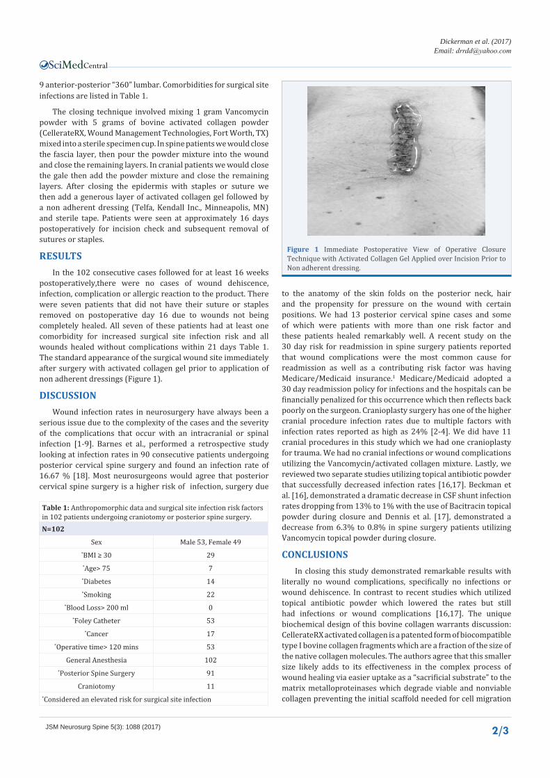
postoperatively,there were no cases of wound dehiscence, infection, complication or allergic reaction to the product. There were seven patients that did not have their suture or staples removed on postoperative day 16 due to wounds not being completely healed. All seven of these patients had at least one comorbidity for increased surgical site infection risk and all wounds healed without complications within 21 days Table 1. The standard appearance of the surgical wound site immediately after surgery with activated collagen gel prior to application of non adherent dressings (Figure 1).
DISCUSSIONWound infection rates in neurosurgery have always been a
serious issue due to the complexity of the cases and the severity of the complications that occur with an intracranial or spinal infection [1-9]. Barnes et al., performed a retrospective study looking at infection rates in 90 consecutive patients undergoing posterior cervical spine surgery and found an infection rate of 16.67 % [18]. Most neurosurgeons would agree that posterior cervical spine surgery is a higher risk of infection, surgery due
to the anatomy of the skin folds on the posterior neck, hair and the propensity for pressure on the wound with certain positions. We had 13 posterior cervical spine cases and some of which were patients with more than one risk factor and these patients healed remarkably well. A recent study on the 30 day risk for readmission in spine surgery patients reported that wound complications were the most common cause for readmission as well as a contributing risk factor was having Medicare/Medicaid insurance.1 Medicare/Medicaid adopted a 30 day readmission policy for infections and the hospitals can be financially penalized for this occurrence which then reflects back poorly on the surgeon. Cranioplasty surgery has one of the higher cranial procedure infection rates due to multiple factors with infection rates reported as high as 24% [2-4]. We did have 11 cranial procedures in this study which we had one cranioplasty for trauma. We had no cranial infections or wound complications utilizing the Vancomycin/activated collagen mixture. Lastly, we reviewed two separate studies utilizing topical antibiotic powder that successfully decreased infection rates [16,17]. Beckman et al. [16], demonstrated a dramatic decrease in CSF shunt infection rates dropping from 13% to 1% with the use of Bacitracin topical powder during closure and Dennis et al. [17], demonstrated a decrease from 6.3% to 0.8% in spine surgery patients utilizing Vancomycin topical powder during closure.
CONCLUSIONSIn closing this study demonstrated remarkable results with
literally no wound complications, specifically no infections or wound dehiscence. In contrast to recent studies which utilized topical antibiotic powder which lowered the rates but still had infections or wound complications [16,17]. The unique biochemical design of this bovine collagen warrants discussion: CellerateRX activated collagen is a patented form of biocompatible type I bovine collagen fragments which are a fraction of the size of the native collagen molecules. The authors agree that this smaller size likely adds to its effectiveness in the complex process of wound healing via easier uptake as a “sacrificial substrate” to the matrix metalloproteinases which degrade viable and nonviable collagen preventing the initial scaffold needed for cell migration
Table 1: Anthropomorphic data and surgical site infection risk factors in 102 patients undergoing craniotomy or posterior spine surgery.N=102
Sex Male 53, Female 49*BMI ≥ 30 29*Age> 75 7*Diabetes 14*Smoking 22
*Blood Loss> 200 ml 0*Foley Catheter 53
*Cancer 17*Operative time> 120 mins 53
General Anesthesia 102*Posterior Spine Surgery 91
Craniotomy 11*Considered an elevated risk for surgical site infection
Figure 1 Immediate Postoperative View of Operative Closure Technique with Activated Collagen Gel Applied over Incision Prior to Non adherent dressing.
Central
Dickerman et al. (2017)Email:
JSM Neurosurg Spine 5(3): 1088 (2017) 3/3
Dickerman R, Ashley SRNP, Winters K (2017) Operative Closure Technique Utilizing Bovine Collagen Fragments in a Prospective Analysis of 102 Consecutive Neurosurgery Patients. JSM Neurosurg Spine 5(2): 1088.
Cite this article
and ultimately prevents the formation of the extracellular matrix and granulation tissue i.e. healing [10-12]. Furthermore, as the bovine collagen is degraded the by-products are known to have chemotactic properties for a variety of cell types required for granulation tissue [10-12]. Thus, the fractionated size of this bovine collagen works directly by sacrificing itself as a substrate to the matrix metalloproteinases which inhibit healing and indirectly by its chemotactic properties attracting the specific cell types required for healing. We have continued the use of activated collagen powder and gel in our cases and recommend a multi-institutional study among several surgical specialties to further define its effectiveness in surgical cases.
ACKNOWLEDGEMENTSWe thank Julie E. Williamson for assistance in manuscript
preparation, data collection and coordinating patient appointments. University of North Texas Health Science Center and Wound Management Technologies for collaborative assistance with research staff and Presbyterian Hospital of Plano for clinical support staff.
REFERENCES1. Bernatz JT, Anderson PA. Thirty-day readmission rates in spine
surgery: systematic review and meta-analysis. Neurosurg Focus. 2015; 39: 7.
2. Chiang HY, Kamath AS, Pottinger JM, Greenlee JD, Howard MA, Cavanaugh JE, et al. Risk factors and outcomes associated with surgical site infections after craniotomy or craniectomy. J Neurosurg. 2014; 120: 509-521.
3. Riordan MA, Simpson VM, Hall WA. Analysis of factors contributing to infections after cranioplasty: A single-institution retrospective chart review. World Neurosurg. 2016; 87: 207-213.
4. von Lehe M, Kim HJ, Schramm J, Simon M. A comprehensive analysis of early outcomes and complication rates after 769 craniotomies in pediatric patients. Childs Nerv Syst. 2013; 29: 781-790.
5. Cheadle WG. Risk factors for surgical site infection. Surg Infect (Larchmt). 2006; 7: 7-11.
6. Fei Q, Li J, Lin J, Li D, Wang B, Meng H, et al. Risk factors for surgical site infection following spinal surgery: A meta-analysis. World Neurosurg.
2015; 5: 137-141.
7. Korol E, Johnston K, Waser N, Sifakis F, Jafri HS, Lo M, et al. A systematic review of risk factors associated with surgical site infections among surgical patients. PLoS One. 2013; 8: 83743.
8. Sørensen LT. Wound healing and infection in surgery: the pathophysiological impact of smoking, smoking cessation, and nicotine replacement therapy: a systematic review. Ann Surg. 2012; 255: 1069-1079.
9. Abdallah DY, Jadaan MM, Mc Cabe JP. Body mass index and risk of surgical site infection following spine surgery: a meta-analysis. Eur Spine J. 2013; 22: 2800-2809.
10. Kolenik SA, Mc Govern TW, Leffell DJ. Use of a lyophilized bovine collagen matrix in postoperative wound healing. Dermatol Surg. 1999; 25: 303-307.
11. Ciapetti G, Verri E, Granchi D, Cenni E, Gamberini S, Benetti D, et al. In vitro assessment of phagocytosis of bovine collagen by human monocytes/macrophages using a spectrophotometric method. Biomaterials. 1996; 17: 1703-1707.
12. Garwood CS, Kim PJ, Matal V, Steinberg JS, Evans KK, Mitnick CD, et al. The use of bovine collagen-glycosaminoglycan matrix for atypical lower extremity ulcers. Wounds. 2016; 28: 298-305.
13. Coban YK, Kalender AM. Treatment of gun-shot defect of the foot with bovine collagen matrix application. Foot (Edinb). 2009; 19: 222-223.
14. Uygur F, Evinc R, Ulkur E, Celikoz B. Use of lyophilized bovine collagen for split-thickness skin graft donor site management. Burns. 2008; 34: 1011-1014.
15. Yazici I, Cavusoglu T, Karakaya EL, Vural AC. Use of gleeful (lyophilized type 1 bovine collagen) pad dressing for spilt-thickenss graft donor area management. J CraniofacSurg. 2010; 21: 1662.
16. Beckman JM, Amankwah EK, Tetreault LL, Tuite GF. Reduction in CSF shunt infection over a 10-year period associated with the application of concentrated topical antibiotic powder directly to surgical wounds prior to closure. J Neurosurg Pediatric. 2015; 16: 648-661.
17. Dennis HH, Wei DT, Darren KZ, Shantakumar JT, Kumar N, Lau LL, et al. Is Intraoperative Local Vancomycin Powder the Answer to Surgical Site Infections in Spine Surgery? Spine (Phila Pa 1976). 2016.
18. Barnes M, Liew S. The incidence of infection after posterior cervical spine surgery: a 10 year review. Global Spine J. 2012; 2: 3-6.