50
Pulmonary Pathology Obstructive Airways Disease
| Date post: | 29-Dec-2015 |
| Category: |
Documents |
| Upload: | irma-phillips |
| View: | 224 times |
| Download: | 0 times |

Pulmonary Pathology
Obstructive Airways Disease

Respiratory disease
• Pulmonary diseases (especially infective) together with gastrointestinal infection are the commonest cause of death in the developing world
• Pulmonary disease is almost entirely environmental rather than genetic

Basic anatomy!
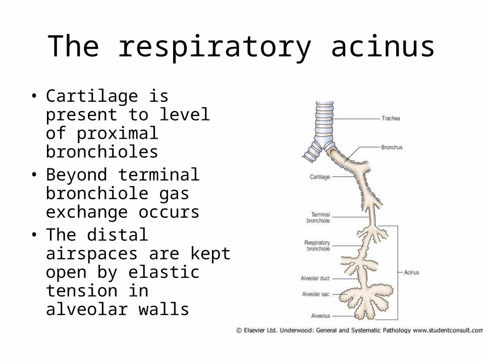
The respiratory acinus
• Cartilage is present to level of proximal bronchioles
• Beyond terminal bronchiole gas exchange occurs
• The distal airspaces are kept open by elastic tension in alveolar walls

Function of lungs….
• Gas exchange (O2, CO2)– Depends on compliance (stretchability) of
lungs– Can only occur in alveoli that are both
ventilated and perfused

Ventilation-perfusion defects
• Alveoli that are ventilated but not perfused is ventilatory “dead space”
• Alveoli that are perfused but not ventilated leads to “shunting” of non-oxygenated blood from pulmonary to systemic circulation ( a mechanism of cyanosis)
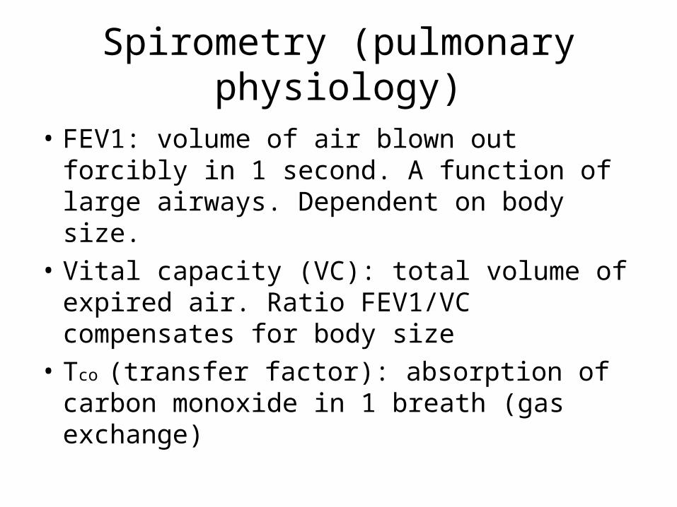
Spirometry (pulmonary physiology)
• FEV1: volume of air blown out forcibly in 1 second. A function of large airways. Dependent on body size.
• Vital capacity (VC): total volume of expired air. Ratio FEV1/VC compensates for body size
• Tco (transfer factor): absorption of carbon monoxide in 1 breath (gas exchange)
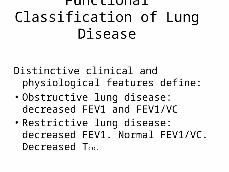
Functional Classification of Lung Disease
Distinctive clinical and physiological features define:
• Obstructive lung disease: decreased FEV1 and FEV1/VC
• Restrictive lung disease: decreased FEV1. Normal FEV1/VC. Decreased Tco.

Respiratory failure (causes)
• Ventilation defects (CNS, neuromuscular defects, drugs)
• Perfusion defects (cardiac failure, pulmonary emboli)
• Gas exchange defects (fibrosis, consolidation, emphysema)
Lead to hypoxia and hypercapnia
Often more than factor one will operate
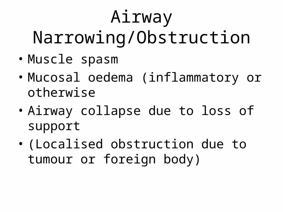
Airway Narrowing/Obstruction
• Muscle spasm
• Mucosal oedema (inflammatory or otherwise
• Airway collapse due to loss of support
• (Localised obstruction due to tumour or foreign body)

Localised obstruction
• Collapse• Lipid pneumonia• Infection• Bronchiectasis (if
longstanding)
Main Categories of (diffuse) Obstructive Disease
• Asthma
• Chronic obstructive pulmonary disease (COPD/COAD/COLD)

Chronic Obstructive Disease
• Chronic bronchitis
• Emphysema
Symptomatic patients often have both

Bronchial Asthma
A chronic inflammatory disorder characterised by hyperreactive airways leading to episodic reversible bronchoconstriction

Asthma
• Extrinsic - response to inhaled antigen
• Intrinsic - non-immune mechanisms (cold, exercise, aspirin)
Immunological Mechanisms
Type hypersensitivity - allergen binds to IgE on surface of mast cells
• Degranulation (histamine)– muscle spasm– inflammatory cell influx (eosinophils)– mucosal inflammation/oedema
• Inflammatory infiltrate tends to chronicity

Pathology of asthma
• Airway inflammation with mucosal oedema• Mucus plugging


Mucosal oedema
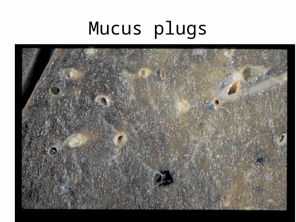
Mucus plugs

Mucus plug/inflammation
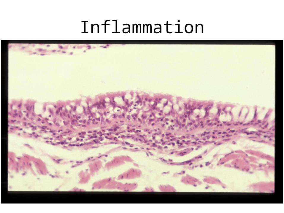
Inflammation
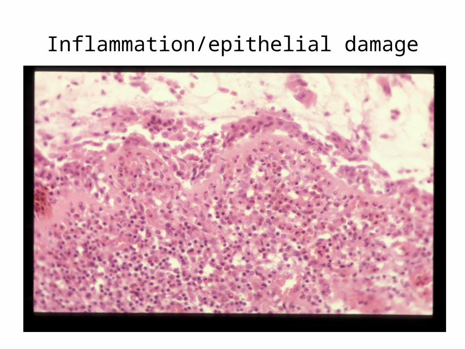
Inflammation/epithelial damage

Chronic Obstructive Pulmonary Disease
• Chronic bronchitis
• Emphysema
A smokers disease
Symptomatic patients usually have both
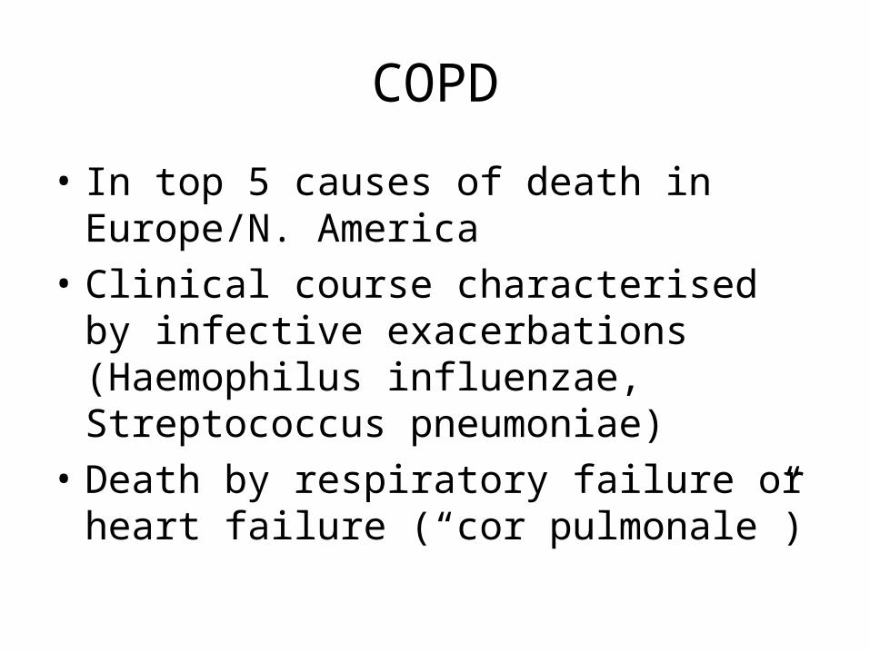
COPD
• In top 5 causes of death in Europe/N. America
• Clinical course characterised by infective exacerbations (Haemophilus influenzae, Streptococcus pneumoniae)
• Death by respiratory failure or heart failure (“cor pulmonale”)

Chronic BronchitisCough productive of sputum on most
days for 3 months of at least 2 successive years
• An epidemiological definition
• Does not imply airway inflammation
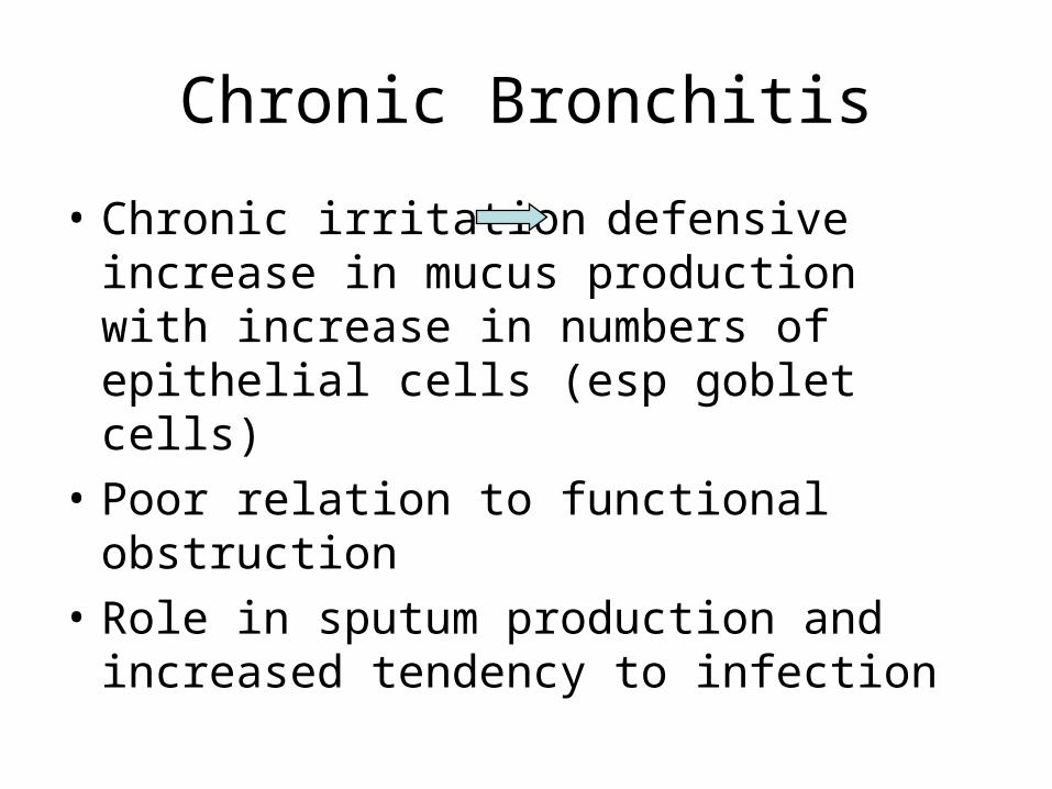
Chronic Bronchitis
• Chronic irritation defensive increase in mucus production with increase in numbers of epithelial cells (esp goblet cells)
• Poor relation to functional obstruction
• Role in sputum production and increased tendency to infection

Chronic Bronchitis
• Non-reversible obstruction
• In some patients there may be a reversible (“asthmatic”) component
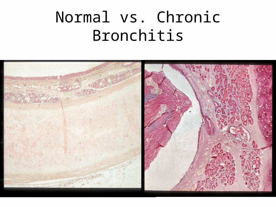
Normal vs. Chronic Bronchitis

Small airways in Chronic Bronchitis
• More important than traditionally realised
• Goblet cell metaplasia, macrophage accumulation and fibrosis around bronchioles may generate functional obstruction
Emphysema
• Increase beyond the normal in the size of the airspaces distal to the terminal bronchiole
• Without fibrosis
The gas-exchanging compartment of the lung
Emphysema (types)
• Centriacinar (centrilobular)
• Panacinar
• Others (e.g. localised around scars in the lung)

Emphysema
• Difficult to diagnose in life (apart from late disease – enl;arged “barrel chest”)
• Radiology (CT) can show changes in lung density
• Correlation with function known from autopsy studies

Emphysema
• “Dilatation” is due to loss of alveolar walls (tissue destruction)
• Appears as “holes” in the lung tissue
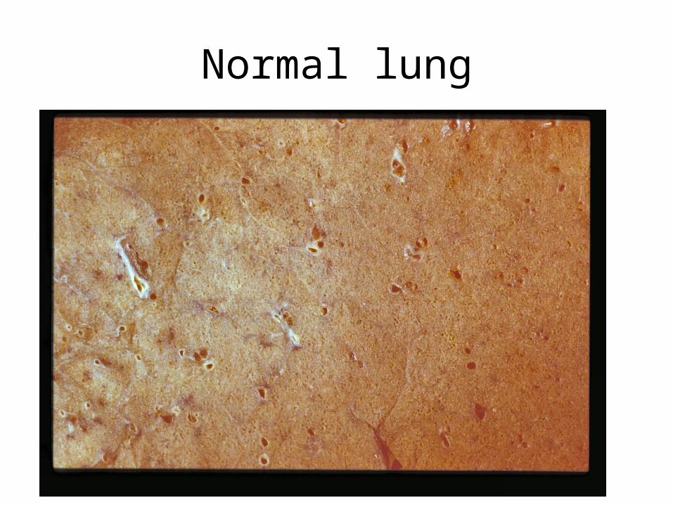
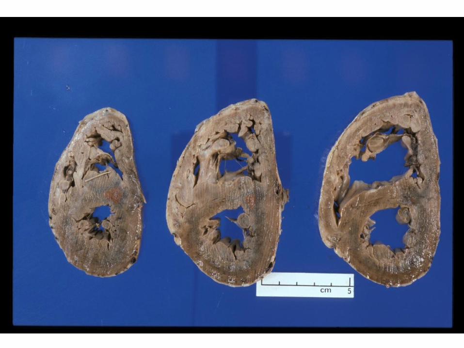
Normal lung

Centriacinar emphysema
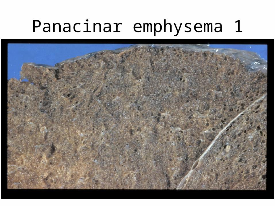
Panacinar emphysema 1
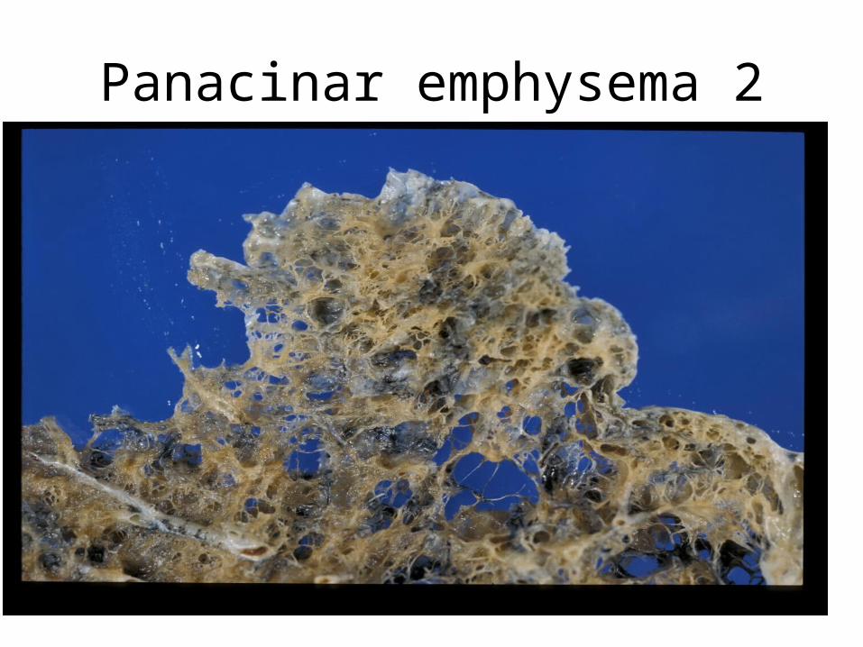
Panacinar emphysema 2
Emphysema
How do these changes relate to functional deficit?
• Poorly at macroscopic level
• Better with microscopic measurement

Normal

Early emphysema
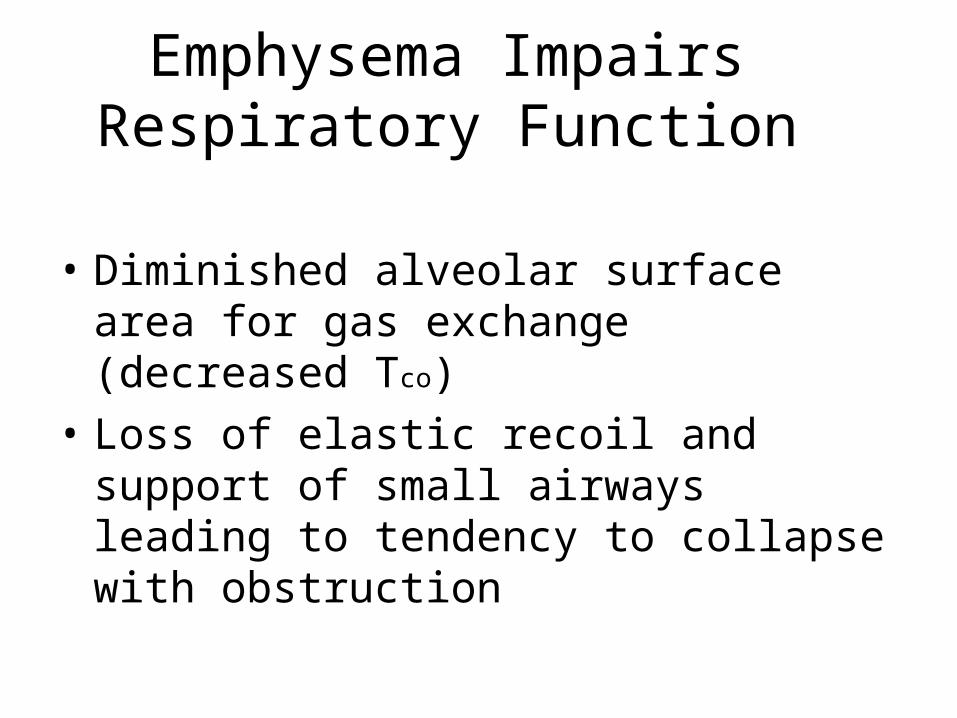
Emphysema Impairs Respiratory Function
• Diminished alveolar surface area for gas exchange (decreased Tco)
• Loss of elastic recoil and support of small airways leading to tendency to collapse with obstruction

Loss of surface area (emphysema)

Loss of support on bronchiolar walls

As disease advances….
Pa O2 leads to:
• Dyspnoea and increased respiratory rate
• Pulmonary vasoconstriction (and pulmonary hypertension)

Epidemiology of COPD
• Smoking
• Atmospheric pollution
• Genetic factors
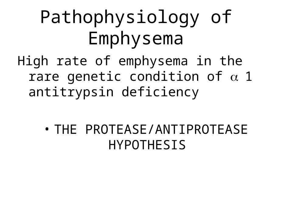
Pathophysiology of Emphysema
High rate of emphysema in the rare genetic condition of 1 antitrypsin deficiency
• THE PROTEASE/ANTIPROTEASE HYPOTHESIS
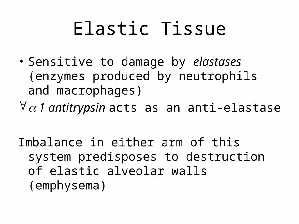
Elastic Tissue
• Sensitive to damage by elastases (enzymes produced by neutrophils and macrophages)
1 antitrypsin acts as an anti-elastase
Imbalance in either arm of this system predisposes to destruction of elastic alveolar walls (emphysema)
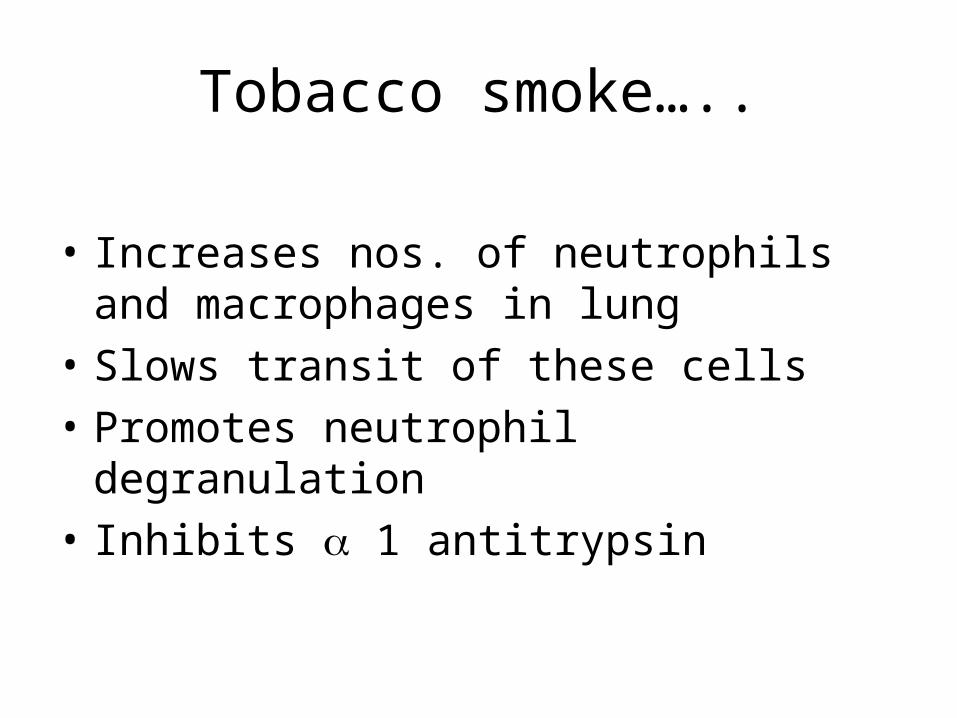
Tobacco smoke…..
• Increases nos. of neutrophils and macrophages in lung
• Slows transit of these cells
• Promotes neutrophil degranulation
• Inhibits 1 antitrypsin


