27
Quality Assurance Methods In Proton Beam Therapy JAMES GOLBOURN

Quality Assurance Methods In Proton Beam TherapyJAMES GOLBOURN
Introduction
u Proton Beam Therapy Background
u Beam characteristics
u What is Quality Assurance?
u QA at UCLH
u Commercially available QA machines and examples of clinical QA procedures
u Methods for measuring beam characteristics

Proton Beam Therapy Background
u An advanced form of radiotherapy that uses proton beams to target cancerous tumours.
u The advantage compared to traditional radiotherapy arises from Bragg curve behaviour of protons.
u Maximum proton energy is deposited at peak of curve.
Taken from T Mitin, AL Zietman, Promise and pitfalls of heavy-particle therapy, J Clin Oncol, 2014 Sep 10, 32(26):2855-63.Taken from Paganetti, H, Proton Therapy Physics, 2012,
CRC Press.

Proton Beam Therapy Background
u A series of pristine Bragg peaks can be combined to treat an extended target volume.
u Result is a spread- out Bragg peak (SOBP).
u Homogenous dose distribution across SOBP.
u Range modulators are used to vary the proton energy.
u Typical proton energy range 70 –250 MeV.
Taken from Trikalinos TA, Terasawa T, Ip S, et al. Particle Beam Radiation Therapies for Cancer [Internet]. Rockville (MD): Agency for Healthcare Research and Quality (US); 2009 Nov. (Comparative Effectiveness Technical Briefs, No. 1.) Figure 1, Depth-dose distributions for a
Taken from Paganetti, H, Proton Therapy Physics, 2012, CRC Press.

Method of Beam Production
Cyclotronsu Nearly continuous beam at
constant energy.
u Protons at the maximum energy are extracted.
u Beam degraded to lower energy.
u Collimators, slits and magnets are required to restore the beam quality.
Synchrotronsu Spills of protons of variable energy
are produced.
u The injected protons are accelerated and then extracted at the desired energy.
u Each proton spill lasts for several seconds.
u Excess protons are decelerated.
Taken from Paganetti, H, Proton Therapy Physics, 2012, CRC Press.
Methods of Beam Productionu Advantages and disadvantages to both methods.
u Synchrotrons are much larger than cyclotrons (about 6-8m in diameter) leading to spacial issues.
u Beam quality has to be degraded for cyclotrons.
u No degradation required for synchrotrons.
u Following acceleration the beams are transported to treatment rooms.
u The beams are focussed by magnets during the transportation phase to maintain beam quality.
Taken from Paganetti, H, Proton Therapy Physics, 2012, CRC Press.

Beam Parameters and Characteristics
u Beam energy – corresponds to beam range in the body.
u Beam energy and angular spread – related to distal fall off and energy straggling in tissue.
u Spot size – area covered by beam in PBS.
u Average beam intensity – relates to the dose patient receives.
Taken from Paganetti, H, Proton Therapy Physics, 2012, CRC Press.

What is Quality Assurance?
u Quality assurance (QA) is necessary to ensure the proton beam system is operating as intended.
u A QA system will find the problems and defects in a system before patient treatment begins.
u QA for X-ray radiotherapy is well established.
u PBT facilities have different QA systems in place with different checks making up the process.
u QA can be split into daily, monthly and annual QA depending on the failure criticality of that particular check.
u This project focusses on daily QA comprising of the most critical tests.

Properties of QA Systems
u An aspect of PBT QA systems is measuring beam properties.
u The QA should therefore be a detector or series of detectors that measure these properties.
u The QA checks should be comprehensive and measurements should be to the required precision.
u The QA system should be integrated and be able to carry out measurements consecutively.
u Needs to ensure the system runs from start to finish as planned.
u Uses patient specific information to carry out tests.
Taken from Paganetti, H, Proton Therapy Physics, 2012, CRC Press.
QA at UCLHu UCLH PBT centre set to open in 2020.u Christie Hospital in Manchester treated first patient in December
2018.u Discussion with Alison Warry, Principal Radiotherapy Physicist, Proton
Beam Therapy at UCLH. u What features they want from a daily QA system.u Criticisms of commercially available QA systems.
Taken from NHS Proton Beam Therapy Website https://www.england.nhs.uk/commissioning/spec-services/highly-spec-services/pbt/

UCLH daily QA
Beam QAu Range consistency
u Spot position
u Spot shape
u Variation with gantry angle
* Each parameter should be checked with 2/3 beam energies.
Machine QAu Safety interlocks
u Communication between components
u Imaging system
u Patient support system

Commercially available machines: Sun Nuclear QA3
u Designed to measure photon and electron beams but can be modified to measure proton beam characteristics.
u Method for modification described by X Ding Et al, A Novel QA system for proton therapy, J Appl Clin Med Phys. 2013 Mar; 14(2): 115–126.
u Can be used for PBS and US.
u Components:u 13 parallel plate ionization chambers – 5 are used.
u In house acrylic phantom
u Blank acrylic compensator
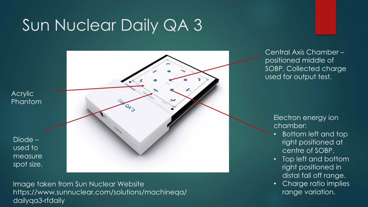
Sun Nuclear Daily QA 3Central Axis Chamber –positioned middle of SOBP. Collected charge used for output test.
Electron energy ion chamber:• Bottom left and top
right positioned at centre of SOBP.
• Top left and bottom right positioned in distal fall off range.
• Charge ratio implies range variation.
Acrylic Phantom
Image taken from Sun Nuclear Website https://www.sunnuclear.com/solutions/machineqa/dailyqa3-rfdaily
Diode –used to measure spot size.

Sun Nuclear Daily QA SummaryMeasurement Component Details Tolerance
Output CAX Charge collected related to MU dose.
<3%
Beam range E chambers Ratio of charges to find fall off
<1.0mm
Symmetry E chambers Compare charge in each chamber.
<3%
• QA system takes less than 20 minutes.• Can be incorporated into a larger QA system for further checks:
• Couch alignment• Laser alignment• Image registration
Criticism and Improvement on Sun Nuclear QA3
u There are complexities associated with PBS.u Need to account for spot size and position variations.
u Due to beam optics
u Daily QA in proton therapy using a single commercially available detector, Jl Lambert Et al describes an improved method.
u Spot size and position can be measured by directing spot at innermost diode of triplet and measuring charge in diodes.
u Similar tolerances.
u Longer QA time of about 30 minutes.
u Mayo Clinic uses this and is discussed in Technical Note: An efficient daily QA procedure for proton pencil beam scanning, J Younkin et al 2018, Phys. 45 1040.

Commercially available machines:IBA Zebra
u 2D ionization chamber made up of 180 parallel plate chambers.
u Developed for X-ray therapy but can be adapted for PBT.u Discussion taken from Quality assurance of proton beams using a
multilayer ionization chamber system, September 2013, Medical Physics 40(9):092102.
u Primarily used to measure the dose distribution in tissue:u Range - depth of 90% of the dose
u SOBP length - proximal 95% and distal 90%
u Distal dose fall off – between distal 80% and 20%
u Can be used for scanned and scattered beams.

IBA ZebraSpecifications:• 180 parallel plate ionization chambers• Each plate has a resolution of 2mm• Spacing of 1mm
Method:• Chambers irradiated for 60s.• Carried out for radiation fields up to a
maximum 18 cm x 18 cm.• Depth dose distribution compared to
measured values:• For scattered beams compared with a
scanned water tank.• For scanned beams compared with data
from a Bragg peak chamber from commissioning.
Image taken from IBA website
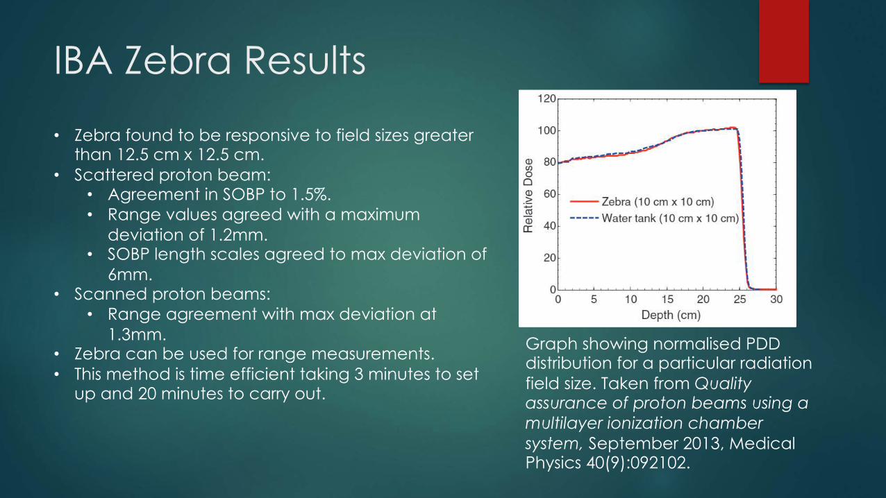
IBA Zebra Results
Graph showing normalised PDD distribution for a particular radiation field size. Taken from Quality assurance of proton beams using a multilayer ionization chamber system, September 2013, Medical Physics 40(9):092102.
• Zebra found to be responsive to field sizes greater than 12.5 cm x 12.5 cm.
• Scattered proton beam:• Agreement in SOBP to 1.5%.• Range values agreed with a maximum
deviation of 1.2mm.• SOBP length scales agreed to max deviation of
6mm.• Scanned proton beams:
• Range agreement with max deviation at 1.3mm.
• Zebra can be used for range measurements.• This method is time efficient taking 3 minutes to set
up and 20 minutes to carry out.
Commercially available machines: IBA Matrixx
u Universal detector array.
u IBA’s fastest and most accurate detector for QA.
u Initially designed for conventional and IMRT
u Composition:u 1020 parallel plate ionization chambers in a 32 x 32 array.
u Highly sensitive: 1.4nC/Gy
u Example method taken from Use of a two-dimensional ionization chamber array for proton therapy beam quality assurance, B Arjomandy Et al, Med Phys. 2008 Sep;35(9):3889-94.

IBA Matrixx
u Method looks at suitability of using parallel plate ion chamber array for PBT.
u Measurements carried out:
u Beam flatness
u Beam Symmetry
u Compared results to those obtained with film dosimetry and ion chamber in water.
u Result:
u Excellent agreement between Matrixx measurement and comparison with other methods.
u Matrixx can be used to measure dosimetric properties.

IBA Matrixx
u Taken from Quality assurance of carbon ion and proton beams: A feasibility study for using the 2D MatriXX detector, M Donetti Et al,Phys Med. 2016 Jun;32(6):831-7. doi: 10.1016/j.ejmp.2016.05.058. Epub 2016 May 28.
u Paper further confirms that Matrixx can be used to replace films for measurements.
u Advantages: Quicker and almost instant results, good agreement with film results.
u There is also possibility to measure dose output but method not explored in detail.
u Conclusion: A good device when combined with others.

Beam Energy
u Beam energy is directly related to the range of the beam in tissue. There are several ways to measure the beam energy.
u Ionization chambers:
u Multilayer ionization chamber
u Thimble chamber
u Calorimeters – e.g graphite.
u PIN diode stacks

Spot Size and Symmetry
u Spot size and symmetry QA can be required for PBS.
u Spot size:
u Ionization chambers – Gaussian produced in chamber and can obtain width [1].
u Plastic scintillator fibre - photon output from a scintillating screen. Better than ionization chambers due to faster response and higher spatial resolution [2].
u Symmetry:
u Using ionization chambers – method discussed using Sun Nuclear QA3 [3].

Scintillation Screens and Ionization Chambers
u Both can be used to check:
u Range
u Spot position
u Shape
u Scintillation screens have a greater resolution but are more time consuming.

Criticisms of commercially available QA systems
u These commercial machines are fragile – cannot withstand knocks and bangs.
u Expensive to then replace, for example IBA machines cost £100,000’s and might have to be replaced twice a year.
u Multiple detectors needed to carry out a thorough QA analysis.
u Often difficult to use.
u Estimate from IBA of about 30 minutes per room for QA: often there are multiple rooms wanting access to beam accelerator so there is time competition.

Ideal UCLH QA System
u Ideal QA for UCLH:u Time efficient – 20 minutes.
u Cost effective - £10,000 to buy.
u Robust – in a container.
u A single detector capable of multiple consecutive measurements.
Taken from UCLH website: https://www.uclh.nhs.uk/News/Pages/UCLHpartnerswithUScompany.aspx

Thank You


