Quinone compounds regulate the level of ROS production by the NADPH oxidase Nox4 Minh Vu Chuong Nguyen a,1, *, Bernard Lardy a,d,1 , Francis Rousset a , Florence Hazane-Puch e , Leilei Zhang a , Candice Trocme ´ d , Lena Serrander b , Karl-Heinz Krause c , Franc ¸oise Morel a a Universite ´ Joseph Fourier, GREPI AGIM FRE 3405, CNRS, EPHE, Grenoble, France b Department of Clinical Microbiology, University Hospital, Linkoping, Sweden c Department of Pathology and Immunology, Medical Faculty and University Hospital, Geneva, Switzerland d Laboratoire de Biochimie Enzyme et Prote ´ine-DBTP, Institut de Biologie et de Pathologie, University Hospital CHU-Grenoble, Grenoble, France e Laboratoire de Biochimie Hormonale et Nutritionnelle-DBTP, Institut de Biologie et de Pathologie, University Hospital CHU-Grenoble, Grenoble, France 1. Introduction Quinone derivative compounds induce a broad spectrum of effects in humans and represent a class of toxicological inter- mediates, which may induce acute cytotoxicity, immunotoxicity, and carcinogenesis, and lead to cellular damage [1]. They can be formed from benzene or polycyclic aromatic hydrocarbons and are toxicologically important components of air pollution [2] and cigarette smoke [3]. Among the enzymes that modulate quinone toxicity, the NAD(P)H quinone oxidoreductase type 1 (NQO1; EC 1.6.99.2) is one of the most important enzymes. NQO1 is a homodimeric flavoenzyme that catalyses the obligatory two- electron reduction of quinones to hydroquinones [4]. NQO1 activity can protect animal cells from the deleterious and carcinogenic effects of quinones by preventing the one-electron reduction of quinones by other reductases; however, prolonged exposure to environmental contamination, which contains qui- nones, damages pulmonary tissue and leads to airway inflamma- tion and pathologies through an oxidative stress mechanism [5,6]. NQO1 is mainly cytosolic but it has been described to be expressed at plasma membrane [7]. Semiquinone radicals of inhaled airborne particulate matter are believed to cause oxidative stress by generating reactive oxygen species (ROS), as reported in the lung [8]. Furthermore, diesel exhaust particles composed of polycyclic Biochemical Pharmacology 85 (2013) 1644–1654 A R T I C L E I N F O Article history: Received 3 February 2013 Accepted 29 March 2013 Available online 9 April 2013 Keywords: NADPH oxidase Nox4 NAD(P)H:quinone oxidoreductase NQO1 Quinones Redox regulation of Nox Reactive oxygen species (ROS) A B S T R A C T NADPH oxidase Nox4 is expressed in a wide range of tissues and plays a role in cellular signaling by providing reactive oxygen species (ROS) as intracellular messengers. Nox4 oxidase activity is thought to be constitutive and regulated at the transcriptional level; however, we challenge this point of view and suggest that specific quinone derivatives could modulate this activity. In fact, we demonstrated a significant stimulation of Nox4 activity by 4 quinone derivatives (AA-861, tBuBHQ, tBuBQ, and duroquinone) observed in 3 different cellular models, HEK293E, T-REx TM , and chondrocyte cell lines. Our results indicate that the effect is specific toward Nox4 versus Nox2. Furthermore, we showed that NAD(P)H:quinone oxidoreductase (NQO1) may participate in this stimulation. Interestingly, Nox4 activity is also stimulated by reducing agents that possibly act by reducing the disulfide bridge (Cys226, Cys270) located in the extracellular E-loop of Nox4. Such model of Nox4 activity regulation could provide new insight into the understanding of the molecular mechanism of the electron transfer through the enzyme, i.e., its potential redox regulation, and could also define new therapeutic targets in diseases in which quinones and Nox4 are implicated. ß 2013 Elsevier Inc. All rights reserved. Abbreviations: AA-861, 2-(12-hydroxydodeca-5,10-diynyl)-3,5,6-trimethyl-p-ben- zoquinone; BQ, benzoquinone; CGD, chronic granulomatous disease; CHX, cycloheximide; DPI, diphenyliodonium; Duroquinone, 2,3,5,6-tetramethyl-1,4- benzoquinone; HQ, hydroquinone; tMetBQ, 2,5-dimethyl-1,4-benzoquinone; LDH, lactate dehydrogenase; 5-LO, 5-lipoxygenase; NQO1, NAD(P)H:quinone oxidoreductase 1; PMA, phorbol 12-myristate 13-acetate; PMN, polymorphonu- clear cells; RLU, relative light unit; ROS, reactive oxygen species; tBuBHQ, 2,5-di- tert-butyl-1,4-benzo-hydroquinone; and tBuBQ, 2,5-di-tert-butyl-1,4-benzoqui- none. * Corresponding author at: GREPI AGIM FRE 3405, CNRS, Universite ´ Joseph Fourier, EPHE, CHU Albert Michallon, BP217, 38043 Grenoble, France. Tel.: +33 4 76 76 57 52; fax: +33 4 76 76 62 51. E-mail address: [email protected](M.V.C. Nguyen). 1 Equal contributors. Contents lists available at SciVerse ScienceDirect Biochemical Pharmacology jo u rn al h om epag e: ww w.els evier.c o m/lo cat e/bio c hem p har m 0006-2952/$ – see front matter ß 2013 Elsevier Inc. All rights reserved. http://dx.doi.org/10.1016/j.bcp.2013.03.023
Transcript
Biochemical Pharmacology 85 (2013) 1644–1654
Quinone compounds regulate the level of ROS production by theNADPH oxidase Nox4
Minh Vu Chuong Nguyen a,1,*, Bernard Lardy a,d,1, Francis Rousset a,Florence Hazane-Puch e, Leilei Zhang a, Candice Trocme d, Lena Serrander b,Karl-Heinz Krause c, Francoise Morel a
a Universite Joseph Fourier, GREPI AGIM FRE 3405, CNRS, EPHE, Grenoble, Franceb Department of Clinical Microbiology, University Hospital, Linkoping, Swedenc Department of Pathology and Immunology, Medical Faculty and University Hospital, Geneva, Switzerlandd Laboratoire de Biochimie Enzyme et Proteine-DBTP, Institut de Biologie et de Pathologie, University Hospital CHU-Grenoble, Grenoble, Francee Laboratoire de Biochimie Hormonale et Nutritionnelle-DBTP, Institut de Biologie et de Pathologie, University Hospital CHU-Grenoble, Grenoble, France
A R T I C L E I N F O
Article history:
Received 3 February 2013
Accepted 29 March 2013
Available online 9 April 2013
Keywords:
NADPH oxidase Nox4
NAD(P)H:quinone oxidoreductase NQO1
Quinones
Redox regulation of Nox
Reactive oxygen species (ROS)
A B S T R A C T
NADPH oxidase Nox4 is expressed in a wide range of tissues and plays a role in cellular signaling by
providing reactive oxygen species (ROS) as intracellular messengers. Nox4 oxidase activity is thought to
be constitutive and regulated at the transcriptional level; however, we challenge this point of view and
suggest that specific quinone derivatives could modulate this activity. In fact, we demonstrated a
significant stimulation of Nox4 activity by 4 quinone derivatives (AA-861, tBuBHQ, tBuBQ, and
duroquinone) observed in 3 different cellular models, HEK293E, T-RExTM, and chondrocyte cell lines. Our
results indicate that the effect is specific toward Nox4 versus Nox2. Furthermore, we showed that
NAD(P)H:quinone oxidoreductase (NQO1) may participate in this stimulation. Interestingly, Nox4
activity is also stimulated by reducing agents that possibly act by reducing the disulfide bridge (Cys226,
Cys270) located in the extracellular E-loop of Nox4. Such model of Nox4 activity regulation could provide
new insight into the understanding of the molecular mechanism of the electron transfer through the
enzyme, i.e., its potential redox regulation, and could also define new therapeutic targets in diseases in
which quinones and Nox4 are implicated.
� 2013 Elsevier Inc. All rights reserved.
Contents lists available at SciVerse ScienceDirect
Biochemical Pharmacology
jo u rn al h om epag e: ww w.els evier .c o m/lo cat e/bio c hem p har m
1. Introduction
Quinone derivative compounds induce a broad spectrum ofeffects in humans and represent a class of toxicological inter-mediates, which may induce acute cytotoxicity, immunotoxicity,
0006-2952/$ – see front matter � 2013 Elsevier Inc. All rights reserved.
http://dx.doi.org/10.1016/j.bcp.2013.03.023
and carcinogenesis, and lead to cellular damage [1]. They can beformed from benzene or polycyclic aromatic hydrocarbons and aretoxicologically important components of air pollution [2] andcigarette smoke [3]. Among the enzymes that modulate quinonetoxicity, the NAD(P)H quinone oxidoreductase type 1 (NQO1; EC1.6.99.2) is one of the most important enzymes. NQO1 is ahomodimeric flavoenzyme that catalyses the obligatory two-electron reduction of quinones to hydroquinones [4]. NQO1activity can protect animal cells from the deleterious andcarcinogenic effects of quinones by preventing the one-electronreduction of quinones by other reductases; however, prolongedexposure to environmental contamination, which contains qui-nones, damages pulmonary tissue and leads to airway inflamma-tion and pathologies through an oxidative stress mechanism [5,6].NQO1 is mainly cytosolic but it has been described to be expressedat plasma membrane [7]. Semiquinone radicals of inhaled airborneparticulate matter are believed to cause oxidative stress bygenerating reactive oxygen species (ROS), as reported in the lung[8]. Furthermore, diesel exhaust particles composed of polycyclic
aromatic hydrocarbons and quinones are involved in a cellular ROSproduction associated with lung function impairment via a Nox4redox-dependent mechanism [9].
Nox4 belongs to the NADPH oxidase family that contains 7members [10–12]. The function of those enzymes is exclusivelydedicated to the production of ROS, which are essential signalingmolecules. Primary discovered in kidney tissue [13], Nox4 appearsto be ubiquitously distributed. Its dysfunction has been linked toseveral pathologies including hypertension [14], diabetes [15],atherosclerosis [16], cancer [17], osteoarthritis [18] and inflam-mation [19], and Nox4 represent a potential therapeutic target[20]. Despite its wide distribution, its activation mechanisms atthe molecular level are unclear. It is of therapeutic interest toelucidate the mechanism of Nox4 NADPH oxidase activity. Whileactivity of Nox1, Nox2, and Nox3 largely depends on the presenceof cytosolic activator or organizer subunits, no well-knownNADPH oxidase partners, beside p22phox, have been identified orshown to activate Nox4 [21–23]. However, two partners of Nox4have been described recently: the protein disulfide isomerase(PDI) and the polymerase DNA directed-delta-interacting protein(Poldip2). Nox4 is unique among other Nox isoenzymes in that itsactivity is constitutive and may depend on a specific conformationof the dehydrogenase DH domain that should allow a spontaneoustransfer of electrons from NADPH to FAD and to the hemes [24,25].Data from various studies indicated clearly that Nox4 activity isregulated at the mRNA level, implying that an increase or decreaseof ROS production by Nox4 is correlated to an up regulation[9,16,26–33] or to a decline [34] of Nox4 transcripts. Although ithas not been reported yet, post-translational regulation of Nox4oxidase activity cannot be excluded.
In this study, we provide the first evidence that Nox4 activitycan be modulated independently of both transcriptional andtranslational processes. We showed a dual effect of quinonecompounds, as being activators or inhibitors of Nox4 ROSproduction depending on their redox potential (E). We suggestthat quinones derivatives could modulate Nox4 activity by a redoxregulation pathway. Moreover, NQO1 is introduced as anintermediate between quinones and Nox4 activation, suggestinga functional interaction between Nox4 and NQO1. The resultsargue in favor of a modulation of Nox4 activity by a new family ofchemical compounds and suggest a potential role of Nox4 inpathologies implying quinone toxicity.
2. Materials and methods
2.1. Materials
Polyclonal antibody against 5-lipoxygenase (5-LO) (Cayman, AnnArbor, USA) was a generous gift from F. Stanke (Grenoble, France).pEF6V5/HisB vector, penicillin, streptomycin, L-glutamine, Dulbec-co’s modified Eagle’s medium (DMEM), fetal bovine serum (FBS),trypsin, Earles’s balanced salts (EBSS), geneticin, Fluo-3/AM, BAPTA/AM, and TRIzol1 were purchased from Invitrogen (Cergy Pontoise,France). Blasticidin was from Funakoshi Co. (Japan); AMV reversetranscriptase was from QBiogene (Illkirch, France). Effectenetransfection reagent was from Qiagen (Courtaboeuf, France).Luminol, ionomycin, horseradish peroxidase, thapsigargin, diphe-nyleneiodonium chloride (DPI), rotenone, Nv-nitro-L-argininemethyl ester hydrochloride (L-NAME), tiron, xanthine, benzoqui-none (BQ), hydroquinone (HQ), 2,5-dimethyl-1,4-benzoquinone(tMetBQ), duroquinone, 2,5-di-tert-butyl-1,4-benzo-hydroquinone(tBuBHQ), 2,5-di-tert-butyl-1,4-benzoquinone (tBuBQ), 2-(12-hydroxydodeca-5,10-diynil)-3,5,6-trimethyl-p-benzoquinone (AA-861), 3,30-methylene-bis-4-hydroxycoumarin (Dicoumarol), cyclo-heximide (CHX) compounds were purchased from Sigma (SaintQuentin Fallavier, France). b-Mercaptoethanol was purchased from
Carlo Erba (Val de Reuil, France). LightCycler FastStart DNA Masterplus SYBR Green I kit, protease inhibitors cocktail tablet, tosyl-lysine-chloromethyl ketone (TLCK), NADPH, NADH, xanthineoxidase, and lactate dehydrogenase (LDH) optimized kits werepurchased from Roche (Meylan, France). Leupeptin, pepstatin, andECL reagents were from GE Healthcare (Orsay, France). UNI-ZAPhuman kidney lambda cDNA library was purchased from Stratagene(La Jolla, CA). Housekeeping gene GAPDH was purchased from BDBioscience (Pont de Claix, France). SuperScriptIII first-strandsynthesis was obtained from Life Technologies (Saint Aubin, France).
2.2. Cell culture
Nox4 T-RExTM cells were a generous gift from K.H. Krause(Department of Pathology and Immunology, Geneve, Suisse) andwere generated from HEK293 cell (Invitrogen). The HEK293E cellline was purchased from Invitrogen (Cergy Pontoise, France). PLB-985 human myeloid cell lines (wild type PLB-985 WT) or knock outfor Nox2 (PLB-985 KO-Nox2) were a generous gift from M. Dinauer(Department of Pediatrics, St. Louis, USA). PLB-985 cells werecultured at 37 8C under a 5% CO2 atmosphere in RPMI 1640medium containing 2 mM L-glutamine supplemented with 10%fetal bovine serum and with 1% penicillin/streptomycin. TheHEK293E cell line was maintained in DMEM supplemented with10% (v/v) fetal bovine serum (FBS), 100 units/mL penicillin,100 mg/mL streptomycin, 2 mM L-glutamine at 37 8C in a 5% CO2
humidified atmosphere. Five mg/mL Blasticidin was added to theculture medium of cells transfected with the mammalianexpression plasmid pEF6V5/HisB.
The T-RExTM system was used for Tetracycline-inducibleexpression of the genes of interest. T-RExTM cells stably expressingTet repressor were selected by 5 mg/mL Blasticidin and maintainedin DMEM supplemented with 10% (v/v) FBS, 2 mM L-glutamine at37 8C in a 5% CO2 humidified atmosphere. T-RExTM Nox4 cells ascompared to T-RExTM WT are able to induce Nox4 expression. Bothcells were selected with 5 mg/mL blasticidin and 400 mg/mL G418.Nox4 expression was induced by the addition of 1 mg/mLtetracycline in the culture medium. Experiments were performedafter the incubation at set times.
2.3. Isolation of human neutrophils
As previously reported [35], human neutrophils were isolatedfrom citrated venous blood of healthy volunteers. Blood sampleswere diluted twice in PBS (137 mM NaCl, 2.7 mM KCl, 1.5 mMKH2PO4, 8 mM Na2HPO4 pH 7.3) containing 1% (w/v) tri-sodiumcitrate, using a 33% (v/v) Hypaque–Ficoll gradient. After 20 min ofcentrifugation at 800 � g at 20 8C, the pellet was submitted to ahypotonic lysis for 5–15 min in ice. After 5 min of centrifugation at350 � g at 4 8C, the neutrophil pellet was collected and washedonce in PBS. Neutrophils were suspended in PBS containing 0.2%(w/v) BSA and 0.5 mM CaCl2 at a concentration of 107 cells/mL andused for superoxide measurement by chemiluminescence andprotein extraction.
2.4. Generation of plasmid constructs for Nox4 and NQO1 isoforms
expression
Two Nox4 cDNAs were characterized by PCR using UNI-ZAPhuman kidney lambda cDNA library (Stratagene) as template.Nox4A corresponds to the full length Nox4 usually described(GenbankTM accession number: AF254621), and Nox4B is the 14spliced exon isoform (GenbankTM accession number: AY288918).Two NQO1 cDNAs were characterized by RT-PCR from mRNAextracts of Nox4 T-RExTM cells, NQO1A (no. access NP_000894.1)and NQO1C (no. access NP_001020605.1). Nox4A, Nox4B, NQO1A,
M.V.C. Nguyen et al. / Biochemical Pharmacology 85 (2013) 1644–16541646
and NQO1C were cloned into pEF6V5/HisB for mammalianexpression as described [18].
2.5. Stable transfection of mammalian expression plasmids
HEK293E cells were trypsinized and counted; 4 � 105 HEK293Ecells were seeded in 6-well plates and allowed to grow for 24 h toreach a 60% confluence in 2 mL of culture medium. Cells weretransfected with 0.4 mg of vectors containing Nox4A or Nox4Baccording to the manufacturing protocol (Effectene transfectionreagent, Qiagen). After 24 h of culture, stable transfected cells wereselected by 5 mg/mL blasticidin for 3 weeks before analysis.
2.6. Real time RT-PCR
Total RNA was extracted using a TRIzol1 reagent kit or RNeasyMini Kit (Qiagen) according to the manufacturer’s instructions.Total RNA was treated with RNase-free DNase I (Qiagen). One or5 mg of RNA were converted to cDNA by reverse transcription with20 U of AMV reverse transcriptase or SuperScriptIII first-strandsynthesis (Life Technologies). Real time PCRs were performed withthe LightCycler FastStart DNA Master plus SYBR Green I kit (Roche)or QuantiTect SYBR Green RT-PCR kit (Qiagen) as described [18].Real time RT-PCR was conducted using the LightCycler1 Carousel-Based System (Roche) or a Stratagene Mx3005P (Stratagene).Briefly, the expression levels of human Nox4, NQO1 andhousekeeping GAPDH, RPL27 and RPL32 mRNAs were determinedusing specific primers chosen to include intron spanning (Table 1).PCRs were carried out for each sample in triplicate. Geneexpression was quantified using the comparative threshold cycle(Ct) method. The amount of target gene, normalized to threeendogenous reference genes (RPL27, RPL32 and GAPDH) wasexpressed relative to the control cells, as indicated in Figures. Thespecificity of the products was confirmed for each fragment by amelting curve analysis and gel electrophoresis.
2.7. Protein extraction
Two methods were used. First, HEK293E cells were harvestedand lysed on ice in 1% (p/v) Triton X-100 buffer (20 mM Tris–HCl,
Table 1Primers used in this study for RT-PCR experiments. tot, total and exo, exogenous.
Name Primers (50. . .30)
Real time RT-PCRNox4 Forward CTGAATGCAGCAAGATACCGAGAT
150 mM NaCl, 1 mM EDTA, pH 7.6), containing a protease inhibitorcocktail containing 2 mM leupeptin, 2 mM pepstatin, and 10 mMTLCK l, for 20 min at 4 8C. The lysate was then centrifuged at10,000 � g for 10 min at 4 8C and supernatant was used forWestern blotting experiments. Second, human neutrophils weresuspended at a concentration of 5 � 108 cells/mL in PBS containingthe protease inhibitor cocktail. The cells were sonicated for3 � 10 s at 4 8C and 40 W using a Branson sonifier. The homogenatewas centrifuged at 1000 � g for 15 min at 4 8C to remove unbrokencells and nuclei. The post-nuclear supernatant was centrifuged at200,000 � g for 1 h at 4 8C. The pellet consisting of crudemembranes was suspended in the same buffer.
2.8. SDS-PAGE and Western blot
The Triton X-100 soluble extract, membrane or cytosol fractionswere loaded on a 7% or 10% SDS-PAGE and electro-transferred tonitrocellulose, as previously described. Immunodetection wasperformed using primary polyclonal antibodies against 5-lipox-ygenase (dilution 1:2000) following by a secondary antibodycombined with horseradish peroxidase. The bound peroxidaseactivity was measured using ECL reagents.
2.9. Determination of ROS production by chemiluminescence
ROS production was measured as previously described [18].Cells were washed twice with PBS, detached with trypsin, andcollected by centrifugation (250 g, 5 min at 20 8C). The cellviability was superior to 90%, as determined by the trypan blueexclusion method. For measurement of Nox4 activity that isconstitutive, 5 � 105 living cells per well (96-well plate) wereresuspended in 250 ml of PBS containing 20 mM luminol and10 U/mL horseradish peroxidase. The phagocyte oxidase (Nox2)activity was initiated by 0.13 mM PMA or by 0.15 mM fMLP inmedium containing 0.9 mM CaCl2, 0.5 mM MgCl2, and 20 mMglucose. Relative luminescence unit (RLU) counts were recordedevery minute for a total of 60 min using a Luminoscan1
luminometer (Labsystems, Helsinki, Finland). In some experi-ments, chemical compounds (quinones) were added just beforeluminescence measurement (Nox4) or before the addition of
stimulating agents (Nox2). For the xanthine-xanthine oxidaseassay, ROS production was measured as previously in a mediumcontaining luminol and horse radish peroxidase plus 1 mMXanthine and 0.75 mUI Xanthine oxidase in the presence or not ofquinones.
2.10. Intracellular free Ca2+ measurement
Intracellular free Ca2+ levels were measured by flowcytometry (FACSCalibur, Becton Dickinson cytometer) usingFluo-3/AM. The fluorescence intensity of Fluo-3/AM dye wasdetected in the gated cell population at 526 nm (FL1 channel),which is proportional to the intracellular free calcium level. Cellswere harvested and then counted. 107 cells/mL, suspended inTyrode’s «free» buffer (10 mM HEPES, 145 mM NaCl, 2.5 mM KCl,10 mM glucose, 1.2 mM MgCl2, pH 7.3) were incubated in a lightfree environment with 10 mM of Fluo-3/AM for 30 min at 37 8Cbefore analysis. After washing cells with the Tyrode’s ‘‘free’’buffer containing 1.5 mM CaCl2 or 1 mM EGTA, basal fluores-cence was measured at 526 nm for 1 min. Chemical compoundswere added to the tubes containing 5 � 106 cells per 500 ml, andmeasurement was continued for 5 more minutes. The dataanalysis was performed as follow: gates (5 s) were created alongthe time axis of the dot plots windows at defined time points, andthe mean fluorescence intensity was statistically analyzed forevery gate (WinMDI 2.8 software).
The activity of LDH was measured in the incubation mediumas an index of plasma membrane integrity. Cells were washedtwice with PBS, detached with trypsin, collected by centrifuga-tion (250 g, 5 min at 20 8C), and counted. In a 96-well plate,5 � 105 cells per well were resuspended in 250 ml of PBS.Cells were incubated with quinone compounds at differentconcentrations for 60 min. At the end of the treatment period,supernatants were collected and assessed for LDH activity on aHitachi/MODULAR automated analyzer using the Roche opti-mized kit. The cell viability was expressed as the percentage ofLDH released into the incubation medium versus total cellactivity measured after cell lysis with 1% (v/v) Triton-X 100.
2.12. Statistical analysis
Data were expressed as the mean � SD. Statistical analysis wasperformed using Mann and Whitney or Kruskal and Wallis tests. Theresults are reported when significantly different (p < 0.05) fromcontrol.
Fig. 1. Characterization of Nox4 expression and constitutive activity in HEK293E transfect
non-transfected WT-, Nox4A-, or Nox4B-HEK293E transfected cells. (B) Measurement of c
chemiluminescence on 5 � 105 intact WT-, Nox4A-, or Nox4B-HEK293E transfected c
overexpressing Nox4A was confirmed by using several inhibitors: DPI (10 mM), rotenone
of RLU measurements for 90 min. Values represent the mean � SD of triplicate determina
(A and B), or cells without inhibitors (C).
3. Results
3.1. Overexpression of Nox4 in HEK293E cell line and constitutive
NADPH oxidase activity
Nox4 was identified for the first time in tubular cells of adultkidney and its oxidase activity was shown to be constitutive [13].We, therefore, performed the functional characterization of Nox4in HEK293E (human embryonic kidney) cell line which expressesNox4 mRNA but not Nox1 and Nox2, (unpublished data). Theoxidase activity of these cells was very low and was not sensitive toPMA (unpublished data). We stably overexpressed two isoforms ofNox4, Nox4A the active isoform (GenbankTM accession number:AF254621), and a splicing variant Nox4B (GenbankTM accessionnumber: AY288918) that is unable to produce ROS due to theabsence of one NADPH binding sites [36]. The results showed asignificant increase of mRNA expression encoding both Nox4isoforms compared to WT cells (Fig. 1A). While the overexpressionof Nox4B failed to produce ROS, Nox4A demonstrated aspontaneous ROS generation compared to WT-HEK293E cells(Fig. 1B) and that corresponded to a NADPH oxidase activity since itwas solely inhibited by DPI (inhibitor of flavoproteins) and Tiron(superoxide scavenger) but not by rotenone (mitochondrialrespiratory chain inhibitor) and L-NAME (competitive inhibitorof NO synthase) (Fig. 1C).
3.2. Stimulation of Nox4 activity by two quinone derivatives
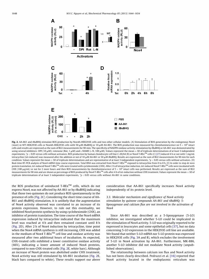
Next, we evaluated the effect of two quinone compounds, AA-861 and tBuBHQ on Nox4 constitutive activity. Incubation ofNox4A-HEK293E cells with AA-861 or tBuBHQ led to an increase ofROS production (Fig. 2A). The stimulated activity reached itsoptimum level at 10 min or 20 min after AA-861 or tBuBHQincubation respectively. The incubation with WT-HEK293E cellsdid not lead to a high level of ROS production. In fact, only smallincrease of ROS was observed reflecting the stimulation of theendogenous Nox4 by those two quinones (Fig. 2A, inset). Thespecificity of a NADPH oxidase activity was confirmed by using DPIwhich had an inhibitory effect whereas rotenone and L-NAME didnot (Fig. 2A).
To confirm that the observed effect of AA-861 and tBuBHQ wasspecifically related to Nox4 proteins, we used two othercharacterized cell lines that over-expressed Nox4: human chon-drocyte C-20/A4 [18] and Nox4 T-RExTM in which Nox4 expressioncould be temporally induced by the addition of tetracycline [27].Consistently, the constitutive ROS production in Nox4 C-20/A4cells or in 4 h-induced Nox4 T-REx cells was enhanced after theaddition of AA-861 or tBuBHQ (Fig. 2B and C). It is noticeable that
ed cells. (A) Real-time RT-PCR analysis of Nox4 mRNA. Total RNA was extracted from
onstitutive ROS production by HEK293E cells. The ROS production was measured by
ells. (C) The specificity of constitutive NADPH oxidase activity of HEK293E cells
(Rot, 1 mM), L-NAME (L-N, 100 mM), tiron (0.5 mM). Results are expressed as the sum
tions of at least 3 independent experiments, *p < 0.05 versus cells without transfection
Fig. 2. AA-861 and tBuBHQ stimulate ROS production by Nox4A-HEK293E cells and two other cellular models. (A) Stimulation of ROS generation by the endogenous Nox4
(inset) in WT-HEK293E cells or Nox4A-HEK293E cells with 50 mM tBuBHQ or 10 mM AA-861. The ROS production was measured by chemiluminescence on 5 � 105 intact
cells and results are expressed as the sum of RLU measurements for 90 min. The specificity of NADPH oxidase activity stimulation by tBuBHQ or AA-861 was demonstrated by
using several inhibitors: DPI (10 mM); rotenone (Rot, 1 mM) and L-NAME (L-N, 100 mM). Values represent the mean � SD of triplicate determinations of at least 3 independent
experiments, *p < 0.05 versus cells without activators. ROS production by human chondrocyte cell line C-20/A4 (B) or Nox4 T-RExTM cells (C) [27] induced 4 h or not with 1 mg/mL
tetracycline (tet-induced) was measured after the addition or not of 10 mM AA-861 or 50 mM tBuBHQ. Results are expressed as the sum of RLU measurements for 90 min for each
condition. Values represent the mean � SD of triplicate determinations and are representative of at least 3 independent experiments, *p < 0.05 versus cells without activators. (D)
Real-time RT-PCR analysis of Nox4 mRNA time course expression. Total RNA was extracted from Nox4 T-RExTM exposed to tetracycline from 0 to 8 h. (E) In order to stop de novo
protein translation, tet-induced Nox4 T-RExTM cells were treated with cycloheximide (CHX). After 2 h of tetracycline induction, tet-induced Nox4 T-RExTM cells were incubated with
CHX (100 mg/mL) or not for 2 more hours and then ROS measurements by chemiluminescence of 5 � 105 intact cells was performed. Results are expressed as the sum of RLU
measurements for 90 min and are shown as percentage of ROS produced by Nox4 T-RExTM cells after 4 h of tet-induction without CHX treatment. Values represent the mean � SD of
triplicate determinations of at least 3 independent experiments, *p < 0.05 versus cells without AA-861 in same conditions.
M.V.C. Nguyen et al. / Biochemical Pharmacology 85 (2013) 1644–16541648
the ROS production of uninduced T-RExTM cells, which do notexpress Nox4, was not affected by AA-861 or by tBuBHQ indicatingthat those two quinones do not produce ROS spontaneously in thepresence of cells (Fig. 2C). Considering the short time course of AA-861 and tBuBHQ stimulation, it is unlikely that the augmentationof Nox4 activity observed was correlated to an increase of itsprotein expression. However, to rule out this eventuality, weinhibited Nox4 protein synthesis by using cycloheximide (CHX), aninhibitor of protein translation. The time course of the Nox4 mRNAexpression induced by tetracycline indicated that the maximumlevel was reached at 4 h and then remained constant until 8 h(Fig. 2D). After 2 h of Nox4 induction by tetracycline, time pointwhen the Nox4 mRNA synthesis is still increasing, CHX was addedin the medium of Nox4 T-RExTM cell line and oxidase activity wasmeasured after two additional hours of incubation. As expected,CHX-treated cells exhibited a lower constitutive oxidase activity(66%), indicating a lower amount of induced Nox4 proteins,compared to non-CHX-treated cells (Fig. 2E, white bars). Althoughan increase of Nox4 protein was abolished under CHX treatment,Nox4 activity was still stimulated by AA-861 incubation (Fig. 2E,black bars compared to white). These results support our above
consideration that AA-861 specifically increases Nox4 activityindependently of its protein level.
3.3. Molecular mechanism and significance of Nox4 activity
stimulation by quinone compounds AA-861 and tBuBHQ: 5-
lipoxygenase and calcium flux are not involved in the activation of
Nox4
Since AA-861 was described as a 5-lipoxygenase (5-LO)inhibitor, we investigated whether 5-LO could be implicated inthe stimulation of Nox4 activity. 5-LO was originally reported to beexpressed in leucocytes and some epithelial cells [37], but no dataconcerning 5-LO expression in the HEK293E cell line are available.We found that neither 5-LO mRNA nor 5-LO protein was expressedin HEK293E cells (Fig. 3A and B), which excludes the involvementof 5-LO in Nox4 activation by AA-861. Furthermore, MK-886,another 5-LO inhibitor did not modulate Nox4 activity (unpub-lished observations).
The relationship between calcium ion flux and Nox4 activityhas not been clearly described. Pedruzzi et al. [16] reported thatNox4 activity located in the endoplasmic reticulum was
Fig. 3. AA-861 or tBuBHQ NADPH oxidase activity stimulation is independent of 5-lipoxygenase and of calcium flux. (A) 5-lipoxygenase mRNA expression in HEK293E cells.
Total RNA was extracted from HEK293E and B lymphocytes (BL), a semi-quantitative RT-PCR was performed using specific primers for 5-lipoxygenase (5-LO). The
housekeeping actin gene (Act) was used as a positive control. (B) 5-lipoxygenase protein expression in HEK293E cells. 150 mg of total proteins from HEK293E WT, and 75 mg
of cytosolic (Cyto) protein fraction (positive control) from neutrophils (PMN) were loaded on a 7% SDS-PAGE gel. 5-lipoxygenase proteins were characterized with a specific
monoclonal antibody against 5-LO. (C) and (D) Calcium flux measurement was assessed by a fluorescent probe in Nox4A-HEK293E cells. Intact cells were incubated with
10 mM Fluo3/AM with or without 20 mM BAPTA, during 30 min at 37 8C, washed and resuspended at 106 cells/mL. Calcium flux visualization was performed by flow
cytometry. The arrow indicates the time of the addition of ionomycin (Iono, 10 mM), AA-861 (10 mM), tBuBHQ (50 mM), or thapsigargin (Thaps, 10 mM) in presence of 1 mM
EGTA (C); or that of tBuBQ (50–200 mM) and tBuBHQ (50–200 mM) (D). Results are expressed as the mean of the fluorescence intensity (FL1 change) measured every 5 s (E).
5 � 105 intact HEK293E cells overexpressing Nox4A were incubated or not with ionomycin (Iono, 10 mM), thapsigargin (Thaps, 10 mM), AA-861 (10 mM), or tBuBHQ (50 mM)
in presence of 1 mM EGTA and then the ROS production was measured by chemiluminescence. Results are expressed as the sum of RLU measurements for 90 min.
concomitant to Ca2+ oscillation after exposure to 7-ketocholes-terol. However in a previous study, we showed that ionomycin(calcium ionophore) had no effect on the Nox4 activity inchondrocytes C-20/A4 cells over-expressing Nox4A [18]. SincetBuBHQ was described as a SERCA pump inhibitor, it is necessaryto evaluate the potential contribution of the Ca2+ flux on Nox4stimulation. To this purpose, we measured the calcium flux inNox4-HEK293E cells by FACs using a fluorescent Fluo-3/AMprobe and compared the calcium changes to Nox4 activation. Asillustrated in the figure 3 (Fig. 3C and D), an increase of cytosoliccalcium from intracellular stores was observed in the presenceof extracellular EGTA after adding ionomycin, thapsigargin ortBuBHQ whereas the addition of AA-861 or tBuBQ, twocompounds that stimulate Nox4 activity were unable to modify
Ca2+ level. Furthermore, thapsigargin or ionomycin could notmodulate the constitutive activity of Nox4 in Nox4-HEK293Ecells (Fig. 3E). Same experiments were performed in thepresence of 1.5 mM CaCl2 in the extracellular medium withoutEGTA and led to the same results (unpublished observations).These data strongly suggest that there is no correlation betweenCa2+ flux and Nox4 activity.
3.4. Quinone structure may be responsible for the modulation of Nox4
activity
AA-861 and tBuBHQ share the same quinone moiety. Toevaluate whether quinone structure was responsible for Nox4activation, the ROS production by Nox4-HEK293E cells was
Table 2Quinone effect on Nox4 ROS production of Nox4A-HEK293 cells. Data were
expressed as the mean � SD.
Effect Structure [C] mM ROS production
NonePBS 9.01 � 0.8
ActivatorsAA-861 1 24.26 � 0.91*
5 34.40 � 1.16*
10 45.70 � 0.77*
50 37.20 � 2.14*
tBuBHQ 1 14.20 � 0.76
10 36.01 � 0.68*
50 61.74 � 0.78*
100 38.90 � 1.82*
tBuBQ 1 13.58 � 0.42
10 32.70 � 0.22*
50 36.50 � 0.63*
100 34.99 � 1.22*
Duroquinone 1 35.69 � 1.65*
10 30.21 � 1.70*
50 29.30 � 1.80*
InhibitorsBenzoquinone 0.01 7.88 � 0.36
0.5 8.09 � 0.37
1 7.69 � 0.51
3 3.63 � 0.15*
30 1.01 � 0.11*
Hydroquinone 0.01 7.65 � 0.50
0.5 8.09 � 0.37
1 8.70 � 0.52
3 4.18 � 0.34*
30 0.12 � 0.07*
tMetBQ 1 10.09 � 1.68
10 12.70 � 0.43
50 12.87 � 0.82
100 1.32 � 0.20*
200 0.38 � 0.01*
Plumbagine 0.01 7.40 � 0.67
0.5 6.64 � 0.27
1 4.00 � 0.22*
5 1.93 � 0.29*
10 1.34 � 0.09*
* p < 0.01 versus PBS treated cells.
Table 3Cytotoxicity of quinones and ROS production in the presence of phosphatidylcho-
line.
Structure [C] mM Toxicity %LDHa ROS productionb
PBS PC
– – 10.80 � 0.29 1.9 � 0.1 2.7 � 0.2
Benzoquinone 1 11.37 � 0.53 ND ND
30 10.72 � 0.53 2.0 � 0.1 2.3 � 0.2
Hydroquinone 1 10.73 � 0.53 ND ND
30 10.99 � 0.80 2.1 � 0.1 2.3 � 0.1
tMetBQ 50 12.79 � 0.97 2.3 � 0.3 2.4 � 0.1
200 12.66 � 1.00 ND ND
tBuBQ 50 12.44 � 0.42 2.3 � 0.1 2.2 � 0.2
tBuBHQ 50 11.93 � 1.41 4.4 � 0.9 6.8 � 0.4
Duroquinone 50 11.03 � 1.13 2.3 � 0.1 2.8 � 0.2
AA-861 10 12.00 � 0.84 2.3 � 0.1 2.4 � 0.3
a Cytotoxicity evaluation of quinone compounds on HEK293E cell line. 5 � 105
intact HEK293E were incubated with chemical compounds and lactate dehydroge-
nase (LDH) activity was assessed 60 min later in the culture medium. Values
represent the mean percentage of LDH activity compared to total LDH enzymatic
activity (cells lyzed by a treatment with 1% triton X-100).b Luminol chemiluminescence measurements of quinone alone (PBS column) or
with only 25 mg of phosphatidylcholine (PC column) demonstrated no spontaneous
ROS production. Values represent the mean percentage of NADPH oxidase activity
compared to constitutive ROS production by Nox4A-HEK293E alone.
M.V.C. Nguyen et al. / Biochemical Pharmacology 85 (2013) 1644–16541650
measured after incubation with various quinone derivatives thatdiffer with respect to their reduced or oxidized state andsubstitution levels. The results shown in Table 2 illustrate twoopposite effects. Duroquinone as well as tBuBHQ, tBuBQ (theoxidative form of tBuBHQ), and AA-861, stimulated Nox4 activityat a starting concentration of 1 mM, whereas BQ (30 mM), HQ(30 mM) and tMetBQ (100 mM) inhibited this activity in a dosedependent manner. We also confirmed the inhibitory effect of thenaphthoquinone, plumbagin, on Nox4 activity as reported [38,39].
To exclude a non-specific spontaneous ROS production byquinone compounds in contact with membrane phospholipids asdescribed for tBuBHQ [40], we measured the ROS synthesis in thepresence of 25 mg phosphatidylcholine. The results illustrated inTable 3 show that there was no ROS enhancement by quinonesafter addition of phospholipids versus control, except for a slightincrease observed as expected with tBuBHQ, as previouslyreported [40]. Furthermore, quinone concentrations used in thisstudy were not cytotoxic as examined by the LDH activity (Table 3).
3.5. Effect of quinones on Nox2
We next evaluated whether those quinones could affect Nox2oxidase activity in isolated human neutrophils which express only
Nox2, the redox core of phagocyte NADPH oxidase. Compared tothe well known activator PMA, no NADPH oxidase activation wasobserved when neutrophils were incubated with AA-861, tBuBQ,or duroquinone (Fig. 4A) as opposed to what was previouslyobserved in Nox4 T-RExTM cells (Table 2). However, slight oxidaseactivation was observed upon the addition of tBuBHQ, probablydue to its effect on the calcium flux since thapsigargin, anotherSERCA pump inhibitor, or ionomycin gave similar results (unpub-lished data). On the contrary, the quinones BQ, HQ, tMetBQ, andplumbagin also inhibited the PMA stimulated neutrophil oxidaseactivity (Fig. 4B). By using an in vitro xanthine/xanthine oxidasesuperoxide production system, we observed that the ROSproduction was abolished in the presence of 30 mM BQ, 30 mMHQ, and 100 mM tMetBQ while no effect on the xanthine/xanthineoxidase ROS production was noticeable with 50 mM duroquinone,50 mM tBuBHQ, 50 mM tBuBQ, or 10 mM AA-861 (unpublisheddata). Using differentiated PLB-985 as another cellular model, weobtained similar results than those observed with human PMN(Fig. 4C). We, next, evaluated whether quinones could increaseNox2 activity already activated by the physiological agonists (theformylated peptide fMLP) in differentiated PLB-985 cells and foundthat AA-861, tBuBQ, and duroquinone did not stimulate Nox2activity (Fig. 4D).
These results suggest that BQ, HQ, and tMetBQ inhibit ROSproduction through an antioxidant property. On the contrary,duroquinone, tBuBHQ, tBuBQ, and AA-861 appear to stimulatespecifically NADPH oxidase activity of Nox4.
3.6. Putative quinone binding site (Q site) on Nox4
Quinones compounds bind to its target proteins through welldescribed ubiquinone binding sites [41]. Based on computationalprediction, a consensus sequence for a predictable quinone bindingsite or Q site was suggested: ‘‘aliphatic-(X)3-H-(X)2-3-(Leu/Thr/Ser)’’ [42]. The analysis of the Nox4 protein sequence showed asimilar putative motif in the fifth transmembrane domain(‘‘203LLTLHVS209’’) that is missing in Nox2 sequence (Fig. 5A).Therefore, quinones could regulate Nox4 activity by bindingdirectly to the protein through this putative domain. To investigatethe relevance of this potential Q site in Nox4 protein, we mutated
Fig. 4. Effect of quinone derivatives on the activation of Nox2 oxidase activity. (A) The reactive oxygen species production was measured by chemiluminescence on 5 � 105
intact PMN cells in the presence of an increasing concentration (1 mM, 10 mM, or 50 mM) of AA-861, tBuBQ, tBuBHQ, or duroquinone. Results are expressed as the sum of RLU
measurements for 60 min and are shown as percentage of ROS produced by PMN stimulated by PMA (0.13 mM). Values represent the mean � SD of triplicate determinations of
at least 3 independent experiments, *p < 0.05 versus cells without PMA activation and quinones. (B) PMN (5 � 105 cells) were stimulated by PMA (0.13 mM) in the presence or not of
BQ (1 mM, 3 mM, and 30 mM), HQ (1 mM, 3 mM, and 30 mM), tMetBQ (10 mM, 50 mM, and 100 mM) or plumbagin (1 mM and 5 mM). Results are expressed as the sum of RLU
measurements for 90 min and are reported as a percentage of ROS produced by PMN stimulated by PMA (0.13 mM). Values represent the mean � SD of triplicate determinations of at
least 3 independent experiments, *p < 0.05 versus cells activated by PMA without quinones. (C) The reactive oxygen species production was measured by chemiluminescence on
5 � 105 intact differentiated PLB-985 cells in the presence of 30 mM BQ, 30 mM HQ, 50 MetBQ, 10 mM AA-861, 50 mM tBuBQ, 50 mM tBuBHQ, or 50 mM duroquinone. Results are
expressed as the sum of RLU measurements for 60 min and are shown as percentage of ROS produced by differentiated PLB-985 cells stimulated by fMLP (0.15 mM). Values represent
the mean � SD of triplicate determinations of at least 3 independent experiments, *p < 0.05 versus cells without fMLP activation and quinones. (D) Differentiated PLB-985
(5 � 105 cells) were stimulated by fMLP (0.15 mM) in the presence or not of 30 mM BQ, 30 mM HQ, 50 MetBQ, 10 mM AA-861, 50 mM tBuBQ, 50 mM tBuBHQ, or 50 mM duroquinone.
Results are expressed as the sum of RLU measurements for 60 min and are reported as a percentage of ROS produced by differentiated PLB-985 cells stimulated by fMLP (0.15 mM).
Values represent the mean � SD of triplicate determinations of at least 3 independent experiments, *p < 0.05 versus cells activated by fMLP without quinones.
key amino acid residues of that sequence, i.e., substitutions ofL203G and S209A (Fig. 5A). Our results showed that thosemutations did not modify the stimulatory effect of the quinonetBuBHQ on Nox4 activity which remained similar to WT Nox4(Fig. 5B). Since our data showed that quinones did not activateNox2 activity nor enhance its activity (Fig. 4), we thereforeexchanged the sequence 201Y. . .Y215 of Nox4, which containsthe putative Q binding of Nox4, with the homologous sequence ofNox2 216F. . .G230. This Nox4/2 chimera protein remained stillstimulated by tBuBHQ (Fig. 5B). Those results suggest that thisputative Q site domain on Nox4 is not involved in the stimulationof Nox4 activity by quinone compounds.
3.7. Redox regulation of Nox4 activity
A recent study [43] showed that structural modifications of theE-loop, by deletion or exchange of cysteine, had an impact on theproduction rate and nature of ROS generated. The authorshypothesized that extracellular cysteines C226 and C270 mightform a disulfide bridge that modulates Nox4 ROS production.Quinones compounds are highly reducing molecules capable ofbreaking disulfide bridges and, therefore, could modulate Nox4activity through a redox regulation. To test this hypothesis, wemeasured the effect of known reducing agents on Nox4 activityand found that in tet-induced Nox4 T-RExTM cells, the addition of80 mM NADPH, NADH, or b-mercaptoethanol increased signifi-cantly the ROS production compared to non-induced anduntreated Nox4 T-RExTM cells (Fig. 5C). These results suggest thatreducing agents stimulate Nox4 activity and point out a possible
mechanism by which quinones might regulate Nox4 activity byreducing the E-loop disulfide bridge.
3.8. Involvement of NQO1 on Nox4 activation by quinone
Quinone compounds are metabolized through different path-ways and one of them involves the NAD(P)H oxidoreductase 1(NQO1). By RT-PCR and sequencing, we revealed that Nox4 T-RExTM cells expressed two types of mRNA encoding for NQO1:N-QO1A, the active form and NQO1C, the inactive form, which lacks 2essential amino acid residues (Tyrosine-126 and -128) necessaryfor its quinone binding capacity [4]. To investigate the potentialrole of NQO1 in the stimulation of Nox4 activity by quinonecompounds, we used dicoumarol to inhibit NQO1. Pre-incubationof the tetracycline induced Nox4 T-RExTM cells with 100 mMdicoumarol inhibited Nox4 constitutive activity as well as itsstimulation by tBuBHQ (Fig. 6A). To further investigate the role ofNQO1, we stably overexpressed NQO1A and NQO1C in Nox4-HEK293 and confirmed the presence of the exogenous NQO1A andNQO1C mRNA in the corresponding transfected cells comparedwith the empty vector control transfected cells (Fig. 6B). Further-more, by real time RT-PCR we showed the mRNA level of NQO1Aand NQO1C increased 2 and 8 fold, respectively, compared to thecontrol Nox4-HEK293 cells (Fig. 6B). We then investigated theconsequences of these overexpressions on Nox4 activity. Resultsshowed that overexpression of NQO1A enhanced twice thestimulatory effect of tBuBHQ on Nox4 activity compared to thecontrol empty vector cells. This is consistent with the increase levelof NQO1A mRNA observed previously (Fig. 6B). On the contrary,
Fig. 5. Putative Q site and redox regulation of Nox4 activity. (A) Schematic
representation of the alignment of partial sequences of Nox4, Nox2 and Nox4
mutants (Nox4L203G/S209A and Nox4/2). Gray box represents the predicted 5th
transmembrane domain. Boxed amino acids on Nox4 sequence correspond to the
putative Q site and bold letters are key amino acids described for the Q site [42].
Underlined amino acids represent amino acids that have been exchanged from
Nox2 sequence to produce the chimera Nox4/2. (B) Measurement of ROS generation
by chemiluminescence on 5 � 105 intact transfected Nox4-, Nox4L203G/S209A- and
Nox4/2-HEK293 cells with or without 50 mM tBuBHQ. (C) Measurement of ROS
generation by chemiluminescence on 5 � 105 intact cells. ROS production by Nox4
T-RExTM cell induced 4 h or not with 1 mg/mL tetracycline (tet-induced) was
measured after the addition or not of NADPH, NADH, b-mercaptoethanol (b-ME)
(80 mM of each), or tBuBHQ (50 mM). Results are expressed as the sum of RLU
measurements for 90 min for each condition. Values represent the mean � SD of
triplicate determinations of at least 3 independent experiments, *p < 0.05 versus cells
without redox compounds.
Fig. 6. Effect of NQO1 on tBuBHQ activation of ROS production by Nox4. (A) Nox4 T-
RExTM cells were induced (or not) with 1 mg/mL tetracycline for 4 h. Dicoumarol
was added in the culture medium 2 h before ROS measurements.
Chemiluminescence assay was done on 106 intact cells per well, activated or not
by 50 mM tBuBHQ. Results are expressed as the sum of RLU measurements for
90 min. Values represent the mean � SD of triplicate determinations of at least 3
independent experiments, *p < 0.05 versus cells in same condition without
dicoumarol. (B) Real-time RT-PCR analysis of exogenous and total NQO1 mRNA in
Nox4A-, Nox4A/NQO1C- and Nox4A/NQO1A-HEK293 cells. Left panel, real time RT-PCR
showing the presence of exogenous NQO1 mRNA in Nox4A/NQO1C- and Nox4A/
NQO1A-HEK293 cells and not in Nox4A-HEK293 transfected with the control pEFb
empty plasmid. N.D., not detected. Right panel, real time RT-PCR showing the increase
of NQO1 mRNA level in Nox4A/NQO1C- and Nox4A/NQO1A-HEK293 cells compared to
Nox4A-HEK293 transfected with the control pEFb empty plasmid. (C) Nox4A-HEK293E
cells were transfected with the empty vector pEFb (Control) or containing NQO1A or
NQO1B. The reactive oxygen species production was measured by chemiluminescence
on 5 � 105 intact cells after the addition of 50 mM tBuBHQ or the same volume of
vehicle (DMSO). Results are expressed as the sum of RLU measurements for 90 min.
Values represent the mean � SD of triplicate determinations of at least 3 independent
experiments, *p < 0.005 versus cells transfected with the control empty vector in same
conditions.
M.V.C. Nguyen et al. / Biochemical Pharmacology 85 (2013) 1644–16541652
although the overexpression of NQO1C is higher than that ofNQOA, it did not modify the stimulatory effect which is comparableto that observed with the control empty plasmid (Fig. 6C).Together, these data suggest the implication of NQO1 enzymes inquinone-stimulated Nox4 activity.
4. Discussion
Our study focuses on the capacity of quinone compounds tomodulate the NADPH oxidase activity of Nox4. Quinones areimportant toxic compounds present in air pollution and abundantin all burnt organic material, including urban air particles, cigarettesmoke, and diesel exhaust particles (DEP) [44]. DEP were reportedto increase Nox4 ROS production after 1 h of incubation,suggesting the implication of Nox4 in the quinone metabolism[9]. Considering the structure/function relationship of the qui-nones used in this study (Table 2), we observed a dual stimulation/inhibition effect that depends on their substitution state: Nox4activity was stimulated by AA-861, duroquinone, tBuBQ, andtBuBHQ whereas other less substituted derivatives such as BQ, HQ,
tMetBQ, and plumbagin, developed antioxidant properties(Table 2). The specificity of AA-861, duroquinone, and tBuBQ forNox4 over Nox2, suggests either a direct effect of quinonederivatives in the molecular dynamics of electron transfermediated by Nox4, or an indirect role on a quinone sensitivefunctional partner of Nox4. In this work, we suggest a new aspectof Nox4 oxidase activity regulation that takes place not onlyat a transcriptional level as usually described [13,27,45] but also ata post-translational level as described above with quinonemolecules.
Quinones are bioreactive molecules that are sensitive to redoxmechanism. Ubiquinone, for example, is a mobile electron carrierof mitochondria and a highly hydrophobic molecule that diffusesinto the core of inner membrane where the electron transferproceeds through a Q cycle on either side of membrane [41,46].A predictable quinone binding site has been suggested [42] andwe identified a similar domain in Nox4 protein sequence‘‘203LLTLHVS209’’. However, the implication of such domainwas not confirmed since the ability of quinones to stimulateNox4 activity was not abolished when appropriate residues ofthis sequence were mutated (Fig. 5A and B).
Alternatively, Takac and his colleagues [43] have shown thatdisulfide bridges formed by two cysteines C226 and C270 may beinvolved in the maintenance of the extracellular E-loop integrity.Indeed, the disruption of the bridge by mutagenesis modifiednotably Nox4 ROS production [43]. Disulfide bridges with astandard redox potential (E0) of �180 mV [47] could be reduced byNADPH, NADH and b-mercaptoethanol that possess a lower E0
(�324, �320, and �253 mV, respectively) [48]. Our results usingthose reducing agents indicated that Nox4 activity could bestimulated by extracellular redox modifications, possibly throughthe reduction of its disulfide bridge. Interestingly, duroquinone,which enhances Nox4 activity, has a low E0 (�260 mV) [49] andthen could theoretically reduce disulfide bridge. Conversely,compounds such as BQ, HQ and tMetBQ that do not stimulateNox4 exhibit a higher E0, 78 mV, 78 mV and �67 mV, respectively,compared to that of the disulfide bridge. Thus, it is possible thatquinones exhibiting an E0 lower than that of disulfide bridge areable to stimulate Nox4 activity.
NAD(P)H-quinone oxidoreductase-1 (NQO1) is a broadlydistributed FAD-dependent flavoprotein that catalyses the reduc-tion of a wide variety of quinone compounds. It reduces quinones,by the transfer of 2 electrons, to the corresponding hydroquinoneswhich are the electron donor state capable to initiate reductivereactions. We showed that NQO1 is expressed in uninduced or tet-induced Nox4 T-RExTM cells; however, the stimulation of ROSproduction by quinones occurs only when Nox4 expression wasinduced. Moreover, the increase of Nox4 ROS production byquinones was inhibited by a well known inhibitor of NQO1,dicoumarol. Noticeably, dicoumarol also decreases Nox4 constitu-tive activity in quinone-untreated Tet-induced Nox4 T-RExTM cells(Fig. 6A), possibly by inhibiting the endogenous NQO1 enzymes.More specifically, only the functional NQO1 enzyme, NQO1A,enhanced the ROS production of Nox4 induced by quinonecompounds (Fig. 6C). Interestingly, NQO1 has been described topartition between cytosol and plasma membrane [7] where Nox4is also located in HEK293 cells [50]. These observations suggest apotential link between NQO1 and Nox4. Such potential link couldbe illustrated in pancreatic cancer. Pancreatic cancer is veryaggressive and unresponsive to treatments due to a resistance ofcells to apoptosis. Vaquero et al. [51] reported that ROS producedby Nox4 are prosurvival and antiapoptotic for human pancreaticadenocarcinoma cell lines, MIA PaCa-2 and PANC-1. In the MIAPaCa-2 cell line, Lewis et al. [52] showed that inhibition of NQO1 bydicoumarol suppresses the malignant phenotype of pancreaticcancer cells and induces cell apoptosis.
Finally, our study described 4 quinones derivatives especiallyAA-861 and tBuBHQ capable of enhancing Nox4 ROS production.We showed that NQO1 may be involved in the stimulation of Nox4ROS production induced by quinones. We further observed thatNox4 activity is also stimulated in the presence of reducing agentsthat may possibly act by reducing the disulfide bridge located inthe extracellular E-loop of Nox4. All together, we hypothesize thatin the presence of quinones, NQO1 may generate hydroquinoneforms, providing, therefore, potential reducing agents near Nox4protein. Depending on the quinone redox potential, Nox4 E-loop
disulfide bridge could be disrupted consequently leading to theincrease of its oxidase activity. However, the direct implication ofthe E-loop disulfide bridge needs further investigations. Theproposed model could provide new insight into the understandingof molecular mechanism of the electron transfer through Nox4 andalso define new therapeutic targets in diseases in which quinonesand Nox4 are implicated.
Acknowledgments
This work was supported by grants from: the ‘Ministere del‘Enseignement superieur de la recherche et la technologie’, Paris,France; the ‘CNRS Institute’; the Association pour la Recherchecontre le Cancer (ARC), Paris, France; the Region Rhone-Alpes,programme ARCUS, France/Chine 2007–2008, programme Emer-gence 2003–2006; the ‘CGD research Trust 2006–2007’, UK; the‘Groupement des Entreprises Francaises de la Lutte contre leCancer’, delegation de Grenoble; the ‘UFR de Medecine, UniversiteJoseph Fourier, Grenoble’; and the ‘Direction Regionale de laRecherche Clinique, Center Hospitalier Universitaire, Grenoble’.We thank Prof. Stanke (Grenoble, France) for kindly providing the5-lipoxygenase polyclonal antibody, Prof. P.E. Cole, Drs M.H.Huynh and M.H. Paclet for English language corrections, S. Berthierfor FACS measurements, A. Dariz-Soldini for real time RT-PCRexperiments and Prof. A. Maitre and Prof. R. Brandes for helpfulreading and discussions.
References
[1] Bolton JL, Trush MA, Penning TM, Dryhurst G, Monks TJ. Role of quinones intoxicology. Chem Res Toxicol 2000;13:135–60.
[2] Ghio AJ, Carraway MS, Madden MC. Composition of air pollution particles andoxidative stress in cells, tissues, and living systems. J Toxicol Environ Health BCrit Rev 2012;15:1–21.
[3] Rodgman A, Smith CJ, Perfetti TA. The composition of cigarette smoke: aretrospective, with emphasis on polycyclic components. Hum Exp Toxicol2000;19:573–95.
[4] Faig M, Bianchet MA, Talalay P, Chen S, Winski S, Ross D, et al. Structures ofrecombinant human and mouse NAD(P)H:quinone oxidoreductases: speciescomparison and structural changes with substrate binding and release. ProcNatl Acad Sci U S A 2000;97:3177–82.
[5] Kovacic P, Somanathan R. Pulmonary toxicity and environmental contamina-tion: radicals, electron transfer, and protection by antioxidants. Rev EnvironContam Toxicol 2009;201:41–69.
[6] Auerbach A, Hernandez ML. The effect of environmental oxidative stress onairway inflammation. Curr Opin Allergy Clin Immunol 2012;12:133–9.
[7] Gray JP, Eisen T, Cline GW, Smith PJ, Heart E. Plasma membrane electrontransport in pancreatic beta-cells is mediated in part by NQO1. Am J PhysiolEndocrinol Metab 2011;301:E113–21.
[8] Squadrito GL, Cueto R, Dellinger B, Pryor WA. Quinoid redox cycling as amechanism for sustained free radical generation by inhaled airborne particu-late matter. Free Radic Biol Med 2001;31:1132–8.
[9] Amara N, Bachoual R, Desmard M, Golda S, Guichard C, Lanone S, et al. Dieselexhaust particles induce matrix metalloprotease-1 in human lung epithelialcells via a NADP(H) oxidase/NOX4 redox-dependent mechanism. Am J PhysiolLung Cell Mol Physiol 2007;293:L170–81.
[10] Morel F. Molecular aspects of chronic granulomatous disease. the NADPHoxidase complex. Bull Acad Natl Med 2007;191:377–90 [discussion 90-2].
[11] Lardy B, Bof M, Aubry L, Paclet MH, Morel F, Satre M, et al. NADPH oxidasehomologs are required for normal cell differentiation and morphogenesis inDictyostelium discoideum. Biochim Biophys Acta 2005;1744:199–212.
[12] Bedard K, Krause KH. The NOX family of ROS-generating NADPH oxidases:physiology and pathophysiology. Physiol Rev 2007;87:245–313.
[13] Geiszt M, Kopp JB, Varnai P, Leto TL. Identification of renox, an NAD(P)Hoxidase in kidney. Proc Natl Acad Sci U S A 2000;97:8010–4.
[14] Djordjevic T, BelAiba RS, Bonello S, Pfeilschifter J, Hess J, Gorlach A. Humanurotensin II is a novel activator of NADPH oxidase in human pulmonary arterysmooth muscle cells. Arterioscler Thromb Vasc Biol 2005;25:519–25.
[15] Mahadev K, Motoshima H, Wu X, Ruddy JM, Arnold RS, Cheng G, et al. TheNAD(P)H oxidase homolog Nox4 modulates insulin-stimulated generation ofH2O2 and plays an integral role in insulin signal transduction. Mol Cell Biol2004;24:1844–54.
[16] Pedruzzi E, Guichard C, Ollivier V, Driss F, Fay M, Prunet C, et al. NAD(P)Hoxidase Nox-4 mediates 7-ketocholesterol-induced endoplasmic reticulumstress and apoptosis in human aortic smooth muscle cells. Mol Cell Biol2004;24:10703–17.
M.V.C. Nguyen et al. / Biochemical Pharmacology 85 (2013) 1644–16541654
[17] Fitzgerald JP, Nayak B, Shanmugasundaram K, Friedrichs W, Sudarshan S, EidAA, et al. Nox4 mediates renal cell carcinoma cell invasion through hypoxia-induced interleukin 6- and 8- production. PLoS ONE 2012;7:e30712.
[18] Grange L, Nguyen MV, Lardy B, Derouazi M, Campion Y, Trocme C, et al.NAD(P)H oxidase activity of Nox4 in chondrocytes is both inducible andinvolved in collagenase expression. Antioxid Redox Signal 2006;8:1485–96.
[19] Park HS, Chun JN, Jung HY, Choi C, Bae YS. Role of NADPH oxidase 4 inlipopolysaccharide-induced proinflammatory responses by human aortic en-dothelial cells. Cardiovasc Res 2006;72:447–55.
[20] Bonner MY, Arbiser JL. Targeting NADPH oxidases for the treatment of cancerand inflammation. Cell Mol Life Sci 2012;69:2435–42.
[21] Ambasta RK, Kumar P, Griendling KK, Schmidt HH, Busse R, Brandes RP. Directinteraction of the novel Nox proteins with p22phox is required for theformation of a functionally active NADPH oxidase. J Biol Chem 2004;279:45935–41.
[22] Hilenski LL, Clempus RE, Quinn MT, Lambeth JD, Griendling KK. Distinctsubcellular localizations of Nox1 and Nox4 in vascular smooth muscle cells.Arterioscler Thromb Vasc Biol 2004;24:677–83.
[23] Martyn KD, Frederick LM, von Loehneysen K, Dinauer MC, Knaus UG. Func-tional analysis of Nox4 reveals unique characteristics compared to otherNADPH oxidases. Cell Signal 2006;18:69–82.
[24] Nguyen MV, Zhang L, Lhomme S, Mouz N, Lenormand JL, Lardy B, et al.Recombinant Nox4 cytosolic domain produced by a cell or cell-free basesystems exhibits constitutive diaphorase activity. Biochem Biophys Res Com-mun 2012;419:453–8.
[25] Nisimoto Y, Jackson HM, Ogawa H, Kawahara T, Lambeth JD. ConstitutiveNADPH-dependent electron transferase activity of the Nox4 dehydrogenasedomain. Biochemistry 2010;49:2433–42.
[26] Ellmark SH, Dusting GJ, Fui MN, Guzzo-Pernell N, Drummond GR. The contri-bution of Nox4 to NADPH oxidase activity in mouse vascular smooth muscle.Cardiovasc Res 2005;65:495–504.
[27] Serrander L, Cartier L, Bedard K, Banfi B, Lardy B, Plastre O, et al. NOX4 activityis determined by mRNA levels and reveals a unique pattern of ROS generation.Biochem J 2007;406:105–14.
[28] Cucoranu I, Clempus R, Dikalova A, Phelan PJ, Ariyan S, Dikalov S, et al. NAD(P)Hoxidase 4 mediates transforming growth factor-beta1-induced differentiation ofcardiac fibroblasts into myofibroblasts. Circ Res 2005;97:900–7.
[29] McKallip RJ, Jia W, Schlomer J, Warren JW, Nagarkatti PS, Nagarkatti M.Cannabidiol-induced apoptosis in human leukemia cells: A novel role ofcannabidiol in the regulation of p22phox and Nox4 expression. Mol Pharmacol2006;70:897–908.
[30] Moe KT, Aulia S, Jiang F, Chua YL, Koh TH, Wong MC, et al. Differentialupregulation of Nox homologues of NADPH oxidase by tumor necrosis fac-tor-alpha in human aortic smooth muscle and embryonic kidney cells. J CellMol Med 2006;10:231–9.
[31] Murillo MM, Carmona-Cuenca I, Del Castillo G, Ortiz C, Roncero C, Sanchez A,et al. Activation of NADPH oxidase by transforming growth factor-beta inhepatocytes mediates up-regulation of epidermal growth factor receptorligands through a nuclear factor-kappaB-dependent mechanism. Biochem J2007;405:251–9.
[32] Touyz RM, Chen X, Tabet F, Yao G, He G, Quinn MT, et al. Expression of afunctionally active gp91phox-containing neutrophil-type NAD(P)H oxidase insmooth muscle cells from human resistance arteries: regulation by angioten-sin II. Circ Res 2002;90:1205–13.
[33] Wingler K, Wunsch S, Kreutz R, Rothermund L, Paul M, Schmidt HH. Upregula-tion of the vascular NAD(P)H-oxidase isoforms Nox1 and Nox4 by the renin-angiotensin system in vitro and in vivo. Free Radic Biol Med 2001;31:1456–64.
[34] Park HS, Jin DK, Shin SM, Jang MK, Longo N, Park JW, et al. Impaired generationof reactive oxygen species in leprechaunism through downregulation of Nox4.Diabetes 2005;54:3175–81.
[35] Campion Y, Paclet MH, Jesaitis AJ, Marques B, Grichine A, Berthier S, et al. Newinsights into the membrane topology of the phagocyte NADPH oxidase:characterization of an anti-gp91-phox conformational monoclonal antibody.Biochimie 2007;89:1145–58.
[36] Goyal P, Weissmann N, Rose F, Grimminger F, Schafers HJ, Seeger W, et al.Identification of novel Nox4 splice variants with impact on ROS levels in A549cells. Biochem Biophys Res Commun 2005;329:32–9.
[37] Luo M, Lee S, Brock TG. Leukotriene synthesis by epithelial cells. HistolHistopathol 2003;18:587–95.
[38] Ding Y, Chen ZJ, Liu S, Che D, Vetter M, Chang CH. Inhibition of Nox-4 activityby plumbagin, a plant-derived bioactive naphthoquinone. J Pharm Pharmacol2005;57:111–6.
[39] Rossary A, Arab K, Steghens JP. Polyunsaturated fatty acids modulate NOX 4anion superoxide production in human fibroblasts. Biochem J 2007;406:77–83.
[40] Tsukii K, Nakahata N, Watanabe K, Tsurufuji S, Ohizumi Y. Comparison oftBuBHQ with chemotactic peptide and phorbol ester in O2- production in HL-60 cells. Eur J Pharmacol 1996;298:87–95.
[41] Lenaz G, Fato R, Genova ML, Bergamini C, Bianchi C, Biondi A. Mitochondrialcomplex I: structural and functional aspects. Biochim Biophys Acta2006;1757:1406–20.
[42] Fisher N, Rich PR. A motif for quinone binding sites in respiratory andphotosynthetic systems. J Mol Biol 2000;296:1153–62.
[43] Takac I, Schroder K, Zhang L, Lardy B, Anilkumar N, Lambeth JD, et al. The E-loop is involved in hydrogen peroxide formation by the NADPH oxidase Nox4. JBiol Chem 2011;286:13304–13.
[44] Schuetzle D, Lee FS, Prater TJ. The identification of polynuclear aromatichydrocarbon (PAH) derivatives in mutagenic fractions of diesel particulateextracts. Int J Environ Anal Chem 1981;9:93–144.
[45] Shiose A, Kuroda J, Tsuruya K, Hirai M, Hirakata H, Naito S, et al. A novelsuperoxide-producing NAD(P)H oxidase in kidney. J Biol Chem 2001;276:1417–23.
[46] Mitchell P. Protonmotive redox mechanism of the cytochrome b-c1 complexin the respiratory chain: protonmotive ubiquinone cycle. FEBS Lett 1975;56:1–6.
[47] Siedler F, Rudolph-Bohner S, Doi M, Musiol HJ, Moroder L. Redox potentials ofactive-site bis(cysteinyl) fragments of thiol–protein oxidoreductases. Bio-chemistry 1993;32:7488–95.
[48] Crans DC, Zhang B, Gaidamauskas E, Keramidas AD, Willsky GR, Roberts CR. Isvanadate reduced by thiols under biological conditions? Changing the redoxpotential of V(V)/V(IV) by complexation in aqueous solution. Inorg Chem2010;49:4245–56.
[49] Schultz TW. Influence of the energy relationship of organic compounds ontoxicity to the cladoceran Daphnia magna and the fish Pimephales promelas.Ecotoxicol Environ Saf 1997;38:336–8.
[50] Zhang L, Nguyen MV, Lardy B, Jesaitis AJ, Grichine A, Rousset F, et al. Newinsight into the Nox4 subcellular localization in HEK293 cells: first monoclo-nal antibodies against Nox4. Biochimie 2011;93:457–68.
[51] Vaquero EC, Edderkaoui M, Pandol SJ, Gukovsky I, Gukovskaya AS. Reactiveoxygen species produced by NAD(P)H oxidase inhibit apoptosis in pancreaticcancer cells. J Biol Chem 2004;279:34643–54.
[52] Lewis A, Ough M, Li L, Hinkhouse MM, Ritchie JM, Spitz DR, et al. Treatment ofpancreatic cancer cells with dicumarol induces cytotoxicity and oxidativestress. Clin Cancer Res 2004;10:4550–8.