65
Radiotherapy for Non-Small Cell Lung Cancer Jürg Heuberger Kantonsspital Aarau I Standard Treatment Options II Radiotherapy Planning
| Date post: | 23-Dec-2015 |
| Category: |
Documents |
| Upload: | egbert-dawson |
| View: | 217 times |
| Download: | 0 times |

Radiotherapy forNon-Small Cell Lung Cancer
Jürg Heuberger
Kantonsspital Aarau
I Standard Treatment Options
II Radiotherapy Planning
TNM Staging System
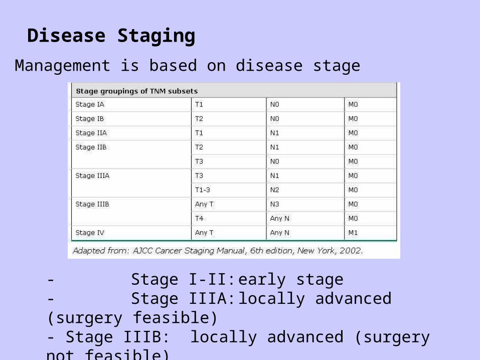
Disease Staging
- Management is based on disease stage
- Stage I-II: early stage- Stage IIIA: locally advanced (surgery feasible)- Stage IIIB: locally advanced (surgery not feasible)- Stage IV: metastatic disease
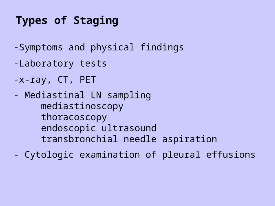
Types of Staging
-Symptoms and physical findings
-Laboratory tests
-x-ray, CT, PET
- Mediastinal LN samplingmediastinoscopythoracoscopyendoscopic ultrasoundtransbronchial needle aspiration
- Cytologic examination of pleural effusions

Staging Algorithm

Lymph Node Map – Nomenclature(American College of Surgeons)

Management of Stage I + II NSCLC
-Surgery alone is the standard treatment choice !
-Lobectomy: optimal procedure-Wedge resection: 3x LR / 30% more mortality (Ginsberg 1995)
but newer series show no worse outcome with limited surgery(Lee 2003, El Sherif 2006)
-Wedge resection for small tumors (<3cm) and elderly patients-No randomized trials, but excellent results
(randomized trial ‘Surgery – Radiotherapy’ underway)
-Adjuvant Cisplatin-based ChT for stage II for stage IB data is conflicting
-No adjuvant radiotherapy after radical surgery (i.e. R0)

Stage I: Outcome after Surgery

Stage I - III: Outcome after Surgery

Definitive Radiotherapy for Stage I + II NSCLC
-Alternative for comorbid patients who are not fit for surgery-For patients who refuse surgery
-60 – 66Gy to primary (+/- 50Gy to part of mediastinum, if feasible)
Review of 26 nonrandomized trials (Powell 2001)
Cancer-specific Survival OS (RT) OS (surgery)
2y 54 – 93% 22 – 72% 67%3y 22 – 56% 17 – 55%5y 13 – 39% 0 – 42% 47%
Non-cancer deaths following RT: 11 – 43% (reflecting the poor health status of pts. treated in these studies)
-Clinical stage I only in 57% pathologic stage I (Lopez 2005)
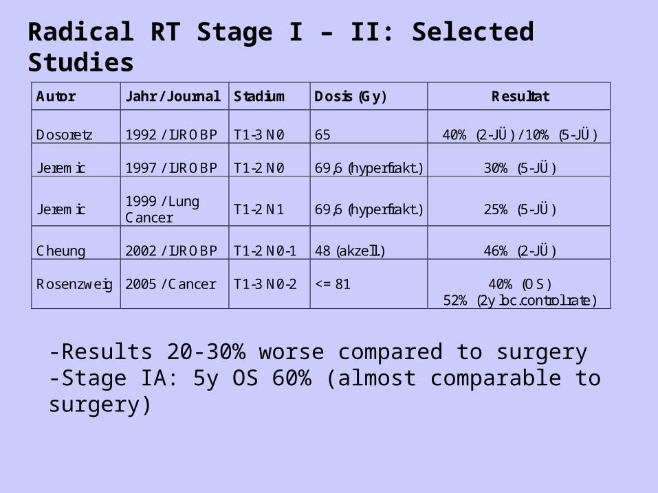
Radical RT Stage I – II: Selected Studies
Autor Jahr / Journal Stadium Dosis (Gy) Resultat
Dosoretz
1992 / IJROBP
T1-3 N0
65
40% (2-JÜ) / 10% (5-JÜ)
Jeremic
1997 / IJROBP
T1-2 N0
69,6 (hyperfrakt.)
30% (5-JÜ)
Jeremic
1999 / Lung Cancer
T1-2 N1
69,6 (hyperfrakt.)
25% (5-JÜ)
Cheung
2002 / IJROBP
T1-2 N0-1
48 (akzell.)
46% (2-JÜ)
Rosenzweig 2005 / Cancer T1-3 N0-2 <= 81
40% (OS) 52% (2y loc.control rate)
-Results 20-30% worse compared to surgery-Stage IA: 5y OS 60% (almost comparable to surgery)
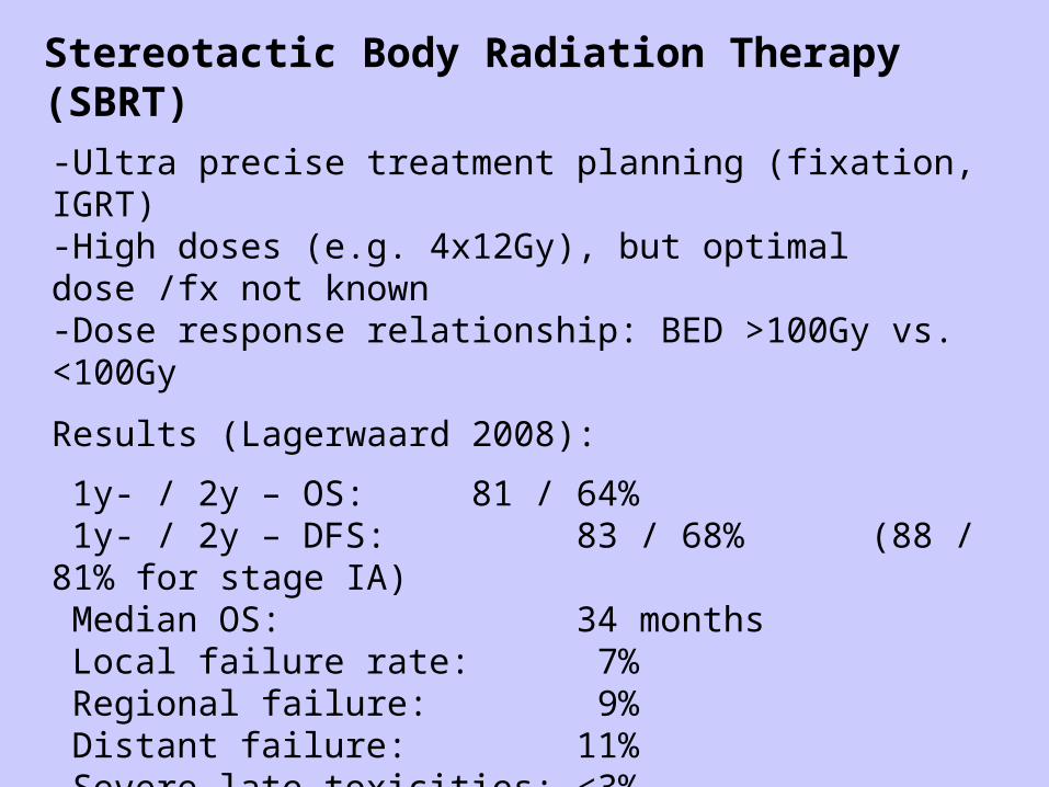
Stereotactic Body Radiation Therapy (SBRT)
-Ultra precise treatment planning (fixation, IGRT)-High doses (e.g. 4x12Gy), but optimal dose /fx not known-Dose response relationship: BED >100Gy vs. <100Gy
Results (Lagerwaard 2008):
1y- / 2y – OS: 81 / 64%1y- / 2y – DFS: 83 / 68% (88 / 81% for stage IA)Median OS: 34 monthsLocal failure rate: 7%Regional failure: 9%Distant failure: 11%Severe late toxicities: <3%
-Results superior to conventional 3D-CRT-For stage IA results near surgery

SBRT – Example-T2 N0-CR after radical radiation-COPD with emphysema

Other Techniques improving Outcome
Hyperfractionation (Jeremic 1997, 1999)Stage I Stage II
Median survival 33mts. 27mts.
5y-OS 30% 25%
Protons (Bush 2004) 3y local control 74%
Disease-specific survival
72%
Pneumonitis, esophageal or late cardiac toxicity
0%
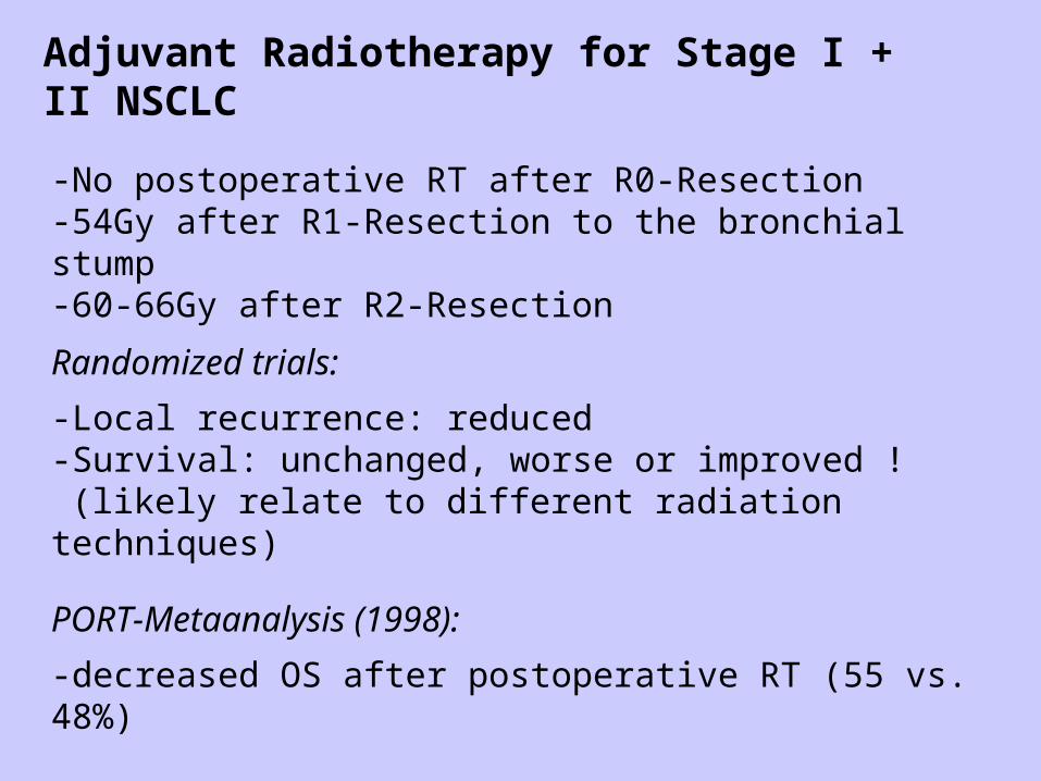
Adjuvant Radiotherapy for Stage I + II NSCLC
-No postoperative RT after R0-Resection-54Gy after R1-Resection to the bronchial stump-60-66Gy after R2-Resection
Randomized trials:
-Local recurrence: reduced-Survival: unchanged, worse or improved !
(likely relate to different radiation techniques)
PORT-Metaanalysis (1998):
-decreased OS after postoperative RT (55 vs. 48%)
Adjuvant Radiotherapy for Stage I + II NSCLC
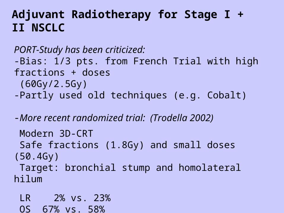
PORT-Study has been criticized:-Bias: 1/3 pts. from French Trial with high fractions + doses
(60Gy/2.5Gy)-Partly used old techniques (e.g. Cobalt)
-More recent randomized trial: (Trodella 2002)
Modern 3D-CRTSafe fractions (1.8Gy) and small doses (50.4Gy)Target: bronchial stump and homolateral hilum
LR 2% vs. 23%OS 67% vs. 58%Long-term toxicity acceptable
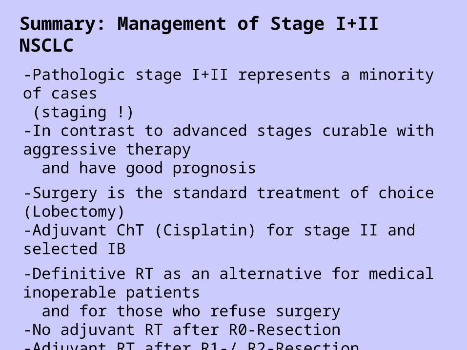
Summary: Management of Stage I+II NSCLC
-Pathologic stage I+II represents a minority of cases(staging !)
-In contrast to advanced stages curable with aggressive therapy and have good prognosis
-Surgery is the standard treatment of choice (Lobectomy)-Adjuvant ChT (Cisplatin) for stage II and selected IB
-Definitive RT as an alternative for medical inoperable patients and for those who refuse surgery-No adjuvant RT after R0-Resection-Adjuvant RT after R1-/ R2-Resection
-Further trials are needed to establish the role of RT in a post- operative setting and its optimal dose/fractionation/technique in a radical setting
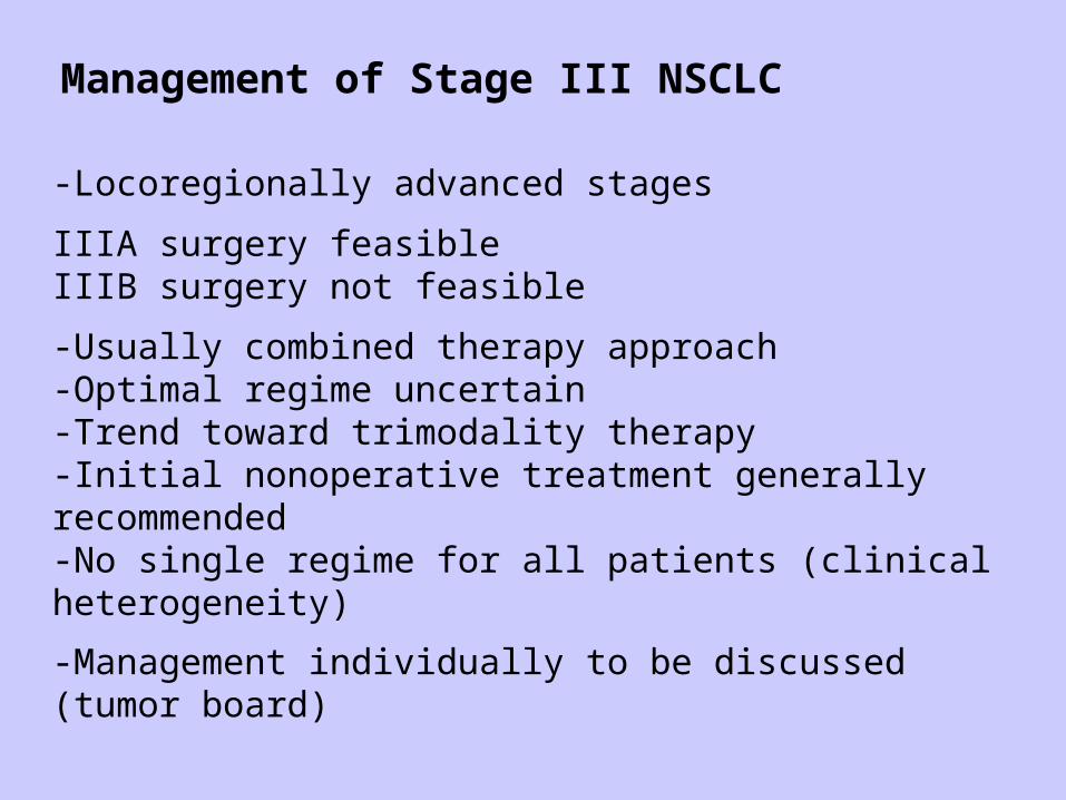
Management of Stage III NSCLC
-Locoregionally advanced stages
IIIA surgery feasibleIIIB surgery not feasible
-Usually combined therapy approach-Optimal regime uncertain-Trend toward trimodality therapy-Initial nonoperative treatment generally recommended-No single regime for all patients (clinical heterogeneity)
-Management individually to be discussed (tumor board)

Radiotherapy for Stage III NSCLC
Definitive radiotherapy alone
-for patients who are not fit for combined treatment-isolated thoracic recurrence after surgery-palliative for patients with poor performance status or stage IV
Early randomized trial: RT vs. Placebo (Roswit 1968) modest but significant survival benefit (18 vs. 14% at 1 year)
RT alone: MS 10mts.5y-OS 5%
Factors associated with improved prognosis:(Basaki 2006, RTOG 93-11 2008)-small primary tumor-small total tumor volume

Radiotherapy for Stage III NSCLC
Definitive radiotherapy alone
Should it be given immediately or deferred ?
Randomized trial: immediate RT vs. RT reserved for symptoms(Falk 2002)
-median survival ns-rate of symptom control similar
Palliative symptomatic care is a valuable option for patients with locoregionally advanced NSCLC who are not candidates for combined modality treatment.
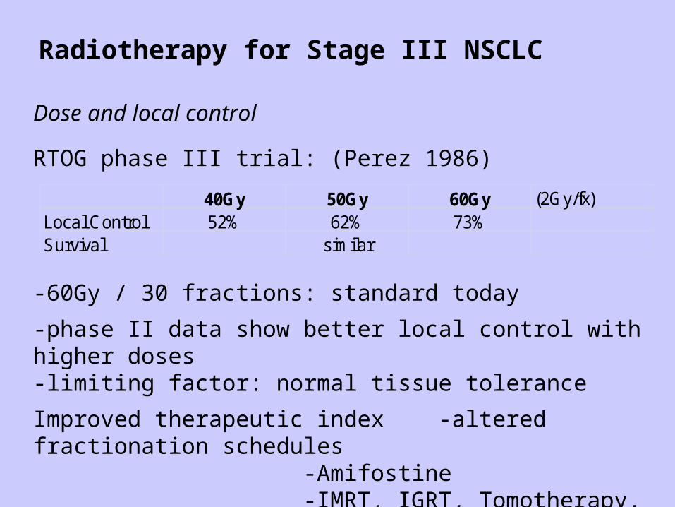
Radiotherapy for Stage III NSCLC
Dose and local control
RTOG phase III trial: (Perez 1986)
40Gy 50Gy 60Gy (2Gy/fx)Local Control 52% 62% 73%Survival similar
-60Gy / 30 fractions: standard today
-phase II data show better local control with higher doses-limiting factor: normal tissue tolerance
Improved therapeutic index -altered fractionation schedules-Amifostine-IMRT, IGRT, Tomotherapy, Protons..
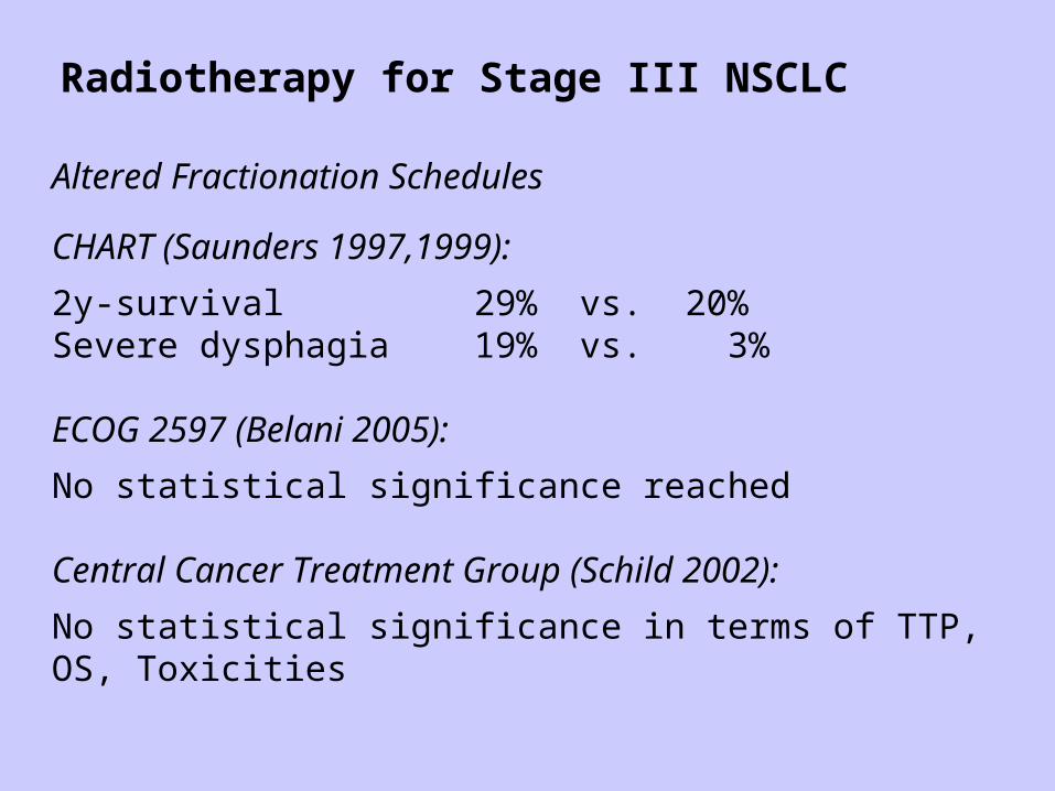
Radiotherapy for Stage III NSCLC
Altered Fractionation Schedules
CHART (Saunders 1997,1999):
2y-survival 29% vs. 20%Severe dysphagia 19% vs. 3%
ECOG 2597 (Belani 2005):
No statistical significance reached
Central Cancer Treatment Group (Schild 2002):
No statistical significance in terms of TTP, OS, Toxicities
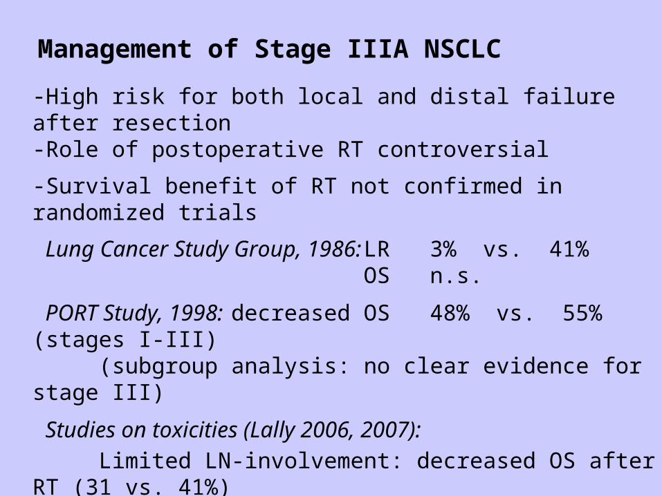
Management of Stage IIIA NSCLC
-High risk for both local and distal failure after resection-Role of postoperative RT controversial
-Survival benefit of RT not confirmed in randomized trials
Lung Cancer Study Group, 1986: LR 3% vs. 41%OS n.s.
PORT Study, 1998: decreased OS 48% vs. 55% (stages I-III)(subgroup analysis: no clear evidence for stage III)
Studies on toxicities (Lally 2006, 2007):
Limited LN-involvement: decreased OS after RT (31 vs. 41%)N2-disease: improved OS after postop. RT (27 vs. 20%)Death from cardiac toxicities: increased for pts. treated in early studies (1983-1988) not increased for those treated after 1989
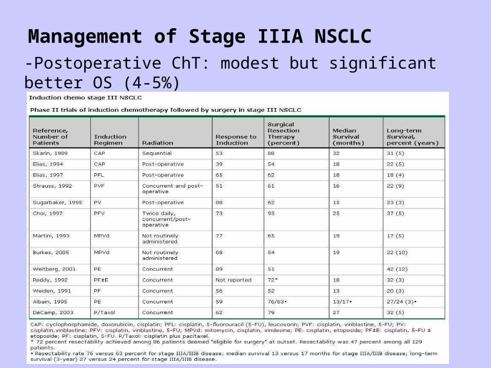
Management of Stage IIIA NSCLC-Postoperative ChT: modest but significant better OS (4-5%)-Promising results from preoperative ChT

Management of Stage IIIA NSCLC
-Better survival after adjuvant ChT-Promising results of phase II data with induction ChT
→ New Protocols:-Role of preoperative RT-ChT (SAKK)-Role of postoperative RT (EORTC)

Summary: Management of Stage IIIA NSCLC
-Pre- or postoperative ChT-No established role of pre- or postoperative RT → RT in Clinical Trials
(e.g. SAKK 16/00: RT/ChT – OP vs. ChT – OP)
-No postoperative RT recommended routinely Postoperative RT recommended: N2 (multilevel)
R1/R2
-Preoperative RT for Pancoast Tumor (45-50Gy)
-Radical RT (+/- ChT) for medically inoperable patients (60Gy)(concomitant better than sequential, see stage IIIB)

Management of Stage IIIB NSCLC
-Long Term OS < 5% ! (Hagen 1997)
-Most patients die from metastasis
-Median survival prolonged 8-10 months with RT-ChT for younger patients with good performance status (Sause 1997)
-Other patients: good palliation by RT
-Combined ChT-RT better survival than RT alone (Pignon 1994)
-Concomitant ChT-RT better than sequential, but more toxicities (Furuse 1999, RTOG 9410)
-Role of surgery uncertain (SAKK 16/01: preoperative ChT-RT)
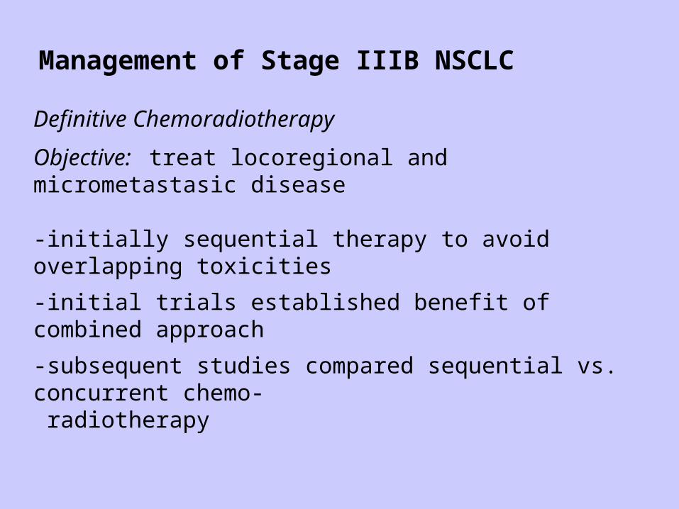
Management of Stage IIIB NSCLC
Definitive Chemoradiotherapy
Objective: treat locoregional and micrometastasic disease
-initially sequential therapy to avoid overlapping toxicities
-initial trials established benefit of combined approach
-subsequent studies compared sequential vs. concurrent chemo- radiotherapy
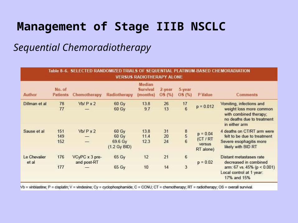
Management of Stage IIIB NSCLC
Sequential Chemoradiotherapy

Management of Stage IIIB NSCLC
Concurrent Chemoradiotherapy
Objective: early treatment of micrometastasesradio-sensitization (better local control)
-randomized trials established this approach as the preferred treatment
-toxicity is increased but manageable

Management of Stage IIIB NSCLC
Concurrent Chemoradiotherapy
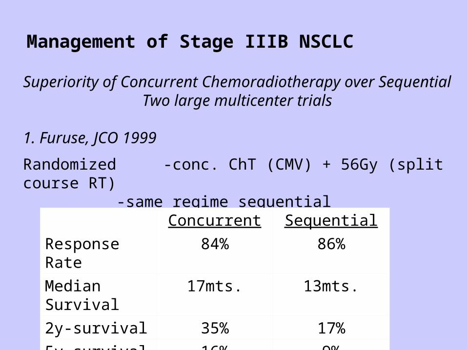
Management of Stage IIIB NSCLC
Superiority of Concurrent Chemoradiotherapy over SequentialTwo large multicenter trials
1. Furuse, JCO 1999
Randomized -conc. ChT (CMV) + 56Gy (split course RT)-same regime sequential
Concurrent Sequential
Response Rate 84% 86%
Median Survival 17mts. 13mts.
2y-survival 35% 17%
5y-survival 16% 9%

Management of Stage IIIB NSCLC
Superiority of Concurrent Chemoradiotherapy over SequentialTwo large multicenter trials
2. RTOG 9410
Randomized -conc. ChT (CV) + 60Gy-same regime sequential
Concurrent Sequential
Median Survival 17mts. 14.6mts.
4y-survival 21% 127%
ToxicityIncreased, but nut increased
treatment related death
Management of Stage IIIB NSCLC
Concurrent low dose Chemoradiotherapy
Objective: improved locoregional controlminimize toxicity
-only one randomized trial demonstrate benefit over RT alone (Schaake-Koning, 1992)
-several other studies failed to demonstrate survival benefit
-no trials comparing low dose vs. standard dose ChT
-option for elderly patients

Management of Stage IIIB NSCLC
Recommendations:
-Concomitant ChT-RTas first choice
-Concomitant daily low-dose Cisplatin + RT 60Gyelderly patients (Schake-Koning, 1992)
-Sequential ChT-RT: Cisplatin + 60Gy (Dillman, 1990)for large tumors
-RT only (30 x 2Gy – 13-15 x 3Gy)poor performance status, palliation
-Surgery only within study protocol or selected patients (e.g. T4 N0-1 after induction therapy)
Summary: Management of Stage IIIB NSCLC
-Heterogeneous group, therapy to be discussed at tumor board-Radical multimodality treatment vs. good palliation-Combined Radio-Chemotherapy is standard treatment-Concomitant better than sequential (survival benefit) but more
toxicities-Sequential Chemo- Radiotherapy or RT alone for unfit patients-Induction Chemotherapy for extensive tumor-volume which can not be encompassed in reasonable RT portals
-Role of Surgery uncertain, only selected patients
-Optimal regime not clear, therapy within clinical trials as possible:Induction-therapy – OPAccelerated RT schemes
New drugs + concomitant RT …..

Management of RT Toxicity - Pneumonitis
Pneumonitis: 4-6 wks. after RT (Fibrosis after 12-24 mts.)
Symptoms: fever, cough, illness
Risk factors:
-Lung function (FEV1)-Treated volume: V20=25% (8% pneumonitis)
V20=37% (39% pneumonitis)V10, V5, …. V30-40 (fibrosis)
-Dmean: <10Gy - very small risk20Gy - 15% risk30Gy - 50% risk
Treatment: Antibiotics (e.g. Roxithromycin) for 10dSteroids (e.g. Prednisone) beginning with high dose for 6wks. (reducing doses)

Management of RT Toxicity - Pneumonitis
Radiation portal (left) with subsequentradiation pneumonitis
Sequential transverse imagesthrough lung showing radiationpneumonitis in right lung
Radiographic finding: diffuse interstitial infiltrate

Management of RT Toxicity - Fibrosis
Rosen, I. I. et al. Radiology 2001;221:614-622

RT-Planning – Definition of Target Volumes
ICRU 50 + 62
Gross Tumour Volume
Clinical Target Volume
Planning Target Volume
= critical step
= weakest link in radiotherapy chain

RT-Planning – Defining the GTV
CT: standard imaging modality
Complementary information by MRI and PET scanning
Limiting factors of CT imaging for lung cancer:
-planning-CT without intravenous contrast so as not to disturb the electron density information interpretation always in conjunction with diagnostic CT
-not routinely possible to distinguish T3 – T4 (MRI some advantages)
-MRI used for imaging apical primary tumours (Pancoast)
-Sensitivity / specificity only 60 / 77% for LN knowledge of normal anatomy (LN levels, hilar anatomy) ! knowledge of patterns of lymphatic drainage

RT-Planning – Defining the GTV
Knowledge of anatomyLN levels(American College of Surgeons)

RT-Planning Defining the GTV
Knowledge of anatomyLN levels - Cross Sectional Anatomy
Murray JG, Eur J Radiol, 1993,17:61-68.


RT-Planning - Defining the GTV
Cross SectionalAnatomy - Suggested Paper

RT-Planning – Defining the GTVKnowledge of lymphatic drainage according to localisation of PT(Hata 1990)

RT-Planning – Defining the GTV
Integrating PET
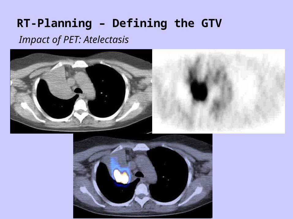
Value of PET for PT: Atelectasis – reduction of irradiated volume
Value of PET for LN staging: Sensitivity 79% Specificity 91% Negative predictive value 95% Positive predictive value 80% (hot spots still require verification)
Value of PET for Metastases: metastases detected in10-15% of surgical candidates
RT-Planning – Defining the GTV
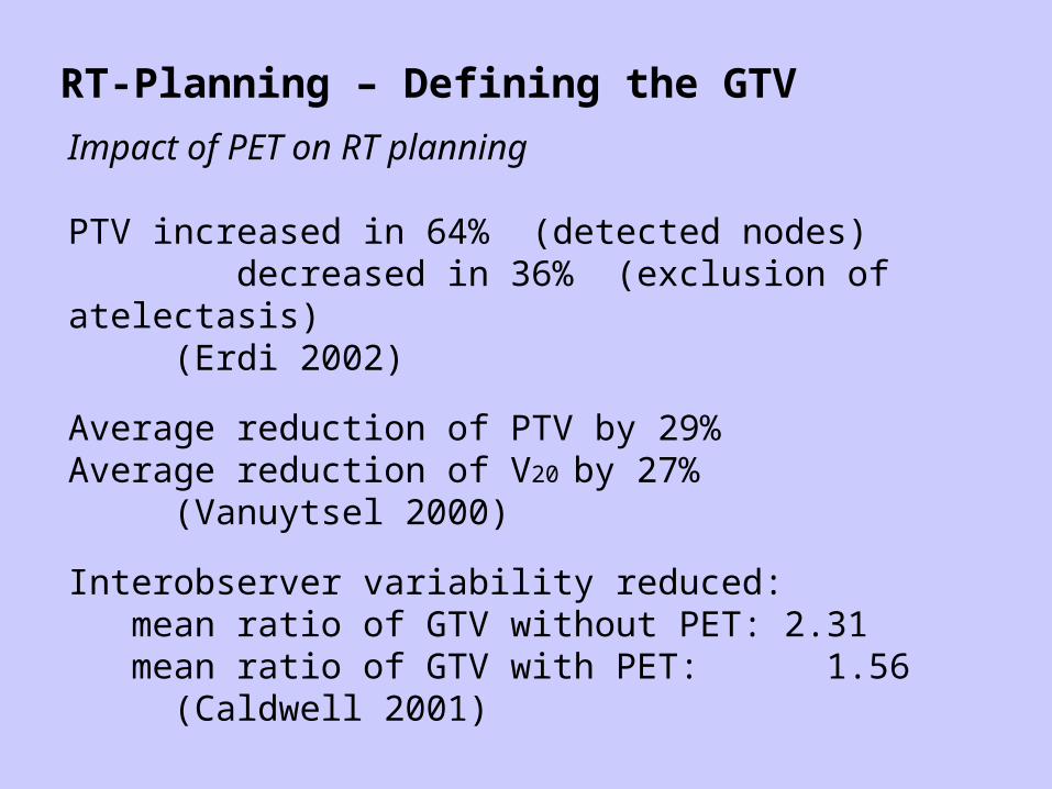
Impact of PET on RT planning
PTV increased in 64% (detected nodes) decreased in 36% (exclusion of atelectasis) (Erdi 2002)
Average reduction of PTV by 29%Average reduction of V20 by 27% (Vanuytsel 2000)
Interobserver variability reduced: mean ratio of GTV without PET: 2.31 mean ratio of GTV with PET: 1.56 (Caldwell 2001)
RT-Planning – Defining the GTV
Impact of PET: Atelectasis

RT-Planning – Defining the GTV
Impact of PET: PTV
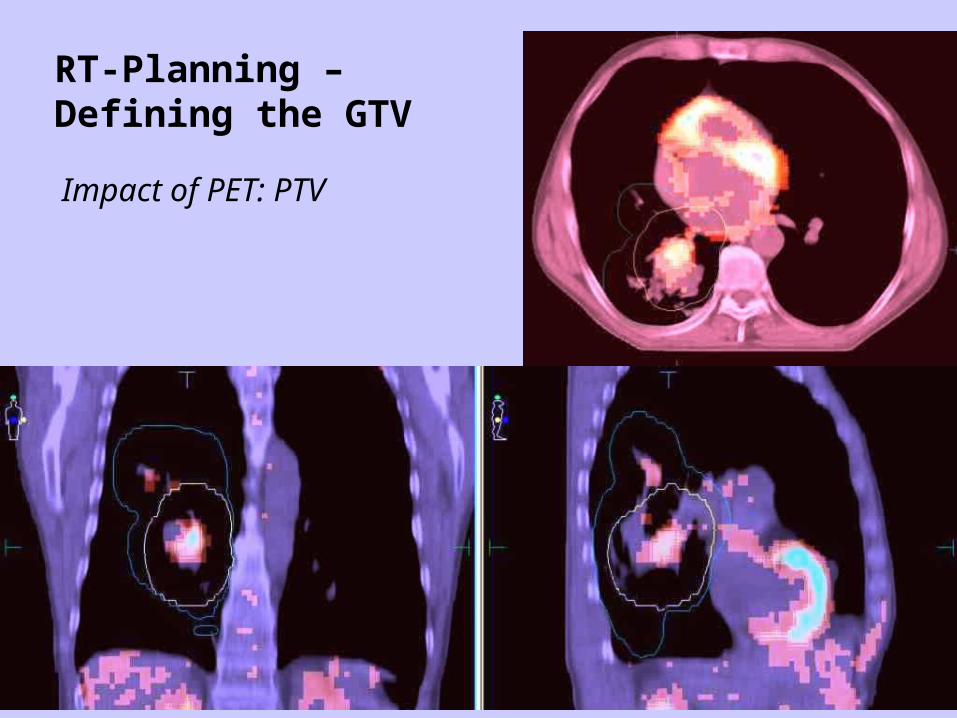
RT-Planning – Defining the GTV
Impact of PET: PTV

RT-Planning – Defining the GTVImpact of PET: PTV – RT Plan

RT-Planning – Defining the GTV
Limiting factors of PET
-Resolution 4-8mm (depending on scanner and institution)
-Registration errors (esp. with software based fusion)
-Threshold value (SUV) individually to be determined
Summary:
PET is a promising complementary tool in RT planning of NSCLC. Its value for staging has been established and preliminary reports suggest that it may lead to more consistent definition of GTV in RT planning. However, it is still not clear, whether this will translate into better survival.

RT-Planning – Defining the CTV
1. Margin around primary tumour (microscopic spread)
Histopathologic quantification of subclinical cancer around the grossly visible primary (Giraud 2000):
Microscopic extension Adeno Squamos mean value 2.69mm 1.48mm 5mm margin covers: 80% 91% margin to cover 95% 8mm 6mm
This data could also be used for IMRT planning:
-define constraint for GTV (dose escalation to primary)-define constraint for subclinical disease (less dose)
-increase therapeutic index

RT-Planning – Defining the CTV
2. Subclinical lymph nodes (ENI)
-high risk of nodal spread in lung cancer-but value of ENI is not proven
Reasons against ENI:
-less than 20% locally controlled 1y after RT with conventional dose (Arriagada 1991)-need for more intense treatment to gross tumour-large volumes prevent dose escalation (normal tissue tolerance)-small primary tumor and small total tumor volume predictive (Basaki 2006, RTOG 93-11 2008)-modern chemotherapy regimens may lead to better control of microscopic disease

RT-Planning – Defining the CTV2. Subclinical lymph nodes (ENI)

RT-Planning – Defining the CTV
2. Subclinical lymph nodes (ENI)
From large ....
“Old“ Standard … (Perez 1997)

RT-Planning – Defining the CTV
2. Subclinical lymph nodes (ENI)
.... to small !
…“New“ Trend (IMRT 2007)

RT-Planning – Defining the PTV
ICRU recommendations
CTV ...
+ Internal Margin (Internal Target Volume) variations in position, size and shape of CTV
(internal reference systemattached to the patient)
+ Set-up Margin variations in relation patient - beam
(external reference systemattached to machine)

RT-Planning – Defining the PTV
Reducing set-up uncertainty:
-Tattoos (instead of skin markers)-Custom immobilisation devices

RT-Planning – Defining the PTVReducing set-up uncertainty:
-Daily EPID: -matching DRR - EPI -distinguish between systematic (needs correction) and random error (no correction needed)

RT-Planning – Defining the PTV
Reducing respiration induced errors:
-Breath - hold -Voluntary (Deep Inspiration Breath Hold) -Forced (Active Breathing Control)
-CT scanning -Slow scanning -Respiration correlated CT -Gating
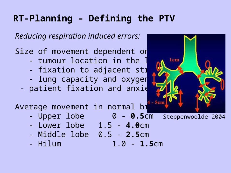
RT-Planning – Defining the PTV
Reducing respiration induced errors:
Size of movement dependent on: - tumour location in the lung - fixation to adjacent structures - lung capacity and oxygenation
- patient fixation and anxiety
Average movement in normal breathing: - Upper lobe 0 - 0.5cm - Lower lobe 1.5 - 4.0cm - Middle lobe 0.5 - 2.5cm - Hilum 1.0 - 1.5cm
Steppenwoolde 2004

RT-Planning – Defining the PTVReducing respiration induced errors:
Gated CT normally reduces the marginPTV - CTV(compared to using published data):

RT-Planning – Defining the PTV
Drawing PTV in gated planning CT:
-Define GTV/CTV for inspiration and expiration phase-Give a margin of 0.5 - 1cm in all directions (setup uncertainty)
Closing Words:
DON’T use dose escalation and highly conformal techniques such as IMRT for lung cancer until tumour motion can be taken into account !
In the meantime ...
-Outline GTV as best as possible-Construct CTV based on the literature-Construct PTV based on measured tumour motion and known setup uncertainty.


