RANDOM ARRAY OF COLOUR FILTERS IN THE EYES OF BUTTERFLIES
KENTARO ARIKAWA1,* AND DOEKELE G. STAVENGA2
1Graduate School of Integrated Science, Yokohama City University, 22-2 Seto, Kanazawa-ku, Yokohama 236, Japanand 2Department of Biophysics, University of Groningen, Groningen, the Netherlands
Accepted 16 July 1997
The compound eye of the Japanese yellow swallowtailbutterfly Papilio xuthus is not uniform. In a combinedhistological, electrophysiological and optical study, wefound that the eye of P. xuthus has at least three differenttypes of ommatidia, in a random distribution. In eachommatidium, nine photoreceptors contribute microvilli tothe rhabdom. The distal two-thirds of the rhabdom lengthis taken up by the rhabdomeres of photoreceptors R1–R4.The proximal third consists of rhabdomeres ofphotoreceptors R5–R8, except for the very basal part, towhich photoreceptor R9 contributes. In all ommatidia, theR1 and R2 photoreceptors have a purple pigmentationpositioned at the distal tip of the ommatidia. The R3–R8
photoreceptors in any one ommatidium all have eitheryellow or red pigmentation in the cell body, concentratednear the edge of the rhabdom. The ommatidia with red-pigmented R3–R8 are divided into two classes: one classcontains an ultraviolet-fluorescing pigment. The differentpigmentations are presumably intimately related to thevarious spectral types found previously inelectrophysiological studies.
Butterflies are often admired for their spectacular colourpatterns. They are therefore generally assumed to possesscolour vision. Behavioural experiments on butterflies haveindicated that they can indeed perceive colour contrast (forexample Hidaka and Yamashita, 1975; Ilse, 1928, 1937, 1941;Kolb and Scherer, 1982; Wehner, 1981). Compared with bees,in which extensive behavioural studies have demonstrated theability to see colour (Menzel and Backhaus, 1989), and flies,which also possess a clear colour discrimination system(Fukushi, 1989; Troje, 1993), convincing behavioural evidencefor colour vision in butterflies is lacking.
A basic physiological requirement for colour vision is theexistence of a set of different spectral receptor types in theretina. This is certainly fulfilled in a number of butterflyspecies, as shown by anatomical (Ribi, 1978, 1987), optical(Bernard, 1979; Bernard and Remington, 1991) andelectrophysiological (Eguchi et al. 1982; Horridge et al. 1984;Kinoshita et al. 1997; Matic 1983; Shimohigashi andTominaga, 1991; Steiner et al. 1987) studies. Presently, thebest characterized retina of a butterfly, in terms of the spectralproperties of the photoreceptors, is that of the Japanese yellowswallowtail butterfly Papilio xuthus. We have demonstratedpreviously that the retina contains at least five types of spectralreceptors, peaking in the ultraviolet, violet, blue, green and redwavelength regions (Arikawa et al. 1987). Subsequent work
has demonstrated that the nine photoreceptors of anommatidium, R1–R9 (see Fig. 1), are spectrallyheterogeneous. R1 and R2 are ultraviolet, violet or bluereceptors, R3 and R4 are green receptors (Bandai et al. 1992)and R5–R9 are either green or red receptors (Arikawa andUchiyama, 1996).
Within this diverse population of photoreceptors, the natureand composition of spectral receptors in the ommatidia has yetto be elucidated. A more complete characterization of theommatidia and their arrangement over the retina is the aim ofthe present paper. Histological, electrophysiological andoptical experiments on the compound eye of P. xuthus haveidentified three different types of ommatidia, randomlydistributed over the retina. This random distribution may becrucial for colour vision.
Materials and methodsAnimals
The Japanese yellow swallowtail butterfly Papilio xuthusLinnaeus was held as a laboratory stock culture. Other specieswere collected in Yokohama, Japan.
Anatomy
For light microscopy, the compound eyes were isolated from
2502 K. ARIKAWA AND D. G. STAVENGA
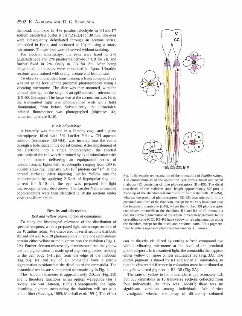
Fig. 1. Schematic representation of the ommatidia of Papilio xuthus.The ommatidium is of the apposition type with a fused and tieredrhabdom (R) consisting of nine photoreceptors (R1–R9). The distaltwo-thirds of the rhabdom (total length approximately 500 µm) ismade up of the rhabdomeral microvilli of four distal cells (R1–R4),whereas the proximal photoreceptors, R5–R8, bear microvilli in theproximal one-third of the rhabdom, except for the very basal part nearthe basement membrane (BM), where the bilobed R9 photoreceptorcontributes microvilli to the rhabdom. R1 and R2 of all ommatidiacontain purple pigmentation at the region immediately proximal to thecrystalline cone (CC). R3–R8 have yellow or red pigmentation alongthe rhabdom except for the distal and proximal parts. R9 is pigment-free. Numbers represent photoreceptor number. C, cornea.
the head, and fixed in 4 % paraformaldehyde in 0.1 mol l−1
sodium cacodylate buffer at pH 7.2 (CB) for 30 min. The eyeswere subsequently dehydrated through an acetone series,embedded in Epon, and sectioned at 10 µm using a rotarymicrotome. The sections were observed without staining.
For electron microscopy, the eyes were fixed in 2 %glutaraldehyde and 2 % paraformaldehyde in CB for 2 h, andfurther fixed in 2 % OsO4 in CB for 2 h. After beingdehydrated, the tissues were embedded in Epon. Ultrathinsections were stained with uranyl acetate and lead citrate.
To observe ommatidial transmission, a fresh compound eyewas cut at the level of the proximal photoreceptors using avibrating microtome. The slice was then mounted, with thecorneal side up, on the stage of an epifluorescent microscope(BX-60, Olympus). The focus was at the corneal surface. First,the transmitted light was photographed with white lightillumination, from below. Subsequently, the ultraviolet-induced fluorescence was photographed (objective 4×,numerical aperture 0.16).
Electrophysiology
A butterfly was mounted in a Faraday cage, and a glassmicropipette, filled with 5 % Lucifer Yellow CH aqueoussolution (resistance 150 MΩ), was inserted into the retinathrough a hole made in the dorsal cornea. After impalement ofthe electrode into a single photoreceptor, the spectralsensitivity of the cell was determined by axial stimulation witha point source delivering an equiquantal series ofmonochromatic lights with wavelengths ranging from 290 to700 nm (maximal intensity 5.0×10¹¹ photons cm−2 s−1 at thecorneal surface). After injecting Lucifer Yellow into thephotoreceptor, by applying 2–5 nA of hyperpolarizing d.c.current for 5–10 min, the eye was prepared for lightmicroscopy as described above. The Lucifer-Yellow-injectedphotoreceptors were then identified in 10 µm sections underviolet epi-illumination.
Results and discussionRed and yellow pigmentation of ommatidia
To study the histological relevance of the distribution ofspectral receptors, we first prepared light microscope sections ofthe P. xuthus retina. We discovered in serial sections that bothR3 and R4 and R5–R8 photoreceptors in any one ommatidiumcontain either yellow or red pigment near the rhabdom (Figs 1,2A). Further electron microscopy demonstrated that the yellowand red pigmentation is made up of pigment granules, residingin the cell body 1–1.5µm from the edge of the rhabdom(Fig. 2B). R1 and R2 of all ommatidia have a purplepigmentation positioned at the distal tip of the ommatidia. Theanatomical results are summarized schematically in Fig. 1.
The rhabdom diameter is approximately 2.0 µm (Fig. 2B)and it therefore functions as an optical waveguide (for areview, see van Hateren, 1989). Consequently, the light-absorbing pigment surrounding the rhabdom will act as acolour filter (Stavenga, 1989; Marshall et al. 1991). This effect
can be directly visualized by cutting a fresh compound eyewith a vibrating microtome at the level of the proximalphotoreceptors. In transmitted light, the ommatidia then appeareither yellow or (more or less saturated) red (Fig. 3A). Thepurple pigment is shared by R1 and R2 in all ommatidia, sothat the observed difference in coloration must be attributed tothe yellow or red pigment in R3–R8 (Fig. 2A).
The ratio of yellow to red ommatidia is approximately 1:3.For 653 ommatidia in 10 transverse sections collected fromfour individuals, the ratio was 166:487; there was nosignificant variation among individuals. We furtherinvestigated whether the array of differently coloured
2503Butterfly colour filters
Fig. 2. Pigmentation in the ommatidia ofPapilio xuthus. (A) Unstained Epon-embeddedsection through the proximal tier of theommatidium, where four proximalphotoreceptors, R5–R8, contribute theirmicrovilli to the rhabdom. The four colouredpatches (arrowheads) are pigment clusters inR5–R8. Red-pigment-containing and yellow-pigment-containing ommatidia are distributedrandomly throughout the retina. Comparisonsof frozen sections with Epon-embeddedsections show that the colours of the pigmentsare virtually unchanged by the short fixationtime (30 min) without osmication used inpreparing these sections for light microscopy.Scale bar, 10 µm. (B) Electron micrograph of ared-pigment-containing ommatidium. Thepigment granules (arrowheads) reside in thecell body area 1–1.5 µm from the edge of therhabdom (R). Numbers indicate photoreceptornumber. Scale bar, 1 µm.
ommatidia has any regularity. To do this, we counted thefrequency of transition, for example from yellow to red or fromred to red, along the three axes of the hexagonal lattice. Itappeared that the transition frequency is independent of thefrequency of the type of its neighbours and only reflects theabsolute probability of the ommatidial type (10 micrographsyielded χ2=2.25, d.f.=4).
To investigate any possible correlation between the spectralreceptor types and the pigmentation, we recorded spectralsensitivities from single photoreceptors and marked the cellsby injecting Lucifer Yellow. Subsequently, we identified thepigmentation of the ommatidia to which the penetratedphotoreceptors belonged by light microscope histology. Wefound that the proximal R5–R8 cells in the yellow-pigment-
containing ommatidia, without exception, appear to be green-sensitive receptors. The R5–R8 cells in the red-pigment-containing ommatidia are always red-sensitive receptors.
Of course, it is virtually impossible to prove conclusivelyusing electrophysiology that cells R5–R8 in a singleommatidium are always identical. A powerful alternative isoffered by histological in situ hybridization to localize differentmRNAs encoding visual pigment opsins. Our preliminaryresults indicate that photoreceptors R5–R8 in a singleommatidium always contain identical opsin mRNA (Kitamotoet al. 1996); that is, it is most likely that R5–R8 in the red-pigment-containing and the yellow-pigment-containingommatidia are all red- and green-sensitive receptors,respectively.
2504 K. ARIKAWA AND D. G. STAVENGA
Fig. 3. (A) Ommatidial pigmentation seen in a slice of fresh eye observed in transmitted light, i.e. under antidromic illumination. (B) Ommatidialfluorescence under ultraviolet epi-illumination of the same preparation as in A. Arrowheads indicate identical ommatidia. Fluorescing ommatidiain B appear very pale or pink in A. Scale bar, 200 µm.
Fluorescing ommatidiaWe also investigated the intact eye of P. xuthus using
fluorescence microscopy and found that a further aspect ofretinal heterogeneity can be directly observed in vivo.Ultraviolet epi-illumination of the P. xuthus eye excites adistinct whitish emission in approximately one-quarter of the
Fig. 4. Photoreceptor pigmentation in lycaenid and satyrid butterfliesPigmented ommatidia (arrowheads) and non-pigmented ommatidia (arro(Lycaenidae), (C) Neope goschkevitschii (Satyridae), (D) Ypthima argu
ommatidia in the ventral half of the eye (Fig. 3B). In sixmicrographs, 28 % of the ommatidia fluoresced. Again, theirdistribution appears to be random. Upon focusing at levelsproximal to the cornea, waveguide mode patterns appear,indicating that the fluorescence originates from the rhabdom(Franceschini et al. 1981; Nilsson et al. 1988; van Hateren,
. Transverse sections of ommatidia were observed without staining.ws) are evident. (A) Zizeeria maha (Lycaenidae), (B) Lycaena phlaeass (Satyridae). Scale bars, 10 µm (A, B, D) and 25 µm (C).
2505Butterfly colour filters
1989). Preliminary photochemical and biochemicalexperiments suggest that the fluorescing pigment is a retinoid,but conclusive results have yet to be obtained. The ultraviolet-fluorescing ommatidia are not seen in the dorsal half of the eye.Their presence, if they are there, is obscured by a distinctcorneal fluorescence.
Combined fluorescence and transmission microscopy of eyeslices revealed that those ommatidia that fluoresce underultraviolet excitation appear a less saturated red, i.e. very paleto pink, in transmitted light (Fig. 3A,B). Furtherelectrophysiological experiments, combined with LuciferYellow staining and fluorescence microscopy, demonstratedthat violet receptors only occur in the fluorescing ommatidia.Ultraviolet and blue receptors were found exclusively in thenon-fluorescing ommatidia.
The sensitivity spectrum of the violet receptor is very sharp-peaked (Arikawa et al. 1987). This suggests the challenginghypothesis that the fluorescing pigment might act as a selectiveultraviolet absorbance filter. The red receptor also has anarrower spectral sensitivity spectrum than predicted from arhodopsin template (Arikawa et al. 1987). This is probably adirect consequence of the strong high-pass filtering by the redphotoreceptor pigmentation (Arikawa et al. 1996).
Comparative aspects
Our studies on P. xuthus show that its retina is a mesh of atleast three types of optically distinguishable ommatidia: onecontaining yellow pigment and two containing red pigment,one type of which contains an ultraviolet-fluorescing pigment.Together with the visual pigments of the photoreceptors, thesepigments are crucial in shaping the spectral sensitivity of thefive receptor types. How P. xuthus connects these receptors toachieve colour discrimination, which might even bepentachromatic, remains an intriguing problem. Behaviouralexperiments detailing the colour discrimination capabilities ofP. xuthus are now in progress.
A mesh of different classes of ommatidia has been foundpreviously in the retina of flies (Franceschini et al. 1981;Hardie, 1986; Chou et al. 1996), where the two centralphotoreceptors determine colour vision (Troje, 1993). Theobserved randomness of the retinal organization in P. xuthusis also shared with primates: a random distribution of M andL cones has, for instance, been demonstrated in talapoinmonkeys (Cercopithecus talapoin) (Mollon and Bowmaker,1992). In contrast, several fish species have a very regularretina (Lythgoe, 1979).
In a survey of the compound eyes of other butterflies(Fig. 4), we found a marked variation in pigmentation aroundthe rhabdom, not only in the species belonging to the familyPapilionidae (Papilio bianor, Papilio protenor, Papiliopolytes, Graphium sarpedon, Parnassius glacialis) but also inLycaenidae (Zizeeria maha, Lycaena phlaeas) and Satyridae(Neope goschkevitschii, Ypthima argus). These results are inagreement with previous optical studies on intact butterfly eyesusing epi-illumination microscopy (Bernard and Miller, 1970;Miller, 1979). These studies revealed a multicoloured
reflection from a tapetal mirror proximal to the rhabdom thatexists in the eye of all butterflies except for the Papilionidae.Although no direct evidence is available so far, the differencein pigmentation and coloration of the tapetal reflection stronglyindicates that the ommatidia are different in terms of thespectral receptor types they contain.
Compound eyes often exhibit considerable regionalization,because different parts of the eye are devoted to specific tasks,such as prey or mate recognition or polarization vision(Stavenga, 1992). Here, we have shown that, within restrictedregions of the butterfly eye, ommatidial characteristics candiffer considerably. Clearly, the concept that compound eyes,at least locally, consist of identical building blocks, theommatidia, does not hold. The random organization of thespectral receptors in the retina of butterflies seems to be auniversal feature, presumably because a diverse set of spectralreceptors is essential for a highly developed colour visionsystem (Bernard, 1979; Bernard and Remington, 1991;Goldsmith, 1990).
We thank P. Brakefield, M. F. Land and G. D. Bernard forreading critically an earlier version of the manuscript. Thiswork was supported by Grants from the Ministry of Education,Science, and Culture of Japan and the Uehara MemorialFoundation to K.A. and from the Japanese Society for thePromotion of Science (JSPS) to D.G.S.
ReferencesARIKAWA, K., INOKUMA, K. AND EGUCHI, E. (1987). Pentachromatic
visual system in a butterfly. Naturwissenschaften 74, 297–298.ARIKAWA, K., SCHOLTEN, D. G. W. AND STAVENGA, D. G. (1996).
Spectral origin of the red receptors in the retina of the butterflyPapilio xuthus. Zool. Sci. 13S, 118.
ARIKAWA, K. AND UCHIYAMA, H. (1996). Red receptors dominate theproximal tier of the retina in the butterfly Papilio xuthus. J. comp.Physiol. A 178, 55–61.
BANDAI, K., ARIKAWA, K. AND EGUCHI, E. (1992). Localization ofspectral receptors in the ommatidium of butterfly compound eyedetermined by polarization sensitivity. J. comp. Physiol. A 171,289–297.
BERNARD, G. D. (1979). Red-absorbing visual pigment of butterflies.Science 203, 1125–1127.
BERNARD, G. D. AND MILLER, W. H. (1970). What does antennaengineering have to do with insect eyes? IEEE Student J. 8, 2–8.
BERNARD, G. D. AND REMINGTON, C. L. (1991). Color vision inLycaena butterflies: Spectral tuning of receptor arrays in relation tobehavioral ecology. Proc. natn. Acad. Sci. U.S.A. 88, 2783–2787.
CHOU, W. H., HALL, K. J., WILSON, D. B., WIDEMAN, C. L., TOWNSON,S. M., CHADWELL, L. V. AND BRITT, S. G. (1996). Identification ofa novel Drosophila opsin reveals specific patterning of the R7 andR8 photoreceptor cells. Neuron 17, 1101–1115.
EGUCHI, E., WATANABE, K., HARIYAMA, T. AND YAMAMOTO, K.(1982). A comparison of electrophysiologically determinedspectral responses in 35 species of Lepidoptera. J. Insect Physiol.28, 675–682.
FRANCESCHINI, N., KIRSCHFELD, K. AND MINKE, B. (1981).Fluorescence of photoreceptor cells observed in vivo. Science 213,1264–1267.
2506 K. ARIKAWA AND D. G. STAVENGA
FUKUSHI, T. (1989). Learning and discrimination of coloured papersin the walking blowfly, Lucilia cuprina. J. comp. Physiol. A 166,57–64.
GOLDSMITH, T. H. (1990). Optimization, constraint and history in theevolution of eyes. Q. Rev. Biol. 65, 281–322.
HARDIE, R. C. (1986). The photoreceptor array of the dipteran retina.Trends Neurosci. 9, 419–423.
HIDAKA, T. AND YAMASHITA, K. (1975). Wing color pattern as thereleaser of mating behavior in the swallowtail butterfly, Papilioxuthus. Appl. Ent. Zool. 10, 263–267.
HORRIDGE, G. A., MARÇELJA, L. AND JAHNKE, R. (1984). Color visionin butterflies. I. Single colour experiments. J. comp. Physiol. A 155,529–542.
ILSE, D. (1928). Über den Farbensinn der Tagfalter. Z. vergl. Physiol.8, 658–691.
ILSE, D. (1937). New observations on responses to colours in egg-laying butterflies. Nature 140, 544–545.
ILSE, D. (1941). The colour vision of insects. Proc. phil. Soc. Glasgow65, 68–82.
KINOSHITA, M., SATO, M. AND ARIKAWA, K. (1997). Spectral receptorsof nymphalid butterflies. Naturwissenschaften 84, 199-201.
KITAMOTO, J., SAKAMOTO, K., OZAKI, K. AND ARIKAWA, K. (1996).The primary structures of butterfly opsins and their distribution inthe retina. Zool. Sci. 13S, 129.
KOLB, G. AND SCHERER, C. (1982). Experiments on wavelengthspecific behavior of Pieris brassicae L. during drumming and egg-laying. J. comp. Physiol. A 149, 325–332.
LYTHGOE, J. N. (1979). The Ecology of Vision. Oxford: ClarendonPress
MARSHALL, N. J., LAND, M. F., KING, C. A. AND CRONIN, T. W. (1991).The compound eyes of mantis shrimps (Crustacea, Hoplocarida,Stomatopoda). 2. Colour pigments in the eyes of stomatopodcrustaceans: polychromatic vision by serial and lateral filtering.Phil. Trans. R. Soc. Lond. B 334, 57–84.
MATIC, T. (1983). Electrical inhibition in the retina of the butterflyPapilio. I. Four spectral types of photoreceptors. J. comp. Physiol.A 152, 169–182.
MENZEL, R. AND BACKHAUS, W. (1989). Color vision in honey bees:
Phenomena and physiological mechanisms. In Facets of Vision (ed.D. G. Stavenga and R. C. Hardie), pp. 281–297. Berlin, Heidelberg,New York, London, Paris, Tokyo: Springer-Verlag.
MILLER, W. H. (1979). Ocular optical filtering. In Handbook ofSensory Physiology, vol. VII/6A (ed. H. Autrum), pp. 69–143.Berlin, Heidelberg, New York: Springer-Verlag.
MOLLON, J. D. AND BOWMAKER, J. K. (1992). The spatial arrangementof cones in the primate fovea. Nature 360, 677–679.
NILSSON, D.-E., LAND, M. F. AND HOWARD, J. (1988). Optics of thebutterfly eye. J. comp. Physiol. A 162, 341–366.
RIBI, W. A. (1978). Ultrastructure and migration of screeningpigments in the retina of Pieris rapae L. (Lepidoptera, Pieridae).Cell Tissue Res. 191, 57–73.
RIBI, W. A. (1987). Anatomical identification of spectral receptortypes in the retina and lamina of the Australian orchard butterfly,Papilio aegeus aegeus D. Cell Tissue Res. 247, 49–59.
SHIMOHIGASHI, M. AND TOMINAGA, Y. (1991). Identification of UV,green and red receptors and their projection to lamina in thecabbage butterfly, Pieris rapae. Cell Tissue Res. 247, 49–59.
STAVENGA, D. G. (1989). Pigments in compound eyes. In Facets ofVision (ed. D. G. Stavenga and R. C. Hardie), pp. 152–172. Berlin,Heidelberg, New York, London, Paris, Tokyo: Springer-Verlag.
STAVENGA, D. G. (1992). Eye regionalization and spectral tuning ofretinal pigments in insects. Trends Neurosci. 15, 213–218.
STEINER, A., PAUL, R. AND GEMPERLEIN, R. (1987). Retinal receptortypes in Aglais urticae and Pieris brassicae (Lepidoptera), revealedby analysis of the electroretinogram obtained with Fourierinterferometric stimulation (FIS). J. comp. Physiol. A 160,247–258.
TROJE, N. (1993). Spectral categories in the learning behaviour ofblowflies. Z. Naturforsch. C 48, 96–104.
VAN HATEREN, J. H. (1989). Photoreceptor optics, theory and practice.In Facets of Vision (ed. D. G. Stavenga and R. C. Hardie), pp.74–89. Berlin, Heidelberg, New York, London, Paris, Tokyo:Springer-Verlag.
WEHNER, R. (1981). Spatial vision in arthropods. In Handbook ofSensory Physiology, vol. VII/6C (ed. H. Autrum), pp. 287–616.Berlin, Heidelberg, New York: Springer-Verlag.