Page 1
REVIEW
Recent Advances in Osteogenesis Imperfecta
Tim Cundy
Received: 23 January 2012 / Accepted: 2 March 2012 / Published online: 27 March 2012
� Springer Science+Business Media, LLC 2012
Abstract ‘‘Osteogenesis imperfecta’’ is a term used to
describe a group of genetic disorders of variable phenotype
usually defined by recurrent fractures, low bone mass, and
skeletal fragility. Most cases are associated with mutations
in one of the type I collagen genes, but in recent years
several other forms have been identified with recessive
inheritance. In most instances the latter result from muta-
tions in genes encoding proteins involved in type I colla-
gen’s complex posttranslational modification or in genes
regulating bone matrix homeostasis. This article reviews
the recent discoveries and an approach to classification and
diagnosis. Bisphosphonates are widely used in patients
with osteogenesis imperfecta, but some important ques-
tions about their optimal usage, their utility in children and
adults with milder phenotypes, and their potential adverse
effects are not yet resolved.
Keywords Bisphosphonate � Matrix protein �Osteogenesis imperfecta � Pediatric bone disease �Type I collagen
Osteogenesis Imperfecta
The term ‘‘osteogenesis imperfecta’’ (OI) encompasses a
group of genetic disorders usually defined by recurrent
fractures, low bone mass, and skeletal fragility. Most cases
are associated with mutations in the genes encoding either
type I collagen (the most abundant and main structural
protein of bone) or (in about 10% of cases) genes encoding
proteins involved in type I collagen’s complex posttrans-
lational modification and intracellular trafficking. How-
ever, they are not synonymous with OI as a number of
genetic disorders affecting type I collagen or its post-
translational modification that have skeletal phenotypes are
not usually included under the OI rubric. These include
Caffey disease and the kyphoscoliosis, arthrochalasia, and
dermatosparaxis types of the Ehlers-Danlos syndrome
(types VI, VIIA/B, and VIIC, respectively). In addition,
some genetic disorders of skeletal fragility included in
classifications of OI are the consequence of mutations in
key osteoblast genes (LRP5 and SP7) that code for proteins
concerned with matrix homeostasis and are not directly
related to collagen metabolism and matrix structure.
Type I Collagen
Type I collagen is initially synthesized in the rough
endoplasmic reticulum as a precursor molecule (type I
procollagen) that combines two proa1(I) and one
proa2(I) peptide chains (coded by COL1A1 and COL1A2,
respectively) in a triple helix. Proa1(I) and proa2(I) have
similar structures, with a core triple-helical domain of
1,014 amino acids composed of uninterrupted Gly-Xaa-
Yaa tripeptide repeats, flanked by propeptides at both the
N- and C-terminal ends. During and after translation the
three chains undergo extensive modification. Prolyl-4-
hydroxylase converts virtually all Y-position proline resi-
dues to 4-hydroxyproline, an alteration that is essential for
thermal stability of the assembled trimer. In the absence of
this modification, the trimer melts (i.e., the individual
The author has stated that there is no conflict of interest.
T. Cundy (&)
Department of Medicine, Faculty of Medical & Health Sciences,
University of Auckland, Private Bag 92019, Auckland,
New Zealand
e-mail: [email protected]
123
Calcif Tissue Int (2012) 90:439–449
DOI 10.1007/s00223-012-9588-3
Page 2
chains unfold from the stable triple helix, at about 27�C,
whereas with full hydroxylation the melting temperature is
about 42�C). Some Y-position lysine residues within the
triple-helical domain are hydroxylated by the enzyme lysyl
hydroxylase-1 and glucose and galactose groups added by
glycosyltransferases. Hydroxylation of these triple-helical
residues is part of the pathway to form stable complex
intermolecular cross-links that provide the tensile strength
in tissues. Most of these modifications are completed dur-
ing translation and occur on individual chains. If there is a
delay in triple-helix folding, the process can continue but
the physical properties of the chains and molecules are
altered and contribute to an OI phenotype.
The three chains—proa1(I)2 and proa2(I)—that form a
trimer interact through regions in the carboxyl-terminal
propeptide of each chain. This creates the unusual situation
in which the full-length chain must be maintained in an
unfolded state while the carboxyl-terminal propeptides
fold, associate, and then begin the process of triple-helix
formation. Propogation of the collagen triple helix requires
a number of enzymes and molecular chaperones to ensure
correct folding and trimerization. These include peptidyl
disulfide isomerase, which also forms part of the prolyl
4-hydroxylase complex and likely involves prolyl peptidyl
cis–trans isomerase B (also known as cyclophilin B). This
protein can act on its own, to assist in the folding around
prolyl residues, such as those in the carboxyl-terminal
propeptide adjacent to cysteine residues, and as part of a
complex that includes two additional proteins, cartilage-
related protein and prolyl 3-hydroxylase, to modify certain
triple-helical prolines. The function of this last process is
not yet entirely clear, but when the complex is missing, the
propagation of the triple helix is altered and modification of
the chains increased.
Disulfide bonds between the carboxyl-terminal region of
the chains require protein-disulfide isomerase and act to
secure the three chains in a trimer. Lysine residues outside
the major triple-helical domain of type I collagen and
needed for the formation of mature intermolecular cross-
links are hydroxylated by lysyl hydroxylase-2. These
complex modifications, which are necessary for correct
folding, assuring thermal stability of the triple helix, and
cross-link formation between collagen molecules once they
are secreted into the matrix, need to take place in an orderly
and timely sequence; and various chaperone proteins,
including HSP47 and FKBP65, help to regulate this pro-
cess. Procollagen trimers are then transported via the Golgi
network and packaged into membrane-bound organelles,
where lateral aggregation, the intial phase of fibril forma-
tion, occurs. As secretion occurs, the procollagen mole-
cules are further processed into mature type I collagen
molecules by proteolytic cleavage of the N- and C-terminal
propeptides (by the enzymes ADAMTS-2 and BMP1,
respectively). Finally, the trimers are assembled into col-
lagen fibrils and fibers [1–3] and anchored in those posi-
tions by intermolecular lysine-derived cross-links in a
process that is begun by modification of specific residues
by lysyl oxidase.
Classification
The Sillence classification, published more than 30 years
ago, was the first systematic classification of OI phenotype
[4]. Although the original numbering system is somewhat
counterintuitive in that the ascending numbers do not
correlate with severity, the distinctions between the non-
deforming (type I), moderate (type IV), severe or pro-
gressively deforming (type III), and perinatal lethal forms
(type II) remains clinically useful [4].
In recent years, the genetic complexity of the molecular
basis of OI has become increasingly evident, and at the
same time the extensive phenotypic variation arising from
single loci has been documented clearly. The International
Skeletal Dysplasia Society has suggested that it is unten-
able to try to maintain tight correlations between ‘‘Sillence
types’’ and their molecular bases. They recommend
retaining the essence of the Sillence classification to
describe the phenotypic severity in OI, to free the clinical
classification from direct molecular reference, and to limit
the proliferation of new numbered ‘‘OI types’’ with each
new genetic discovery [5]. When faced with a patient with
possible OI, the clinician should firstly carefully define the
phenotype and the inheritance; taken together these will
usually point to the path by which one can identify the
likely genetic defect.
Defining the Phenotype
The important points to be taken from the history and
physical examination include a detailed family history of
bone disease; the presence of fractures detected in utero or
in the neonatal period; the nature, chronology, and outcome
of subsequent fractures; growth velocity and current height
and skeletal proportions; the presence and progression of
long bone deformity, acetabular protrusion, and kypho-
scoliosis. The distinction between mild, moderate, and
severe phenotypes is broadly based on the number of
fractures, the degree of deformity and growth impairment,
and the age at which abnormalities are first recognized.
Other phenotypic features of importance are head circum-
ference, joint mobility (measured, e.g., on the Beighton
scale) or joint contractures, scleral color, hearing, cardiac
murmurs, and tooth abnormalities, particularly dentino-
genesis imperfecta (Fig. 1a–c).
440 T. Cundy: Recent Advances in OI
123
Page 3
The standard biochemical tests of bone turnover are
generally unhelpful in diagnosis. There are a few exceptions:
plasma concentrations of procollagen-1 N-propeptide and
procollagen-1 C-propeptide are low in patients with hap-
loinsufficiency [6], and the ratios of pyridinoline to deoxy-
pyrodinoline in urine are altered in the Bruck syndrome
variants of OI (due to mutations in FKBP10 or PLOD2)
where the formation of lysine cross-links is impaired. Plain
radiography is very useful in defining the phenotype, but
bone density testing is of little diagnostic value and can be
difficult to perform and interpret when there is deformity or
short stature. It is, however, commonly used in monitoring
responses to bisphosphonate treatment.
Dominantly Inherited OI
Most patients with OI (* 90%) have mutations in one of
the type I collagen genes, COL1A1 or COL1A2. These are
large genes (51 and 52 exons, respectively), and disease-
causing mutations occur all along both. COL1A1 and
COL1A2 mutations are dominantly inherited, and the
phenotype can vary from the very mild to in utero lethal. It
is generally not necessary to undertake in vitro studies of
type I collagen or to sequence these genes if the diagnosis
is clear from the family history and phenotype. However,
further investigation is indicated if there are atypical fea-
tures or if there is uncertainty about the diagnosis or a need
for genetic counseling about recurrence risk, prenatal
diagnosis, or preimplantation genetic diagnosis.
There are two general classes of mutations in the type I
collagen genes that result in OI: those that cause a quanti-
tative defect with synthesis of structurally normal type I
procollagen at about half the normal amount (haploinsuffi-
ciency) and those that result in synthesis of a structurally
abnormal collagen. The former are usually the result of
premature termination codons in one COL1A1 molecule that
initiate nonsense-mediated decay of the mRNA from the
affected allele. These generally result in a mild, nonde-
forming phenotype with blue sclerae (type I in the original
Sillence classification) [1]. The class of mutations that
change the protein sequence in the triple-helical domain has
Fig. 1 Clinical features of OI in
adults. a Blue sclerae in a
woman and her daughter with
OI of mild phenotype. The
scleral color can vary
substantially between patients
and is commonly darker in
infancy. b Dentinogenesis
imperfecta in an adult patient
with a mild phenotype. The
lower teeth are commonly more
severely affected than the upper.
As is typically the case with
dentinogenesis imperfecta, skin
fibroblast studies indicated the
production of both normal and
abnormal collagen.
c Hypermobility in an adult
patient with a moderate-to-
severe OI phenotype. d OI
presenting as ‘‘postpartum
osteoporosis.’’ This patient had
a mild phenotype associated
with a missense mutation in
COL1A2. Four months after her
first pregnancy she sustained a
single vertebral fracture. Three
weeks after her second
pregnancy, age 38, she
developed severe back pain with
height loss. The radiograph
shows multiple vertebral
compression fractures
T. Cundy: Recent Advances in OI 441
123
Page 4
a wide phenotypic range (from mild to lethal). The most
prevalent mutations result in substitution for one of the
invariant glycine residues that have a critical role in helix
formation (glycine is the only amino acid residue small
enough to be accommodated in the sterically restricted inner
aspect of the helix). Other mutations cause alterations in
splice sites that can lead to exon skipping, intronic inclusion,
or activation of cryptic sites in introns or exons. Mutations
affecting the C-propeptides of either of the chains are also
common.
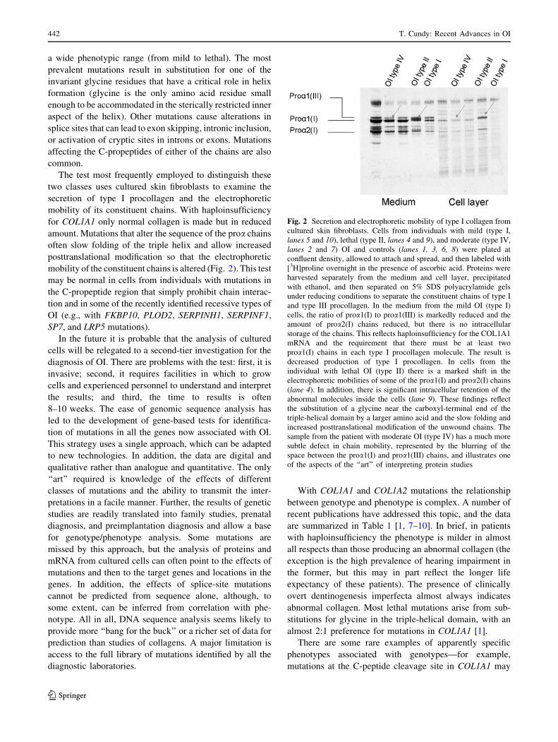
The test most frequently employed to distinguish these
two classes uses cultured skin fibroblasts to examine the
secretion of type I procollagen and the electrophoretic
mobility of its constituent chains. With haploinsufficiency
for COL1A1 only normal collagen is made but in reduced
amount. Mutations that alter the sequence of the proa chains
often slow folding of the triple helix and allow increased
posttranslational modification so that the electrophoretic
mobility of the constituent chains is altered (Fig. 2). This test
may be normal in cells from individuals with mutations in
the C-propeptide region that simply prohibit chain interac-
tion and in some of the recently identified recessive types of
OI (e.g., with FKBP10, PLOD2, SERPINH1, SERPINF1,
SP7, and LRP5 mutations).
In the future it is probable that the analysis of cultured
cells will be relegated to a second-tier investigation for the
diagnosis of OI. There are problems with the test: first, it is
invasive; second, it requires facilities in which to grow
cells and experienced personnel to understand and interpret
the results; and third, the time to results is often
8–10 weeks. The ease of genomic sequence analysis has
led to the development of gene-based tests for identifica-
tion of mutations in all the genes now associated with OI.
This strategy uses a single approach, which can be adapted
to new technologies. In addition, the data are digital and
qualitative rather than analogue and quantitative. The only
‘‘art’’ required is knowledge of the effects of different
classes of mutations and the ability to transmit the inter-
pretations in a facile manner. Further, the results of genetic
studies are readily translated into family studies, prenatal
diagnosis, and preimplantation diagnosis and allow a base
for genotype/phenotype analysis. Some mutations are
missed by this approach, but the analysis of proteins and
mRNA from cultured cells can often point to the effects of
mutations and then to the target genes and locations in the
genes. In addition, the effects of splice-site mutations
cannot be predicted from sequence alone, although, to
some extent, can be inferred from correlation with phe-
notype. All in all, DNA sequence analysis seems likely to
provide more ‘‘bang for the buck’’ or a richer set of data for
prediction than studies of collagens. A major limitation is
access to the full library of mutations identified by all the
diagnostic laboratories.
With COL1A1 and COL1A2 mutations the relationship
between genotype and phenotype is complex. A number of
recent publications have addressed this topic, and the data
are summarized in Table 1 [1, 7–10]. In brief, in patients
with haploinsufficiency the phenotype is milder in almost
all respects than those producing an abnormal collagen (the
exception is the high prevalence of hearing impairment in
the former, but this may in part reflect the longer life
expectancy of these patients). The presence of clinically
overt dentinogenesis imperfecta almost always indicates
abnormal collagen. Most lethal mutations arise from sub-
stitutions for glycine in the triple-helical domain, with an
almost 2:1 preference for mutations in COL1A1 [1].
There are some rare examples of apparently specific
phenotypes associated with genotypes—for example,
mutations at the C-peptide cleavage site in COL1A1 may
Fig. 2 Secretion and electrophoretic mobility of type I collagen from
cultured skin fibroblasts. Cells from individuals with mild (type I,
lanes 5 and 10), lethal (type II, lanes 4 and 9), and moderate (type IV,
lanes 2 and 7) OI and controls (lanes 1, 3, 6, 8) were plated at
confluent density, allowed to attach and spread, and then labeled with
[3H]proline overnight in the presence of ascorbic acid. Proteins were
harvested separately from the medium and cell layer, precipitated
with ethanol, and then separated on 5% SDS polyacrylamide gels
under reducing conditions to separate the constituent chains of type I
and type III procollagen. In the medium from the mild OI (type I)
cells, the ratio of proa1(I) to proa1(III) is markedly reduced and the
amount of proa2(I) chains reduced, but there is no intracellular
storage of the chains. This reflects haploinsufficiency for the COL1A1
mRNA and the requirement that there must be at least two
proa1(I) chains in each type I procollagen molecule. The result is
decreased production of type I procollagen. In cells from the
individual with lethal OI (type II) there is a marked shift in the
electrophoretic mobilities of some of the proa1(I) and proa2(I) chains
(lane 4). In addition, there is significant intracellular retention of the
abnormal molecules inside the cells (lane 9). These findings reflect
the substitution of a glycine near the carboxyl-terminal end of the
triple-helical domain by a larger amino acid and the slow folding and
increased posttranslational modification of the unwound chains. The
sample from the patient with moderate OI (type IV) has a much more
subtle defect in chain mobility, represented by the blurring of the
space between the proa1(I) and proa1(III) chains, and illustrates one
of the aspects of the ‘‘art’’ of interpreting protein studies
442 T. Cundy: Recent Advances in OI
123
Page 5
result in a relatively high bone mass with skeletal fragility
[11], and substitutions for glycine near the very end of the
triple-helical domain of proa2(I) encoded in exon 49 of the
COL1A2 gene have been associated with brachydactyly
and intracerebral hemorrhage, in addition to a severe OI
phenotype [12].
To date, only one dominantly inherited OI variant not
related to COL1A1 and COL1A2 mutations has been
described. This variant (type V) has a mild-to-moderate
phenotype and is distinguished clinically by hyperplastic
callus formation (particularly in the lower limbs), calcifi-
cation of the forearm interosseous membrane (causing
limitation of pronation/supination), anterior dislocation of
the radial head and a radiodense metaphyseal band
immediately adjacent to the growth plate in growing
patients, white sclerae, and no dentinogenesis imperfecta.
Bone biopsies reveal irregular, mesh-like lamellation [13,
14]. The underlying genetic locus has not yet been
identified.
Recessively Inherited OI
This is a fast-moving field; in the past decade the genetic
bases of 10 new OI variants have been discovered, seven
(or possibly eight) of which result from mutations in genes
encoding proteins involved in the posttranslational modi-
fication of type I procollagen [15–28]. The rate of dis-
covery shows no sign of slowing, with the technique of
exome sequencing now permitting the identification of
genetic causes in families of small size. One of the main
interests in these variants is that they help to define the
minimal degree of change to type I collagen that can result
in an OI phenotype, and that could eventually lead to
effective therapies. Phenotypic data on the recessively
inherited forms of OI is summarized in Table 2, although it
should be noted that for some of these disorders very few
individuals have been described, so our appreciation of the
clinical spectrum is likely to change as further cases are
found.
Collagen-Related Genes
In patients at the severe-to-lethal end of the OI spectrum
the question of dominant inheritance is, for obvious rea-
sons, rarely put to the test; and in Sillence’s original
classification the perinatal lethal and progressive deform-
ing types (types II and III) were thought to be recessively
inherited. However, it was not until 2007 that mutations in
CRTAP were identified in patients without mutations in
COL1A1 or COL1A2 but with excess posttranslational
modification of type I collagen, indicative of delayed
folding of the triple helix [15, 16]. CRTAP encodes carti-
lage-related protein, part of the three-protein complex
responsible for the 3-hydroxylation of proline at position
986 of the triple helix in the proa1(I) chain. Very soon
after, patients with similar phenotypes with mutations in
the genes for the other components of this complex,
Table 1 Genotype–phenotype relationships in OI resulting from COL1A1 or COL1A2 mutations
Haploinsufficiency: compared to helical-domain mutations
Nonlethal, nondeforming, taller
Blue sclerae usual
Normal life expectancy
Fewer fractures—fracture rate falls after adolescence
No dentinogenesis imperfecta
Fewer skull-base abnormalities
Sensorineural deafness common
Greater size-adjusted lumbar spine areal BMD
Greater cortical bone width
Wormian bones less common (28% vs. 81%)
Helical-domain mutations—with production of an abnormal collagen
82% are glycine substitutions: ? serine, arginine, or cysteine are the most common (*78% of proa1(I) and 63% of proa2(I) substitutions)
33% of glycine substitutions are lethal in proa1(I), 20% lethal in proa2(1)
Substitutions of large amino acid residues (arginine, valine, glutamic acid, aspartic acid) for glycine in proa1(I)
beyond position *200 are usually lethal
Glycine substitutions at N-terminal end of either proa1(I) or proa2(I) are nonlethal and not associated with dentinogenesis imperfecta
18% are splice-site, exon-skipping, intronic inclusion, or activation of cryptic sites—rarely lethal
C-propeptide mutations
Mutations in proa1(I) severe to lethal, in proa2 (I) mild to moderate phenotype
Data compiled from references 1, 7–10, 30
T. Cundy: Recent Advances in OI 443
123
Page 6
Ta
ble
2A
uto
som
alre
cess
ive
form
so
fO
I
Gen
eL
ocu
sP
rote
inS
yn
on
ym
Sk
elet
al
ph
eno
typ
e
Wo
rmia
n
bo
nes
Scl
eral
colo
r
Den
tin
og
enes
is
imp
erfe
cta
Dea
fnes
sO
ther
feat
ure
sR
ef.
Gen
esre
gu
lati
ng
mat
rix
stru
ctu
re
CR
TA
P3
p2
2C
arti
lag
e-as
soci
ated
pro
tein
OI-
VII
Let
hal
–m
od
erat
e*
40
%L
igh
tb
lue
No
No
Rh
izo
mel
ia,
po
pco
rn
met
aph
yse
s;h
ead
circ
um
fere
nce
smal
l
15
,17
LE
PR
E1
1p
34
Pro
lyl
3-h
yd
rox
yla
se1
OI-
VII
IL
eth
al–
sev
ere
Yes
Lig
ht
blu
eN
oN
oIm
pai
red
calv
aria
l
calc
ifica
tio
n,
po
pco
rn
met
aph
yse
s
16
,17
PP
IB1
5q
21
Pep
tid
yl-
pro
lyl
iso
mer
ase
B
OI-
IXS
ever
e?
Gra
yN
o?
–1
8
FK
BP
10
17
q2
1F
K5
06
-bin
din
gp
rote
in
10
(FK
BP
65
)
Bru
ck1
Sev
ere–
mo
der
ate
10
0%
Wh
ite
No
No
Co
ntr
actu
res,
sco
lio
sis,
acet
abu
lar
pro
tru
sio
n
19
-21
PL
OD
23
q2
3T
elo
pep
tid
ely
syl
hy
dro
xy
lase
Bru
ck2
Sev
ere–
mo
der
ate
Yes
Gra
yN
oN
o?
Co
ntr
actu
res
22
SE
RP
INH
11
1q
13
Hea
tsh
ock
pro
tein
47
OI-
XS
ever
e?
Blu
eY
esN
o?
Ren
alst
on
es2
3
BM
P1
8p
21
Bo
ne
mo
rph
og
enet
ic
pro
tein
1
–S
ever
eY
es?
No
No
Hy
per
exte
nsi
bil
ity
24
SE
RP
INF
11
7p
13
Pig
men
t-d
eriv
ed
epit
hel
ium
fact
or
OI-
VI
Sev
ere
Infr
equ
ent
Wh
ite
No
No
Hy
per
ost
eoid
osi
sw
ith
abn
orm
alla
mel
lati
on
25
,26
Gen
esre
gu
lati
ng
mat
rix
ho
meo
stas
is
LR
P5
11
q1
3L
DL
rece
pto
r-re
late
d
pro
tein
5
Ost
eop
oro
sis
pse
ud
og
lio
ma
Mil
d–
mo
der
ate
No
Wh
ite
No
No
Bli
nd
nes
s;
het
ero
zyg
ote
sh
ave
low
bo
ne
mas
s
27
,28
SP
71
2q
13
Ost
erix
OI-
XI
Mo
der
ate
Yes
Wh
ite
No
No
Del
ayed
too
ther
up
tio
n2
9
444 T. Cundy: Recent Advances in OI
123
Page 7
LEPRE1 (prolyl 3-hydroxylase, PH3) and PPIB (cyclo-
philin B), were identified [17, 18]. It is not clear that the
importance of this enzyme complex is due solely to its
effect on prolyl 3-hydroxylation. The genetic test of
substituting the proline 986 residue has not been done
either by nature or in the laboratory. The 3-hydroxylation
of additional X-position proline residues may be important
to make normal molecules, and cyclophilin B, a prolyl cis–
trans isomerase, may have other targets in type I procol-
lagen chains; thus, the effects of mutations in any of these
genes are likely to be biochemically complex.
Bruck syndrome describes the occurrence of neonatal
fractures with contractures of the legs (and, in some cases,
the arms), which is occasionally misclassified as arthro-
gryposis. Two causative genes, FKBP10 (Bruck 1) and
PLOD2 (Bruck 2), have been identified [19–22]. FKBP10
mutations seem to be the more common, and as more cases
have come to light, a milder phenotype presenting in late
childhood or adolescence with long bone fractures, ace-
tabular protrusion, and scoliosis has been recognized [21].
FKBP65, the protein product of FKBP10, is a prolyl cis–
trans isomerase which seems to have multiple substrates,
among them being lysyl hydroxylase 2 (encoded by
PLOD2) and perhaps LH1 and HSP47 (see the following).
The effects of mutations in each gene probably share
phenotypic features because of this interaction. One child
with a severe phenotype has been described with homo-
zygosity for a missense mutation in the gene SERPINH1,
which encodes another collagen chaperone, HSP47 [23].
Among the most recent discoveries are the association of
mutations in the gene SERPINF1 with a variant known as type
VI OI [24, 25]. SERPINF1 encodes pigment epithelium-
derived factor (PEDF), a secreted glycoprotein of uncertain
function in bone; and it is not clear at present whether it acts on
collagen metabolism or in some other way. PEDF can be
measured in normal serum and is undetectable in the serum of
patients with this variant of OI. Bone from these individuals has
the distinctive feature of a marked increase in osteoid, with an
unusual ‘‘fish-scale’’ pattern when viewed under polarized
light. In addition, the response to bisphosphonate therapy in
affected children may be disappointing in terms of reducing
fracture rates and improving mobility [25].
Recessive disorders occur most commonly in areas of
the world where consanguineous marriage takes place or
where there are mutations in relatively small founder
populations. Many instances of the latter are seen in
recessively inherited OI. Examples include the splice-site
mutation in LEPRE1 (IVS5 ? 1G-T) seen both in West
Africa and in people of African descent dispersed to the
Americas (through the Atlantic slave trade) [15, 16] and
the insertion mutation in FKBP10 (c.948dupT) seen in
Samoa [21]. Knowledge of such locally occurring muta-
tions can significantly aid diagnosis.
Non-Collagen-Related Genes
The osteoporosis pseudoglioma syndrome (originally des-
ignated the ocular form of OI) is an example of how genetic
discoveries can advance understanding of bone metabolism
and open up therapeutic possibilities. This disorder is due to
mutations in the gene LRP5, which encodes lipoprotein
receptor protein 5 (LRP5), a key regulator of osteoblast
function. LRP5 affects bone accrual during growth and is
important for the establishment of peak bone mass but is not
directly related to type I collagen metabolism. The skeletal
phenotype of the osteoporosis pseudoglioma syndrome is
relatively mild, but the distinguishing feature is the ocular
involvement, which is due to failure of the normal involution
of the hyaloid vasculature [27]. The remnant forms a fibrous
mass (‘‘pseudoglioma’’) in the globe, causing retinal
detachment and blindness; nearly all subjects have near
complete loss of vision by the age of 25 years [28]. Heter-
ozygosity for LRP5 loss-of-function mutations differs from
other recessive forms of OI in that carriers clearly have an
osteopenic phenotype [28]. Some patients with familial,
idiopathic, or juvenile osteoporosis are heterozygous for
LRP5 mutations. Of note is that dominant gain-of-function
mutations in LRP5 give rise to a dense bone phenotype
because of activation of the same pathway in bone that is
diminished by the recessive loss-of-function mutations.
More recently, a child with a moderate OI phenotype
has been identified with homozygous mutations in SP7.
This gene encodes osterix, a transcription factor specifi-
cally expressed in osteoblasts in the developing skeleton
[29].
OI in Adults
Children with severe phenotypes have significantly reduced
life expectancy, predominantly because of problems asso-
ciated with respiratory insufficiency and pulmonary
hypertension, secondary to kyphoscoliosis and small lung
volumes. Although survival of patients with severe phe-
notypes into adulthood is not uncommon only * 20%
survive beyond the age of 40 [30]. Thus, most patients who
survive into adulthood have a mild or moderate phenotype.
In these patients the fracture rate typically falls after pub-
erty (as it does for most forms of childhood osteoporosis;
the strength of long bones is proportional to the fourth
power of their external radius, so bones get stronger as they
grow wider). Vertebral fractures can occur in the post-
partum period (Fig. 1d), and the fracture rate for both
vertebral and long bones increases after the menopause in
women [31] and in later life in men.
Deafness is a common problem for adults, particularly
those with a mild phenotype due to haploinsufficiency.
T. Cundy: Recent Advances in OI 445
123
Page 8
About one-third of such patients are affected by the age of
30 and one-half by the age of 50, although the proportion
affected does not change much after that age. Hearing
impairment shows distinct familial trends, being common
in some families and infrequent in others [32]. Cardiac
valve dysfunction (aortic or mitral regurgitation) is a rec-
ognized but uncommon feature seen in people with OI, but
the mechanistic relationship between the two remains
unclear. Valve replacement may be necessary, but the
surgery is often complicated and should be undertaken in
centers with experience [33].
Bisphosphonate therapy in adults with OI has been
explored in three relatively small randomized controlled
trials: using intravenous neridronate, given 3-monthly for
2 years [34]; oral alendronate, given daily for 3 years [35];
or oral risedronate, given weekly for 2 years [36]. All of
these studies showed that bisphosphonate treatment had
statistically significant effects, increasing bone density at
the spine and hip, and decreasing bone turnover markers;
but there were was no difference in the fracture rate, which
was generally low. Given the relative rarity of OI, it will be
difficult to demonstrate efficacy at fracture reduction for
any intervention.
Bisphosphonate Therapy in Children and Adolescents
Physiotherapy, rehabilitation, and orthopedic surgery are
the mainstay of treatment for children and adolescents with
OI; and the best results are obtained when undertaken by
skilled multidisciplinary teams. Intravenous bisphospho-
nates were first suggested as treatment to improve bone
fragility in children with severe OI 25 years ago [37], and
although not subjected to randomized, placebo-controlled
trials, bisphosphonate treatment has rapidly become
established as a standard of care. Compared to historical
controls, intravenous bisphosphonate therapy is associated
with improvements in bone pain, well-being, longitudinal
growth and muscle strength, and vertebral and long bone
mass as well as with a reduced fracture rate [38]. In an
important series of studies the Montreal group has shown
clearly how bisphosphonate therapy has effects on both
trabecular and cortical bone mass. In OI, trabeculae are
reduced in number and abnormally thin. With bisphosph-
onate treatment, the number (but not the thickness) of
trabeculae is increased. During endochondral growth most
primary trabeculae are lost in the conversion of primary
into secondary spongiosa, but bisphosphonate treatment
inhibits the resorption of primary trabeculae, permitting
more to survive as secondary trabeculae [38]. Because of
slow periosteal bone formation, the long bones in OI are
typically narrow (although this is often partially compen-
sated by a relative narrowing of the marrow cavity that
conserves cortical bone width). During normal growth,
cortical width is determined by bone modeling in which
bone resorption at the endosteal surface takes place in
parallel with periosteal new bone formation. Bisphospho-
nates inhibit the former process (but not the latter), per-
mitting an increase in cortical thickness and, thus,
improvement in bone strength [38]. Bisphosphonates do
not increase bone width, and of course, if an abnormal
collagen is produced, then bone quality will also remain
unaffected. In adults, bisphosphonates increase bone min-
eralization and decrease bone turnover [39]; but in OI the
bones are already hypermineralized [40], so this mecha-
nism is unlikely to contribute to any improvement in bone
strength.
The mode of action of bisphosphonates is the same in all
forms of OI and, indeed, probably across all varieties of
childhood osteoporosis. Intravenous pamidronate is the
most widely used bisphosphonate; but the effects are
generic, and regimens employing longer-acting agents such
as zoledronate are being used increasingly.
Bisphosphonate therapy is not without side effects. In
addition to the well-known first dose reaction, uveitis has
been reported; and in children prolonged bisphosphonate
use can impair metaphyseal modeling [41]. Intermittent
intravenous therapy produces the characteristic metaphy-
seal lines, where remnants of calcified cartilage from the
growth plate have accumulated (Fig. 3a–c). Low bone
turnover induced by bisphosphonates can impair bone
healing, particularly after corrective osteotomy [42]. Sev-
eral important questions concerning bisphosphonate treat-
ment of moderate to severe OI in children remain
unresolved. These include how treatment should be mon-
itored, whether the regimen should be modified for long-
term use, and if and when treatment should be
discontinued.
Debate continues on the use of bisphosphonates in
children with mild OI and whether oral administration is
just as effective as intravenous. Children with mild OI have
less to gain from treatment and potentially more to lose
from adverse events. Reports on two randomized con-
trolled trials of oral bisphosphonates in children with pre-
dominantly (but not exclusively) mild forms of OI have
been recently published [43, 44]. These showed that, at the
doses given, both oral risedronate and alendronate
increased spinal bone density and reduced bone turnover
markers, but there was no improvement in fracture rate
over a 2-year period. A randomized controlled trial of oral
risedronate in children with moderate or severe OI had
similar results, but there was a suggestion that the active
treatment slowed the progression of bone deformity [45].
The authors pointed out that in both their control and bis-
phosphonate-treated groups the fracture rate fell with
increasing age, emphasizing the challenge that future
446 T. Cundy: Recent Advances in OI
123
Page 9
studies will need to be powered adequately to demonstrate
a fracture outcome.
The use of anabolic agents to increase bone mass and
size in children with severe forms of OI is an attractive
theoretical option, but there are formidable practical
problems. Parathyroid hormone is contraindicated in
childhood because of concerns about the possibility of
inducing osteosarcoma, so to date most attention has been
focused on growth hormone. Anabolic agents generally
increase bone turnover, which makes the development of
deformity more likely, so the coadministration of an
inhibitor of bone resorption would most likely be neces-
sary. One small trial has looked at the effects of 1-year
growth hormone treatment in conjunction with neridronate
vs. neridronate alone [46]. The growth hormone–treated
subjects had greater increases in bone mass at various sites
Fig. 3 Effects of prolonged
bisphosphonate treatment on
bone. a Femoral radiograph
from a 2.5-year-old boy with
severe OI who was treated with
intravenous bisphosphonates
from the age of 2 months. The
femoral diaphysis is narrow
(typical in OI), but the
metaphysis is wide because of
bisphosphonate-associated
impairment of bone modeling.
Horizontal white lines coincide
with his pamidronate infusions.
b Upper tibia and femur from
the same child age 10. He had
been treated with pamidronate
infusions from age 2 months to
6 years and with zoledronate
infusions from the age of 8
onward. The bone laid down in
the 2 years off bisphosphonate
treatment is notably less dense.
The junction between treated
and nontreated bone may be
vulnerable to fracture [47]. c A
transiliac bone biopsy from the
same child aged 10
(hematoxylin and eosin stain)
showing extensive retention of
mineralized cartilage (bluestain) with relatively little bone
tissue (pink stain). Calcified
cartilage contributes to greater
‘‘bone density’’ but may not be
resistant to fracture. Note also
the giant osteoclasts detached
from the surface of bone
(arrows) that are a common
finding in bisphosphonate-
treated bone [48]
T. Cundy: Recent Advances in OI 447
123
Page 10
and improved growth velocity, but the study was under-
powered to detect any difference in fracture rates. Exper-
imental approaches such as bone marrow transplantation,
stem cell transplantation, and correction of the mutated
gene may eventually come to fruition but are not currently
ready for clinical trial.
Conclusions
There have been substantial advances in the understanding
of OI in recent years. The main progress has been in
understanding the genetic bases of this heterogenous group
of disorders that has provided better information for
genetic counseling and opportunities for prenatal diagnosis.
The mode of action and risks and benefits of bisphospho-
nate therapy are being clarified. Particular challenges for
future OI research will be to design therapeutic trials that
can convincingly demonstrate effects on deformity and
fracture and to determine the role of mutations in deter-
mining responses to treatment.
Acknowledgement My sincere thanks go to Dr. Peter Byers, Uni-
versity of Washington, for his great help in preparing this article and
for providing Fig. 2. The radiographs and the histology in Fig. 3 are
reproduced with kind permission of Dr. Paul Hofman and Dr. Michael
Dray, respectively.
References
1. Marini JC, Forlino A, Cabral WA, Barnes AM, San Antonio JD,
Milgrom S, Hyland JC, Korkko J, Prockop DJ, De Paepe A,
Coucke P, Symoens S, Glorieux FH, Roughley PJ, Lund AM,
Kuurila-Svahn K, Hartikka H, Cohn DH, Krakow D, Mottes M,
Schwarze U, Chen D, Yang K, Kuslich C, Troendle J, Dalgleish
R, Byers PH (2007) Consortium for osteogenesis imperfecta
mutations in the helical domain of type I collagen: regions rich in
lethal mutations align with collagen binding sites for integrins
and proteoglycans. Hum Mutat 28:209–221
2. Canty EG, Kadler KE (2005) Procollagen trafficking, processing
and fibrillogenesis. J Cell Sci 118:1341–1353
3. Krane SM (2008) The importance of proline residues in the
structure, stability and susceptibility to proteolytic degradations
of collagens. Amino Acids 35:703–710
4. Sillence DO, Senn A, Danks DM (1979) Genetic heterogeneity in
osteogenesis imperfecta. J Med Genet 16:101–116
5. Warman ML, Cormier-Daire V, Hall C, Krakow D, Lachman R,
LeMerrer M, Mortier G, Mundlos S, Nishimura G, Rimoin DL,
Robertson S, Savarirayan R, Sillence D, Spranger J, Unger S,
Zabel B, Superti-Furga A (2011) Nosology and classification of
genetic skeletal disorders: 2010 revision. Am J Med Genet
155A:943–968
6. Cundy T, Horne A, Bolland M, Gamble G, Davidson J (2007)
Bone formation markers in adults with mild osteogenesis im-
perfecta. Clin Chem 53:1109–1114
7. Rauch F, Lalic L, Roughley P, Glorieux FH (2010) Genotype–
phenotype correlations in nonlethal osteogenesis imperfecta
caused by mutations in the helical domain of collagen type I. Eur
J Hum Genet 18:642–647
8. Rauch F, Lalic L, Roughley P, Glorieux FH (2010) Relationship
between genotype and skeletal phenotype in children and ado-
lescents with osteogenesis imperfecta. J Bone Miner Res 25:
1367–1374
9. Semler O, Cheung MS, Glorieux FH, Rauch F (2010) Wormian
bones in osteogenesis imperfecta: correlation to clinical findings
and genotype. Am J Med Genet A 152:1681–1687
10. Cheung MS, Arponen H, Roughley P, Azouz ME, Glorieux FH,
Waltimo-Siren J, Rauch F (2011) Cranial base abnormalities in
osteogenesis imperfecta: phenotypic and genotypic determinants.
J Bone Miner Res 26:405–413
11. Faqeih E, Roughley P, Glorieux FH, Rauch F (2009) Osteogen-
esis imperfecta type III with intracranial hemorrhage and brach-
ydactyly associated with mutations in exon 49 of COL1A2. Am J
Med Genet A 149:461–465
12. Lindahl K, Barnes AM, Fratzl-Zelman N, Whyte MP, Hefferan
TE, Makareeva E, Brusel M, Yaszemski MJ, Rubin CJ, Kindmark
A, Roschger P, Klaushofer K, McAlister WH, Mumm S, Leikin
S, Kessler E, Boskey AL, Ljunggren O, Marini JC (2011)
COL1A1 C-propeptide cleavage site mutations cause high bone
mass osteogenesis imperfecta. Hum Mutat 32:598–609
13. Glorieux FH, Rauch F, Plotkin H, Ward L, Travers R, Roughley
P, Lalic L, Glorieux DF, Fassier F, Bishop NJ (2000) Type V
osteogenesis imperfecta: a new form of brittle bone disease.
J Bone Miner Res 15:1650–1658
14. Cheung MS, Glorieux FH, Rauch F (2007) Natural history of
hyperplastic callus formation in osteogenesis imperfecta type V.
J Bone Miner Res 22:1181–1186
15. Morello R, Bertin TK, Chen Y, Hicks J, Tonachini L, Monticone
M, Castagnola P, Rauch F, Glorieux FH, Vranka J, Bachinger
HP, Pace JM, Schwarze U, Byers PH, Weis M, Fernandes RJ,
Eyre DR, Yao Z, Boyce BF, Lee B (2006) CRTAP is required for
prolyl 3- hydroxylation and mutations cause recessive osteo-
genesis imperfecta. Cell 127:291–304
16. Cabral WA, Chang W, Barnes AM, Weis M, Scott MA, Leikin S,
Makareeva E, Kuznetsova NV, Rosenbaum KN, Tifft CJ, Bulas
DI, Kozma C, Smith PA, Eyre DR, Marini JC (2007) Prolyl
3-hydroxylase 1 deficiency causes a recessive metabolic bone
disorder resembling lethal/severe osteogenesis imperfecta. Nat
Genet 39:359–365
17. Baldridge D, Schwarze U, Morello R, Lennington J, Bertin TK,
Pace JM, Pepin MG, Weis M, Eyre DR, Walsh J, Lambert D,
Green A, Robinson H, Michelson M, Houge G, Lindman C,
Martin J, Ward J, Lemyre E, Mitchell JJ, Krakow D, Rimoin DL,
Cohn DH, Byers PH, Lee B (2008) CRTAP and LEPRE1
mutations in recessive osteogenesis imperfecta. Hum Mutation
29:1435–1442
18. Barnes AM, Carter EM, Cabral WA, Weis M, Chang W, Mak-
areeva E, Leikin S, Rotimi CN, Eyre DR, Raggio CL, Marini JC
(2010) Lack of cyclophilin B in osteogenesis imperfecta with
normal collagen folding. N Engl J Med 362:521–528
19. Alanay Y, Avaygan H, Camacho N, Utine GE, Boduroglu K,
Aktas D, Alikasifoglu M, Tuncbilek E, Orhan D, Bakar FT, Zabel
B, Superti-Furga A, Bruckner-Tuderman L, Curry CJ, Pyott S,
Byers PH, Eyre DR, Baldridge D, Lee B, Merrill AE, Davis EC,
Cohn DH, Akarsu N, Krakow D (2010) Mutations in the gene
encoding the RER protein FKBP65 cause autosomal-recessive
osteogenesis imperfecta. Am J Hum Genet 87:551–559
20. Kelley BP, Malfait F, Bonafe L, Baldridge D, Homan E,
Symoens S, Willaert A, Elcioglu N, Van Maldergem L, Verellen-
Dumoulin C, Gillerot Y, Napierala D, Krakow D, Beighton P,
Superti-Furga A, De Paepe A, Lee B (2011) Mutations in
FKBP10 cause recessive osteogenesis imperfecta and Bruck
syndrome. J Bone Miner Res 26:666–672
21. Schwarze U, Cundy T, Pyott S, Christiansen H, Hegde MR, Bank
R, Pals G, Ankala A, Connelly K, Seaver L, Yandow S, Raney H,
448 T. Cundy: Recent Advances in OI
123
Page 11
Babovich-Vicsanovic D, Stoler J, Ben-Neriah Z, Segal R, Al-Aqeel
A, Siderius L, Hannibal M, Hudgins L, McPherson E, Clemens M,
Sussman MD, Steiner R, Mahan J, Smith R, Anyane-Yeboa K,
Chong K, Uster T, Aftimos S, Sutton VR, Davis EC, Weis MA,
Eyre D, Byers PH (2012) Mutation in FKBP10, which encodes a
65kD FK506 binding prolyl cis-trans isomerase, results in reces-
sive forms of osteogenesis imperfecta and is the first Bruck syn-
drome (contractures and fractures) locus. Submitted for publication
22. Van der Slot AJ, Zuurmond A-M, Bardoe AFJ, Wijmenga C,
Pruijs HEH, Sillence DO, Brinckmann J, Abraham DJ, Black
CM, Verzijl N, DeGroot J, Hanemaaijer R, TeKoppele JM, Hu-
izinga TWJ, Bank RA (2003) Identification of PLOD2 as telo-
peptide lysyl hydroxylase, an important enzyme in fibrosis. J Biol
Chem 278:40967–40972
23. Christiansen HE, Schwarze U, Pyott SM, AlSwaid A, Al Balwi M,
Alrasheed S, Pepin MG, Weis MA, Eyre DR, Byers PH (2010)
Homozygosity for a missense mutation in SERPINH1, which
encodes the collagen chaperone protein HSP47, results in severe
recessive osteogenesis imperfecta. Am J Hum Genet 86:389–398
24. Martinez-Glez V, Valencia M, Caparros-Martin JA, Aglan M,
Temtamy S, Tenorio J, Pulido V, Lindert U, Rohrbach M, Eyre
D, Giunta C, Lapunzina P, Ruiz-Perez VL (2012) Identification
of a mutation causing deficient BMP1/mTLD proteolytic activity
in autosomal recessive osteogenesis imperfecta. Hum Mutat
33:343–350
25. Becker J, Semler O, Gilissen C, Li Y, Bolz HJ, Giunta C,
Bergmann C, Rohrbach M, Koerber F, Zimmermann K, de Vries
P, Wirth B, Schoenau E, Wollnik B, Veltman JA, Hoischen A,
Netzer C (2011) Exome sequencing identifies truncating muta-
tions in human SERPINF1 in autosomal-recessive osteogenesis
imperfecta. Am J Hum Genet 88:362–371
26. Homan EP, Rauch F, Grafe I, Lietman C, Doll JA, Dawson B,
Bertin T, Napierala D, Morello R, Gibbs R, White L, Miki R,
Cohn DH, Crawford S, Travers R, Glorieux FH, Lee B (2011)
Mutations in SERPINF1 cause osteogenesis imperfecta type VI.
J Bone Miner Res 26:2798–2803
27. Gong Y, Slee RB, Fukai N, Rawadi G, Roman–Roman S, Reg-
inato AM, Wang H, Cundy T, Glorieux FH, Lev D, Zacharin M,
Oexle K, Marcelino J, Suwairi W, Heeger S, Sabatakos G, Apte
S, Adkins WN, Allgrove J, Arslan-Kirchner M, Batch JA,
Beighton P, Black GC, Boles RG, Boon LM, Borrone C, Brunner
HG, Carle GF, Dallapiccola B, De Paepe A, Floege B, Halfhide
ML, Hall B, Hennekam RC, Hirose T, Jans A, Juppner H, Kim
CA, Keppler-Noreuil K, Kohlschuetter A, Lacombe D, Lambert
M, Lemyre E, Letteboer T, Peltonen L, Ramesar RS, Romanengo
M, Somer H, Steichen-Gersdorf E, Steinmann B, Sullivan B,
Superti-Furga A, Swoboda W, van den Boogaard MJ, Van Hul
W, Vikkula M, Votruba M, Zabel B, Garcia T, Baron R, Olsen
BR, Warman ML (2001) LDL receptor–related protein 5 (LRP5)
affects bone accrual and eye development. Cell 107:513–523
28. Ai M, Heeger S, Bartels CF, Schelling DK, Osteoporosis-Pseu-
doglioma Collaborative Group (2005) Clinical and molecular
findings in osteoporosis-pseudoglioma syndrome. Am J Hum
Genet 77:741–753
29. Lapunzina P, Aglan M, Temtamy S, Caparros-Martin JA,
Valencia M, Leton R, Martinez-Glez V, Elhossini R, Amr K,
Vilaboa N, Ruiz-Perez VL (2010) Identification of a frameshift
mutation in osterix in a patient with recessive osteogenesis im-
perfecta. Am J Hum Genet 87:110–114
30. Paterson CR, Ogston SA, Henry RM (1996) Life expectancy in
osteogenesis imperfecta. BMJ 1996:312–351
31. Paterson CR, McAllion S, Stellman JL (1984) Osteogenesis im-
perfecta after the menopause. N Engl J Med 310:1694–1696
32. Paterson CR, Monk EA, McAllion SJ (2001) How common is
hearing impairment in osteogenesis imperfecta? J Laryngol Otol
115:280–282
33. Bonita RE, Cohen IS, Berko BA (2010) Valvular heart disease in
osteogenesis imperfecta: presentation of a case and review of the
literature. Echocardiography 27:69–73
34. Adami S, Gatti D, Colapietro F, Fracassi E, Braga V, Rossini M,
Tato L (2003) Intravenous neridronate in adults with osteogenesis
imperfecta. J Bone Miner Res 18:126–130
35. Chevrel G, Schott A-M, Fontanges E, Charrin JE, Lina-Granade
G, Duboeuf F, Garnero P, Arlot M, Raynal C, Meunier PJ (2006)
Effects of oral alendronate on BMD in adult patients with
osteogenesis imperfecta: a 3-year randomized placebo-controlled
trial. J Bone Miner Res 21:300–306
36. Bradbury LA, Barlow S, Geoghegan F, Hannon RA, Stuckey SL,
Wass JAH, Russell RGG, Brown MA, Duncan EL (2012)
Risedronate in adults with osteogenesis imperfecta type I:
increased bone mineral density and decreased bone turnover, but
high fracture rate persists. Osteoporos Int 23:285–294
37. Devogelaer JP, Malghem J, Maldague B, Nagant de Deuxchaines
C (1987) Radiological manifestations of bisphosphonate treat-
ment with APD in a child suffering from osteogenesis imperfecta.
Skel Radiol 16:360–363
38. Rauch F, Glorieux FH (2004) Osteogenesis imperfecta. Lancet
363:1377–1385
39. Gamsjaeger S, Buchinger B, Zwettler E, Recker R, Black D, Gasser
JA, Eriksen EF, Klaushofer K, Paschalis EP (2011) Bone material
properties in actively bone-forming trabeculae in postmenopausal
women with osteoporosis after three years of treatment with once-
yearly zoledronic acid. J Bone Miner Res 26:12–18
40. Roschger P, Fratzl-Zelman N, Misof BM, Glorieux FH, Klau-
shofer K, Rauch F (2008) Evidence that abnormal high bone
mineralization in growing children with osteogenesis imperfecta
is not associated with specific collagen mutations. Calcif Tissue
Int 82:263–270
41. Land C, Rauch F, Glorieux FH (2006) Cyclical intravenous
pamidronate treatment affects metaphyseal modeling in growing
patients with osteogenesis imperfecta. J Bone Miner Res
21:374–379
42. Munns CF, Rauch F, Zeitlin L, Fassier F, Glorieux FH (2004)
Delayed osteotomy but not fracture healing in pediatric osteo-
genesis imperfecta patients receiving pamidronate. J Bone Miner
Res 19:1779–1786
43. Rauch F, Munns CF, Land C, Cheung M, Glorieux FH (2009)
Risedronate in the treatment of mild pediatric osteogenesis im-
perfecta: a randomized placebo-controlled study. J Bone Miner
Res 24:1282–1289
44. Ward LM, Rauch F, Whyte MP, D’Astous J, Gates PE, Grogan
D, Lester EL, McCall RE, Pressly TA, Sanders JO, Smith PA,
Steiner RD, Sullivan E, Tyerman G, Smith-Wright DL, Verb-
ruggen N, Heyden N, Lombardi A, Glorieux FH (2011) Alendr-
onate for the treatment of pediatric osteogenesis imperfecta: a
randomized placebo-controlled study. J Clin Endocrinol Metab
96:355–364
45. Bishop N, Harrison R, Ahmed F, Shaw N, Eastell R, Campbell
M, Knowles E, Hill C, Hall C, Chapman S, Sprigg A, Rigby A
(2010) A randomized, controlled dose-ranging study of risedro-
nate in children with moderate and severe osteogenesis imper-
fecta. J Bone Miner Res 25:32–40
46. Antoniazzi F, Monti E, Venturi G, Franceschi R, Doro F, Gatti D,
Zamboni G, Tato L (2010) Growth hormone in combination with
bisphosphonate treatment in osteogenesis imperfecta. Eur J
Endocrinol 163:479–487
47. Rauch F, Cornibert S, Cheung M, Glorieux FH (2007) Long-bone
changes after pamidronate discontinuation in children and ado-
lescents with osteogenesis imperfecta. Bone 40:821–827
48. Cheung MS, Glorieux FH, Rauch F (2009) Large osteoclasts in
pediatric osteogenesis imperfecta patients receiving intravenous
pamidronate. J Bone Miner Res 24:669–674
T. Cundy: Recent Advances in OI 449
123