Page 1
1
Reducing the Environmental Sensitivity of Yellow Fluorescent Protein: Mechanism and
Applications
Oliver Griesbeck‡§, Geoffrey S. Baird¶§||, Robert E. Campbell¶, David A. Zacharias‡, and
Roger Y. Tsien‡¶**
From the ‡Howard Hughes Medical Institute, ¶Department of Pharmacology, and ||Medical
Scientist Training Program and Biomedical Sciences Graduate Program, University of
California, San Diego, La Jolla, CA 92093-0647, USA
*This work was supported by the Howard Hughes Medical Institute and NIH (NS-27177) and a
postdoctoral fellowship from the Canadian Institutes of Health Research to R.E.C.
§These authors contributed equally to this work.
**To whom correspondence should be addressed: Dept. of Pharmacology and HHMI, Univ.
California San Diego, La Jolla, CA 92093-0647. Tel.: (858)534-4891; Fax (858)534-5270; E-
mail [email protected] .
Running title: Environmentally Insensitive Yellow Fluorescent Protein
Copyright 2001 by The American Society for Biochemistry and Molecular Biology, Inc.
JBC Papers in Press. Published on May 31, 2001 as Manuscript M102815200 by guest on M
ay 18, 2018http://w
ww
.jbc.org/D
ownloaded from
Page 2
2
SUMMARY
Yellow mutants of the green fluorescent protein (YFP) are crucial constituents of
genetically encoded indicators of signal transduction and fusions to monitor protein-protein
interactions. However, previous YFPs show excessive pH-sensitivity, chloride interference, poor
photostability, or poor expression at 37 °C. Protein evolution in E. coli has produced a new YFP
named Citrine, in which the mutation Q69M confers a much lower pKa (5.7) than for previous
YFPs, indifference to chloride, twice the photostability of previous YFPs, and much better
expression at 37 °C and in organelles. The halide resistance is explained by a 2.2-Å x-ray crystal
structure of Citrine, showing that the methionine side-chain fills what was once a large halide-
binding cavity adjacent to the chromophore. Insertion of calmodulin within Citrine or fusion of
cyan fluorescent protein, calmodulin, a calmodulin-binding peptide and Citrine has generated
improved calcium indicators. These chimeras can be targeted to multiple cellular locations and
have permitted the first single-cell imaging of free [Ca2+] in the Golgi. Citrine is superior to all
previous YFPs except when pH- or halide-sensitivity is desired and is particularly advantageous
within genetically encoded fluorescent indicators of physiological signals.
by guest on May 18, 2018
http://ww
w.jbc.org/
Dow
nloaded from
Page 3
3
INTRODUCTION
Yellow Fluorescent Proteins (YFPs) were created (1) by mutating Thr203 of the
Aequorea victoria Green Fluorescent Protein (GFP) (2) to aromatic amino acids, typically Tyr.
The resulting π-π stacking and increased local polarizability immediately adjacent to the
chromophore are believed to be responsible for the ~20 nm shift to longer excitation and
emission wavelengths (3). However, the changes in internal hydrogen bonding and steric packing
also made the fluorescence more vulnerable to photobleaching (4, 5), decolorization by
protonation (6-10), and quenching by many anions (10-12), of which chloride is the
physiologically most relevant. These sensitivities can be exploited for specialized applications
such as measuring fluorescence recovery after photobleaching and sensing pH and halide
concentrations, but are deleterious for using YFPs either as simple fusion tags or as acceptors for
fluorescence resonance energy transfer (FRET). YFPs are becoming very popular in such roles,
particularly as partners for cyan fluorescent protein (CFP) mutants of GFP (2, 5, 13-15). CFPs
and YFPs are spectroscopically well enough separated to be easily distinguishable in either
excitation or emission spectra, yet the emission wavelengths of CFPs and excitation wavelengths
of YFPs overlap well enough to make them good partners for FRET. They have largely
superseded the initial pairing of blue mutants and improved green forms of GFP (16), because
the blue mutants were too dim and photobleachable, and because shorter wavelengths generically
excite more autofluorescence and raise more concerns of phototoxicity.
Measurements of FRET between CFP and YFP are becoming increasingly common to
monitor protein-protein interactions nondestructively in live cells (5, 13, 17). The potential
partners are fused to CFP and YFP respectively and coexpressed in cells. Because FRET requires
that the CFP and YFP be within a few nm of each other, it can detect proximity at molecular
by guest on May 18, 2018
http://ww
w.jbc.org/
Dow
nloaded from
Page 4
4
dimensions, with two orders of magnitude higher spatial resolution than simple colocalization of
the two colors. This approach has been used to monitor interactions of nuclear receptors and
coactivators (18), nuclear transport factors (19), protein kinase A and anchoring proteins (20), G-
protein subunits (21), G-protein-coupled receptors (22), and cytokine receptors (23). FRET can
also detect intramolecular conformational changes, particularly within genetically encoded
fluorescent indicators for a wide variety of intracellular analytes and processes such as Ca2+ (8,
24-26), (Ca2+)4-CaM (27), Zn2+ (5), NO (28), cGMP (29, 30), protease activation (31, 32) and
protein kinase A-dependent phosphorylation (33).
Genetically encoded indicators offer the major advantages of versatile and modular
construction, applicability to intact transgenic organisms, and precise targetability to specific
tissues, organelles, and subcellular microenvironments. These advantages are particularly
important for Ca2+ indicators, which have been the subject of more effort than any of the other
indicator classes. Both ratiometric and non-ratiometric indicators of Ca2+ have been constructed
from CFP, GFP, or YFP (2) as fluorophores and calmodulin as calcium binding moiety in several
configurations. In cameleons (26), an N-terminal CFP is fused to calmodulin, the calmodulin
binding peptide M13 from myosin light chain kinase, and a C-terminal YFP. Binding of Ca2+ to
calmodulin leads to a conformational change that enhances the fluorescence resonance energy
transfer (FRET) from the shorter wavelength emitting CFP to the longer wavelength emitting
YFP. Subsequent modifications in the YFP acceptor protein led to improved cameleons with
decreased sensitivity to cytosolic pH-changes (8). The YFP portion of these improved cameleons
(termed EYFP V68L/Q69K) had a pKa of 6.1, rendering it largely insensitive to pH-changes near
neutrality. However, due to poor folding at 37 °C, specific targeting was hard to achieve.
by guest on May 18, 2018
http://ww
w.jbc.org/
Dow
nloaded from
Page 5
5
In a different approach, calmodulin was directly inserted into the backbone of YFP in
place of Tyr145 to generate a medium affinity Ca2+ indicator termed camgaroo-1 that increased
fluorescence intensity approximately 8-fold upon saturation with Ca2+ (34). A problem with this
non-ratiometric indicator was that the fluorescence of the indicator in transfected cells at resting
Ca2+ levels was almost zero, making it difficult to identify transfected cells for experiments.
Also, the protein did not express well at 37°C.
In an effort to overcome these problems, we undertook an expression screen in E. coli
and identified an improved mutant of YFP, consisting of GFP with mutations
S65G/V68L/Q69M/S72A/T203Y. For brevity we have named this mutation Citrine to reflect its
yellow color and acid resistance. Citrine folds well at 37°C, can be targeted to subcellular
compartments, and has a pKa of 5.7. Some aspects of Citrine’s photophysics, including two-
photon spectra, light-driven flickering, excitation state decay kinetics, and translational and
rotational diffusion, were recently described (35), but these measurements were wholly in vitro,
did not document Citrine’s superiority over previous YFPs, and did not explain why the Q69M
mutation conferred beneficial properties. Using Citrine, we have now constructed new genetic
indicators of cellular Ca2+ dynamics and assessed their properties with respect to pH-
interference, folding, and targeting in mammalian cells. In addition, we have determined the 2.2
Å x-ray structure of Citrine and propose a structural explanation for the various improvements
conferred upon Citrine by the Q69M mutation.
EXPERIMENTAL PROCEDURES
Error-prone PCR and bacterial colony screening-cDNA encoding camgaroo-1 (34) in
the vector pRSETB (Invitrogen) was subjected to error-prone PCR using Taq polymerase. The 5’
by guest on May 18, 2018
http://ww
w.jbc.org/
Dow
nloaded from
Page 6
6
primer included a BamHI site and ended at the starting Met of the GFP, and the 3’ primer
included an EcoRI site and ended at the stop codon, theoretically allowing mutagenesis of every
base of camgaroo-1 other than Met1. The PCR (38 cycles with annealing at 55°C) was run in
four 100 µL batches, each containing 10 µL of 10× PCR buffer with Mg2+ (Boehringer), 150 µM
Mn2+, 250 µM of three nucleotides, 50 µM of the remaining nucleotide, and 5 ng of template.
Mutagenic PCR products were combined, purified by agarose gel electrophoresis, digested with
BamHI and EcoRI, and repurified by QiaQuick columns (Qiagen). The resulting fragment was
ligated into pRSETB, and the crude ligation mix was transformed into E. coli BL21(DE3) Gold
(Stratagene) by electroporation. Bacteria plated on LB/agar plates were imaged as described
(34), and colonies that became fluorescent after overnight incubation at 37°C were grown in
liquid culture and the plasmid DNA obtained by Miniprep (Qiagen). Protein was expressed and
purified as previously described (34). Spectroscopy of purified protein was typically performed
in 100 mM KCl, 10 mM MOPS, pH 7.25, in a fluorescence spectrometer (Fluorolog-2, Spex
Industries). pH-titrations were performed as described (34). All DNA sequencing was performed
by the Molecular Pathology Shared Resource, UCSD Cancer Center, which is funded in part by
NCI Cancer Center Support Grant #5P0CA23100-16.
Gene construction and in vitro characterization-Mutations Q69M (Citrine), C48L, and
C70V were introduced into EYFP V68L/Q69K by site-directed mutagenesis (QuikChange,
Stratagene). To generate yellow cameleon-2.3 (YC2.3), and YC3.3, Citrine was inserted into the
previously described cameleons YC2 and YC3 (26) in the cloning vector pUC119, and then
subcloned into the mammalian expression vector pcDNA3 (Invitrogen). Targeting to the
endoplasmic reticulum (ER) was achieved by the calreticulin signal peptide and the KDEL ER-
retention sequence (36). Targeting to the medial/trans-Golgi was achieved using the type II
by guest on May 18, 2018
http://ww
w.jbc.org/
Dow
nloaded from
Page 7
7
membrane-anchored protein galactosyltransferase (GT), which has been used to target GFP to
this organelle (6). Mitochondrial targeting of camgaroo-2 was achieved by replacing ECFP with
the camgaroo-2 coding sequence in the pECFP-Mito vector (Clontech), which uses the targeting
sequence of subunit VIII of cytochrome C oxidase. In order to evaluate targeted expression of
YFP mutants, identical amounts of DNA (20 µg) of EYFP V68L/Q69K-ER, Citrine-ER or
CitrineC48L/C70V-ER in pcDNA3 were transfected into HeLa cells (3 × 10⁵ per 35 mm dish)
with lipofectin (GIBCO). After 2 days of expression cells were suspended in Hanks-buffered
saline solution (HBSS), normalized at OD 600 and measured in the fluorescence spectrometer.
Single cell imaging-Single HeLa cells were imaged with a charge-coupled device camera
(Photometrics, Tucson, AZ) as described (26) at room temperature 1-5 days after transfection.
The excitation filter for ratiometric imaging was 440DF10 with a 455DCLP dichroic mirror. The
emission filters were 480DF30 (CFP) or 535DF25 (Citrine). Experiments were processed
digitally using Metafluor software version 2.75 or 4.01 (Universal Imaging, West Chester, PA).
For imaging camgaroo-2, a 480DF30 excitation filter was used in combination with a fluorescein
dichroic mirror and emission filter 535DF25.
Crystallization and Data Collection-Citrine in vector pRSETB was expressed in E. Coli
JM109(DE3) and the protein purified as previously described (34). Following enterokinase
(Invitrogen) catalyzed proteolysis of the 6-his tag, 1 mL of Ni-NTA agarose (Qiagen) was added
to bind residual uncleaved protein and 6-his peptides and the solution was gently agitated (4 °C
for 2 h). Agarose resin was removed by filtration and the protein was concentrated to 20 mg/mL
with a Micron-30 (Amicon). Citrine was crystallized by hanging drop vapor diffusion at 4 °C by
addition of equal volumes of protein and crystallization buffer (7 % PEG 3400, 50 mM
NH4OAc, 50 mM NaOAc, pH 5.0). Crystals were visible after 3-4 days and grew to
by guest on May 18, 2018
http://ww
w.jbc.org/
Dow
nloaded from
Page 8
8
approximately 0.5 × 0.2 × 0.2 mm within 14 days. The crystals belong to space group P212121
with unit cell dimensions of a = 52.50 Å, b = 61.76 Å, and c = 70.68 Å and one monomer per
asymmetric unit. X-ray intensity data on a single crystal were collected at room temperature on a
Mar 345 image plate detector (Mar Research) with a graphite monochromated CuKα beam from
a Rigaku RU-200 rotating anode X-ray generator with mirrors. The crystal diffracted to 2.2 Å
resolution with an Rmerge of 5.5 % and 99.3 % completeness with 4.8 fold redundancy. All data
were integrated and scaled with DENZO/SCALEPACK (37). The Wilson B factor is 29.7 Å2.
Refinement and Analysis- The atomic coordinates of the Protein Data Bank (PDB) entry
2YFP (3) with all solvent molecules, the chromophore, and residue Gln 69 removed were used as
the starting model for refinement. The B factor for all atoms was set to 25 Å2. One round of rigid
body refinement, simulated annealing, and individual B factor refinement in CNS (38) resulted in
an Rfactor = 24 % and an Rfree = 29 %. Refinement proceeded with alternate rounds of manual
adjustment in XTALVIEW (39) and simulated annealing/B factor refinement in CNS. The
stereochemistry of the model was evaluated with PROCHECK (40). The most favored regions
of the Ramachandran plot contained 89.6 % of the nonglycine residues with the remaining 10.4
% in the additional allowed regions. Cavity volumes were determined with MSMS (41).
RESULTS
Our newest and best YFP arose from efforts to improve camgaroo-1 (34), a genetically
encoded Ca2+ indicator consisting of Xenopus calmodulin inserted in place of residue 145 of
EYFP-Q69K (2). Camgaroo-1 had the desirable feature of a rather large (~7-fold) increase in
fluorescence in response to Ca2+ binding, but it unfortunately expressed poorly at 37°C and could
not be targeted to organelles such as mitochondria (34). We therefore randomly mutated the
by guest on May 18, 2018
http://ww
w.jbc.org/
Dow
nloaded from
Page 9
9
cDNA encoding camgaroo-1 by error-prone PCR, transformed the resulting library into E. coli,
and screened colonies grown at 37°C for maximal fluorescence. Sequencing of the brightest
clones (camgaroo-2) revealed just one new mutation, replacement of residue 69 (Gln in wild
type, Lys in EYFP V68L/Q69K) by Met. The excitation and emission maxima as well as the
response to in vitro titration with Ca2+ (5.3 ± 0.3 µM apparent dissociation constant, Hill
coefficient 1.24, fluorescence enhancement of ~7-fold) (Fig. 1A) were much the same as for
camgaroo-1. However camgaroo-2 produced far brighter expression in HeLa cells grown at 37
°C, where it filled the cytosol and nucleus uniformly (Fig. 1B). Stimulation of the cells with
histamine produced only about 5 % intensity increase (Fig. 1C), consistent with the bias of
camgaroos towards higher amplitude [Ca2+] transients. A saturating elevation of cytosolic [Ca2+]
induced with ionomycin increased the fluorescence about 6-fold (Fig. 1C). We also targeted
camgaroo-2 to mitochondria using the pECFP-Mito vector (Clontech), which uses the targeting
sequence of subunit VIII of cytochrome C oxidase. Transfected cells showed a pattern typical of
mitochondria (Fig. 1D), indistinguishable from that of the accepted mitochondrial marker
rhodamine 123 (data not shown). Camgaroo-2 is functional in mitochondria because a response
to histamine was detected and ionomycin produced a significant fluorescence increase, though
lower in dynamic range than in the cytosol (Fig. 1E).
The desirable effects of mutation Q69M in camgaroo-2 prompted transfer of this same
mutation into EYFP V68L/Q69K not containing any inserted proteins. This improved variant of
YFP, i.e. Citrine, has excitation and emission peaks of 516 and 529 nm respectively, a quantum
yield of 0.76 and an extinction coefficient of 7.7 × 10⁴ (Table 1). These properties are
comparable to those of previous YFPs. One unexpected spectroscopic difference is that Citrine
photobleaches at about half the rate as EYFP V68L/Q69K (Fig. 2A). Based on the illumination
by guest on May 18, 2018
http://ww
w.jbc.org/
Dow
nloaded from
Page 10
10
intensity of 1.9 W/cm2, we estimate the photobleaching quantum yield of Citrine to be about 2.3
× 10-5, in surprisingly good agreement with an estimate of 2.6 × 10⁻⁵ obtained at much higher
illumination intensities (35). The corresponding value for EYFP V68L/Q69K is 5 × 10⁻⁵ from
Fig. 2A and (42). Citrine also has a considerably lower pKa, 5.7, than previous YFPs such as
EYFP V68L/Q69K (Fig. 2B and Table 1) making it less sensitive to fluctuations in intracellular
pH. Cytosolic pH can range from approximately 7.3 to 6.8, depending on cell type and
stimulation (43), so cytosolic Citrine should not be expected to vary in fluorescence during
normal physiological stimulation. Furthermore, pH titrations were the same in 100 mM
potassium chloride and 100 mM sodium gluconate (Fig. 2B), indicating that Citrine is not
perturbed by chloride. The pKas of all previous YFPs increase with increasing halide
concentrations (10-12). For example, Fig. 2B also shows the chloride dependence of EYFP
V68L/Q69K, which is actually one of the less halide-sensitive YFPs. Citrine folded efficiently at
37°C, and with appropriate targeting sequences, could be expressed in the endoplasmic reticulum
of HeLa cells. In contrast, EYFP V68L/Q69K did not tolerate attachment of ER-targeting
sequences, and remained mostly non-fluorescent, with sporadic cells showing cytosolic
fluorescence (data not shown). In addition, circular permutations of Citrine were observed to
develop fluorescence at 37°C (Table 1), in contrast to comparable permutations of EYFP
V68L/Q69K that become fluorescent only at 20°C or less. In summary, the Q69M mutation
improves many of the shortcomings of YFP including pH and chloride sensitivity as well as the
inability to fold well in organelles or as a circular permutation.
To investigate why the mutation Q69M improves YFP’s chloride and pH-resistance, we
determined the x-ray structure of Citrine at 2.2 Å resolution (Table II and PDB accession code
1HUY). As expected, the effect of the Q69M mutation on the overall structure of YFP is minor.
by guest on May 18, 2018
http://ww
w.jbc.org/
Dow
nloaded from
Page 11
11
The r.m.s. deviation between Citrine and the same protein with Gln at 69 (PDB accession code
1YFP) is 0.32 Å (3). In the immediate vicinity of the chromophore and the adjacent Met69
residue, small positional shifts (on the order of 0.3 Å when compared to 1YFP) resulting from
the introduction of the bulky methionine side chain are apparent (Fig. 3A). There is a localized
slight outward displacement of the two closest strands of the β-barrel due to steric contact of the
side chains of residues Val 150 and Leu 201 with the methionine. Additional residues in the local
environment, including the chromophore and its π-stacked partner Tyr 203, have undergone
compensatory shifts and thus the majority of the packing interactions and hydrogen bond
network are unchanged.
In previous YFPs, the pKa of the chromophore and the halide binding constant are
interdependent such that protonation and halide binding facilitate each other. To explain this
effect, it has been proposed that in the presence of halide, the anionic form of the chromophore is
destabilized through suppressed delocalization of the negative charge (10). Conversely,
neutralization of the chromophore would reduce electrostatic repulsion of an adjacent anion.
Previous x-ray structural studies on YFP have shown that iodide binds in a large cavity adjacent
to the chromophore and in close contact to the heterocyclic carbonyl oxygen of the chromophore
(10). In the absence of halide, the binding cavity (55 Å3) is partially occupied by the side chain
of Gln69 (Fig. 3B). In order to form the anion binding cavity, the side chain of this residue must
undergo a conformational change and swing out of the cavity thereby expanding the cavity size
(91 Å3) and positioning the nitrogen of the carboxamide such that it can hydrogen bond to the
anion (Fig. 3C). In Citrine, Gln69 has been replaced with a Met that effectively fills the halide-
binding cavity such that it is no longer accessible to a sphere with radius 1.2 Å (Fig. 3A). In the
x-ray structure of Citrine, the Met is well ordered (Bav = 17.5 Å3) and there is no unexplained
by guest on May 18, 2018
http://ww
w.jbc.org/
Dow
nloaded from
Page 12
12
difference density in the region of the cavity. This suggests that the Met side chain is tightly
packed into the cavity and likely unable to undergo a conformational change that would be
analogous to that observed for Gln69 between the free and iodide bound forms of EYFP (10).
Even if such a conformational change was permitted, it is unlikely that the thioether side chain of
Met could contribute to the formation of a halide-binding site since it is incapable of hydrogen
bonding in the same manner as the carboxamide nitrogen of a Gln side chain. The benefits of
Q69M are not generalizable across GFP colors, because this mutation prevents CFPs from
becoming fluorescent (results not shown). CFPs have bulkier chromophores based on Trp rather
than Tyr at position 66, so their intolerance of increased adjacent bulk at position 69 is not
surprising.
We wondered whether removal of the two cysteines in GFPs could further improve
folding in the oxidative environment of the secretory pathway. For this purpose we introduced
the mutations C48L and C70V into GFP mutants. These mutations had previously been found to
be the least injurious replacements for the cysteines in GFP itself (R. Ranganathan, personal
communication). When introduced into CFP or EYFP V68L/Q69K, these mutants retained
fluorescence but became extremely temperature-sensitive and developed bright fluorescence
only after overnight growth at 4°C or room temperature. Cysteine-less Citrine was brightly
fluorescent, folded well at 37°C and had spectroscopic properties similar to Citrine itself, with
only a slight decrease in quantum yield and extinction coefficient (Table 1). However, in HeLa
cells, ER-targeted cysteine-less Citrine gave less fluorescence intensity and lower expression
than ER-targeted Citrine containing cysteines, as verified by Western blot analysis. Therefore,
the cysteines were left in Citrine for all subsequent constructs for either cytosolic or targeted
expression.
by guest on May 18, 2018
http://ww
w.jbc.org/
Dow
nloaded from
Page 13
13
We then set out to construct a series of improved genetic indicators that incorporated
Citrine in place of previous YFPs. Yellow cameleons YC2.3 and YC3.3 are new ratiometric
indicators of high and medium calcium affinity based on previous cameleons (8, 26), but
incorporating Citrine as the FRET acceptor protein. The spectral changes in the emission of
purified YC3.3 from 100 µM EGTA to calcium saturation were as expected (Fig. 4A), indicating
that substitution of EYFP V68L/Q69K with Citrine did not alter the Ca2+-dependent FRET
changes. The ratio of 528/476 nm emissions was stable down to approximately pH 6.5, and then
decreased with further acidification (Fig. 4B). The pH effects were greatest at saturating Ca2+, at
which FRET from the relatively pH-insensitive CFP to the still somewhat pH-sensitive Citrine is
maximal. Nevertheless YC2.3 and 3.3 are more resistant than any other cameleon to acidic pH.
YC2.3 and YC3.3 were brightly fluorescent when expressed in the cytosol of HeLa cells and
were homogenously distributed in the cytosol with the nucleus excluded, as expected of a 74 kDa
protein without targeting sequences. Responses to submaximal doses of histamine were readily
detected, and the maximal ratio change obtained in cells was around 2-fold (data not shown),
similar to the results from previous cameleons (8).
The lower pKa of Citrine compared to previous YFPs should allow imaging of free
calcium transients in more acidic compartments that so far have been inaccessible to cameleons.
For example, the Golgi was reported to have a pH of 6.58 (6), which should still be in the
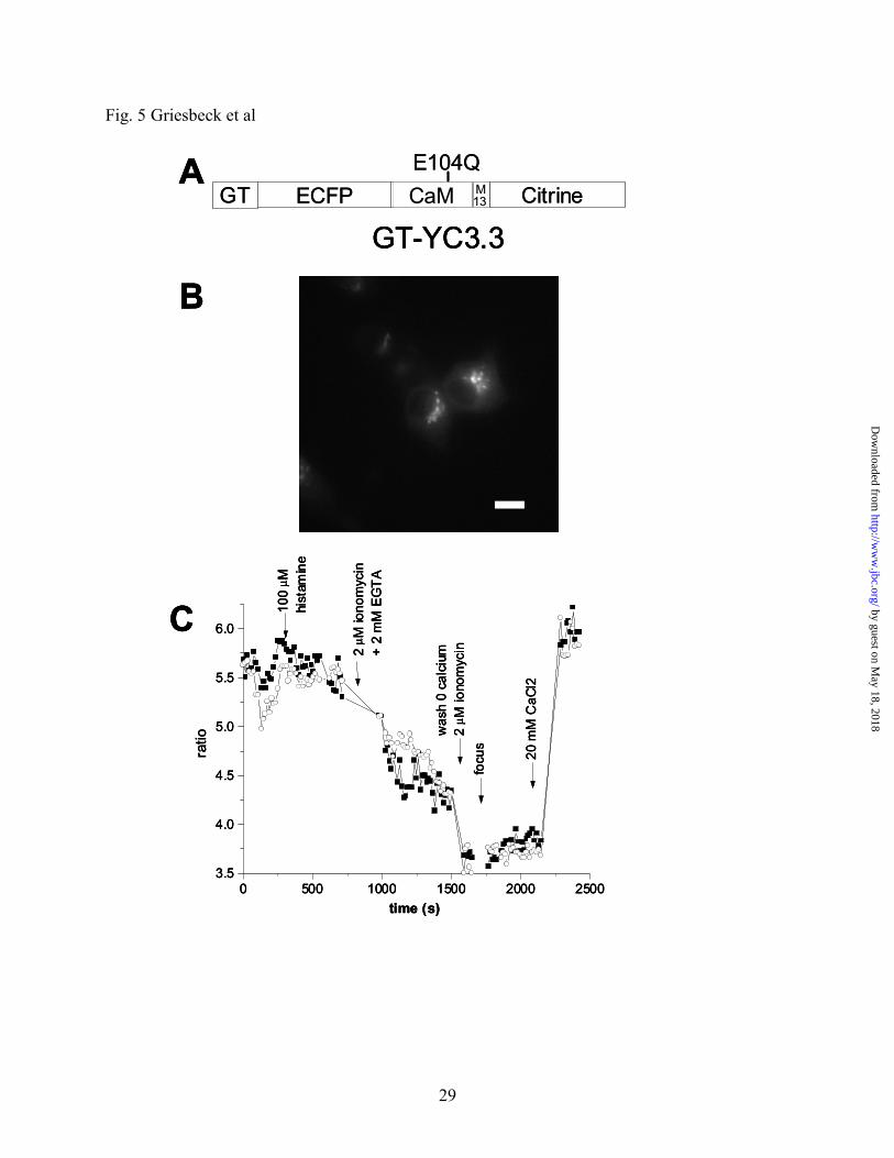
working range of our new cameleons. To test this we targeted YC3.3 to the Golgi by fusing the
81 N-terminal amino acids of human galactosyltransferase type II to YC3.3 and thereby
generating GT-YC3.3 (Fig. 5A). Transfection of HeLa cells resulted in bright punctate labeling
of Golgi stacks in a juxtanuclear position (Fig. 5B), identical to cells transfected with GT-EYFP
or stained for the medial/trans-Golgi marker α-mannosidase II (6). GT-YC3.3 was saturated at
by guest on May 18, 2018
http://ww
w.jbc.org/
Dow
nloaded from
Page 14
14
resting conditions (Fig. 5C), indicating a high concentration of free Ca2+ in the Golgi. Histamine
(100 μM) caused a very small decrease. The Golgi calcium store could be depleted with several
washes of ionomycin/EGTA and was refilled upon readmitting extracellular calcium (Fig. 5C),
demonstrating the feasibility of single cell imaging of free calcium concentrations in the Golgi of
mammalian cells. It has to be kept in mind that ionomycin does not perform optimally in acidic
compartments. Also it should be noted that YC3.3 is near its lower pH-limit under these
conditions. Further improvements in pH-resistance are still desirable, especially if one wants to
study even more acidic compartments of interest such as secretory vesicles. YC3.3 was similarly
well expressed in the ER (data not shown).
DISCUSSION
Citrine represents a third generation of YFPs or yellow mutants of Green Fluorescent
Protein. The first generation was exemplified by S65G/S72A/T203Y (26) and “10C” (1),
S65G/V68L/S72A/T203Y. These proteins proved to be quite sensitive to pH (e.g. pKa 6.9-7.1)
(6), halides such as Cl- (11) and partially reversible photobleaching (4). These sensitivities have
been useful for particular purposes such as quantifying cytosolic pH (6), [Cl-] (12, 44), or FRET
efficiency (18), but are considerable nuisances whenever one simply wants to use YFP as a
reliable label or FRET acceptor. In a second generation, the mutation Q69K was introduced into
10C to give S65G/V68L/Q69K/S72A/T203Y(8) or “EYFP V68L/Q69K”, which lowered the pKa
to 6.1 with little effect on the other sensitivities. We speculated that the positively charged lysine
might electrostatically hinder chromophore protonation (8). The Q69K mutation also introduced
a disadvantage: folding became noticeably more difficult, especially in organelles at 37ºC. In the
most recent version, Citrine, replacement of Q69K by Q69M lowered the pKa yet further to 5.7,
by guest on May 18, 2018
http://ww
w.jbc.org/
Dow
nloaded from
Page 15
15
eliminated the halide sensitivity, doubled the photostability, and improved the folding. The
improvement in folding efficiency was particularly apparent in difficult cases such as functional
expression at 37ºC in organelles or with an internally inserted calmodulin, i.e. camgaroo-2. The
crystal structure provides some reasonable rationalizations for these improved properties, in that
the Met side chain nicely plugs what had been a sizable halide-binding cavity next to the
chromophore. The poorer folding of Q69K might well be due to the extra length of a Lys side
chain making an uncomfortable fit within the cavity, or the electrostatic penalty for burying a
positive charge, or both. Thus a good steric fit with a neutral side chain seems far more effective
at lowering the chromophore pKa than an awkward fit with a positively charged side chain. The
apparent photobleaching of YFPs probably consists of two components, a reversible proton
redistribution or tautomerization and a truly irreversible covalent reaction (4, 5). Either or both
would be hindered by better packing of the hydrophobic core and elimination of a cavity next to
the chromophore.
Despite the inferiority of Q69K, it was an essential stepping stone in the evolution of
better properties by random mutagenesis and screening, because direct alteration of the Gln
codon CAG to the Met codon ATG would require two base changes in a single codon, a very
unlikely event. It was fortunate that there was an easy evolutionary path from CAG to the Lys
codon AAG and then to ATG. Many other examples of optimal sequences may remain relatively
inaccessible to random mutation due to barriers created by the genetic code.
We have demonstrated the application of Citrine in a series of genetically encoded Ca2+
indicators based on Citrine, all of which were improved in relation to their predecessors.
Camgaroo-2 may constitute an alternative to cameleons in confocal microscopy given that it can
be conveniently excited at the 488 nm argon laser line, or in cases in which targeting of
by guest on May 18, 2018
http://ww
w.jbc.org/
Dow
nloaded from
Page 16
16
cameleons are not successful. For example, we and others have found targeting of cameleons to
mitochondria to be difficult (45), whereas camgaroo-2 was easy to send to the mitochondria with
the targeting sequence of cytochrome C oxidase subunit VIII. Single cell imaging of
mitochondrial calcium offers exciting new prospects for studying its dynamics in this organelle
as well as to address aspects of heterogeneity of the mitochondrial population (46). Camgaroos
lack a CaM-binding peptide and therefore have lower Ca2+ affinities than the newest generic
design of GFP-based Ca2+ indicators, “G-CaMP” (47) or “pericams” (45). These indicators are
chimeras of the CaM-binding peptide M13, circularly permuted GFP or YFP, and CaM.
However, many of these molecules still do not express well at 37ºC, so annealing mutations
corresponding to Q69M might well be worth incorporating.
Our new improved cameleons expressed well at 37°C and were successfully targeted to
the ER and Golgi. Cytosolic pH fluctuations are readily transmitted to the ER (48), therefore it
was important to be able to express a pH-resistant functional indicator in this organelle, which
had not been possible with previous versions of cameleons. Similarly, previous cameleons did
not allow imaging free calcium in the Golgi due to the mild acidity of the compartment, which
quenched other YFPs. Little is known about calcium regulation in the Golgi. One study using
targeted aequorin identified the Golgi as a major calcium store within the cell (49), but aequorin
has many disadvantages, such as lack of intrinsic fluorescence and requirement for an exogenous
cofactor, that limit its use as a reliable calcium probe. We believe that Citrine should supersede
previous YFPs within fusions for multicolor observation of protein trafficking, protein-protein
interaction, and intramolecular conformational change, especially within genetically encoded
Ca2+ indicators.
by guest on May 18, 2018
http://ww
w.jbc.org/
Dow
nloaded from
Page 17
17
Acknowledgements-We would like to thank Qing Xiong for skillful technical assistance
and Nick Nguyen for assistance in X-ray data collection.
REFERENCES
1. Ormö, M., Cubitt, A. B., Kallio, K., Gross, L. A., Tsien, R. Y., and Remington, S. J. (1996)
Science 273, 1392-1395
2. Tsien, R. Y. (1998) Ann. Rev. Biochem. 67, 509-544
3. Wachter, R. M., Elsliger, M.-A., Kallio, K., Hanson, G. T., and Remington, S. J. (1998)
Structure 6, 1267-1277
4. Dickson, R. M., Cubitt, A. B., Tsien, R. Y., and Moerner, W. E. (1997) Nature 388, 355-
358
5. Miyawaki, A. and Tsien, R. Y. (2000) Methods Enzymol. 327, 472-500
6. Llopis, J., McCaffery, J. M., Miyawaki, A., Farquhar, M. G., and Tsien, R. Y. (1998) Proc.
Natl. Acad. Sci. USA 95, 6803-6808
7. Matsuyama, S., Llopis, J., Deveraux, Q. L., Tsien, R. Y., and Reed, J. C. (2000) Nature
Cell Biol. 2, 318-325
8. Miyawaki, A., Griesbeck, O., Heim, R., and Tsien, R. Y. (1999) Proc. Natl. Acad. Sci. USA
96, 2135-2140
9. Elsliger, M.-A., Wachter, R. M., Hanson, G. T., Kallio, K., and Remington, S. J. (1999)
Biochemistry 38, 5296-5301
10. Wachter, R. M., Yarbrough, D., Kallio, K., and Remington, S. J. (2000) J. Mol. Biol. 301,
157-171
11. Wachter, R. M. and Remington, S. J. (1999) Curr. Biol. 9, R628-R629
by guest on May 18, 2018
http://ww
w.jbc.org/
Dow
nloaded from
Page 18
18
12. Jayaraman, S., Haggie, P., Wachter, R. M., Remington, S. J., and Verkman, A. S. (2000) J.
Biol. Chem. 275, 6047-6050
13. Tsien, R. Y. and Miyawaki, A. (1998) Science 280, 1954-1955
14. Ellenberg, J., Lippincott-Schwartz, J., and Presley, J. F. (1999) Trends Cell Biol. 9, 52-56
15. Green, G., Kain, S. R., and Angres, B. (2000) Methods Enzymol. 327, 89-94
16. Heim, R. and Tsien, R. Y. (1996) Curr. Biol. 6, 178-182
17. Zacharias, D. A., Baird, G. S., and Tsien, R. Y. (2000) Curr. Opin. Neurobio. 10, 416-421
18. Llopis, J., Westin, S., Ricote, M., Wang, J., Cho, C. Y., Kurokawa, R., Rose, D. W.,
Rosenfeld, M. G., Tsien, R. Y., and Glass, C. K. (2000) Proc. Natl. Acad. Sci. USA 97,
4363-4368
19. Damelin, M. and Silver, P. A. (2000) Mol. Cell 5, 133-140
20. Ruehr, M. L., Zakhary, D. R., Damron, D. S., and Bond, M. (1999) J. Biol. Chem. 274,
33092-33096
21. Janetopoulos, C., Jin, T., and Devreotes, P. (2001) Science 291, 2408-2411
22. Overton, M. C. and Blumer, K. J. (2000) Curr. Biol. 10, 341-344
23. Siegel, R. M., Frederiksen, J. K., Zacharias, D. A., Chan, F. K. M., Johnson, M., Lynch, D.,
Tsien, R. Y., and Lenardo, M. J. (2000) Science 288, 2354-2357
24. Romoser, V. A., Hinkle, P. M., and Persechini, A. (1997) J. Biol. Chem. 272, 13270-13274
25. Persechini, A., Lynch, J. A., and Romoser, V. A. (1997) Cell Calcium 22, 209-216
26. Miyawaki, A., Llopis, J., Heim, R., McCaffery, J. M., Adams, J. A., Ikura, M., and Tsien,
R. Y. (1997) Nature 388, 882-887
27. Persechini, A. and Cronk, B. (1999) J. Biol. Chem. 274, 6827-6830
by guest on May 18, 2018
http://ww
w.jbc.org/
Dow
nloaded from
Page 19
19
28. Pearce, L. L., Gandley, R. E., Han, W., Wasserloos, K., Stitt, M., Kanai, A. J., McLaughlin,
M. K., Pitt, B. R., and Levitan, E. S. (2000) Proc. Natl. Acad. Sci. USA 97, 477-482
29. Sato, M., Hida, N., Ozawa, T., and Umezawa, Y. (2000) Anal. Chem. 72, 5918-5924
30. Honda, A., Adams, S. R., Sawyer, C. L., Lev-Ram, V., Tsien, R. Y., and Dostmann, W. R.
G. (2001) Proc. Natl. Acad. Sci. USA 98, 2437-2442
31. Mahajan, N. P., Harrison-Shostak, D. C., Michaux, J., and Herman, B. (1999) Chem. Biol.
6, 401-409
32. Vanderklish, P. W., Krushel, L. A., Holst, B. H., Gaily, J. A., Crossin, K. L., and Edelman,
G. M. (2000) Proc. Natl. Acad. Sci. USA 97, 2253-2258
33. Nagai, Y., Miyazaki, M., Aoki, R., Zama, T., Inouye, S., Hirose, K., Iino, M., and
Hagiwara, M. (2000) Nat. Biotechnol. 18, 313-316
34. Baird, G. S., Zacharias, D. A., and Tsien, R. Y. (1999) Proc. Natl. Acad. Sci. USA 96,
11241-11246
35. Heikal, A. A., Hess, S. T., Baird, G. S., Tsien, R. Y., and Webb, W. W. (2000) Proc. Natl.
Acad. Sci. USA 97, 11996-12001
36. Kendall, J. M., Dormer, R. L., and Campbell, A. K. (1992) Biochem. Biophys. Res. Comm.
189, 1008-1016
37. Otwinowski, Z. and Minor, W. (1997) Processing of x-ray diffraction data collected in
oscillation mode. In Carter, C. W. Jr. and Sweet, R. M., editors. Macromolecular
Crystallography, Academic Press, San Diego
38. Brunger, A. T., Adams, P. D., Clore, G. M., DeLano, W. L., Gros.P., Grosse-Kunstleve, R.
W., Jiang, J. S., Kuszewski, J., Nilges, M., Pannu, N. S., Read, R. J., Rice, L. M.,
Simonson, T., and Warren, G. L. (1998) Acta Cryst. D. Biol. Cryst. 54, 905-921
by guest on May 18, 2018
http://ww
w.jbc.org/
Dow
nloaded from
Page 20
20
39. McRee, D. E. (1999) J. Struct. Biol. 125, 156-165
40. Laskowski, R. A., MacArthur, M. W., Moss, D. S., and Thornton, J. M. (1993) J. Appl.
Cryst. 26, 283-291
41. Sanner, M. F., Olson, A. J., and Spehner, J. C. (1996) Biopolymers 38, 305-320
42. Baird, G. S., Zacharias, D. A., and Tsien, R. Y. (2000) Proc. Natl. Acad. Sci. USA 97,
11984-11989
43. Chesler, M. and Kaila, K. (1992) Trends Neurosci. 15, 396-402
44. Kuner, T. and Augustine, G. J. (2000) Neuron 27, 447-459
45. Nagai, T., Sawano, A., Park, E., and Miyawaki, A. (2001) Proc. Natl. Acad. Sci. USA 98,
3197-3202
46. Rizzuto, R., Pinton, P., Carrington, W., Fay, F. S., Fogarty, K. E., Lifshitz, L. M., Tuft, R.
A., and Pozzan, T. (1998) Science 280, 1763-1766
47. Nakai, J., Ohkura, M., and Imoto, K. (2001) Nature Biotechnology 19, 137-141
48. Kim, J. H., Johannes, L., Goud, B., Antony, C., Lingwood, C. A., Daneman, R., and
Grinstein, S. (1998) Proc. Natl. Acad. Sci. USA 95, 2997-3002
49. Pinton, P., Pozzan, T., and Rizzuto, R. (1998) EMBO J. 17, 5298-5308
50. Lawrence, M. C. and Bourke, P. D. (2000) J. Appl. Cryst. 33, 990-991
51. Kraulis, P. J. (1991) J. Appl. Cryst. 24, 946-950
FOOTNOTES
The atomic coordinates (code 1HUY) have been deposited in the Protein Data Bank,
Research Collaboration for Structural Bioinformatics, Rutgers University, New Brunswick, NJ
(http://www.rcsb.org).
by guest on May 18, 2018
http://ww
w.jbc.org/
Dow
nloaded from
Page 21
21
1The abbreviations and nonstandard terms used are: GFP, green fluorescent protein; CFP,
cyan fluorescent protein; YFP, yellow-emission variants of GFP; EYFP V68L/Q69K, GFP with
mutations S65G/V68L/Q69K/S72A/T203Y; Citrine, GFP with mutations
S65G/V68L/Q69M/S72A/T203Y; FRET, fluorescence resonance energy transfer; cameleon or
YC, a protein construct consisting of CFP, calmodulin, M13, and YFP fused in sequence;
camgaroo, a YFP with calmodulin inserted at position 145; ER, endoplasmic reticulum.
FIGURE LEGENDS
FIG. 1. Camgaroo-2 in vitro and in mammalian cells. A: Fluorescence intensity (528 nm
emission, pH 7.25) as a function of free Ca2+ concentrations. B: Unstimulated HeLa cells
transfected with cytosolic camgaroo-2, imaged at 480 nm excitation (30 nm bandwidth) with a
fluorescein dichroic mirror, and emission at 535 nm (25 nm bandwidth). C: Fluorescence
changes in a HeLa cell expressing cytosolic camgaroo-2 after given stimulations. The
fluorescence F normalized by the prestimulus fluorescence Fo is plotted. D: HeLa cells
transfected with camgaroo-2 targeted to mitochondria at resting calcium levels. E: Fluorescence
changes in a HeLa cell expressing mitochondria targeted camgaroo-2 after given stimulations.
FIG. 2. Photobleaching and pH/Cl--dependency of Citrine vs. EYFP V68L/Q69K. A:
Photobleaching curves for microdroplets of EYFP V68L/Q69K or Citrine under oil, observed on
a fluorescence microscope with 1.9 W/cm2 centered at 490 nm. Time constants for single or
double-exponential fits to the bleaching curves are listed. B: pH-dependence of the fluorescence
(at 528 nm) of Citrine in the presence ( ) or absence (×) of 100 mM Cl-, and likewise of EYFP
V68L/Q69K ( , ♦).
by guest on May 18, 2018
http://ww
w.jbc.org/
Dow
nloaded from
Page 22
22
FIG. 3. Detailed view of residue 69 and surrounding residues from the x-ray structures of
Citrine, EYFP-H148G (PDB accession code 1F0B), and EYFP-H148G with bound iodide
(PDB accession code 1F09). A. Stereoview of residue Q69M of Citrine with the chromophore
(Cro 66) and surrounding residues (Arg 96, Val 150, Ile 152, Val 163, Phe 165, Gln 183, Leu
201, and Tyr 203). The electron density represents a Fo - Fc omit map contoured at 3σ calculated
with the final refined coordinates in which the occupancy of Q69 was set to zero. B. The apo
form of EYFP-H148G (10) in the same orientation as A with the cavity (55 Å3) shown in green.
The cavity represents the volume accessible to a sphere of probe radius 1.2 Å. C. The iodide
bound form of EYFP-H148G (10) with the iodide ion represented as a purple sphere that is not
meant to represent its van der Waals radius. The total volume (91 Å3) of the halide-binding
cavity is represented in yellow. The programs CNS (38), MSMS (41), CONSCRIPT (50), and
MOLSCRIPT (51) were used to construct this figure.
FIG. 4. Dependence of YC3.3 fluorescence on Ca2+ and pH in vitro. A. Emission spectrum of
YC3.3 in the presence of 100 µM EGTA (solid line, -Ca2+) or 100 µM CaCl2 (dotted line, +Ca2+)
at pH 7.25. Excitation was at 432 nm. B. Emission ratio of YC3.3 (528/476 nm) in the presence
of 100 µM CaCl2 ( ) or 100 µM EGTA (●) were measured at the given pHs. Corresponding
ratios for YC2.1 (8) in 100 µM CaCl2 (□) or 100 µM EGTA (○) are shown for comparison.
FIG. 5. Calcium measurements with improved cameleons targeted to the Golgi of HeLa
cells. A. Schematic structure of GT-YC3.3. GT, 81 N-terminal amino acids of human
galactosyltransferase type II. CaM, calmodulin. The E104Q substitution within calmodulin raises
by guest on May 18, 2018
http://ww
w.jbc.org/
Dow
nloaded from
Page 23
23
the apparent dissociation constant for Ca2+ to 1.5 μM (8). B. HeLa cells transfected with GT-
YC3.3. Excitation was at 440 nm (10 nm bandwidth), the dichroic mirror transition was at 455
nm, and emission was collected at 535 nm (25 nm bandwidth). Scale bar 10 µM. C. Emission
ratios of two HeLa cells expressing GT-YC3.3 after stimulations with agonists. CFP emission
was collected at 480 nm (30 nm bandwidth) with excitation and YFP emission as described in B.
by guest on May 18, 2018
http://ww
w.jbc.org/
Dow
nloaded from
Page 24
24
TABLE I Spectral properties and pKa of selected YFP variants λex
a λemb εc quantum
yield pKa
d (147 mM Cl-)
pKae
(no Cl-)
Citrine 516 529 77 × 103 0.76 5.7 5.7 Citrine–C48L/C70V 516 529 69 × 103 0.72 5.7 n.d.h cpCitrine f 506 524 20 × 103 0.10 7.7 n.d. EYFP V68L/Q69K 516 529 62 × 103 0.71 6.1 6.0 cpEYFP V68L/Q69K g 506 524 18 × 103 0.09 8.9 n.d. aExcitation maximum (nm). bEmission maximum (nm). cExtinction coefficient (M-1cm-1). dDetermined in 147 mM chloride. eDetermined in 147 mM gluconate. fCircularly permuted Citrine. gCircularly permuted EYFP V68L/Q69K. hn.d., not determined.
TABLE II Data collection and refinement statistics
Data Collection Resolution (Å)a 28.3-2.2 (2.28-2.20) No. of Reflections a 12106 (1181) Completeness (%)a 99.3 (99.7) Rmerge (%)a,b 5.5 (14.4)
Refinement Statistics Rcryst (%)a 16.4 (19.5) Rfree (%)a,c 20.8 (26.0) No. of solvent molecules 97 Average B factor (Å2)
Main chain 26.3 Side chain 29.4 Solvent 35.5
Stereochemistry r.m.s.d. bond length (Å) 0.009 r.m.s.d. bond angle (°) 1.3
a Numbers in parentheses refer to the highest resolution shell. bRmerge = Σ|Ihkl – Iav|/ΣIav. cRfree was calculated using about 10 % of the reflections which were omitted from the refinement.
by guest on May 18, 2018
http://ww
w.jbc.org/
Dow
nloaded from
Page 25
25
Fig. 1 Griesbeck et al
-9 -8 -7 -6 -5 -4 -3 -2
0.0
0.2
0.4
0.6
0.8
1.0
Kd = 5.3 +/- 0.3 µMHill Coeff. = 1.24(F
-F0)
/(Fm
ax-F
0)
log [Ca2+]
A
B
-200 0 200 400 600 800 1000 1200 14000
1
2
3
4
5
6
7
2 µ M
iono
myc
in +
1 m
M E
GTA
2 µ M
iono
myc
in
+ 20
mM
CaC
l2
100
µ M h
ista
min
e
F/F 0
time (s)
C
D
0 200 400 600 800 1000
0.9
1.0
1.1
1.2
1.3
1.4
1.5
1.6
2 µM
iono
myc
in+
1m
MEG
TA
2 µM
iono
myc
in+
10m
MC
aCl 2
100 µ
M h
ista
min
e
F/F 0
time (s)
E
-9 -8 -7 -6 -5 -4 -3 -2
0.0
0.2
0.4
0.6
0.8
1.0
Kd = 5.3 +/- 0.3 µMHill Coeff. = 1.24(F
-F0)
/(Fm
ax-F
0)
log [Ca2+]
A
-9 -8 -7 -6 -5 -4 -3 -2
0.0
0.2
0.4
0.6
0.8
1.0
Kd = 5.3 +/- 0.3 µMHill Coeff. = 1.24(F
-F0)
/(Fm
ax-F
0)
log [Ca2+]
(F-F
0)/(F
max
-F0)
log [Ca2+]
A
BB
-200 0 200 400 600 800 1000 1200 14000
1
2
3
4
5
6
7
2 µ M
iono
myc
in +
1 m
M E
GTA
2 µ M
iono
myc
in
+ 20
mM
CaC
l2
100
µ M h
ista
min
e
F/F 0
time (s)
C
-200 0 200 400 600 800 1000 1200 14000
1
2
3
4
5
6
7
2 µ M
iono
myc
in +
1 m
M E
GTA
2 µ M
iono
myc
in
+ 20
mM
CaC
l2
100
µ M h
ista
min
e
F/F 0
time (s)
C
DD
0 200 400 600 800 1000
0.9
1.0
1.1
1.2
1.3
1.4
1.5
1.6
2 µM
iono
myc
in+
1m
MEG
TA
2 µM
iono
myc
in+
10m
MC
aCl 2
100 µ
M h
ista
min
e
F/F 0
time (s)
E
by guest on May 18, 2018
http://ww
w.jbc.org/
Dow
nloaded from
Page 26
26
Fig. 2 Griesbeck et al
0 100 200 300 400 5000.0
0.2
0.4
0.6
0.8
1.0 observed for EYFP Q69K single exponential fit (τ=79 s) double exponential fit (τ1=44 s, τ2=176 s)
observed for Citrine single exponential fit (τ=140 s) double exponential fit (τ1=105 s, τ2=316 s)
Frac
tion
of fl
uore
scen
ce re
mai
ning
Cumulative bleaching time (s)
A
B
4 5 6 7 8 9
0.0
0.2
0.4
0.6
0.8
1.0
Citrine/0 Cl-
Citrine/100 mM Cl-
Q69K/0 Cl-
Q69K/100 mM Cl-
norm
aliz
ed fl
uore
scen
ce
pH
by guest on May 18, 2018
http://ww
w.jbc.org/
Dow
nloaded from
Page 27
27
Fig. 3 Griesbeck et al
by guest on May 18, 2018
http://ww
w.jbc.org/
Dow
nloaded from
Page 28
28
Fig. 4 Griesbeck et al
440 460 480 500 520 540 560 580 6000.0
0.2
0.4
0.6
0.8
1.0
-Ca2+
+Ca2+
-Ca2+
+Ca2+
norm
aliz
ed e
mis
sion
inte
nsity
wavelength (nm)
A
B
5.5 6.0 6.5 7.0 7.51.2
1.6
2.0
2.4
2.8
3.2 yc2.1, high Ca2+
yc2.1, 0 Ca2+
yc3.3, high Ca2+
yc3.3, 0 Ca2+
528/
476
nm e
msi
ssio
n ra
tio
pH
4 4 0 460 480 500 520 540 560 580 6000.0
0.2
0.4
0.6
0.8
1.0
-Ca2+
+Ca2+
-Ca2+
+Ca2+
norm
aliz
ed e
mis
sion
inte
nsity
wavelength (nm)
A
B
5.5 6.0 6.5 7.0 7.51.2
1.6
2.0
2.4
2.8
3.2 yc2.1, high Ca2+
yc2.1, 0 Ca2+
yc3.3, high Ca2+
yc3.3, 0 Ca2+
528/
476
nm e
msi
ssio
n ra
tio
pH
by guest on May 18, 2018
http://ww
w.jbc.org/
Dow
nloaded from
Page 29
29
Fig. 5 Griesbeck et al
0 500 1000 1500 2000 25003.5
4.0
4.5
5.0
5.5
6.0
20 m
M C
aCl2
focu
s
was
h 0
calc
ium
2
µ M io
nom
ycin2
µ M io
nom
ycin
+
2 m
M E
GTA
100
µ Mhi
stam
ine
ratio
time (s)
A
C
GT-YC3.3
B
ECFP CaM M13 Citrine
E104QGT
0 500 1000 1500 2000 25003.5
4.0
4.5
5.0
5.5
6.0
20 m
M C
aCl2
focu
s
was
h 0
calc
ium
2
µ M io
nom
ycin2
µ M io
nom
ycin
+
2 m
M E
GTA
100
µ Mhi
stam
ine
ratio
time (s)
A
C
GT-YC3.3
B
0 500 1000 1500 2000 25003.5
4.0
4.5
5.0
5.5
6.0
20 m
M C
aCl2
focu
s
was
h 0
calc
ium
2
µ M io
nom
ycin2
µ M io
nom
ycin
+
2 m
M E
GTA
100
µ Mhi
stam
ine
ratio
time (s)
A
C
GT-YC3.3
B
ECFP CaM M13 Citrine
E104QGT
by guest on May 18, 2018
http://ww
w.jbc.org/
Dow
nloaded from
Page 30
TsienOliver Griesbeck, Geoffrey S. Baird, Robert E. Campbell, David A. Zacharias and Roger Y.
applicationsReducing the environmental sensitivity of yellow fluorescent protein: mechanism and
published online May 31, 2001J. Biol. Chem.
10.1074/jbc.M102815200Access the most updated version of this article at doi:
Alerts:
When a correction for this article is posted•
When this article is cited•
to choose from all of JBC's e-mail alertsClick here
by guest on May 18, 2018
http://ww
w.jbc.org/
Dow
nloaded from