Research ArticleEffects of UV-Vis Irradiation on VanadiumEtioporphyrins Extracted from Crude Oil andthe Role of Nanostructured Titania
Debra Jene Kirkconnell Reyes,1 Andrés García Saravia Ortiz de Montellano,1
Rudy Amilcar Trejo Tzab,2 Gerko Oskam,1 and Juan José Alvarado Gil1
1 Department of Applied Physics, CINVESTAV-Unidad Merida, Antigua Carretera a Progreso Km. 6, 97310 Merida, YUC, Mexico2 Facultad de Ingenierıa Quımica, Universidad Autonoma de Yucatan, Periferico Norte Km. 33.5, Tablaje Catastral 13615,Col. Chuburna de Hidalgo Inn, 97310 Merida, YUC, Mexico
Correspondence should be addressed to Debra Jene Kirkconnell Reyes; [email protected]
Received 14 December 2013; Revised 7 February 2014; Accepted 8 February 2014; Published 2 April 2014
The role of UV-irradiation on oil and its derivatives is particularly important for analyzing the degradability of specific oilcompounds. Also, nanostructured-TiO
2is one of the most promising photocatalysts so it is expected to be useful in their
degradation. However the complexity of crude oil, as well as that of the reactions involved, is such that the effect of the presenceof TiO
2under illumination is not well understood. In this paper, the influence of UV-Vis irradiation on vanadium etioporphyrins,
extracted from crude oil fromDos Bocas, Tabasco,Mexico, is studied using UV-Vis spectrophotometry in the absence and presenceof nanostructured TiO
2or nitrogen-doped TiO
2modified with copper (N-TiO
2/Cu). It is shown that the addition of water shortens
the time to start photodegradation. However, once this process has initiated, the system enters a second stage, that is very similarfor samples with or without water. It is also shown that the use of N-TiO
2/Cu induces an important delay in the initiation of the
porphyrins’ photodegradation process. Additionally, it has been found that the presence of TiO2in samples extracted with water
induces a small reduction in the photodegradation duration and, hence, that TiO2can catalyze the degradation of petroporphyrins.
1. Introduction
The many substances forming part of crude oil can havea limited lifetime due to various external factors. One ofthe most important degrading factors affecting oil is UVlight. The photochemical transformation of a given moleculeinto lower molecular weight fragments occurs usually via anoxidation process [1]. Photodegradation of petroleum prod-ucts both in laboratory and the field have been extensivelyexplored [2]. In particular, the effect of photolysis has beenstudied for a variety of pure hydrocarbons under solar andsimulated solar conditions [3, 4]. It has been shown that,depending on temperature and using light intensities in therange from200 to 750W/m2, somewater-soluble compoundssuch as naphthalenes and phenanthrenes can degrade rapidly.In contrast, other aromatic compounds such as alkylated
benzenes did not exhibit photodegradation under theseconditions [4].
In general, it is important to know under which condi-tions certain substances remain in the oil, stable or not, underUV illumination. This can be helpful in the development ofefficient methods to attend the problems generated by spills,and the vulnerability of specific components is also importantfor commercial or industrial purposes [5–7]. However, alarge number of substances still remain to be explored andunderstood under diverse conditions.
Porphyrins are among the most interesting substancesforming part of crude oil. In particular, the specific chemicalstructure of the porphyrins provides a fingerprint of theorigin of the oil. They are mainly present as vanadyl andnickel chelated compounds, although metal-free porphyrinshave also been isolated from immature sediments [8].
Hindawi Publishing CorporationInternational Journal of PhotoenergyVolume 2014, Article ID 401239, 9 pageshttp://dx.doi.org/10.1155/2014/401239
2 International Journal of Photoenergy
The identification and characterization of metalloporphyrinsin crude oils and source rock are of fundamental interestwithin a geochemical context, because they may providea better understanding of the geochemical origin of thepetroleum via diagenetic and catagenetic oil formation andmaturation of organic matter. Another interesting featurerelated to metalloporphyrins is their significant presence inheavy crude oil, which can cause problems as these metalshave a deleterious effect on the hydrogenation catalysts usedin upgrading and refining processes [9].
A common material used in cleaning the environment istitania (TiO
2), one of the best-known photocatalytic materi-
als due to its high photoactivity andphotostability.Thephoto-catalytic activity of titania depends on its physical properties,such as crystal structure, surface area, particle size, particleshape, among others [10]. One of the main disadvantagesof TiO
2is its large band gap of 3.2 eV (anatase), which
implies that it only absorbs UV light, corresponding to about5% of the solar spectrum. However, nanostructured TiO
2
is expected to be useful in the degradation of crude oil aswell as some of their compounds [11]. On the other hand,there are strong limitations in degrading crude oil; maybe themost important limitation is related to the opacity of crudeoil to UV light, which does not allow light to successfullydegrade oil even if adequate photocatalysts are present.Whencrude oil is mixed with solvents, a partial extraction ofsome components occurs; however, the effects of light andcatalysts on the extracts are not well understood. Oil extractscontaining porphyrins are common, and the analysis oftheir degradability using TiO
2nanoparticles is an interesting
challenge.In recent years, it has been shown that the photocatalytic
action of TiO2can be enhanced by impregnation with metals
such as copper. For example, the degradation of methyleneblue is accelerated using nitrogen-doped TiO
2modified
with copper (N-TiO2/Cu) nanomaterials prepared by plasma
discharge techniques in the presence of metallic copper andgaseous nitrogen [12]. In this paper, the effects of UV-Visirradiation on crude oil extracts containing petroporphyrinsare analyzed in the absence and presence of nanostructuredTiO2. In addition, the degradation of the metalloporphyrins
in the presence of N-TiO2/Cu nanomaterials is determined,
and the role of water in the degradation processes is consid-ered.
2. Materials and Methods
For the experiments we usedMaya crude oil fromDos Bocas,Tabasco, Mexico, which is an oil with ∘API = 21.57, 3.4 wt%sulfur, and 0.32 wt% nitrogen. The following two extractionmethods were used.
In the first method, 1.7mL of heavy crude oil was mixedwith 60mL of acetone in a beaker, and then the followingprocedure was performed.
(a) The mixture was stirred during 2 minutes, usinga Sonics ultrasonic vibrating cell (model TMVCX130PB) operating at 10W.
(b) The obtained sample was covered with a black metal-lic foil and a plastic bag and left for 24 hours to obtainthe extract.
(c) The supernatant, now with a yellow color, was trans-ferred to a second beaker labeled COE (crude oilextract).
The second extraction procedure was performed bymixing 1.7mL of heavy crude oil in a beaker with 60mL ofacetone and 5mL of distilled water, maintaining the propor-tion 12 : 1 of acetone to distilled water. Afterwards, the samplewas treated following the same procedure as described insteps (a), (b), and (c). The solution obtained in the secondextraction procedure was labeled COEW (crude oil extractwith water). Additionally, a standard solution was preparedby dissolving 0.1mg of vanadium (IV) etioporphyrin IIIoxide (C
32H36N4OVSantaCruz Biotechnology, CAS number
15709-03-2) in 60mL of acetone.Two types of nanostructured TiO
2powder were used,
both based on Degussa P25: pure TiO2and N-TiO
2/Cu.
This last compound was obtained by a plasma treatment ofDegussa P25 in a nitrogen atmosphere in the presence ofmetallic copper [12]. Figure 1 shows the XPS analysis as wellas the X-ray diffraction patterns of the N-TiO
2/Cu material.
X-ray diffraction exhibits the well-known proportion of 80%anatase and 20% rutile in titania Degussa P25 but it doesnot have the sensitivity to detect the presence of copper inthe material. However there are small differences betweenboth spectra that can be observed; some peaks are slightlydisplaced while others are wider or higher, but all of themare closely related to the ones observed in untreated DegussaP25 (see Figure 1(a)). These small changes can be related tochanges in the nanoparticles’ size due to the copper impreg-nation process.
In contrast, XPS measurements are able to detect thepresence of copper in N-TiO
2/Cu (see Figure 1(b)); in these
measurements two bands associated with copper, as well asthe characteristic bands of TiO
2(Degussa P25) and a band
related to nitrogen, can be observed. Additional analyses haveshown that the plasma-treated samples contain about 4.4wt%of nitrogen and 0.4% of copper [12].
Using the COE and COEW extracts, a set of samples wasprepared by adding 25mg of TiO
2or N-TiO
2/Cu, labeled
COE-TiO2, COE-(N-TiO
2/Cu), COEW-TiO
2, and COEW-
(N-TiO2/Cu), respectively. These samples were submitted
to an additional mixing process using a Sonics ultrasonicvibrating mixer cell operated at 10W for 2 minutes and leftto settle for 60 minutes with the samples covered to preventevaporation. For all the solutions, COE, COEW, COE-TiO
2,
COEW-TiO2, COE-(N-TiO
2/Cu), and COEW-(N-TiO
2/Cu)
aliquots of about 10mL were taken from the beakers andlabeled as “time zero” experiments.The remaining solution ineach one of the beakers was then poured into an Erlenmeyerflask integrated with a cooling system and placed at 400 rpmusing magnetic stirring. For the photodegradation studies,the light of a 1000W Xenon lamp (Oriel 6269), operated at400W, was used to illuminate the Erlenmeyer reaction vesselfrom above. This lamp provided 370W/m2 on the surface ofthe sample, with most of the intensity in the visible region
International Journal of Photoenergy 3
100
80
60
40
20
0
20 25 30 35 40 45 50
Inte
nsity
(a.u
.)
N-TiO2/CuDegussa P25
2𝜃 (∘)
(a)
402 400 398 396 394 392
Binding energy (eV)
N(E
) (a.u
.)
N(E
) (a.u
.)
955 950 945 940 935 930
Binding energy (eV)
Copper
Cu2p1/2
Cu2p3/2
Nitrogen−
−
Binding energy (eV)465 460 455
N(E
) (a.u
.)
Ti2p1/2
Ti2p3/2TiO2−
N 1 s
(b)
Figure 1: (a) X-ray diffraction spectra of TiO2(Degussa P25) and N-TiO
2/Cu. (b) XPS of N-TiO
2/Cu.
4 International Journal of Photoenergy
2.0
1.5
1.0
0.5
0.0
350 400 450 500
Wavelength (nm)
N-TiO2/CuDegussa P25
Abso
rptio
n (a
.u.)
Figure 2: Absorption of TiO2andN-TiO
2/Cu obtained fromdiffuse
reflectance measurements using the Kubelka-Munk method.
of light, a good emission of UVA and UVB light, and a verylow fraction of UVC. During irradiation the samples in thejacketed flask weremaintained at a temperature of 30∘Cusinga circulating water system. Aliquots of 10mL were takenfrom the Erlenmeyer flask every hour for five hours andtransmission optical spectra for the samples were measuredin quartz cuvettes using a fiber UV-Vis spectrophotometer inthe range from 200 nm to 1100 nm using an AvaSpec-2048fiber optic spectrometer with a deuterium lamp as the lightsource.
3. Results and Discussion
Figure 2 shows the absorption spectra, obtained from diffusereflectance measurements on the powder by applying theKubelka-Munk method, of the Degussa P25 and N-TiO
2/Cu
powders used in the experiments, and an absorption incre-ment for N-TiO
2/Cu in visible light can be observed.
The COE and COEW samples were illuminated over aperiod of 5 hours, and Figure 3 shows the correspondingUV-Vis spectra obtained before illumination (time zero) andafter every hour. Before irradiation the spectra for COE andCOEW samples show a broad band from 220 nm to 450 nmwith three small peaks at 300 nm, 350 nm, and 400 nm andalso a shoulder at 250 nm. Additionally, two smaller bandscan be observed at 530 nm and 569 nm. The spectra remainlargely unchanged upon irradiation; however, as illustrated inthe insets, the peaks at 530 nm and 569 nm disappear uponprolonged illumination indicating a degradation process.
The COE-TiO2and COEW-TiO
2as well as the COE-(N-
TiO2/Cu) and COEW-(N-TiO
2/Cu) samples show the same
bands as the samples without TiO2; however, the shoulder at
250 nm appears more clearly, as illustrated in Figures 4 and 5.Similar to the samples without TiO
2, the two bands at 530 nm
and 569 nm are also observed.It is well known that crude oil contains petroporphyrins
[13] so it is possible that the small bands at 530 nm and
569 nm correspond to such type of compounds. Comparingthe experimental UV-Vis spectra from the crude oil extractswith databases of spectra for petroporphyrins, we concludethat the vanadiumetioporphyrins are the best fit for our crudeoil samples. In Figure 6, the UV-Vis spectrum of a standardsolution of vanadium etioporphyrin in acetone is presented.In this spectrum, the wide band from 200 nm to 270 nm isdue to acetone [14] while a narrow band appearing at 414 nmcan be identified as the Soret band. Additionally, two smallerbands are clearly observed at 530 nm and 569 nm. From theseresults, it can be inferred that the peaks of the spectra ofour extracts appearing at 530 nm and 569 nm correspond tovanadium etioporphyrins as previously reported for crude oilfrom the same geographic location [9].
The UV-Vis spectrum of petroporphyrins show bandsthat correspond to a strong electronic transition to the secondexcited state (𝑆
0→ 𝑆2) at about 400 nm (the Soret or B
band) and a weak transition to the first excited state (𝑆0→
𝑆1) at about 550 nm (the Q band). Internal conversion from𝑆2to 𝑆1is rapid and, as a consequence, fluorescence is
only detected from 𝑆1. The B and the Q bands both arise
from 𝜋-𝜋∗ transitions and can be explained by consideringthe four frontier orbitals (HOMO and LUMO orbitals) (theGouterman four orbital model) [15]. Gouterman’s “four-orbital” model of the 𝜋-𝜋∗ transitions in porphyrins is firmlyestablished as a simple unifying theory of porphyrin spectra.It provides parametric expressions for the frequencies andintensities of the 𝛼-𝛽 (Q) and Soret (B) bands of theporphyrin chromophore. Spectral shifts caused by systematicvariation of ring substituents and changes of the encapsulatedmetal ion, axial ligation, or even dimer formation havesuccessfully been rationalized based on these grounds [16, 17].The electronic transition to the higher energymixed state, the𝑆2state, is strongly allowed, whereas the electronic transition
to the lower energy mixed state, the 𝑆1state, is only weakly
allowed.The band in the UV-Vis absorption spectrum due toa transition to the 𝑆
2state is the Soret band, and the band
due to a transition to the vibrationless 𝑆1state is the 𝛼 band.
The greater the degree of mixing, the less intense the 𝛼 bandrelative to the Soret band. In the UV-visible spectrum ofporphyrin, there is also a vibronic band, the 𝛽 band, whichappears at slightly lower wavelengths than the 𝛼 band. The𝛽 band is due to transitions to higher vibrational levels inthe 𝑆1state and serves as a normalization band in porphyrin
absorption spectra [15]. In summary, the three characteristicbands appearing in the spectra of the nonirradiated samplescorrespond to the bands of vanadium etioporphyrins: at407 nm the Soret band and the 𝛽 and 𝛼 bands at 533 nm and572 nm, respectively [18]. When the intensity of the 𝛼 band islarger than that of the𝛽 band, themetal forms a stable square-planar complex with the porphyrin [19].
In order to determine the concentration of vanadiumetioporphyrins in our samples, we prepared dilutions of thestandard solution (vanadium IV etioporphyrin III oxide)and determined the absorbance as a function of concen-tration. The relationship between the absorbance (𝐴) andconcentration (𝐶), according to the Lambert-Beer law, isgiven by 𝐴 = 𝜀𝑑𝐶, where 𝑑 (cm) and 𝜀 (L cm−1mg−1)are the thickness and extinction coefficient of the sample,
International Journal of Photoenergy 5
Abso
rban
ce (a
.u.)2.0
1.5
1.0
0.5
0.0
Abso
rban
ce (a
.u.)
Wavelength (nm)
Wavelength (nm)0 200 400 600 800 1000 1200
0hours1hour2hours
3hours4hours5hours
0.15
0.10
0.05
0.00510 540 570 600
(a)
2.5
2.0
1.5
1.0
0.5
0.0
Wavelength (nm)0 200 400 600 800 1000 1200
0hours1hour2hours
3hours4hours5hours
Abso
rban
ce (a
.u.)
Wavelength (nm)510 540 570 600
Abso
rban
ce (a
.u.)
0.15
0.20
0.10
0.05
0.00
(b)
Figure 3: UV-Vis absorbance spectra as a function of illumination time of the crude oil extracts: (a) COE and (b) COEW. The insets showthe bands observed at 530 nm and 569 nm with the background subtracted.
Wavelength (nm)0 200 400 600 800 1000 1200
0hours1hour2hours
3hours4hours5hours
2.5
2.0
1.5
1.0
0.5
0.0
Abso
rban
ce (a
.u.)
Abso
rban
ce (a
.u.)
Wavelength (nm)
0.15
0.10
0.05
0.00510 540 570 600
(a)
Wavelength (nm)0 200 400 600 800 1000 1200
0hours1hour2hours
3hours4hours5hours
2.0
1.5
1.0
0.5
0.0
Abso
rban
ce (a
.u.)
Abso
rban
ce (a
.u.)
Wavelength (nm)
0.10
0.05
0.00510 540 570 600
(b)
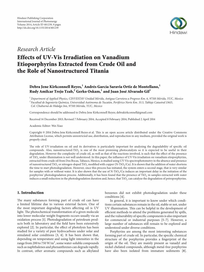
Figure 4: UV-Vis absorbance spectra as a function of illumination time in the presence of the TiO2nanomaterial with the crude oil extracts:
(a) COE-TiO2and (b) COEW-TiO
2. The insets show the bands observed at 530 nm and 569 nm.
respectively. In Figure 7, the absorbance maximum of the 𝛼peak versus concentration is presented, in which the fit of theexperimental data provides a value of 0.116 L cm−1mg−1 forthe extinction coefficient.
Using these results, the concentration of the vanadiumetioporphyrin in ourCOEcan be estimated as being 3.8mg/L,which is in agreement with generally observed concentra-tions in crude oil from the oil from the same geographicallocation [9].
Due to the UV-Vis irradiation treatment, there arechanges in intensity in various bands in the spectra; however,we will focus our attention on the bands in the range from500 nm to 620 nm because they are associated directlywith the vanadium etioporphyrins. In order to performa nonambiguous analysis of the bands appearing in allspectra, we separated the 𝛼 and 𝛽 bands absorbance fromthe main background absorbance by fitting a cubic splinein the smooth region from 425 nm to 750 nm, excluding
6 International Journal of Photoenergy
Wavelength (nm)0 200 400 600 800 1000 1200
0hours1hour2hours
3hours4hours5hours
2.5
2.0
1.5
1.0
0.5
0.0
0.4
0.5
0.3
0.2
0.1
0.0
Abso
rban
ce (a
.u.)
Abso
rban
ce (a
.u.)
Wavelength (nm)510 540 570 600
(a)
Wavelength (nm)0 200 400 600 800 1000 1200
0hours1hour2hours
3hours4hours5hours
2.0
1.5
1.0
0.5
0.0
Abso
rban
ce (a
.u.)
Wavelength (nm)510 540 570 600
Abso
rban
ce (a
.u.)
0.15
0.10
0.05
0.00
(b)
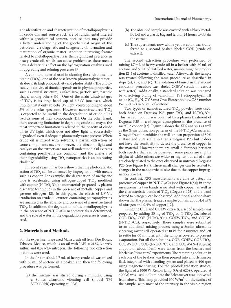
Figure 5: UV-Vis absorbance spectra as a function of illumination time in the presence of the N-TiO2/Cu nanomaterial with the crude oil
extracts: (a) COE-(N-TiO2/Cu) and (b) COEW-(N-TiO
2/Cu).The insets show the bands observed at 530 nm and 569 nmwith the background
subtracted.
Wavelength (nm)0 200 400 600 800 1000 1200
0hours1hour2hours
3hours4hours5hours
1.0
0.8
0.6
0.4
0.2
0.0
0.05
0.04
0.03
0.02
0.01
0.0
Abso
rban
ce (a
.u.)
Abso
rban
ce (a
.u.)
Wavelength (nm)510 540 570 600
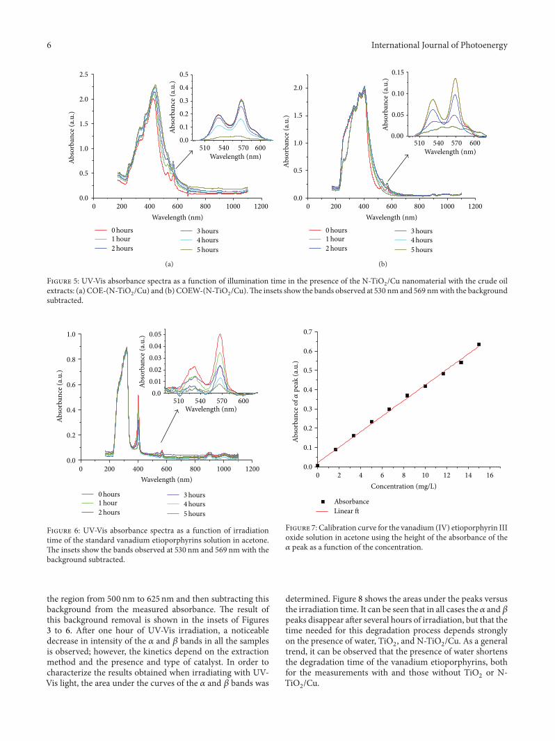
Figure 6: UV-Vis absorbance spectra as a function of irradiationtime of the standard vanadium etioporphyrins solution in acetone.The insets show the bands observed at 530 nm and 569 nm with thebackground subtracted.
the region from 500 nm to 625 nm and then subtracting thisbackground from the measured absorbance. The result ofthis background removal is shown in the insets of Figures3 to 6. After one hour of UV-Vis irradiation, a noticeabledecrease in intensity of the 𝛼 and 𝛽 bands in all the samplesis observed; however, the kinetics depend on the extractionmethod and the presence and type of catalyst. In order tocharacterize the results obtained when irradiating with UV-Vis light, the area under the curves of the 𝛼 and 𝛽 bands was
0.7
0.6
0.5
0.4
0.3
0.2
0.1
0.0
0 2 4 6 8 10 12 14 16
Concentration (mg/L)
AbsorbanceLinear ft
Abso
rban
ce o
f𝛼pe
ak (a
.u.)
Figure 7: Calibration curve for the vanadium (IV) etioporphyrin IIIoxide solution in acetone using the height of the absorbance of the𝛼 peak as a function of the concentration.
determined. Figure 8 shows the areas under the peaks versusthe irradiation time. It can be seen that in all cases the 𝛼 and 𝛽peaks disappear after several hours of irradiation, but that thetime needed for this degradation process depends stronglyon the presence of water, TiO
2, and N-TiO
2/Cu. As a general
trend, it can be observed that the presence of water shortensthe degradation time of the vanadium etioporphyrins, bothfor the measurements with and those without TiO
2or N-
TiO2/Cu.
International Journal of Photoenergy 7
1.0
1.2
0.8
0.6
0.4
0.2
0.0
Are
a of t
he p
orph
yrin
ban
ds
COECOEW
2
COE TiO2
2COEW-TiO
0 1 2 3 4 5
Time (hours)
COE-(N-TiO /Cu)
2COEW-(N-TiO /Cu)
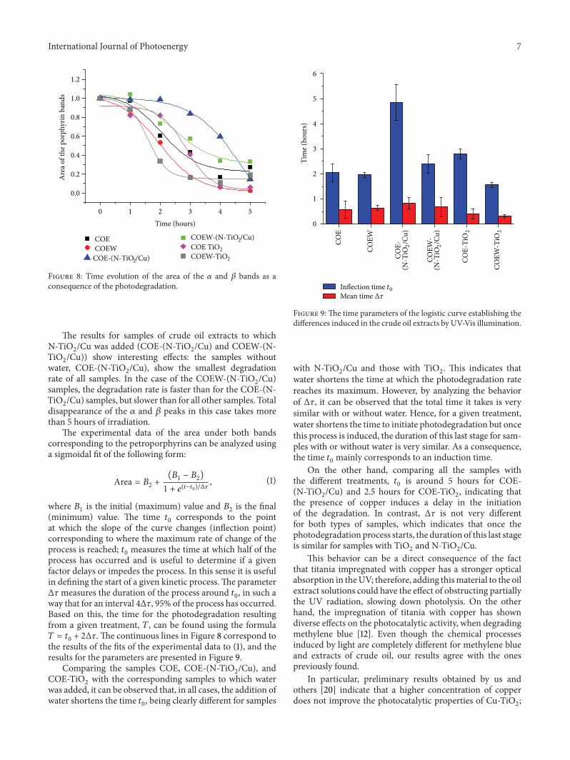
Figure 8: Time evolution of the area of the 𝛼 and 𝛽 bands as aconsequence of the photodegradation.
The results for samples of crude oil extracts to whichN-TiO
2/Cu was added (COE-(N-TiO
2/Cu) and COEW-(N-
TiO2/Cu)) show interesting effects: the samples without
water, COE-(N-TiO2/Cu), show the smallest degradation
rate of all samples. In the case of the COEW-(N-TiO2/Cu)
samples, the degradation rate is faster than for the COE-(N-TiO2/Cu) samples, but slower than for all other samples. Total
disappearance of the 𝛼 and 𝛽 peaks in this case takes morethan 5 hours of irradiation.
The experimental data of the area under both bandscorresponding to the petroporphyrins can be analyzed usinga sigmoidal fit of the following form:
Area = 𝐵2+(𝐵1− 𝐵2)
1 + 𝑒(𝑡−𝑡0)/Δ𝜏, (1)
where 𝐵1is the initial (maximum) value and 𝐵
2is the final
(minimum) value. The time 𝑡0corresponds to the point
at which the slope of the curve changes (inflection point)corresponding to where the maximum rate of change of theprocess is reached; 𝑡
0measures the time at which half of the
process has occurred and is useful to determine if a givenfactor delays or impedes the process. In this sense it is usefulin defining the start of a given kinetic process.The parameterΔ𝜏measures the duration of the process around 𝑡
0, in such a
way that for an interval 4Δ𝜏, 95% of the process has occurred.Based on this, the time for the photodegradation resultingfrom a given treatment, 𝑇, can be found using the formula𝑇 = 𝑡0+ 2Δ𝜏. The continuous lines in Figure 8 correspond to
the results of the fits of the experimental data to (1), and theresults for the parameters are presented in Figure 9.
Comparing the samples COE, COE-(N-TiO2/Cu), and
COE-TiO2with the corresponding samples to which water
was added, it can be observed that, in all cases, the addition ofwater shortens the time 𝑡
0, being clearly different for samples
COE
COEW
Tim
e (ho
urs)
COE-
(N-T
iO2/C
u)
COEW
-(N
-TiO
2/C
u)
COE-
TiO
2
COEW
-TiO
2
6
5
4
3
2
1
0
Inflection time t0Mean time Δ𝜏
Figure 9: The time parameters of the logistic curve establishing thedifferences induced in the crude oil extracts by UV-Vis illumination.
with N-TiO2/Cu and those with TiO
2. This indicates that
water shortens the time at which the photodegradation ratereaches its maximum. However, by analyzing the behaviorof Δ𝜏, it can be observed that the total time it takes is verysimilar with or without water. Hence, for a given treatment,water shortens the time to initiate photodegradation but oncethis process is induced, the duration of this last stage for sam-ples with or without water is very similar. As a consequence,the time 𝑡
0mainly corresponds to an induction time.
On the other hand, comparing all the samples withthe different treatments, 𝑡
0is around 5 hours for COE-
(N-TiO2/Cu) and 2.5 hours for COE-TiO
2, indicating that
the presence of copper induces a delay in the initiationof the degradation. In contrast, Δ𝜏 is not very differentfor both types of samples, which indicates that once thephotodegradation process starts, the duration of this last stageis similar for samples with TiO
2and N-TiO
2/Cu.
This behavior can be a direct consequence of the factthat titania impregnated with copper has a stronger opticalabsorption in theUV; therefore, adding thismaterial to the oilextract solutions could have the effect of obstructing partiallythe UV radiation, slowing down photolysis. On the otherhand, the impregnation of titania with copper has showndiverse effects on the photocatalytic activity, when degradingmethylene blue [12]. Even though the chemical processesinduced by light are completely different for methylene blueand extracts of crude oil, our results agree with the onespreviously found.
In particular, preliminary results obtained by us andothers [20] indicate that a higher concentration of copperdoes not improve the photocatalytic properties of Cu-TiO
2;
8 International Journal of Photoenergy
therefore, if the photodegradation of the porphyrinswould bethe objective, a possible solution could be to use nanoparticleswith a lower content of copper concentration. Additionally,𝑡0and Δ𝜏 are smaller for COEW-TiO
2than for COEW
samples. This implies that Degussa P-25 TiO2is showing
photocatalytic activity in presence of water. In contrast, thisis harder to observe in samples without water, where 𝑡
0is
2 hours for COE-TiO2and 2.7 hours for COE; although
there is a slight tendency for Δ𝜏 to being shorter for COE-TiO2than for COE, the spread in the values as represented
by the error bars does not allow to unequivocally state thatthere is a difference between the values. Hence, it appearsthat direct absorption of UV-light by the crude oil extractsis capable of the photodegradation of petroporphyrins andthat adding a TiO
2-based catalyst, in the absence of water,
does not significantly accelerate photodegradation. However,the presence of water results in a faster initiation of thephotodegradation processes.
4. Conclusions
The effects of UV-Vis illumination on crude oil extracts con-taining petroporphyrins have been monitored as a functionof time. Analysis of the UV-Vis spectra showed that the UV-Vis illumination induces the systematic reduction of the sizeof the 𝛼 and 𝛽 bands, corresponding to the degradation ofthe petroporphyrins. It has been shown that this reductioncan be parameterized as a second-order kinetics process.Theanalysis of the parameters of the degradation kinetics allowsinferring that the addition of water to the crude oil extractsshortens the time to induce the photodegradation. However,once this process has initiated, the duration of the last stageof the photodegradation for samples with or without water isvery similar. It has also been shown that the use ofN-TiO
2/Cu
induces an important delay in the initiation of the process ofphotodegradation of the porphyrins. Additionally, it has beenshown that the presence of TiO
2in the samples extracted
with water induces a small reduction in the duration ofthe photodegradation; hence, Degussa P-25 can catalyze thedegradation of petroporphyrins.
Conflict of Interests
The authors declare that there is no conflict of interestsregarding the publication of this paper.
Acknowledgments
Debra Jene Kirkconnell Reyes and Andres Garcıa Saraviathank Conacyt for a scholarship. The authors wish to thankJ. Ordonez-Miranda, J. M. Yanez-Limon, C. Vales-Pinzon,D. Macıas, J. Bante-Guerra, Gaspar Euan, N.W. Pech-May,and B.E. Heredia-Cervera for their useful suggestions andenlightening discussions during the development of thiswork. This work was partially funded by Conacyt projects105816, 178510 and 193850 and FOMIX-Yucatan project170120.
References
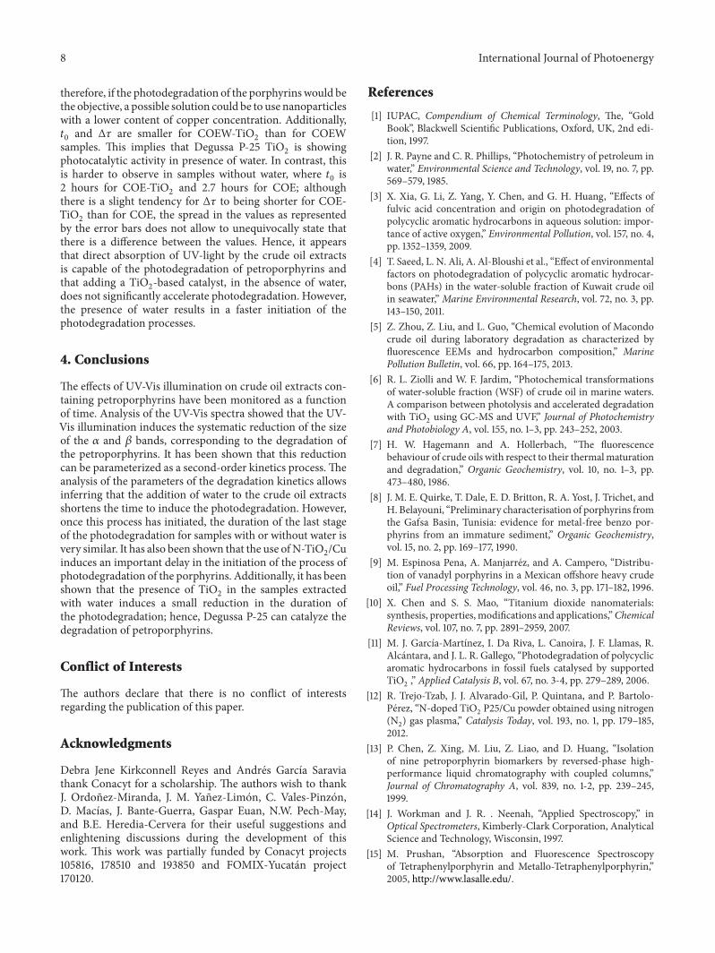
[1] IUPAC, Compendium of Chemical Terminology, The, “GoldBook”, Blackwell Scientific Publications, Oxford, UK, 2nd edi-tion, 1997.
[2] J. R. Payne and C. R. Phillips, “Photochemistry of petroleum inwater,” Environmental Science and Technology, vol. 19, no. 7, pp.569–579, 1985.
[3] X. Xia, G. Li, Z. Yang, Y. Chen, and G. H. Huang, “Effects offulvic acid concentration and origin on photodegradation ofpolycyclic aromatic hydrocarbons in aqueous solution: impor-tance of active oxygen,” Environmental Pollution, vol. 157, no. 4,pp. 1352–1359, 2009.
[4] T. Saeed, L. N. Ali, A. Al-Bloushi et al., “Effect of environmentalfactors on photodegradation of polycyclic aromatic hydrocar-bons (PAHs) in the water-soluble fraction of Kuwait crude oilin seawater,” Marine Environmental Research, vol. 72, no. 3, pp.143–150, 2011.
[5] Z. Zhou, Z. Liu, and L. Guo, “Chemical evolution of Macondocrude oil during laboratory degradation as characterized byfluorescence EEMs and hydrocarbon composition,” MarinePollution Bulletin, vol. 66, pp. 164–175, 2013.
[6] R. L. Ziolli and W. F. Jardim, “Photochemical transformationsof water-soluble fraction (WSF) of crude oil in marine waters.A comparison between photolysis and accelerated degradationwith TiO
2using GC-MS and UVF,” Journal of Photochemistry
and Photobiology A, vol. 155, no. 1–3, pp. 243–252, 2003.[7] H. W. Hagemann and A. Hollerbach, “The fluorescence
behaviour of crude oils with respect to their thermalmaturationand degradation,” Organic Geochemistry, vol. 10, no. 1–3, pp.473–480, 1986.
[8] J. M. E. Quirke, T. Dale, E. D. Britton, R. A. Yost, J. Trichet, andH. Belayouni, “Preliminary characterisation of porphyrins fromthe Gafsa Basin, Tunisia: evidence for metal-free benzo por-phyrins from an immature sediment,” Organic Geochemistry,vol. 15, no. 2, pp. 169–177, 1990.
[9] M. Espinosa Pena, A. Manjarrez, and A. Campero, “Distribu-tion of vanadyl porphyrins in a Mexican offshore heavy crudeoil,” Fuel Processing Technology, vol. 46, no. 3, pp. 171–182, 1996.
[10] X. Chen and S. S. Mao, “Titanium dioxide nanomaterials:synthesis, properties, modifications and applications,”ChemicalReviews, vol. 107, no. 7, pp. 2891–2959, 2007.
[11] M. J. Garcıa-Martınez, I. Da Riva, L. Canoira, J. F. Llamas, R.Alcantara, and J. L. R. Gallego, “Photodegradation of polycyclicaromatic hydrocarbons in fossil fuels catalysed by supportedTiO2,” Applied Catalysis B, vol. 67, no. 3-4, pp. 279–289, 2006.
[12] R. Trejo-Tzab, J. J. Alvarado-Gil, P. Quintana, and P. Bartolo-Perez, “N-doped TiO
2P25/Cu powder obtained using nitrogen
(N2) gas plasma,” Catalysis Today, vol. 193, no. 1, pp. 179–185,
2012.[13] P. Chen, Z. Xing, M. Liu, Z. Liao, and D. Huang, “Isolation
of nine petroporphyrin biomarkers by reversed-phase high-performance liquid chromatography with coupled columns,”Journal of Chromatography A, vol. 839, no. 1-2, pp. 239–245,1999.
[14] J. Workman and J. R. . Neenah, “Applied Spectroscopy,” inOptical Spectrometers, Kimberly-Clark Corporation, AnalyticalScience and Technology, Wisconsin, 1997.
[15] M. Prushan, “Absorption and Fluorescence Spectroscopyof Tetraphenylporphyrin and Metallo-Tetraphenylporphyrin,”2005, http://www.lasalle.edu/.
International Journal of Photoenergy 9
[16] M. Gouterman, G. H. Wagniere, and L. C. Snyder, “Spectra ofporphyrins. Part II. Four orbital model,” Journal of MolecularSpectroscopy, vol. 11, no. 1–6, pp. 108–127, 1963.
[17] A. Ceulemans, W. Oldenhof, C. Gorller-Walrand, and L. G.Vanquickenborne, “Gouterman’s “four-orbital” model and theMCD spectra of high-symmetry metalloporphyrins,” Journal ofthe American Chemical Society, vol. 108, no. 6, pp. 1155–1163,1986.
[18] H. Xu, G. Que, D. Yu, and J. R. Lu, “Characterization of petro-porphyrins using ultraviolet-visible spectroscopy and laser des-orption ionization time-of-flight mass spectrometry,” Energyand Fuels, vol. 19, no. 2, pp. 517–524, 2005.
[19] L. R.Milgrom,TheColours of Life: An Introduction to the Chem-istry of Porphyrins and Related Compounds, Oxford UniversityPress, 1997.
[20] B. Xin, P. Wang, D. Ding, J. Liu, Z. Ren, and H. Fu, “Effectof surface species on Cu-TiO
2photocatalytic activity,” Applied
Surface Science, vol. 254, no. 9, pp. 2569–2574, 2008.