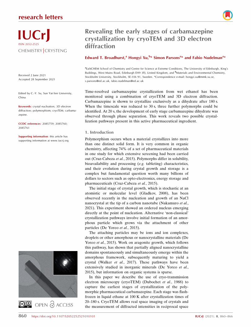
research letters 860 https://doi.org/10.1107/S2052252521010101 IUCrJ (2021). 8, 860–866 IUCrJ ISSN 2052-2525 CHEMISTRY j CRYSTENG Received 2 June 2021 Accepted 28 September 2021 Edited by C.-Y. Su, Sun Yat-Sen University, China Keywords: crystal nucleation; 3D electron diffraction; polymorphism; cryoTEM; carbama- zepine. CCDC references: 2085759; 2085760; 2085761 Supporting information: this article has supporting information at www.iucrj.org Revealing the early stages of carbamazepine crystallization by cryoTEM and 3D electron diffraction Edward T. Broadhurst, a Hongyi Xu, b * Simon Parsons a * and Fabio Nudelman a * a EaStCHEM School of Chemistry and Centre for Science at Extreme Conditions, The University of Edinburgh, King’s Buildings, West Mains Road, Edinburgh EH9 3FJ, United Kingdom, and b Materials and Environmental Chemistry, Stockholm University, Stockholm, SE-106 91, Sweden. *Correspondence e-mail: [email protected], [email protected], [email protected]Time-resolved carbamazepine crystallization from wet ethanol has been monitored using a combination of cryoTEM and 3D electron diffraction. Carbamazepine is shown to crystallize exclusively as a dihydrate after 180 s. When the timescale was reduced to 30 s, three further polymorphs could be identified. At 20 s, the development of early stage carbamazepine dihydrate was observed through phase separation. This work reveals two possible crystal- lization pathways present in this active pharmaceutical ingredient. 1. Introduction Polymorphism occurs when a material crystallizes into more than one distinct solid form. It is very common in organic chemistry, affecting 74% of a set of pharmaceutical materials in one study for which extensive screening had been carried out (Cruz-Cabeza et al., 2015). Polymorphs differ in solubility, bioavailability and processing (e.g. tableting) characteristics, and their evolution during crystal growth and storage is a complex but fundamental question worth many billions of dollars to sectors such as opto-electronics, energy storage and pharmaceuticals (Cruz-Cabeza et al., 2015). The initial stage of crystal growth, which is stochastic at an atomistic or molecular level (Gladkov, 2008), has been observed recently in the nucleation and growth of an NaCl nanocrystal at the tip of a carbon nanotube (Nakamuro et al. , 2021). This experiment showed an ordered nucleus emerging directly at the point of nucleation. Alternative ‘non-classical’ crystallization pathways involve initial formation of an amor- phous particle which grows via the attachment of other particles (De Yoreo et al. , 2015). The attaching particles may be ions and ion complexes, droplets or other amorphous or nanocrystalline materials (De Yoreo et al. , 2015). Work on aragonite growth, which follows this pathway, has shown that partially aligned nanocrystalline domains spontaneously and simultaneously emerge within the amorphous framework, subsequently maturing to yield a crystal (Walker et al., 2017). These pathways have been extensively studied in inorganic minerals (De Yoreo et al. , 2015), but information on organic systems is sparse. In this paper we describe the use of cryo-transmission electron microscopy (cryoTEM) (Dubochet et al., 1988) to capture the earliest stages of crystallization of the poly- morphic pharmaceutical carbamazepine. Each stage was flash- frozen in liquid ethane at 100 K after crystallization times of 20–180 s. CryoTEM allows real space imaging of crystals and the measurement of diffracted intensities in reciprocal space
amide] is a neuralgic drug which is often used as a model for
polymorphism studies. There are five unsolvated polymorphs
(Table S1 of the supporting information): triclinic form I
(Grzesiak et al., 2003), trigonal form II (Lowes et al., 1987),
monoclinic form III (Fernandes et al., 2007) and IV (Lang et
al., 2002) and the most recent polymorph discovered orthor-
hombic form V (Arlin et al., 2011). The relative stability is III
> I > V > IV > II (Table S2). In the presence of water, either
aqueous solution or wet solvents such as bench ethanol, a
dihydrate (CBZDH) is formed (Kahela et al., 1983; Kaneniwa
et al., 1987; Young & Suryanarayanan, 1991; Kobayashi et al.,
2000). CBZDH is usually described as monoclinic, though
recent work has defined a disordered orthorhombic model
(Sovago et al., 2016). Form II also contains cavities which can
accommodate solvent (Fabbiani et al., 2007; Cabeza et al.,
2007).
2. Experimental
CBZ was obtained from Alfa Aesar, with a sample purity
quoted as ACS reagent grade (�98.5%); impurities were
identified using ICP-MS. Ethanol was obtained from Fisher
Chemical at analytical reagent grade (�99.8%). The char-
acterization of water content was achieved by Karl–Fischer
titration, carried out using a Mettler Toledo C30S Coulometric
KF titrator equipped with a Mettler Toledo DM 143-SC
electrode. Hydranal Coulomat AD was used as the solvent.
Details of the ICP-MS and Karl–Fischer titrations are avail-
able in the supporting information.
In a typical experiment, a saturated solution of CBZ
(0.1125 g) in ethanol (25.6257 g, water content 0.03% by Karl–
Fischer titration, Table S3) was filtered under gravity to
remove any undissolved CBZ. Fresh solutions were made for
each study. Aliquots of 3 ml solution were pipetted onto a
cryoTEM grid (Quantifoil R2/2) which had been previously
plasma-treated using a PELCO Easiglow discharge cleaning
system for 45 s to improve hydrophilicity. The grids were
allowed to stand under ambient conditions (298 K, 21%
humidity) for periods between 20 and 180 s. The ethanol was
removed by pressure-assisted blotting (Zhao et al., 2021) at
different time-points and the sample immediately vitrified in
liquid ethane to arrest further crystallization and protect the
crystals from beam and vacuum damage when under the
microscope. Fig. S1 of the supporting information summarizes
the procedure, in which rapid blotting was accomplished using
a disk of filter paper secured with a rubber band over the top
of a Buchner flask connected to a water aspirator.
A Tecnai F20 FEG transmission electron microscope
operating at 200 kV (� = 0.02508 A) equipped with a CMOS
TVIPS F816 camera (8k� 8k pixels) was used for imaging and
3D ED in selected area electron diffraction (SAED) mode. A
Gatan tomography cryo-transfer holder was used, operating at
100 K. During typical 3D ED data collection, diffraction
patterns were collected while rotating the crystal continuously,
going from�40 to +20� (Dubochet et al., 1988; Clabbers & Xu,
2020; Broadhurst et al., 2020; Gemmi et al., 2019; Huang et al.,
2021). The exposure time (0.2 s) and rotation speed (0.95� s�1)
were chosen so that individual diffraction images were inte-
grated over 0.21� of reciprocal space. The estimated dose rate
was 2 e A�2 s�1, with each collection taking on average 240 s,
the total estimated dose per 3D ED data collection was
480 e A�2. The diffraction patterns were indexed and inte-
grated with the programs REDp (Wan et al., 2013), XDS
(Kabsch, 2010) and PETS (Palatinus et al., 2019). Details of
structure analysis and listings of crystal and refinement data
are available in the supporting information.
Samples were also prepared after 3 min and 30 s and
measured using powder X-ray diffraction, further details are
provided in the supporting information.
3. Results and discussion
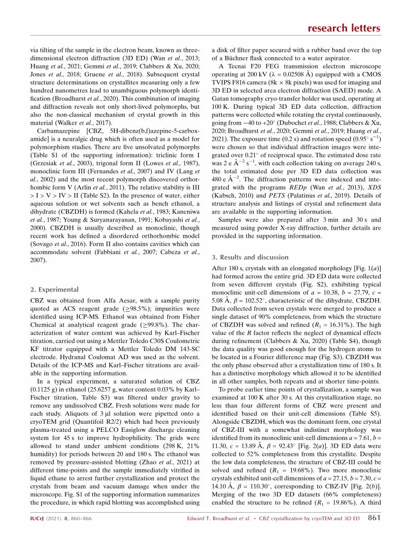
After 180 s, crystals with an elongated morphology [Fig. 1(a)]
had formed across the entire grid. 3D ED data were collected
from seven different crystals (Fig. S2), exhibiting typical
monoclinic unit-cell dimensions of a = 10.38, b = 27.79, c =
5.08 A, � = 102.52�, characteristic of the dihydrate, CBZDH.
Data collected from seven crystals were merged to produce a
single dataset of 90% completeness, from which the structure
of CBZDH was solved and refined (R1 = 16.31%). The high
value of the R factor reflects the neglect of dynamical effects
during refinement (Clabbers & Xu, 2020) (Table S4), though
the data quality was good enough for the hydrogen atoms to
be located in a Fourier difference map (Fig. S3). CBZDH was
the only phase observed after a crystallization time of 180 s. It
has a distinctive morphology which allowed it to be identified
in all other samples, both repeats and at shorter time-points.
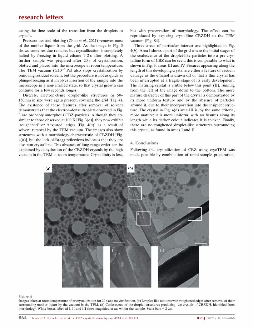
To probe earlier time points of crystallization, a sample was
examined at 100 K after 30 s. At this crystallization stage, no
less than four different forms of CBZ were present and
identified based on their unit-cell dimensions (Table S5).
Alongside CBZDH, which was the dominant form, one crystal
of CBZ-III with a somewhat indistinct morphology was
identified from its monoclinic unit-cell dimensions a = 7.61, b =
11.30, c = 13.89 A, � = 92.43� [Fig. 2(a)]. 3D ED data were
collected to 52% completeness from this crystallite. Despite
the low data completeness, the structure of CBZ-III could be
solved and refined (R1 = 19.68%). Two more monoclinic
crystals exhibited unit-cell dimensions of a = 27.15, b = 7.30, c =
14.10 A, � = 110.30�, corresponding to CBZ-IV [Fig. 2(b)].
Merging of the two 3D ED datasets (66% completeness)
enabled the structure to be refined (R1 = 19.86%). A third
research letters
IUCrJ (2021). 8, 860–866 Edward T. Broadhurst et al. � CBZ crystallization by cryoTEM and 3D ED 861
crystalline form present in the same sample exhibited rhom-
bohedral unit-cell dimensions of a = 36.24, c = 5.31 A, � = � =
90�, � = 120� indicative of CBZ form II [Fig. 2(c)]. Although
the quality of data obtained precluded structure solution, the
crystal form was unambiguously identified from the unit-cell
dimensions.
research letters
862 Edward T. Broadhurst et al. � CBZ crystallization by cryoTEM and 3D ED IUCrJ (2021). 8, 860–866
Figure 1(a) Low-magnification image of CBZDH crystallized after 3 min on the TEM grid at 100 K showing an elongated morphology. Scale bar = 5 mm.(b) Selected crystal of CBZDH used for collection of 3D ED data. Scale bar = 2 mm. Inset shows a diffraction image from the dataset. (c) Disorderedstructure of CBZDH.
Figure 2(a)–(c) Crystals of form III, IV and II, respectively, found in a sample after crystallization for 30 s on a TEM grid. Scale bars = 2 mm. The insets showrepresentative frames from the data collections. (d)–( f ) Corresponding 3D ED reciprocal lattice reconstructions all viewed along b*. All images weremeasured at 100 K.
The identification of forms II, III and IV is the first time that
any polymorph other than the dihydrate has been observed in
the crystallization of CBZ from (wet) ethanol. The fact that
they were only present as tiny fractions of samples otherwise
consisting exclusively of the expected dihydrate demonstrates
the ability of cryoTEM and 3D ED to identify minor phases
and thier potential importance in polymorph discovery. The
results obtained after 180 s, which showed the presence only of
CBZDH, showed that these minor phases are short-lived and
re-dissolve or transform into CBZDH in a matter of minutes.
To obtain bulk measurements of the crystals forming in the
solution after 3 min and 30 s of crystallization, powder X-ray
diffraction data were collected. At both time points, the only
form detected was CBZDH (Figs. S6 and S7). The absence of
the other forms at 30 s is possibly due to them being present as
crystallites that are too small and too few in numbers to be
detected by powder X-ray diffraction. These findings do
illustrate the power of ED for phase identification within a
small quantity of sample. Further details are provided in the
supporting information.
To investigate even earlier stages of the crystallization,
samples were examined after 20 s (Fig. 3). No crystals were
observed. Instead, dark droplet-like structures ca 150–200 nm
in diameter were contained within a thinner film (area I in Fig.
3). Area II shows the droplet-like particles which lack a film of
the mother liquor surrounding them and are slightly darker,
suggesting that they are further developed particles of CBZ
(see below). Areas III and IV show these droplet-like struc-
tures coalescing inside and outside the electron-dense film.
The absence of Bragg reflections in the diffraction images of
these particles showed that they were amorphous, and that the
sample had been captured at a pre-crystallization stage.
The images showed that CBZDH crystallizes via a non-
classical phase separation mechanism that begins with the
formation of an initial film which separates into droplet-like
structures (areas I and II). The coalescence of these droplet-
like structures (areas III and IV) then signals the next stage of
crystal growth and development. The time difference between
the images in Fig. 3 and those consisting of CBZDH, along
with CBZ forms II, III and IV (Fig. 2) is only 10 s, indi-
research letters
IUCrJ (2021). 8, 860–866 Edward T. Broadhurst et al. � CBZ crystallization by cryoTEM and 3D ED 863
Figure 3Images taken at 100 K after crystallization for 20 s and vitrification. Scale bar = 2 mm. I shows the droplet-like structures with a surrounding film ofmother liquor and II shows the same structures without the surrounding film. III and IV show these structures coalescing inside and outside the film.
cating the time scale of the transition from the droplets to
crystals.
Pressure-assisted blotting (Zhao et al., 2021) removes most
of the mother liquor from the grid. As the image in Fig. 3
shows, some residue remains, but crystallization is completely
halted by freezing in liquid ethane 1–2 s after blotting. A
further sample was prepared after 20 s of crystallization,
blotted and placed into the microscope at room temperature.
The TEM vacuum (’10�5 Pa) also stops crystallization by
removing residual solvent, but the procedure is not as quick as
plunge-freezing as it involves insertion of the sample into the
microscope in a non-vitrified state, so that crystal growth can
continue for a few seconds longer.
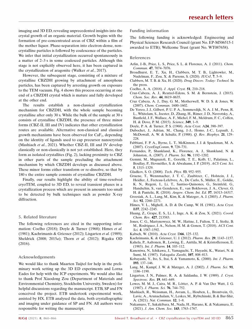
Discrete, electron-dense droplet-like structures ca 50–
150 nm in size were again present, covering the grid (Fig. 4).
The existence of these features after removal of solvent
demonstrates that the electron-dense droplets observed in Fig.
3 are probably amorphous CBZ particles. Although they are
similar to those observed at 100 K [Fig. 3(b)], they now exhibit
‘roughened’ or ‘textured’ edges [Fig. 4(a)] as a result of
solvent removal by the TEM vacuum. The images also show
structures with a morphology characteristic of CBZDH [Fig.
4(b)], but the lack of Bragg reflections indicates that they are
also non-crystalline. This absence of long-range order can be
explained by dehydration of the CBZDH crystals by the high
vacuum in the TEM at room temperature. Crystallinity is lost,
but with preservation of morphology. The effect can be
reproduced by exposing crystalline CBZDH to the TEM
vacuum (Fig. S4).
Three areas of particular interest are highlighted in Fig.
4(b). Area I shows a part of the grid where the initial stages of
the coalescence of the droplet-like particles into a pre-crys-
talline form of CBZ can be seen, this is comparable to what is
shown in Fig. 3, areas III and IV. Fissures appearing along the
length of this developing crystal are either a feature of vacuum
damage as the ethanol is drawn off or that a thin crystal has
been interrupted at a fragile stage of its early development.
The maturing crystal is visible below this point (II), running
from the left of the image down to the bottom. The more
mature character of this part of the crystal is demonstrated by
its more uniform texture and by the absence of particles
around it, due to their incorporation into the insipient struc-
ture. The crystal in Fig. 4(b) area III is, by the same criteria,
more mature: it is more uniform, with no fissures along its
length while its darker colour indicates it is thicker. Finally,
there are no roughened droplet-like structures surrounding
this crystal, as found in areas I and II.
4. Conclusions
Following the crystallization of CBZ using cryoTEM was
made possible by combination of rapid sample preparation,
research letters
864 Edward T. Broadhurst et al. � CBZ crystallization by cryoTEM and 3D ED IUCrJ (2021). 8, 860–866
Figure 4Images taken at room temperature after crystallization for 20 s and no vitrification. (a) Droplet-like features with roughened edges after removal of theirsurrounding mother liquor by the vacuum in the TEM. (b) Coalescence of the droplet structures producing two crystals of CBZDH, identified frommorphology. White boxes labelled I, II and III show magnified areas within the sample. Scale bars = 2 mm.
imaging and 3D ED, revealing unprecedented insights into the
crystal growth of an organic material. Growth begins with the
formation of pre-concentrated areas of CBZ within a film of
the mother liquor. Phase-separation into electron-dense, non-
crystalline particles is followed by coalescence of the particles.
We infer that initial crystallization occurred spontaneously in
a matter of 2–3 s in some coalesced particles. Although this
stage is not explicitly observed here, it has been captured in
the crystallization of aragonite (Walker et al., 2017).
However, the subsequent stage, consisting of a mixture of
crystalline CBZDH growing by attachment of amorphous
particles, has been captured by arresting growth on exposure
to the TEM vacuum. Fig. 4 shows this process occurring at one
end of a CBZDH crystal which is mature and fully developed
at the other end.
The results establish a non-classical crystallization
mechanism for CBZDH, with the whole sample becoming
crystalline after only 30 s. While the bulk of the sample at 30 s
consists of crystalline CBZDH, the presence of three minor
forms (CBZ-II, III and IV) indicates that other crystallization
routes are available. Alternative non-classical and classical
growth mechanisms have been observed for CaF2, depending
on the identity of ligands used to cap precursor nanocrystals
(Mashiach et al., 2021). Whether CBZ-II, III and IV develop
classically or non-classically is not yet established. Here, they
form as isolated crystallites (Fig. 2), lacking proximity to CBZ
in other parts of the sample precluding the attachment
mechanism by which CBZDH develops as discussed above.
These minor forms either transform or re-dissolve, so that by
180 s the entire sample consists of crystalline CBZDH.
Finally, our results highlight the ability of time-resolved
cryoTEM, coupled to 3D ED, to reveal transient phases in a
crystallization process which are present in amounts too small
to be detected by bulk techniques such as powder X-ray
diffraction.
5. Related literature
The following references are cited in the supporting infor-
mation: Coelho (2018); Doyle & Turner (1968); Himes et al.
(1981); Kachrimanis & Griesser (2012); Lisgarten et al. (1989);
Sheldrick (2008; 2015a); Thorn et al. (2012); Rigaku OD
(2016).
Acknowledgements
We would like to thank Maarten Tuijtel for help in the preli-
minary work setting up the 3D ED experiments and Lorna
Eades for help with the ICP experiments. We would also like
to thank Prof Xiaodong Zou (Department of Materials and
Environmental Chemistry, Stockholm University, Sweden) for
helpful discussions regarding the manuscript. ETB, SP and FN
conceived the project. ETB undertook experimental work,
assisted by HX. ETB analysed the data, both crystallographic
and imaging under guidance of SP and FN. All authors were
responsible for writing the manuscript.
Funding information
The following funding is acknowledged: Engineering and
Physical Sciences Research Council (grant No. EP-M506515-1
awarded to ETB); Wellcome Trust (grant No. WT087658).
References
Arlin, J.-B., Price, L. S., Price, S. L. & Florence, A. J. (2011). Chem.Commun. 47, 7074–7076.
Broadhurst, E. T., Xu, H., Clabbers, M. T. B., Lightowler, M.,Nudelman, F., Zou, X. & Parsons, S. (2020). IUCrJ, 7, 5–9.
Clabbers, M. T. B. & Xu, H. (2020). Drug Discov. Today: Technol. Inthe press.
Coelho, A. A. (2018). J. Appl. Cryst. 51, 210–218.Cruz-Cabeza, A. J., Reutzel-Edens, S. M. & Bernstein, J. (2015).
Chem. Soc. Rev. 44, 8619–8635.Cruz Cabeza, A. J., Day, G. M., Motherwell, W. D. S. & Jones, W.
(2007). Chem. Commun. 1600–1602.De Yoreo, J. J., Gilbert, P. U. P. A., Sommerdijk, N. A. J. M., Penn, R.
L., Whitelam, S., Joester, D., Zhang, H., Rimer, J. D., Navrotsky, A.,Banfield, J. F., Wallace, A. F., Michel, F. M., Meldrum, F. C., Colfen,H. & Dove, P. M. (2015). Science, 349, 1–9.
Doyle, P. A. & Turner, P. S. (1968). Acta Cryst. A24, 390–397.Dubochet, J., Adrian, M., Chang, J.-J., Homo, J.-C., Lepault, J.,
McDowall, A. W. & Schultz, P. (1988). Q. Rev. Biophys. 21, 129–228.
Fabbiani, F. P. A., Byrne, L. T., McKinnon, J. J. & Spackman, M. A.(2007). CrystEngComm, 9, 728–731.
Fernandes, P., Shankland, K., Florence, A. J., Shankland, N. &Johnston, A. (2007). J. Pharm. Sci. 96, 1192–1202.
Gemmi, M., Mugnaioli, E., Gorelik, T. E., Kolb, U., Palatinus, L.,Boullay, P., Hovmoller, S. & Abrahams, J. P. (2019). ACS Cent. Sci.5, 1315–1329.
Gladkov, S. O. (2008). Tech. Phys. 53, 952–955.Gruene, T., Wennmacher, J. T. C., Zaubitzer, C., Holstein, J. J.,
Heidler, J., Fecteau-Lefebvre, A., De Carlo, S., Muller, E., Goldie,K. N., Regeni, I., Li, T., Santiso-Quinones, G., Steinfeld, G.,Handschin, S., van Genderen, E., van Bokhoven, J. A., Clever, G.H. & Pantelic, R. (2018). Angew. Chem. Int. Ed. 57, 16313–16317.
Grzesiak, A. L., Lang, M., Kim, K. & Matzger, A. J. (2003). J. Pharm.Sci. 92, 2260–2271.
Himes, V. L., Mighell, A. D. & De Camp, W. H. (1981). Acta Cryst.B37, 2242–2245.
Huang, Z., Grape, E. S., Li, J., Inge, A. K. & Zou, X. (2021). Coord.Chem. Rev. 427, 213583.
Jones, C. G., Martynowycz, M. W., Hattne, J., Fulton, T. J., Stoltz, B.M., Rodriguez, J. A., Nelson, H. M. & Gonen, T. (2018). ACS Cent.Sci. 4, 1587–1592.
Kabsch, W. (2010). Acta Cryst. D66, 125–132.Kachrimanis, K. & Griesser, U. J. (2012). Pharm. Res. 29, 1143–1157.Kahela, P., Aaltonen, R., Lewing, E., Anttila, M. & Kristoffersson, E.
(1983). Int. J. Pharm. 14, 103–112.Kaneniwa, N., Ichikawa, J., Yamaguchi, T., Hayashi, K., Watari, N. &
Sumi, M. (1987). Yakugaku Zasshi, 107, 808–813.Kobayashi, Y., Ito, S., Itai, S. & Yamamoto, K. (2000). Int. J. Pharm.
193, 137–146.Lang, M., Kampf, J. W. & Matzger, A. J. (2002). J. Pharm. Sci. 91,
1186–1190.Lisgarten, J. N., Palmer, R. A. & Saldanha, J. W. (1989). J. Cryst.
Spectrosc. Res. 19, 641–649.Lowes, M. M. J., Caira, M. R., Lotter, A. P. & Van Der Watt, J. G.
(1987). J. Pharm. Sci. 76, 744–752.Mashiach, R., Weissman, H., Avram, L., Houben, L., Brontvein, O.,
Lavie, A., Arunachalam, V., Leskes, M., Rybtchinski, B. & Bar-Shir,A. (2021). Nat. Commun. 12, 1–8.
Nakamuro, T., Sakakibara, M., Nada, H., Harano, K. & Nakamura, E.(2021). J. Am. Chem. Soc. 143, 1763–1767.
research letters
IUCrJ (2021). 8, 860–866 Edward T. Broadhurst et al. � CBZ crystallization by cryoTEM and 3D ED 865
Sheldrick, G. M. (2008). Acta Cryst. A64, 112–122.Sheldrick, G. M. (2015a). Acta Cryst. A71, 3–8.Sovago, I., Gutmann, M. J., Senn, H. M., Thomas, L. H., Wilson, C. C.
& Farrugia, L. J. (2016). Acta Cryst. B72, 39–50.Thorn, A., Dittrich, B. & Sheldrick, G. M. (2012). Acta Cryst. A68,
448–451.
Walker, J. M., Marzec, B. & Nudelman, F. (2017). Angew. Chem. Int.Ed. 56, 11740–11743.
Wan, W., Sun, J., Su, J., Hovmoller, S. & Zou, X. (2013). J. Appl. Cryst.46, 1863–1873.
Young, W. W. L. & Suryanarayanan, R. (1991). J. Pharm. Sci. 80, 496–500.