Li et al. Botanical Studies 2013, 54:52http://www.as-botanicalstudies.com/content/54/1/52
RESEARCH Open Access
Detection and identification of the phytoplasmaassociated with China ixeris (Ixeridium chinense)fasciationZheng-Nan Li1†, Ping Liu1†, Lei Zhang2 and Yun-Feng Wu1*
Abstract
Background: Phytoplasmas are always associated with symptoms in host plants such as stunting of stems,witches’-broom, yellowing of leaves, formation of sterile-deformed flowers, virescence and phyllody. Recently alsosymptom of fasciation was reported associated with phytoplasma presence. In the present work, China ixerisfasciation was observed associated with phytoplasmas in Guanzhong Area, Shaanxi, China.
Results: Phytoplasma-like bodies were observed under transmission electron microscope in stem tissues ofsymptomatic samples. The 16S rRNA operon and tuf genes from phytoplasmas were amplified by PCR assays.Phylogenetic trees were calculated respectively based on sequences data of these two genes. The pattern ofrestriction fragment length polymorphism (RFLP) was generated via digesting the PCR products of 16S rRNA genewith eight restriction enzymes.
Conclusion: The presence of phytoplasma in China ixeris exhibiting fasciation symptom was confirmed by theresults of TEM observation and PCR testing. Based on sequence data, phylogeny analysis and actual restrictionfragment length polymorphism (RFLP) analysis, the associated phytoplasma was classified as related to 16SrI-Csubgroup. This was the first record of phytoplasmas in China ixeris.
Keywords: Transmission electron microscopy; 16S rRNA and tuf genes; PCR; Phylogeny analysis; RFLP
BackgroundFasciation is a result of an aberrant growth developmentin plants. A fasciated plant often forms flattened, widerthan normal stems similar to a multiple stem fusion. Thisabnormal phenotype is observable in many plant speciesin nature, and spontaneous genetic mutations, bacterialinfections, insect or mite attacks, physically wounding orherbicide sprays were reported causing plant fasciations(Goethals et al., 2001). More recently, phytoplasma weredetected in some plants exhibiting fasciation symptom(Bertaccini et al., 2005; Poncarová‐Vorácková et al., 1998;Fránová and Petrzik, 2010; Kumar et al., 2010; Wu et al.,2012; Li et al., 2012).
* Correspondence: [email protected]†Equal contributors1State Key Laboratory of Crop Stress Biology for Arid Areas/Key Laboratory ofIntegrated Pest Management on Crop in Northwestern Loess Plateau,Ministry of Agriculture/Key Laboratory of Plant Protection Resources and PestManagement, Ministry of Education, College of Plant Protection, NorthwestA&F University, Yangling 712100, Shaanxi Province, P. R. ChinaFull list of author information is available at the end of the article
Phytoplasmas were assigned to a provisional genus,‘Candidatus Phytoplasma’ within the class Mollicutes(IRPCM, 2004). They are cell-wall-less prokaryotes thatinhabit plant phloem system, associated with symptomssuch as stunting of stems, proliferation of axillary shoots(witches’-broom), yellowing of leaves, formation of sterile-deformed flowers, greening of floral tissues (virescence)and forming of leaf-like flower organs (phyllody). Sincephytoplasmas have just been cultured under axenic condi-tions recently (Contaldo et al., 2012), their diagnosis stilldepends on molecular analysis of several evolutionarilyconserved genes like 16S ribosomal RNA (rRNA), riboso-mal protein (rp) and elongation factor TU (tuf ). So far, atotal of 31 phytoplasma groups and over 50 subgroupswere found based on analysis of 16S rRNA gene sequences(Wei et al., 2007; Zhao et al., 2009; Lee et al., 2011).China ixeris [Ixeridium chinense (Thunb.) Tzvel.] is a
perennial herb of the family Compositae, which could beused as traditional Chinese herbal medicine for treat-ment of enteritis and cholecystitis (Liu et al. 2010).
en access article distributed under the terms of the Creative Commonsg/licenses/by/2.0), which permits unrestricted use, distribution, and reproductionroperly cited.
Li et al. Botanical Studies 2013, 54:52 Page 2 of 6http://www.as-botanicalstudies.com/content/54/1/52
In this report, China ixeris fasciation disease occurrence isdescribed and a phytoplasma related to 16SrI-C subgroupwas associated with the symptomatology described.
MethodsSample collectionIn May 2009, China ixeris fasciation was observed inGuanzhong Area, Shaanxi Province, China. To verifydisease aetiology, 30 symptomatic and three asymptom-atic plants were collected in field. Some China ixerisplants were also transplanted into an insect-proof green-house for observation of symptom development.
Transmission electron microscopyFasciated stems of ca. 2 × 2 mm2 from symptomaticsamples were processed for transmission electron micro-scope examination. The tissues were fixed in a buffer(pH 7.2) containing 3% (v/v) glutaraldehyde and 4% (v/v)paraformaldehyde, incubated at 4°C for 4 h, and subse-quently in 1% (v/v) osmium tetroxide at room temperaturefor 2 h. Then the fixed samples were dehydrated in con-centration gradients of ethanol (10-70%) and acetone(0-100%), and finally were embedded in Epon 812 Kamińskaet al., (2001). The ultra-thin sections were stained withuranyl acetate and led citrate, and then examined.
PCR amplificationTotal DNA of each sample was extracted following cetyl-trimethyl ammonium bromide (CTAB) method (Kollaret al., 1990), and used as template in PCR assays. TotalDNA of samples infected with a phytoplasma related towheat blue dwarf phytoplasma (WBDp) (16SrI-C sub-group) (Wu et al., 2010) and chinaberry witches’ broomphytoplasma (CWBp) (16SrI-B subgroup) (Wu et al., 2010)were as positive controls.Primers P1 (Deng and Hiruki, 1991) and P7 (Schneider
et al., 1995) were used in amplification of phytoplasma16S rRNA gene, spacer region between 16S and 23SrRNA genes and the start of 23S rRNA gene. The PCRproducts were diluted 1: 29 with sterile double-distilledwater prior to the nested amplification using the generalprimer pair R16F2n/R2 (Gundersen and Lee, 1996). EachPCR mixture (25 μL) contained: 2 μL DNA template(10 ng/uL), 1 μL (10 pM) of each primer, 2 μL dNTP(2.5 mM), 2 μL MgCl2 (25 mM), 2.5 μL 10 × Taq bufferand 1 U Taq DNA polymerase (Thermo Fisher ScientificInc.), and sterile double-distilled water to the final volume.The PCR amplification program was as follows: preheatingat 94°C for 3 min, and then subjected them to 35 amplifi-cation cycles, of denaturation at 94°C for 1 min, annealingat 50°C for 1 min, and extension at 72°C for 1 min, with afinal elongation of 72°C for 10 min.Amplification of phytoplasma tuf gene was primed by
primer pair fTufu/rTufu (Schneider and Gibb, 1997).
The reaction mixture was set as above. After 3 minutes’preheating at 94°C, 30 amplification cycles were carriedout: denaturation at 94°C for 30 s, annealing at 50°C for30 s and extension at 72°C for 1 min, with a final elong-ation of 72°C for 10 min.PCR products were separated in 1% agarose gel by
electrophoresis, stained with ethidium bromide and visu-alized using UV transilluminator.
Cloning, sequencing and sequence analysisPCR products of 1.8 kb (phytoplasma 16S rRNA gene)and 0.8 kb (tuf gene) were purified using a commercialPCR Purification Kit (Bio Teke Corporation, Beijing,China) and cloned. The clones contained recombinantplasmid were selected by blue-white screen and for eachsample, three clones were selected and sequenced byTaKaRa Biotechnology (Dalian) Co., Ltd. The primers usedfor sequencing of 16S rRNA gene were M13F(-47): CGCCAGGGTTTTCCCAGTCACGAC/M13R(-48): AGCGGATAACAATTTCACACAGGA and of tuf gene wereM13F(-77): GATGTGCTGCAAGGCGATTA/M13R(-48),which were designed based on the sequence of pMD18-Tsimple vector and offered by the TaKaRa BiotehnologyCo. Ltd.Sequences were aligned using the Lasergene software
(version 7.0; DNASTAR, Madison, USA) and used forsearching against the database of National Center forBiotechnology Information (NCBI) by BLASTn.Published phytoplasma sequences were retrived from
GenBank; 33 sequences of 16S rRNA gene and 17 se-quences of tuf genes from groups 16SrI, -III, -V, -X and -XIIwere selected. Phylogenetic trees were built by neighbor-joining (16S rRNA) or maximum parsimony (tuf gene)methods with a 1000-replicate bootstrap search usingMEGA4 (Saitou and Nei, 1987; Tamura et al., 2007).The nested PCR products of 16S rRNA gene (1.2 kb)
from symptomatic samples were concentrated, and digestedwith eight restriction enzymes AluI, BfaI, HaeIII, HhaI,HpaII, KpnI, MseI and RsaI (Lee et al., 1998). The digestedPCR products were separated in 8% polyacrylamide gel byelectrophoresis and visualized using UV tranilluminatorafter ethidium bromide staining.
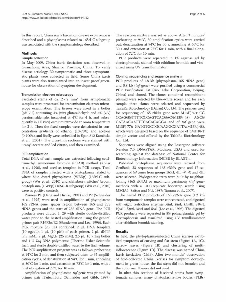
ResultsIn field, the phytoplasma-infected China ixerises exhib-ited symptoms of curving and flat stem (Figure 1A, 1C),narrow leaves (Figure 1B) and clustering of multi-inflorescence (Figure 1D). The disease was named ChinaIxeris fasciation (ChIF). After two months’ observationof field-collected China Ixerises for symptom develop-ment in green house, the flat stem did not broaden andthe abnormal flowers did not seed.In ultra-thin sections of fasciated stems from symp-
tomatic samples, many phytoplasma-like bodies (PLBs)
Figure 1 Symptoms of phytoplasma-infected China ixeris. The symptoms are curving stem (A); flat stem, shorting stalk (C); narrowing leaves(B) and clustering of multi-inflorescence (D), compared to healthy one (E).
Li et al. Botanical Studies 2013, 54:52 Page 3 of 6http://www.as-botanicalstudies.com/content/54/1/52
were identified based on the spherical and dumbbell-shaped structures under TEM (Figure 2). The PLBsranged from 330 to 700 nm in diameter and containedribosome-like bodies and a central region of fibrillar ma-terial, presumed to contain DNA.
Figure 2 Transmission electron micrograph of phytoplasma-likebodies (indicated by black narrow) in stem tissues of symptomaticChina ixeris. Scale bar =1 μm.
From all 30 total DNA of symptomatic samples andpositive controls, PCR products of 0.8 kb (partial tufgene), 1.8 kb and 1.2 kb (partial 16S rRNA gene) weregenerated, but no PCR product was obtained from theasymptomatic samples.The sequences of 1,840 bp generated from symptomatic
samples were identical between each other and depositedunder GenBank under accession no. HM990973. The ob-tained sequences of 842 bp (tuf gene), share 100% of iden-tity and were deposited under accession no. HM990972.NCBI BLASTn program analysis indicated that the se-quences of 1,840 bp and 842 bp were homogenous to se-quences of group 16SrI phytoplasmas. The HM990973shared the highest identity of 99.67% (1,834 bp/1,840 bp)with clover phyllody phytoplasma strain CPh (accessionno. AF222065) and Poa stunt phytoplasma (accession no.DQ640501), and the HM990972 had the identity of 100%with clover phyllody phytoplasma strain KV (accession no.L46369) and Wheat blue dwarf phytoplasma (accessionno. DQ507200).As indicated by the phylogenetic trees inferred from 16S
rRNA genes (Figure 3) and tuf genes (Figure 4), the ChIFpand phytoplasmas of group 16SrI clustered together.ChIFp yielded the same RFLP pattern with WBDp, but
differ from CWBp in HaeIII and HhaI sites (Figure 5).After digested with enzyme HhaI, ChIFp and WBDpgenerated two bands of 872 and 310 bp, while CWBpgot three bands of 872, 118 and 72 bp. And both ChIFpand WBDp yielded the bands of 1,078 and 118 bp after
Figure 4 Phylogenetic tree based on tuf gene sequences ofChIF phytoplasma and related 17 phytoplasmas, constructedby the maximum parsimony method. Numbers on the branchesare confidence values obtained for 1000 replicates.
Figure 3 Phylogenetic tree based on 16S rRNA gene sequencesof the ChIF phytoplasma and 33 related phytoplasmas.Numbers on the branches are confidence values obtained for1000 replicates.
Li et al. Botanical Studies 2013, 54:52 Page 4 of 6http://www.as-botanicalstudies.com/content/54/1/52
digestion with enzyme HaeIII, whereas the CWBp gotbands of 1,078 and 72 bp. The results suggest closecorrelation of ChIp and WBDp with phytoplasmas insubgroup 16SrI-C although no identical to publishedreference strains.
DiscussionBased on TEM examination, PCR assays, sequencing,phylogeny and RFLP, phytoplasmas related to 16SrI-Csubgroup were detected as associated with fasciationin China ixeris. There are other reports of similar as-sociation such as fasciation in lilies (Lilium martagon;Liliaceae) (Bertaccini et al., 2005; Poncarová‐Voráckováet al., 1998), Asparagus officinalis (Liliaceae) (Fránová andPetrzik, 2010), Crotalaria spectabilis Roth. (Fabaceae)(Kumar et al., 2010), Sunshine tree (Senna surattensisBurm. Caesapiniaceae) (Wu et al., 2012) and Puna chicory(Cichrium intybus L.) (Li et al., 2012). All the cases sug-gested that fasciation is a significant symptom for phyto-plasma disease.
Phytoplasmas related to 16SrI-C subgroup were foundin wheat (Wu et al., 2010) and so far, only two other hostplants have been reported to host this kind of pathogen inChina: peach trees (Zhang et al., 2013) and China ixerisand all reported in Shaanxi Province. Sequences analysisindicated that the identities of 16S rRNA genes of ChIFp(accession no. HM990973) and WBDp (accession no.DQ078304) [or peach red leaf phytoplasma (PRLp) (acces-sion no. JX481781)] were 99.84% (1,244 bp/1,246 bp) [or99.60% (1,241 bp/1,246 bp)], and the tuf genes of ChIFp(accession no. HM990972) and WBDp (accession no.DQ507200) were identical to each other. China ixeris iswidely distributed as a volunteer plant and always grew inor around cultivated fields like wheat field. So it can be asignificant alternative host plant for this phytoplasma.China ixeris is also a new host plant for phytoplasmas.
ConclusionChina ixeris fasciation was confirmed associated withphytoplasmas based on results of TEM observation andPCR testing. Analyzing with the sequences of 16S rRNAgene and tuf gene of the phytoplasma suggested the phy-toplasma belongs to group 16SrI, After digesting witheight enzymes, the fragments of 16S rRNA gene of thephytoplasma yielded special patterns which were consist-ent with the patterns for phytoplasmas of subgroup16SrI-C in eight enzymes HpaII, HhaI, HaeIII, BfaI,KpnI and RsaI sites, but different in AluI and MseI sites.The restriction fragment length polymorphism (RFLP)analysis supported that the associated phytoplasma wasclassified as related to 16SrI-C subgroup. This was thefirst record of phytoplasmas in China ixeris.
Figure 5 RFLP pattern of 16S rRNA gene digested with eight enzymes HpaII, HhaI, HaeIII, BfaI, AluI, KpnI, MseI and RsaI. Lane M: ΦX174DNA ladder. 1, chinaberry witches’-broom. 2, China Ixeris fasciation. 3, wheat blue dwarf.
Li et al. Botanical Studies 2013, 54:52 Page 5 of 6http://www.as-botanicalstudies.com/content/54/1/52
AbbreviationsrRNA: Ribosomal RNA; tuf: Elongation factor TU; CTAB: Cetyltrimethylammonium bromide; WBDp: Wheat blue dwarf phytoplasma;CWBp: Chinaberry witches’ broom phytoplasma; ChIF: China ixeris fasciation;UV: Ultra violet; TEM: Transmission electron microscope.
Competing interestsThe authors declare that they have no competing interests.
Authors’ contributionsZ-NL and PL collected the samples, carried out the molecular experimentsand analyzed with the sequence data, and drafted the manuscript. LZ carriedout the observation with transmission electron microscope and processed allthe pictures. Y-FW funded and directed the experiment. All authors read andapproved the final manuscript.
AcknowledgementsWe thank Dr. Frnak M You of Cereal Research Centre, Agriculture and Agri-Food Canada for revisions and suggestions to the manuscript. This researchwas supported by the 111 project (Grant no. B07049) and the Ph.D. ProgramFoundation from the Ministry of Education of China (Grant no. 20100204110004),National Natural Science Foundation of China (Grant no. 31371913) and PhDStudent Academic Newcomer Award of China Ministry of Education (Grant no.Z107021202).
Author details1State Key Laboratory of Crop Stress Biology for Arid Areas/Key Laboratory ofIntegrated Pest Management on Crop in Northwestern Loess Plateau,Ministry of Agriculture/Key Laboratory of Plant Protection Resources and PestManagement, Ministry of Education, College of Plant Protection, NorthwestA&F University, Yangling 712100, Shaanxi Province, P. R. China. 2College ofForestry, Northwest A&F University, Yangling, Shaanxi 712100, P. R. China.
Received: 23 May 2013 Accepted: 23 October 2013Published: 31 October 2013
ReferencesBertaccini A, Fránová J, Botti S, Tabanelli D (2005) Molecular characterization of
phytoplasmas in lilies with fasciation in the Czech Republic. FEMS MicrobiolLett 249:79–85
Contaldo N, Bertaccini A, Paltrinieri S, Windsor HM, Windsor GD (2012) Axenicculture of plant pathogenic phytoplasmas. Phytopathol Mediterr 51:607–617
Deng S, Hiruki C (1991) Amplification of 16S rRNA genes from culturable andnonculturable Mollicutes. J Microbiol Methods 14:53–61
Fránová J, Petrzik K (2010) Asparagus officinalis: a new host of ‘Candidatusphytoplasma asteris’. J Phytopathol 158:317–320
Goethals K, Vereecke D, Jaziri M, Van Montagu M, Holsters M (2001) Leafy gallformation by Rhodococcus fascians. Annu Rev Phytopathol 39:27–52
Gundersen DE, Lee IM (1996) Ultrasensitive detection of phytoplasmas by nested-PCR assays using two universal primer pairs. Phytopathol Mediterr35:144–151
IRPCM Phytoplasma/Spiroplasma Working Team – Phytoplasma Taxonomy Group(2004) ‘Candidatus Phytoplasma’, a taxon for the wall-less, non-helicalprokaryotes that colonize plant phloem and insects. Int J Syst Evol Microbiol54:1243–1255
Kamińska M, Dziekanowska D, Rudzińska-Langwald A (2001) Detection ofphytoplasma infection in rose, with degeneration symptoms. J Phytopathol149:3–10
Kollar A, Seemüller E, Bonnet F, Saillard C, Bove J (1990) Isolation of the DNA ofvarious plant pathogenic mycoplasmalike organisms from infected plants.Phytopathology 80:233–237
Kumar S, Singh V, Lakhanpaul S (2010) First report of Crotalaria spectabilisfasciation associated with ‘Candidatus Phytoplasma asteris’ in India. Plant Dis94:1265, 1265
Lee IM, Gundersen-Rindal DE, Davis RE, Bartoszyk IM (1998) Revised classificationscheme of phytoplasmas based on RFLP analyses of 16S rRNA and ribosomalprotein gene sequences. Int JSyst Bacteriol 48:1153–1169
Lee IM, Bottner-Parker KD, Zhao Y, Villalobos W, Moreira L (2011) ‘CandidatusPhytoplasma costaricanum’ a novel phytoplasma associated with anemerging disease in soybean (Glycine max). Int J Syst Evol Microbiol61:2822–2826
Li ZN, Zhang L, Bai YB, Liu P, Wu YF (2012) Detection and identification of theelm yellows group phytoplasma associated with Puna chicory flat stem inChina. Can J Plant Pathol 34:34–41
Liu SM, Xie WD, Meng FJ (2010) Research progress in chemical compositions andpharmacologica activities of ixeris Cass. Lishizhen Medicine And MaterialMedica Research 21:975–977
Poncarová-Vorácková Z, Fránová J, Válová P, Mertelik J, Navrátil M, Nebesárová J(1998) Identification of phytoplasma infecting Lilium martagon in the CzechRepublic. J Phytopathol 146:609–612
Saitou N, Nei M (1987) The neighbor-joining method: a new method for recon-structing phylogenetic trees. Mol Biol Evol 4:406–425
Schneider B, Gibb KS (1997) Sequence and RFLP analysis of the elongation factorTu gene used in differentiation and classification of phytoplasmas.Microbiology 143:3381–3389
Schneider B, Seemüller E, Smart C, Kirkpatrick B (1995) Phylogenetic classificationof plant pathogenic mycoplasma-like organisms or phytoplasmas. In: Razin S,Tully JG (eds) Molecular and diagnostic procedures in mycoplasmology.Academic, New York, pp 369–380
Tamura K, Dudley J, Nei M, Kumar S (2007) MEGA4: molecular evolutionarygenetics analysis (MEGA) software version 4.0. Mol Biol Evol 24:1596–1599
Wei W, Davis RE, Lee M, Zhao Y (2007) Computer-simulated RFLP analysis of 16SrRNA genes: identification of ten new phytoplasma groups. Int J Syst EvolMicrobiol 57:1855–1867
Wu Y, Hao X, Li Z, Gu P, An F, Xiang J, Wang H, Luo Z, Liu J, Xiang Y (2010)Identification of the phytoplasma associated with wheat blue dwarf diseasein China. Plant Dis 94:977–985
Li et al. Botanical Studies 2013, 54:52 Page 6 of 6http://www.as-botanicalstudies.com/content/54/1/52
Wu W, Cai H, Wei W, Davis R, Lee IM, Chen H, Zhao Y (2012) Identification of twonew phylogenetically distant phytoplasmas from Senna surattensis plantsexhibiting stem fasciation and shoot proliferation symptoms. Ann Appl Biol160:25–34
Zhang L, Li ZN, Zhang HW, Tao Y, Wu YF (2013) Detection and identification ofaster yellows group phytoplasma (16SrI-C) associated with peach red leafdisease. J Phytopathol 161:359–362
Zhao Y, Wei W, Lee M, Shao J, Suo X, Davis RE (2009) Construction of aninteractive online phytoplasma classification tool, iPhyClassifier, and itsapplication in analysis of the peach X-disease phytoplasma group (16SrIII).Int J Syst Evol Microbiol 59:2582–2593
doi:10.1186/1999-3110-54-52Cite this article as: Li et al.: Detection and identification of thephytoplasma associated with China ixeris (Ixeridium chinense) fasciation.Botanical Studies 2013 54:52.
Submit your manuscript to a journal and benefi t from: