Michael et al. Molecular Cancer 2012, 11:89http://www.molecular-cancer.com/content/11/1/89
RESEARCH Open Access
Highly efficient site-specific transgenesis in cancercell linesIacovos P Michael1, Claudio Monetti1, Anthony C Chiu1, Puzheng Zhang1, Takeshi Baba1, Koichiro Nishino1,Siamak Agha-Mohammadi2, Knut Woltjen1, Hoon-Ki Sung1 and Andras Nagy1,3*
Abstract
Background: Transgenes introduced into cancer cell lines serve as powerful tools for identification of genesinvolved in cancer. However, the random nature of genomic integration site of a transgene highly influences thefidelity, reliability and level of its expression. In order to alleviate this bottleneck, we characterized the potentialutility of a novel PhiC31 integrase-mediated site-specific insertion system (PhiC31-IMSI) for introduction oftransgenes into a pre-inserted docking site in the genome of cancer cells.
Methods: According to this system, a “docking-site” was first randomly inserted into human cancer cell lines andclones with a single copy were selected. Subsequently, an “incoming” vector containing the gene of interest wasspecifically inserted in the docking-site using PhiC31.
Results: Using the Pc-3 and SKOV-3 cancer cell lines, we showed that transgene insertion is reproducible andreliable. Furthermore, the selection system ensured that all surviving stable transgenic lines harbored the correctintegration site. We demonstrated that the expression levels of reporter genes, such as green fluorescent proteinand luciferase, from the same locus were comparable among sister, isogenic clones. Using in vivo xenograft studies,we showed that the genetically altered cancer cell lines retain the properties of the parental line. To achievetemporal control of transgene expression, we coupled our insertion strategy with the doxycycline inducible systemand demonstrated tight regulation of the expression of the antiangiogenic molecule sFlt-1-Fc in Pc-3 cells.Furthermore, we introduced the luciferase gene into the insertion cassette allowing for possible live imaging ofcancer cells in transplantation assays. We also generated a series of Gateway cloning-compatible intermediatecassettes ready for high-throughput cloning of transgenes and demonstrated that PhiC31-IMSI can be achieved in ahigh throughput 96-well plate format.
Conclusions: The novel PhiC31-IMSI system described in this study represents a powerful tool that can facilitate thecharacterization of cancer-related genes.
IntroductionMutations and polymorphisms in various genes and/ortheir regulatory elements are implicated in tumor ini-tiation, progression and drug resistance [1-4]. Elucidationof the consequences of these genetic changes relies largelyon the availability of genetic and cancer models. Stabletransgene expression is one of the most powerful and
* Correspondence: [email protected] Lunenfeld Research Institute, Mount Sinai Hospital, Toronto, OntarioM5G 1X5, Canada3Department of Obstetrics & Gynaecology, University of Toronto, Toronto,Ontario M5S 1A8, CanadaFull list of author information is available at the end of the article
informative genetic tools. The generation of stable lines byrandom integration of a transgene is appropriate whenexamining the effect of a single transgene. However, whena comparative analysis of a series of transgenes is required,the generation of stable lines using random integration isinefficient. Foremost, expression levels between clonesvary significantly due to chromosomal position effects andfrequent copy number variation [5]. Thus, the screeningand identification of various stable lines with the desiredcharacteristics is extremely laborious. In addition, randomintegration may lead to genome alterations, such as inacti-vation of endogenous genes, which may alter cellular
l Ltd. This is an Open Access article distributed under the terms of the Creativeommons.org/licenses/by/2.0), which permits unrestricted use, distribution, andiginal work is properly cited.
Michael et al. Molecular Cancer 2012, 11:89 Page 2 of 11http://www.molecular-cancer.com/content/11/1/89
phenotype. Inserting the transgene to a predeterminedgenomic locus, site-specific integration, can eliminate allof the aforementioned confounding factors.Site-specific integration can be achieved by homologous
recombination-based targeting of a specific genomic po-sition [6-9], by recombinase-mediated cassette exchange(RMCE) [10] or integrase-mediated site-specific insertion(IMSI) [11-13]. Homologous recombination, althoughwidely used for gene targeting in mouse embryonic stemcells [14], becomes laborious, time consuming andinefficient in case of human cell lines, due to difficultiesinvolving the design of targeting vectors and the lack ofisogenicity required for efficient homologous recom-bination [9,15,16]. Furthermore, in the case of cancer celllines, homologous recombination becomes even more diffi-cult due to the fact that these cells acquire many geneticalterations during tumorigenesis [17]. These aberrationsfurther increase during in vitro propagation since cancercells are inherently genetically unstable [18]. Recently, alter-native techniques, such as the use of recombinant adeno-associated virus and the introduction of double-strandbreaks for stimulation of targeting have been developed,however the targeting efficiency remains very low, generallybetween 1% and 5%, and often even lower [19-21].RMCE and IMSI overcome the aforementioned limita-
tions, allowing for both higher efficiency and easier vectorconstruction [11,12]. Current methods involve the intro-duction of the transgene using bacterial or yeast DNArecombinases into a pre-generated chromosomal locus.The majority of the described strategies use the tyrosinerecombinases Cre or Flp and their recognition sites loxPand FRT, respectively [22-26]. Although Cre recombinasehas the advantage of being more efficient, it does share adisadvantageous feature with Flp in that both are bi-direc-tional. Since they recognize their recombination productsas a substrate, the inserted transgene is often subsequentlyexcised again [27]. In order to overcome this limitation,chimeric Cre recombinases or mutated loxP sites have beengenerated, such that the recombination products cannot berecognized by Cre recombinase [28-31]. This however, hasresulted in decreased efficiency of recombination [29]. Analternative solution is to utilize integrases that only facilitatethe insertion reaction.Phage PhiC31 integrase catalyzes unidirectional re-
combination between two heterotypic sites, attP andattB (attachment site for Phage/Bacteria [32]); therefore,this enzyme is the desired candidate to facilitate the in-sertion of a foreign expression cassette into a preciselocus. The resulting sites, attL and attR, are no longersubstrates for PhiC31, thus the integration reaction isnot reversible [33,34]. PhiC31 has previously been usedfor integration of an attB-containing plasmid into attPsites pre-inserted into the mammalian genome [35-37].Studies have also shown that pseudo attP sites exist in
both the human and mouse genomes, and that it is pos-sible to achieve PhiC31-mediated integration into thesesites as well [35,36,38].In this study, we characterized our unique IMSI system
that utilizes the PhiC31 integrase (PhiC31-IMSI) for spe-cific integration of the transgene in a pre-inserted genomiclanding site (docking site) in various cancer cell lines. Wedemonstrate that the selection system has 100% fidelityand that sister clones express comparable levels of variousreporter genes. In addition, we coupled the PhiC31-IMSIto a doxycycline inducible system allowing for tight androbust control of the expression of the transgene. We alsointroduced the luciferase gene that could be used forin vivo live imaging. Finally, we constructed a series ofGateway compatible intermediate incoming vectors andshowed that this system can be applied for high-throughput applications as well.
Materials and methodsPlasmid vector constructionWe modified our basic docking site vector (DockZ) wasdescribed by Monetti et al. [39]. This new vector de-signated as DZL contains a fusion gene comprising puro-mycin N-acetyl-transferase (PAC) and luciferase linked viathe T2A peptide [40], i.e. PAC-T2A-luciferase (Figure 1A).The T2A junction along with part of the C-terminus ofPAC and N-terminus of luciferase was synthesized andcloned into pBluescript between BstAPI and ClaI sites.This fragment was then subcloned into DockZ usingBstAPI and ClaI. The C-terminus of luciferase was sub-cloned from a pBluescript (pBS) luciferase-containingvector after digestion with BsrGI and EcoNI.The basic incoming vector, Inc-basic, as well as Inc-
CAG-MCS-pA and Inc-CAG-EGFP-pA were described byMonetti et al. [39] and shown in Figure 1B. After digestionof Inc-CAG-MCS-pA with HpaI, the Gateway RfA cassettewas inserted to generate Inc-CAG-Gateway-pA. Inc-TRE-EGFP-pA was constructed by excision of the pCAGGspromoter and insertion of second-generation tetracycline-regulated promoters, TRE [41] by BamHI and HindIII di-gestion. Subsequent excision of EGFP by Bsu36I and NheIand cloning of MCS gave rise to Inc-TRE-MCS-pA. TheGateway RfA cassette was inserted after HpaI digestion inorder to generate Inc-TRE-Gateway-pA. The IRES-EGFPdigested with EcoRI (polished) and ClaI was inserted intoInc-TRE-MCS-pA after Bsu36I (polished) and ClaI diges-tion generating Inc-TRE-MCS-IRES-EGFP-pA. The Gate-way RfA cassette was inserted after HpaI digestion,constructing Inc-TRE-Gateway-IRES-EGFP-pA.The reverse-transactivator of tetracycline [rtTA; Tet-
ON Advance [42] was subcloned from the pTet-OnAdvanced vector (Clontech) into pBluescript using EcoRIand BamHI sites. Subsequently, it was subcloned into
Figure 1 Docking-incoming system. (A) The basic docking site (DockZ) was modified to contain luciferase as a fusion transgene withpuromycin N-acetyl-transferase (PAC), deriving DZL. (B) PhiC31 integrase-mediated site-specific insertion of the incoming vector. Correctintegrants are selected based on resistance to neomycin. (C) A series of incoming vectors with different promoters and reporter genes. IncBasic ispromoter-less and contains a multiple cloning site (MCS). IncCAP contains the pCAGGs promoter allowing for constitutive expression of thetransgene as well as the Gateway cassette with reading frame A (RfA). IncTAP and IncTAG contain the second-generation tetracycline-regulatedpromoter (TRE) allowing for inducible expression. Inc-TAG allows for indirect monitoring of the expression of the transgene through a bicistronicarrangement with an IRES followed by EGFP.
Michael et al. Molecular Cancer 2012, 11:89 Page 3 of 11http://www.molecular-cancer.com/content/11/1/89
pcDNA6 digested with HindIII and SacII derivingpcDNA6-rtTA.
Cell cultureThe cancer cell lines Pc-3 (prostate cancer), Du145(prostate cancer) and SKOV-3 (ovarian cancer) werepurchased from ATCC and maintained in RPMI-1640media containing 10% fetal bovine serum (Invitrogen).Cells were transfected with plasmids using ExGen500(Fermentas) according to the manufacturer's protocol.Stable clones were selected using 1.0 μg/ml puromycin(Sigma), 7.0 μg/mL blasticidin (Sigma) and 750 μg/mLG418 (Invitrogen). 2.0 μg/mL of doxycycline was used to
induce gene expression under the control of the TREpromoter. Cell lines were incubated at 37°C with 5% CO2.
Generation and screening of stable cell linesStables lines for DockZ and DZL were derived by trans-fection of 106 cells (Pc-3 for DockZ; DU145 and SKOV-3for DZL) with 10 μg DNA linearized with Eam1105I.Forty-eight hours after transfection, cells were trypsinizedand replated at limited dilutions. Resistant colonies wereselected with puromycin and picked using cloning cylin-ders after two weeks of selection.Single-copy stable lines were screened by southern
blot. Ten micrograms of genomic DNA was digestedwith EcoRI overnight, resolved by gel electrophoresis
Michael et al. Molecular Cancer 2012, 11:89 Page 4 of 11http://www.molecular-cancer.com/content/11/1/89
and transferred to Hybond N+ (GE Healthcare). Singlecopy integrants were detected using a neomycin phos-photransferase (neoR) probe.
PhiC31-mediated recombination and selection for IMSIderivativesRecombination was performed by cotransfection of 106
cells with 7.5 μg of incoming vector and 2.5 μg ofpCAGGS-PhiC31 using ExGen 500 (Fermentas). Forty-eight hours after transfection, resistant colonies wereselected with G418. For 96-well format PhiC31-mediatedintegration, 0.66 μg of incoming vector and 0.33 μg ofpCAGGS-PhiC31 were used. Correct integrants were veri-fied with standard PCR using primers recognizing the attLsite (CCAGGGCGTGCCCTTGAGTTCTCTC) and neoR
gene (CGATGAATCCAGAAAAGCGGCCATTTTTC)and/or southern using the thymidine kinase probe(Figure 2A).
Reporter assaysThe Luciferase assay (Promega) was performed accor-ding to the manufacturer’s protocol. In brief, cells grownon 24-well plates were washed twice with phosphate-buffered saline (PBS) and incubated in 200 μl lysis bufferfor 30 min on ice. Twenty microliters of cell lysate wasmixed with 25 μl of luciferase reagent and luciferaseactivity was read on the luminometer. Luciferase activitywas normalized to total protein measured using theBCA kit (Pierce).The FACSAria™ cell sorter (BD biosciences) was used
for single cell analysis of fluorescent expression. Cellsgrown on 6-well dishes were trypsinized and suspendedin PBS. 7-AAD or propidium iodide, diluted 1:100 inPBS, was used for detection of apoptotic cells in the caseof GFP and dsRed stably transfected cells, respectively.Ten thousands cells were analyzed per sample.Soluble Flt1-Fc and free human VEGF were measured
using commercially available sandwich ELISAs (R&D
Figure 2 Characterization of luciferase containing docking site (DZL).SKOV-3 (A, n=3) and DU145 lines (B, n=3).
systems, catalog # MVR100 and DVE00, respectively)following the manufacturer’s protocol.
Xenograft assaysFor xenograft assays, 5×10⁶ cells were suspended into150 μl RPMI-1640 media containing 33% of matrigeland injected subcutaneously in both dorsal flanks ofSCID mice. Doxycycline was administered usingdoxycycline-containing pellets (0.625g/kg, HarlanLaboratories). Tumour size was monitored using cali-pers and the volume was calculated using the formulaV= (LxWxH)π/6.
ResultsPhiC31-mediated site-specific transgene integrationsystem: validation of its fidelity in cancer cell linesIn this study, we modified our recently developed PhiC31integrase-mediated transgene insertion docking site, DockZ[39] to derive DZL (Figure 1A). Both docking sites allow forintroduction of a transgene in their predetermined genomicintegration of the desired cell line. Cell with docking siteinsertion can be identified with puromycin selection. Thedocking site also carries an inverted promoter-less neomy-cin phosphotransferase (neoR) gene at the 5’ end of the attPsite. The transgene is inserted with the incoming vectorthrough PhiC31-mediated integration. The incoming vectorcontains an attB site (Figure 1B) that is subjected to recom-bination along with the attP site of the docking site.Selection for the correct integrants is achieved through ac-tivation of the promoter-less neoR gene in the docking site.The incoming vector contains a promoter that becomespositioned upstream of the neoR gene upon correct integra-tion and starts its expression (Figure 1B). In addition, theoriginal system allows for removal of all selection markersthrough Flp recombinase-mediated deletion [39] and nega-tive selection for removal of the thymidine kinase gene bymeans of selection for FIAU resistance (Figure 1B). In ourcancer cell lines, however, we did not perform this
Normalized luciferase levels of single-copy DZL-containing clones of
Michael et al. Molecular Cancer 2012, 11:89 Page 5 of 11http://www.molecular-cancer.com/content/11/1/89
resolution step since our aim was to take advantage of abuilt-in positive and negative selectable marker system forfuture drug selection for or against the cancer cells. In theDZL docking site we added a luciferase transgene linked tothe puromycin N-acetyl-transferase, in a bicistronic mannerthrough a T2A peptide [40]. Thus the luciferase allowsin vivo imaging of tumours. Both proteins were expected toremain functional despite the addition of the T2A peptideresulted 10 amino acids at the carboxy terminus of PACand a proline at the amino terminus of luciferase genes. Totest the docking site and establish the properties of this sys-tem, we used a series of incoming vectors (Figure 1C) andthree human cancer cell lines (Pc-3, DU145 and SKOV-3).After establishing stable lines for the docking site trans-
gene, single transgene insertion colonies were identifiedby Southern blotting using a neoR probe on genomicDNA cut with the diagnostic EcoR1 restriction enzyme(Figure 1A). For the Pc-3 line, a total of 36 clones werescreened, three of which contained a single copy ofDockZ (data not shown). For the SKOV-3 and DU145lines, 8 out of 48 and 12 out of 48 clones, respectively,contained a single copy of DZL (data not shown), show-ing that the PAC was active. All single-copy containingclones showed the same proliferation rate as the originallines, indicating that the insertion did not have anydetrimental effects (data not shown). Furthermore, we
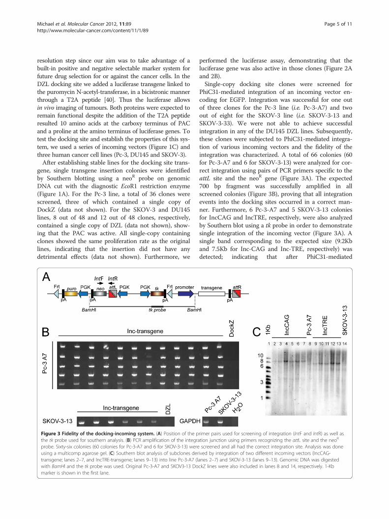
Figure 3 Fidelity of the docking-incoming system. (A) Position of the pthe tk probe used for southern analysis. (B) PCR amplification of the integraprobe. Sixty-six colonies (60 colonies for Pc-3-A7 and 6 for SKOV-3-13) wereusing a multicomp agarose gel. (C) Southern blot analysis of subclones dertransgene; lanes 2–7, and IncTRE-transgene; lanes 9–13) into line Pc-3-A7 (lwith BamHI and the tk probe was used. Original Pc-3-A7 and SKOV3-13 Domarker is shown in the first lane.
performed the luciferase assay, demonstrating that theluciferase gene was also active in those clones (Figure 2Aand 2B).Single-copy docking site clones were screened for
PhiC31-mediated integration of an incoming vector en-coding for EGFP. Integration was successful for one outof three clones for the Pc-3 line (i.e. Pc-3-A7) and twoout of eight for the SKOV-3 line (i.e. SKOV-3-13 andSKOV-3-33). We were not able to achieve successfulintegration in any of the DU145 DZL lines. Subsequently,these clones were subjected to PhiC31-mediated integra-tion of various incoming vectors and the fidelity of theintegration was characterized. A total of 66 colonies (60for Pc-3-A7 and 6 for SKOV-3-13) were analyzed for cor-rect integration using pairs of PCR primers specific to theattL site and the neoR gene (Figure 3A). The expected700 bp fragment was successfully amplified in allscreened colonies (Figure 3B), proving that all integrationevents into the docking sites occurred in a correct man-ner. Furthermore, 6 Pc-3-A7 and 5 SKOV-3-13 coloniesfor IncCAG and IncTRE, respectively, were also analyzedby Southern blot using a tk probe in order to demonstratesingle integration of the incoming vector (Figure 3A). Asingle band corresponding to the expected size (9.2Kband 7.5Kb for Inc-CAG and Inc-TRE, respectively) wasdetected; indicating that after PhiC31-mediated
rimer pairs used for screening of integration (IntF and IntR) as well astion junction using primers recognizing the attL site and the neoR
screened and all had the correct integration site. Analysis was doneived by integration of two different incoming vectors (IncCAG-anes 2–7) and SKOV-3-13 (lanes 9–13). Genomic DNA was digestedckZ lines were also included in lanes 8 and 14, respectively. 1-Kb
Michael et al. Molecular Cancer 2012, 11:89 Page 6 of 11http://www.molecular-cancer.com/content/11/1/89
integration, a single recombination event between theattP site of the docking site and the attB site of the in-coming vector occurred (Figure 3C).
Uniform expression of the transgenesWe used two different reporter assays in order to examinethe expression levels between clones derived from the sameparental line. The incoming vector, expressing luciferaseunder the control of pCAGGs, was introduced into theDockZ docking site containing Pc-3-A7 line and nineclones were isolated. Luciferase activity was measured andnormalized to total protein (Figure 4A). One-way ANOVAanalysis indicated that the expression levels were similarbetween isogenic clones (P=0.509) in contrast to the re-markable expression differences observed between SKOV-3and DU145 lines (Figure 2A and B) with different genomicintegration for the luciferase transgene.In order to further examine the expression between
isogenic clones, we used an incoming vector encodingEGFP or dsRed under the control of pCAGGs either asa single or bicistronic gene, coupled with an IRES to atruncated form of CD4 (tCD4), missing the intracellularand extracellular domains responsible for interactionwith other proteins (I. P. Michael and A. Nagy, unpub-lished data) (Figure 4B). Using flow cytometry for singlecell analysis, we assessed the GFP expression after aperiod of 2-month continuous culture of isogenic clones.We showed uniform GFP expression in each clone(Figure 4Bi and 4Bii, Additional file 1: Figure S1) as wellas a similar mean fluorescent value between isogenicclones derived from Pc-3-A7 and SKOV-3-13 (Table 1and data not shown). Furthermore, essentially all cells(99%) were positive for GFP (Table 1).Mean GFP expression was one order of magnitude
lower when EGFP was expressed as a bicistronic gene
Figure 4 Expression of reporter genes from isogenic clones. (A) Normof an incoming luciferase containing plasmid under the control of pCAGGsdifference in luciferase activity in isogenic clones (P=0.509, n=3). Error barsexpression of ten isogenic clones derived after integration of (i) IncCAP-EGwere an order of magnitude higher (Table 1) when EGFP was expressed asPc-3-A7 lines stably integrated with (i) IncCAP-EGFP and (ii) IncCAP-tCD4-IRmaintained in vivo as well.
using IRES (Table 1). After establishing xenografts, wewere able to show that the difference in expression levelbetween pCAGGs EGFP and pCAGGs tCD4-IRES-EGFPwas maintained at the tumor site as well (Figures 4Ci and4Cii). Similar results were obtained when dsRed was usedinstead of EGFP (data not shown).
Generation of the doxycycline-inducible transgeneexpression systemTo obtain temporal control on transgene expression, wecoupled the IMSI strategy with the doxycycline-induciblesystem. We created a range of incoming vectors containingsecond-generation tetracycline responsive elements coupledwith the minimal CMV promoter (TRE [41]) (Figure 1C).The second-generation reverse tetracycline transactivatorwas randomly integrated in lines Pc-3-A7 and SKOV-3-13using linearized pcDNA6-rtTA-Adv (Figure 5A) and clonesresistant to blasticidin were selected. An incoming vectorfor EGFP under the control of TRE was introducedthrough PhiC31-IMSI. After addition of doxycycline, strongGFP expression was observed for both Pc-3-A7 and SKOV-3-13 rtTA lines, while no GFP was expressed in the absenceof doxycycline (Figure 5B and Additional file 1: Figure S2).Withdrawal of doxycycline resulted in the disappearance ofGFP expression, indicating that this system is tightly regu-lated (Additional file 1: Figure S3).Then an incoming vector, encoding a secreted protein
able to trap Vascular Endothelial Growth Factor (VEGFtrap), i.e. Flt1-Fc [43,44], under the control of the TREpromoter and coupled to EGFP through IRES was stablyintroduced into line Pc-3-A7-23. Using flow cytometry,we showed that after 72 h of doxycycline administration,uniform expression of GFP was observed in three differ-ent sister clones (Figure 5C). Furthermore, using a sensi-tive ELISA for Flt1-Fc, we demonstrated that its
alized luciferase levels of nine isogenic clones derived after integration(IncCAP-luciferase) into line Pc-3-A7. There was no significantshow the standard deviation of the mean. (B) Histogram plots of EGFPFP and (ii) IncCAP- tCD4-IRES-EGFP into the Pc-3-A7. The GFP levelsa single gene. (C) Fluorescent images of xenografts derived fromES-EGFP. The relative ratio of EGFP levels of the two vectors was
Table 1 Characterization of EGFP expression of isogenic clones
GFP IRES-EGFP
Clone GFP+ median % GFP + Clone GFP+ median % GFP +
1 5970 99.2 1 461 98.7
2 5492 99.4 2 440 99.5
3 5655 98.5 3 502 99.3
4 5512 99.2 4 446 98.8
5 6751 99.6 5 538 99.5
6 5332 99.7 6 386 99.2
7 5990 99.5 7 359 98.7
8 5290 99.2 8 358 98.3
9 7031 99.6 9 371 98.9
10 5122 99.6 10 459 98.9
Average 5814.5 99.35 Average 432.0 98.98
Figure 5 Generation of an inducible system. (A) Plasmid encoding the second generation of rtTA under the control of CMV. (B) EGFPexpression after integration of IncTRE-EGFP into Pc-3-A7 rtTA sublines #23 and #25, before and after induction with dox for 24 h and 48 h. bf;brightfield, gfp; fluorescent. (C) Histogram plots of EGFP expression of three isogenic clones derived after integration of IncTRE-Flt1-Fc-IRES-EGFPinto Pc-3-A7-23. (D) Expression levels of (i) Flt1-Fc and (ii) VEGF in the supernatant of Pc-3-A7-23 stably transfected with IncTRE-EGFP or IncTRE-Flt1Fc-IRES-EGFP, before (white columns) and 48h after (grey columns) dox induction (n=3). (E) Tumour volume of Pc-3-A7-23 xenografts stablytransfected with IncTRE-EGFP (empty grey squares, n=4) or IncTRE-Flt1Fc-IRES-EGFP (filled blue squares, n=4). Arrow indicates the time point atwhich the mice were switched from normal chow to doxycycline containing food pellets.
Michael et al. Molecular Cancer 2012, 11:89 Page 7 of 11http://www.molecular-cancer.com/content/11/1/89
Table 2 Integration frequency in a 96-well plate format
Number of colonies per well
Well # EGFP dsRed Luciferase
1 5 5 3
2 1 3 6
3 3 3 4
4 3 4 3
5 3 6 6
6 0 4 6
7 5 5 6
8 1 3 4
9 4 4 5
10 4 3 6
11 2 3 3
12 3 7 4
13 5 4 4
14 5 4 8
15 7 4 4
16 8 5 2
Average 3.9 4.2 4.6
Positive wells* 93.75% 100% 100%
On average, each well was containing 4.2 resistant colonies. In total, 98% ofthe wells were containing at least one resistant colony.
Michael et al. Molecular Cancer 2012, 11:89 Page 8 of 11http://www.molecular-cancer.com/content/11/1/89
expression was tightly regulated; no protein was detectedin the absence of doxycycline while high levels weremeasured after 72h of dox administration (Figure 5Di).Flt1-Fc can also trap VEGF secreted by Pc-3 cells. Byusing an ELISA that detects only free VEGF, we wereable to show that after 72h of doxycycline treatment andsecretion of Flt1-Fc, VEGF was ablated from the super-natant, while no reduction of its levels was observed inthe absence of doxycycline (Figure 5Dii). Inducibleexpression of EGFP alone did not affect the levels ofVEGF in the media (Figure 5Dii).In order to examine if inducibility is maintained in vivo,
we established xenografts of Pc-3-A7-23 expressing Flt1-Fcor EGFP under the control of TRE. Since previous studiesindicate that Flt1-Fc can slow tumour growth throughinhibition of angiogenesis [43], we used this as a functionalassay to test the stability and inducibility of our systemin vivo. After establishing xenografts for Pc3-A7-23transfected with Inc-TRE-EGFP (control) and Pc-3-A7-23transfected with Inc-TRE-Flt1Fc-IRES-EGFP, no differencein tumour growth rate was observed during the first fiveweeks, indicating that tight regulation is maintained(Figure 5E). After inducing the expression of the transgeneby feeding the xenograft bearing animals with doxycyclinecontaining pellets, a rapid reduction in tumour size wasobserved for Pc-3-A7-23 stably transfected with Flt1-Fc,indicating that inducibility is maintained in the xenograftas well (Figure 5E).
Feasibility of a high-throughput systemThe generation of a series of incoming vectors for deri-vation of isogenic clones would require both the cloningof GOIs in the desired vector as well as the establishmentof stable cell lines. To facilitate and accelerate this pro-cedure, we coupled our system with the Gateway cloningsystem [45]. We inserted reading frame A (RfA) of theGateway system into three different incoming vectors,generating Inc-pCAGGs-RfA-pA (Inc-CAP), Inc-TRE-RfA-pA (Inc-TAP) and Inc-TRE-RfA-IRES-EGFP-pA(Inc-TAG) (Figure 1C). This allows for high-throughputinsertion of cDNA libraries into the desired incomingvector.We then examined if it was feasible to achieve
PhiC31-mediated integration in a 96-well format. Usingthe Pc-3-A7 clone, we were able to establish stableclones using three different incoming vectors expressingEGFP, dsRed and luciferase. On average, four G418 resist-ant colonies were observed per well (Table 2 and Additionalfile 1: Figure S3). All of the colonies transfected with EGFPor dsRed were positive, while the luciferase colonies werenot characterized (data not shown). One-way ANOVAanalysis indicated that the number of resistant colonies wasindependent from the transgene that was introduced(P=0.298). Out of 48 wells, 47 had at least one resistant
colony (98%, Table 2). Similar results were observed whenan incoming vector for Flt-1Fc-IRES-EGFP under thecontrol of TRE was introduced in line Pc-3-A7-23, with allcolonies expressing EGFP upon addition of doxycycline(data not shown).
DiscussionIn this study, we characterized a PhiC31-IMSI system incancer cell lines that allows for the control and preciseinsertion of a transgene in a predetermined genomiclocus. To broaden the utility of this system, we com-bined it with a doxycycline-inducible expression systemof the transgene as well as a luciferase reporter that fea-tures in vivo live imaging in animal models.The phage serine recombinase PhiC31 is adopted in
this system in order to facilitate the integration of atransgene into the host genome. First, a docking sitecarrying an attP site upstream of a promoter-less neoR
gene is randomly inserted as a single copy into the hostgenome by means of selection for puromycin resistance.The incoming vector, carrying an attB site in front of apromoter, is subsequently inserted via PhiC31-mediatedintegration into the genome (Figure 1A). The incomingvector also carries the transgene under the control ofthe desired promoter. In addition, using the Flp recom-binase and thymidine kinase for negative selection, theneo and puro selection markers can be excised leaving
Michael et al. Molecular Cancer 2012, 11:89 Page 9 of 11http://www.molecular-cancer.com/content/11/1/89
behind only a clean integration of the transgene [39].However, this aspect was not examined in the presentstudy since maintenance of the selection markers offersthe advantage for easy isolation of pure populations ofcancer cells from tumour models that, for example, resistor develop resistance to various therapeutic schemes. Inorder to further advance the IMSI, we introduced theexpression of luciferase that allows for in vivo live imagingand monitoring of tumour growth and metastasis. Weimplemented this feature by constitutively expressingluciferase as a fusion transgene with the PAC gene of thedocking site (Figures 1 and 2). As far as we know, this isthe first luciferase-containing IMSI.Using this system, we were able to show that all of the
resistant colonies in two different cancer cell lines, Pc-3and SKOV-3, were a result of the integration of the in-coming vector in the desired genomic locus. The DU145cancer line was not permissive to PhiC31-mediated inte-gration. Our unpublished data (K. Nishino, A. Nagy)indicate that this might be either the result of methyla-tion of the AttP site or due to the fact that the AttP ismissing as a result of endogenous nuclease activity.Taken our combined experiences with IMSI in cancerand Embryonic Stem cell lines [13,39] we do not expectconsiderable limitations in the use of this method. Inagreement with previous studies describing pseudo-attPsites in human cell lines, we did not observe any mul-tiple integrations in the same clone [35,38]. It is possiblethat integration at pseudo-attP sites may occur yet G418selection selects against these events.Insertion of various transgenes in the same genomic
locus resulted in similar expression levels when isogenicclones were compared (Figure 4). Such reproducibleexpression allows for various applications, such as thecharacterization of methylation and transgene orientationon transgene expression, as well as the characterization ofpromoter and enhancer elements [46-49]. In this study, wecompared the levels of EGFP expressed either directlyunder the control of a strong promoter or as a bicistronicgene under the control of the same promoter. The GFPlevels were different in the two cases, with the levels drop-ping an order of magnitude when EGFP was expressed in abicistronic fashion under the control of an IRES sequence(Figure 4 and Table 1). We were also able to show that allthe aspects of expression levels characterized in vitro weremaintained after establishment of in vivo xenografts, im-plying that this system is robust and can be utilized forin vivo studies (Figure 4).Many studies involving transgenes require temporal
control of their expression. Thus, we coupled our systemwith the doxycycline inducibility. In addition to the tem-poral expression, this system also allows for control of theexpression levels of the transgene [42]. After creatingstable lines of the docking site, we derived sublines
constitutively expressing rtTA-Advance [42]. In combi-nation with incoming vectors containing the TRE pro-moter [41], we showed that this system allows for tightand inducible regulation of EGFP. Furthermore, in aproof-of-principle experiment, we derived stable linesexpressing soluble VEGF trap, Flt1-Fc, in a doxycyclineinducible manner [43,44]. We showed that the expressionof Flt1-Fc was tightly regulated in vitro. Furthermore, thistight regulation was also maintained in xenograft assays,where the transgene (in our case Flt1-Fc) could exert itsbiological activity (Figure 5).Finally, in an attempt to make this system more fle-
xible, we combined it with Gateway technology (Figure 1and Table 2). All incoming vectors were made Gatewaycompatible, which allows for fast and reliable generationof incoming-expression libraries. This, in combinationwith the fact that this system can be used in a 96-wellset-up, could allow for high-throughput generation ofisogenic stable lines for the expression of gene libraries.
ConclusionsPrecise control of chromosomal insertion and expressionof various genes involved in tumor progression areessential for the creation of future cancer models and tofurther our understanding of the intriguing maze ofbiological processes underlying complex diseases ascancer. The IMSI described here offers reliable anddirectional integration of a transgene into a specific locusof the genome of cancer cell lines utilizing the PhiC31recombinase. It also allows for reproducible and inducibleexpression of the transgene as well as derivation ofexpression libraries in a high-throughput manner. Thesecharacteristics were maintained in vivo during xenograftassays, which along with its live imaging feature due toluciferase expression, makes this system a powerful toolfor broad aspects of cancer research.
Additional file
Additional file 1: Highly efficient site-specific transgenesis in cancercell lines.
Competing interestsThe authors declare that they have no competing interests.
Authors' contributionIPM design the overall study, conducted the majority of experiments,collected and analyzed the data. CM, KN, TB, KW, HS contributed toexperimental design. CM, ACC, PZ, HS conducted experiments. SAM offeredreagents. AN guided the study. IPM and AN wrote the manuscript. Allauthors discussed the results and commented on the manuscript. All authorsread and approved the final manuscript.
AcknowledgementsWe thankfully acknowledge Kristina Nagy for critical reading of themanuscript. This work was supported by a grant to A.N. from the CanadianCancer Society Research Institute.
Michael et al. Molecular Cancer 2012, 11:89 Page 10 of 11http://www.molecular-cancer.com/content/11/1/89
Author details1Samuel Lunenfeld Research Institute, Mount Sinai Hospital, Toronto, OntarioM5G 1X5, Canada. 2Hillman Cancer Center, University of Pittsburgh MedicalCenter, Pittsburgh, PA 15261, USA. 3Department of Obstetrics & Gynaecology,University of Toronto, Toronto, Ontario M5S 1A8, Canada.
Received: 25 September 2012 Accepted: 6 December 2012Published: 11 December 2012
References1. Whibley C, Pharoah PD, Hollstein M: p53 polymorphisms: cancer
implications. Nat Rev Cancer 2009, 9(2):95–107.2. Soussi T, Wiman KG: Shaping genetic alterations in human cancer:
the p53 mutation paradigm. Cancer Cell 2007, 12(4):303–312.3. Sur S, Pagliarini R, Bunz F, Rago C, Diaz LA Jr, Kinzler KW, Vogelstein B,
Papadopoulos N: A panel of isogenic human cancer cells suggests atherapeutic approach for cancers with inactivated p53. Proc Natl AcadSci USA 2009, 106(10):3964–3969.
4. Cimoli G, Malacarne D, Ponassi R, Valenti M, Alberti S, Parodi S:Meta-analysis of the role of p53 status in isogenic systems tested forsensitivity to cytotoxic antineoplastic drugs. Biochim Biophys Acta 2004,1705(2):103–120.
5. Recillas-Targa F: Multiple strategies for gene transfer, expression,knockdown, and chromatin influence in mammalian cell lines andtransgenic animals. Mol Biotechnol 2006, 34(3):337–354.
6. Soriano P: Generalized lacZ expression with the ROSA26 Cre reporterstrain. Nat Genet 1999, 21(1):70–71.
7. Belteki G, Haigh J, Kabacs N, Haigh K, Sison K, Costantini F, Whitsett J,Quaggin SE, Nagy A: Conditional and inducible transgene expression inmice through the combinatorial use of Cre-mediated recombination andtetracycline induction. Nucleic Acids Res 2005, 33(5):e51.
8. Thompson S, Clarke AR, Pow AM, Hooper ML, Melton DW: Germ linetransmission and expression of a corrected HPRT gene produced bygene targeting in embryonic stem cells. Cell 1989, 56(2):313–321.
9. Sakurai K, Shimoji M, Tahimic CG, Aiba K, Kawase E, Hasegawa K, Amagai Y,Suemori H, Nakatsuji N: Efficient integration of transgenes into a definedlocus in human embryonic stem cells. Nucleic Acids Res 2010, 38(7):e96.
10. Sorrell DA, Kolb AF: Targeted modification of mammalian genomes.Biotechnol Adv 2005, 23(7–8):431–469.
12. Branda CS, Dymecki SM: Talking about a revolution: The impact of site-specificrecombinases on genetic analyses in mice. Dev Cell 2004, 6(1):7–28.
13. Belteki G, Gertsenstein M, Ow DW, Nagy A: Site-specific cassette exchangeand germline transmission with mouse ES cells expressing phiC31integrase. Nat Biotechnol 2003, 21(3):321–324.
14. van der Weyden L, Adams DJ, Bradley A: Tools for targeted manipulationof the mouse genome. Physiol Genomics 2002, 11(3):133–164.
15. Vasquez KM, Marburger K, Intody Z, Wilson JH: Manipulating themammalian genome by homologous recombination. Proc Natl AcadSci USA 2001, 98(15):8403–8410.
16. Irion S, Luche H, Gadue P, Fehling HJ, Kennedy M, Keller G: Identificationand targeting of the ROSA26 locus in human embryonic stem cells.Nat Biotechnol 2007, 25(12):1477–1482.
17. Vogelstein B, Kinzler KW: The multistep nature of cancer.Trends Genet 1993, 9(4):138–141.
19. Rago C, Vogelstein B, Bunz F: Genetic knockouts and knockins in humansomatic cells. Nat Protoc 2007, 2(11):2734–2746.
20. Porteus M: Using homologous recombination to manipulate the genomeof human somatic cells. Biotechnol Genet Eng Rev 2007, 24:195–212.
21. Kim JS, Bonifant C, Bunz F, Lane WS, Waldman T: Epitope tagging ofendogenous genes in diverse human cell lines. Nucleic Acids Res 2008,36(19):e127.
22. Hoess RH, Ziese M, Sternberg N: P1 site-specific recombination: nucleotidesequence of the recombining sites. Proc Natl Acad Sci USA 1982,79(11):3398–3402.
23. Hamilton DL, Abremski K: Site-specific recombination by thebacteriophage P1 lox-Cre system. Cre-mediated synapsis of two lox sites.J Mol Biol 1984, 178(2):481–486.
25. Bode J, Schlake T, Iber M, Schubeler D, Seibler J, Snezhkov E, Nikolaev L:The transgeneticist's toolbox: novel methods for the targetedmodification of eukaryotic genomes. Biol Chem 2000, 381(9–10):801–813.
26. Nagy A: Cre recombinase: the universal reagent for genome tailoring.Genesis 2000, 26(2):99–109.
27. Baer A, Bode J: Coping with kinetic and thermodynamic barriers: RMCE,an efficient strategy for the targeted integration of transgenes. Curr OpinBiotechnol 2001, 12(5):473–480.
28. Zhang Z, Lutz B: Cre recombinase-mediated inversion using lox66 andlox71: method to introduce conditional point mutations into theCREB-binding protein. Nucleic Acids Res 2002, 30(17):e90.
29. Oberdoerffer P, Otipoby KL, Maruyama M, Rajewsky K: UnidirectionalCre-mediated genetic inversion in mice using the mutant loxP pairlox66/lox71. Nucleic Acids Res 2003, 31(22):e140.
30. Wong ET, Kolman JL, Li YC, Mesner LD, Hillen W, Berens C, Wahl GM:Reproducible doxycycline-inducible transgene expression at specific locigenerated by Cre-recombinase mediated cassette exchange.Nucleic Acids Res 2005, 33(17):e147.
31. Warren D, Laxmikanthan G, Landy A: A chimeric Cre recombinase withregulated directionality. Proc Natl Acad Sci USA 2008, 105(47):18278–18283.
32. Rausch H, Lehmann M: Structural analysis of the actinophage phi C31attachment site. Nucleic Acids Res 1991, 19(19):5187–5189.
33. Thorpe HM, Smith MC: In vitro site-specific integration of bacteriophageDNA catalyzed by a recombinase of the resolvase/invertase family.Proc Natl Acad Sci USA 1998, 95(10):5505–5510.
34. Thorpe HM, Wilson SE, Smith MC: Control of directionality in the site-specificrecombination system of the Streptomyces phage phiC31. Mol Microbiol 2000,38(2):232–241.
40. Szymczak AL, Vignali DAA: Development of 2A peptide-based strategiesin the design of multicistronic vectors. Expert Opin Biol Ther 2005,5(5):627–638.
41. Agha-Mohammadi S, O'Malley M, Etemad A, Wang Z, Xiao X, Lotze MT:Second-generation tetracycline-regulatable promoter: repositioned tetoperator elements optimize transactivator synergy while shorter minimalpromoter offers tight basal leakiness. J Gene Med 2004, 6(7):817–828.
42. Urlinger S, Baron U, Thellmann M, Hasan MT, Bujard H, Hillen W: Exploringthe sequence space for tetracycline-dependent transcriptional activators:novel mutations yield expanded range and sensitivity. Proc Natl Acad SciUSA 2000, 97(14):7963–7968.
43. Kendall RL, Thomas KA: Inhibition of vascular endothelial cell growthfactor activity by an endogenously encoded soluble receptor. Proc NatlAcad Sci USA 1993, 90(22):10705–10709.
44. Barleon B, Totzke F, Herzog C, Blanke S, Kremmer E, Siemeister G, Marmé D,Martiny-Baron G: Mapping of the sites for ligand binding and receptordimerization at the extracellular domain of the vascular endothelialgrowth factor receptor FLT-1. J Biol Chem 1997, 272(16):10382–10388.
45. Hartley JL, Temple GF, Brasch MA: DNA cloning using in vitro site-specificrecombination. Genome Res 2000, 10(11):1788–1795.
47. Feng YQ, Lorincz MC, Fiering S, Greally JM, Bouhassira EE: Position effectsare influenced by the orientation of a transgene with respect to flankingchromatin. Mol Cell Biol 2001, 21(1):298–309.
Michael et al. Molecular Cancer 2012, 11:89 Page 11 of 11http://www.molecular-cancer.com/content/11/1/89
49. Feng YQ, Warin R, Li T, Olivier E, Besse A, Lobell A, Fu H, Lin CM, AladjemMI, Bouhassira EE: The human beta-globin locus control region cansilence as well as activate gene expression. Mol Cell Biol 2005,25(10):3864–3874.
doi:10.1186/1476-4598-11-89Cite this article as: Michael et al.: Highly efficient site-specifictransgenesis in cancer cell lines. Molecular Cancer 2012 11:89.
Submit your next manuscript to BioMed Centraland take full advantage of:
• Convenient online submission
• Thorough peer review
• No space constraints or color figure charges
• Immediate publication on acceptance
• Inclusion in PubMed, CAS, Scopus and Google Scholar
• Research which is freely available for redistribution
Submit your manuscript at www.biomedcentral.com/submit