+ All Categories
Home > Documents > Respiratory System Anatomy - Med Study...

Respiratory System Anatomy - Med Study...

Date post: 31-Mar-2020
Category:
Upload: others
View: 6 times
Download: 1 times
Share this document with a friend
12
Respiratory System Anatomy Lecture 4 1 PTERYGOPALATINE FOSSA Outline Anatomical Structure and Boundaries Foramina and Communications with other spaces and cavities Contents Pterygopalatine Ganglion Especial emphasis on certain arteries and nerves related to the fossa e.g. maxillary artery and maxillary nerve Venous Drainage
Transcript

Respiratory System Anatomy Lecture 4

1

PTERYGOPALATINE FOSSA

Outline

Anatomical Structure and Boundaries

Foramina and Communications with other spaces and

cavities

Contents

Pterygopalatine Ganglion

Especial emphasis on certain arteries and nerves related

to the fossa e.g. maxillary artery and maxillary nerve

Venous Drainage

Respiratory System Anatomy Lecture 4

2

Anatomical Structure and Boundaries

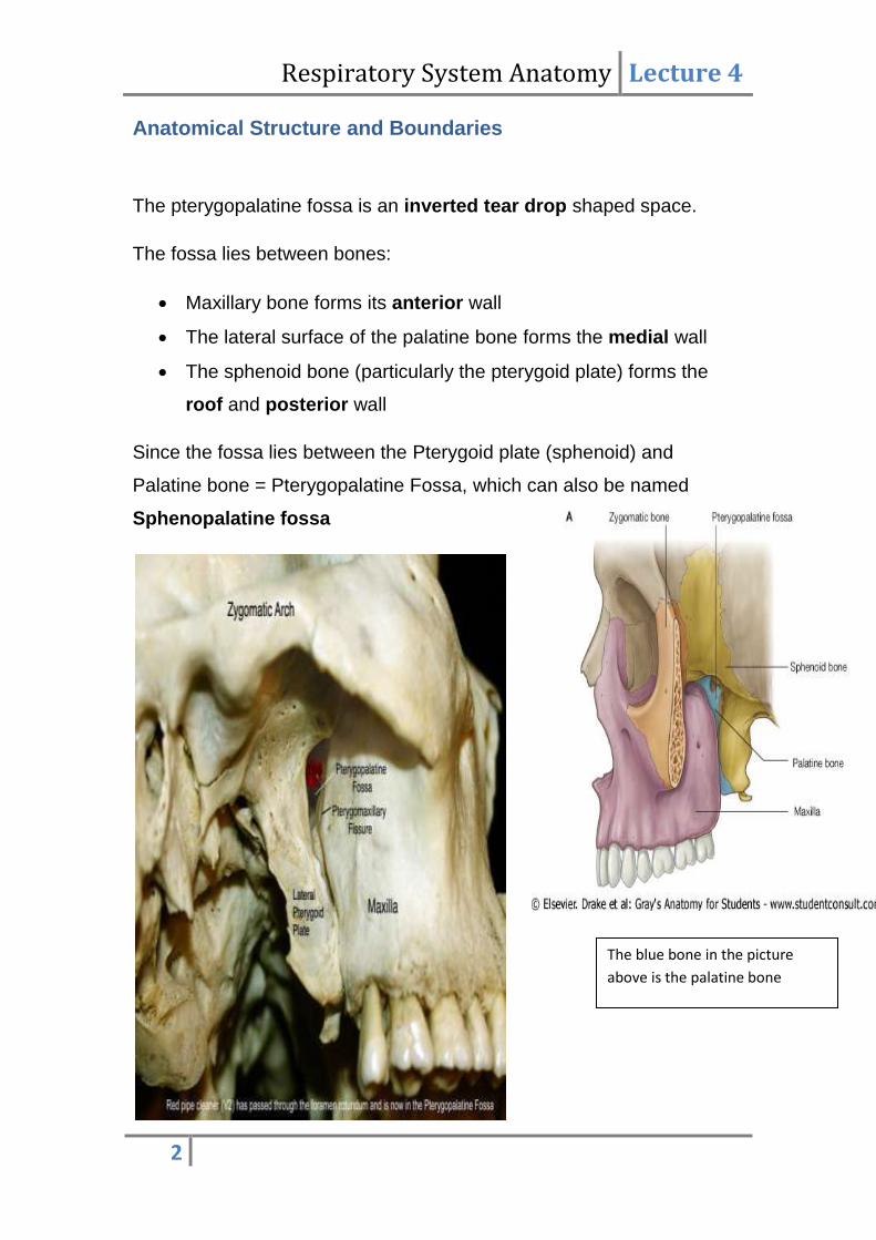
The pterygopalatine fossa is an inverted tear drop shaped space.

The fossa lies between bones:

Maxillary bone forms its anterior wall

The lateral surface of the palatine bone forms the medial wall

The sphenoid bone (particularly the pterygoid plate) forms the

roof and posterior wall

Since the fossa lies between the Pterygoid plate (sphenoid) and

Palatine bone = Pterygopalatine Fossa, which can also be named

Sphenopalatine fossa

The blue bone in the picture

above is the palatine bone

Respiratory System Anatomy Lecture 4

3

Foramina and Communications with other spaces and

cavities

Foramina and Fissures, and their Communications

1) Foramen Rotundum (Large foramen in sphenoid bone)

Communication: with Middle Cranial Fossa

2) Pterygoid Canal (foramen in sphenoid bone): Below and medial to

Foramen Rotundum and at the roof of the Foramen Lacerum (which is

covered by hyaline cartilage)

Communication: with Middle Cranial Fossa

3) Sphenopalatine foramen (foramen in palatine bone)

Communication: Nasal Cavity

4) Palatovaginal Canal

Communication: Nasopharynx

5) Palatine Canal (open as greater and

lesser palatine foramina in the oral cavity)

Communication: Oral Cavity

6) Pterygomaxillary Fissure

Communication: Infratemporal Fossa

7) Inferior Orbital Fissure

Communication: Orbital Cavity

Respiratory System Anatomy Lecture 4

4

Contents: it is what makes the Pterygopalatine fossa

important

1) Maxillary Nerve

2) Terminal part (3rd part) of maxillary artery

3) Nerve of Pterygoid Canal (also called Vidian nerve)

4) Pterygopalatine Ganglion

5) Veins

6) Lymphatics (distributed with blood vessels and

nerves)

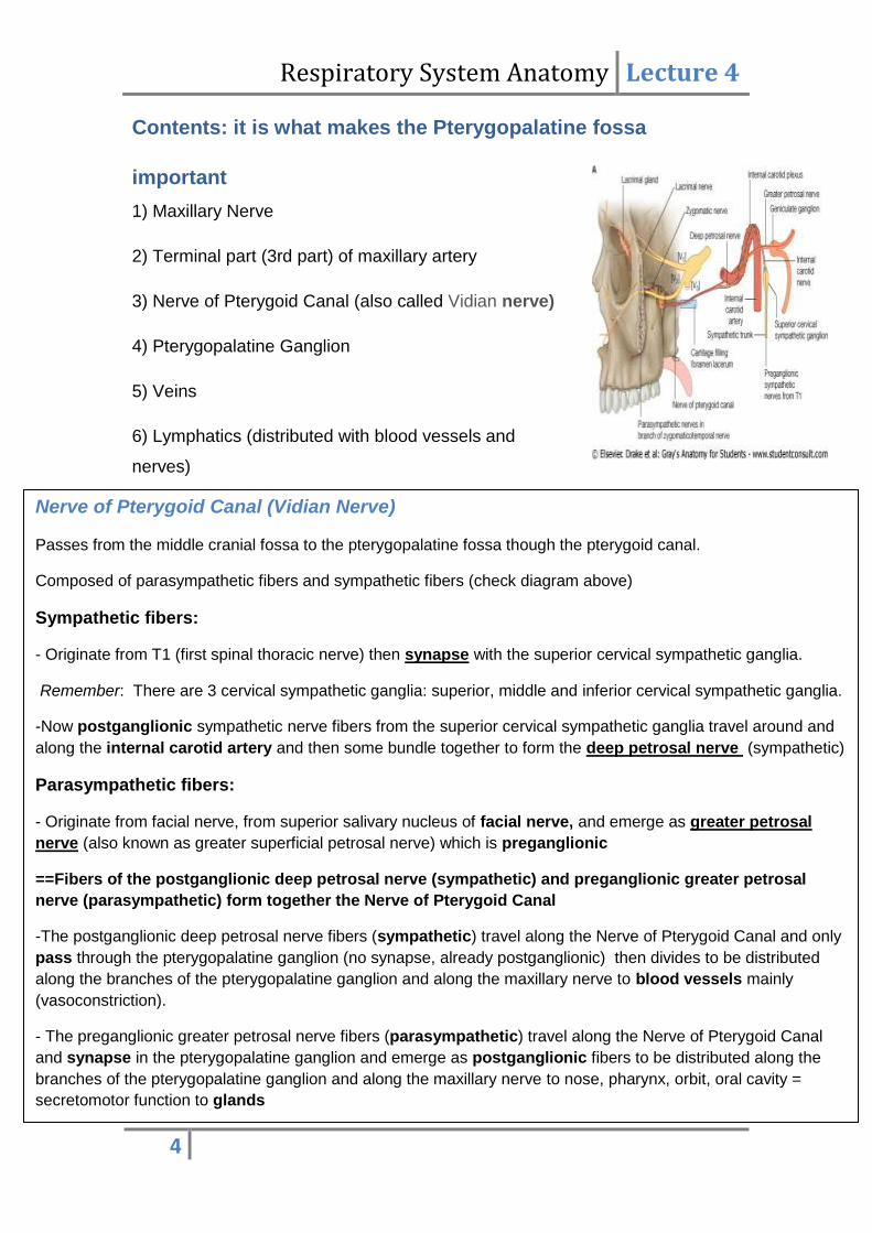
Nerve of Pterygoid Canal (Vidian Nerve)

Passes from the middle cranial fossa to the pterygopalatine fossa though the pterygoid canal.

Composed of parasympathetic fibers and sympathetic fibers (check diagram above)

Sympathetic fibers:

- Originate from T1 (first spinal thoracic nerve) then synapse with the superior cervical sympathetic ganglia.

Remember: There are 3 cervical sympathetic ganglia: superior, middle and inferior cervical sympathetic ganglia.

-Now postganglionic sympathetic nerve fibers from the superior cervical sympathetic ganglia travel around and

along the internal carotid artery and then some bundle together to form the deep petrosal nerve (sympathetic)

Parasympathetic fibers:

- Originate from facial nerve, from superior salivary nucleus of facial nerve, and emerge as greater petrosal

nerve (also known as greater superficial petrosal nerve) which is preganglionic

==Fibers of the postganglionic deep petrosal nerve (sympathetic) and preganglionic greater petrosal

nerve (parasympathetic) form together the Nerve of Pterygoid Canal

-The postganglionic deep petrosal nerve fibers (sympathetic) travel along the Nerve of Pterygoid Canal and only

pass through the pterygopalatine ganglion (no synapse, already postganglionic) then divides to be distributed

along the branches of the pterygopalatine ganglion and along the maxillary nerve to blood vessels mainly

(vasoconstriction).

- The preganglionic greater petrosal nerve fibers (parasympathetic) travel along the Nerve of Pterygoid Canal

and synapse in the pterygopalatine ganglion and emerge as postganglionic fibers to be distributed along the

branches of the pterygopalatine ganglion and along the maxillary nerve to nose, pharynx, orbit, oral cavity =

secretomotor function to glands

Respiratory System Anatomy Lecture 4

5

Pterygopalatine Ganglion

Just like the submandibular ganglia, the pterygopalatine ganglion is a

parasympathetic ganglion where preganglionic parasympathetic

fibers synapse in and emerge as postganglionic fibers.

-The Nerve of Pterygoid Canal joins the ganglia (carrying sympathetic

and parasympathetic fibers) and the Maxillary nerve gives off "twigs"

(around 2 twigs) sensory to the ganglion, so the nerve branches

leaving the ganglion will contain sensory (originally from maxillary

nerve), sympathetic and parasympathetic fibers.

Remember: The pterygopalatine ganglion also gives parasympathetic and

sympathetic fibers to the maxillary nerve.

The nerve branches of the pterygopalatine ganglion:

1) Orbital Branches: pass through inferior orbital fissure to the orbit

- supply periosteum of orbit and lacrimal gland as well as ethmoidal and

sphenoidal air sinuses

2) Palatine Branches: pass through palatine canal to oral cavity

- Divided to greater and lesser palatine nerves which pass through greater

and lesser palatine foramina respectively

- Lesser palatine nerve supplies the soft palate

-Greater palatine nerve supplies hard palate then pass upward and supply

nasal cavity

3) Pharyngeal Branches: pass through the palatovaginal canal to the

nasopharynx

-supplies the nasopharynx especially the glands there

4) Nasal Branches: pass through sphenopalatine foramen to nasal cavity

- 7 or 8 small nerves

Respiratory System Anatomy Lecture 4

6

- most important ones are the short sphenopalatine and long sphenopalatine

nerves (which is also called nasopalatine nerve)

Check diagram below for the pterygopalatine branches

Revision:- Nasal Cavity( mentioned in previous lectures) in this lecture:

The nasal cavity is divided into lateral and medial walls, and the lateral wall is divided into 4

quadrants: upper anterior, upper posterior, inferior anterior, inferior posterior

-Major nerve and blood supply of nasal cavity is from pterygopalatine fossa through sphenopalatine

foramen as from the maxillary artery and maxillary nerve

- 4 main nervous supply to the nasal cavity*:

-Long sphenopalatine nerve, also known as nasopalatine nerve (mainly to medial wall/

septum of nasal cavity and is its major nervous supply)

- Short sphenopalatine nerve

- Greater and lesser palatine nerves

- Anterior and Posterior ethmoidal nerves from ophthalmic nerve

Epistaxis (Nose-Bleed)

There is an area on the nasal septum (upper 2/3 and lower 1/3 meeting) called Kiesselbach's area

that is site of anastomosis and potential bleeding. Anastomosis mainly involves the nasopalatine

artery and superior labial branch of facial artery, and if we are to name one major responsible for

epistaxis then it is the superior labial branch of facial artery.

Respiratory System Anatomy Lecture 4

7

Especial emphasis on certain arteries and nerves related to

the fossa

Maxillary Nerve

- It is originally a pure sensory nerve

- It supplies all the upper teeth, sinuses such as maxillary sinus, and the

maxilla (skin over it as well)

Its course:

- It is a branch of the trigeminal ganglia in middle cranial fossa

- In the middle cranial fossa, the maxillary nerve gives off a branch called

meningeal branch then the maxillary nerve enters the pterygopalatine fossa

through foramen rotundum

- In the pterygopalatine fossa, it gives "twigs" sensory to the

pterygopalatine ganglia

- The maxillary nerve then descends down along the pterygomaxillary fissure

to the infratemporal fossa.

- In the infratemporal fossa, the maxillary nerve gives off a sensory branch

called posterior superior alveolar nerve which supplies the upper molars

and the maxillary air sinus. (Gray's and the

slides say that post. sup. alveolar nerve

branches off the maxillary nerve in

pterygopalatine fossa then enters the

infratemporal fossa)

- The maxillary nerve then ascends and

enters the inferior orbital fissure and just

as it enters, it gives off a branch called

zygomatic branch (branched off maxillary

Respiratory System Anatomy Lecture 4

8

in the pterygopalatine fossa). The zygomatic branch divides into

zygomaticofacial and zygomaticotemporal nerve branches.

The zygomaticofacial supplies the skin of the zygoma (refers to zygomatic

bone) while the zygomaticotemporal nerve which contains parasympathetic

postganglionic nerve fibers reaches the temporal region where it gives the

major supply of parasympathetic fibers to the lacrimal nerve (branch of

ophthalmic nerve) which innervates the lacrimal gland.

- Then, the maxillary nerve enters the orbital cavity through inferior orbital

fissure, it continues along the floor of the orbit in the infraorbital canal. Some

references name the nerve now as infraorbital nerve, while other references

name it infraorbital nerve only when it comes out the orbit through the

infraorbital foramen; both terminologies are acceptable.

- The infraorbital/ maxillary nerve in the orbital floor gives off middle superior

alveolar and anterior superior alveolar nerves.

Remember: the posterior superior alveolar nerve was a branch of the

maxillary nerve in the infratemporal fossa

The middle superior alveolar nerve supplies premolars, while the anterior

superior alveolar nerve supplies canines, incisors, and ascends to the nasal

cavity (gives sensory to the nose)

- At last the nerve emerges from the infraorbital foramen, and the infraorbital

nerve then divides to 3 terminal branches:

1) Palpebral branch: supply skin of lower eyelid

2) Nasal branch: supply skin of nose

3) Superior labial: supply upper lip

Maxillary Artery

- External carotid artery divides in the parotid gland to maxillary artery

and superficial temporal artery

Respiratory System Anatomy Lecture 4

9

- The lateral pterygoid muscle divides the maxillary artery into three:

- 1st part: before the muscle

- 2nd part: related to the muscle either superficial or deep to it

- 3rd part: after the muscle (terminal part in infratemporal and

pterygopalatine fossa)

First Part

5 branches and all of them pass through foramina or fissures:

1) Auditory Artery*: to external auditory canal (however, after checking

external references, it is the deep auricular artery not auditory artery)

2) Tympanic Artery: to tympanic membrane in the ear

-so first 2 branches related to the ear

3) Inferior Alveolar Artery:

Note: the inferior alveolar nerve originates from mandibular nerve while

the inferior alveolar artery originates from maxillary artery, and both of them

enter mandibular foramen

- The Inferior Alveolar Artery gives incisive branches to the lower teeth

and then exits through the mental foramen as mental artery

- The Inferior Alveolar Artery before entering the mandibular canal

gives off a branch called mylohyoid artery at the mylohyoid groove on the

mandible which supplies the mylohyoid muscle

4) Middle meningeal Artery

- Pass through foramen spinosum to middle cranial fossa

- Forms a groove on the skull on the inside, and is liable to rupture by trauma

causing extradural intracranial hemorrhage/ hematoma

5) Accessory Middle meningeal artery

Both

ascend

upwards

deep to

the lateral

pterygoid

Respiratory System Anatomy Lecture 4

10

Second Part

5 branches: 4 of them are related to mastication and 1 buccal artery:

1) Masseteric Artery: to masseter muscle

2) Deep Temporal Artery: to temporalis muscle

3) Medial Pterygoid Artery: to medial pterygoid muscle

4) Lateral Pterygoid Artery: to lateral pterygoid muscle

5) Buccal Artery: to buccinator region

The maxillary artery then enters the infratemporal fossa and ascends

upwards along the pterygomaxillary fissure (opposite in direction to the

maxillary nerve) toward the pterygopalatine fossa and then it enters the

inferior orbital fissure to the orbital cavity and ends as Infraorbital Artery

(just like the nerve).

Third Part (terminal part): the part that reaches the pterygopalatine fossa,

Important in blood supply of the nose and pharynx:

1) Sphenopalatine Arteries

-Divides to long and short sphenopalatine with long sphenopalatine

(nasopalatine) supplying mainly the nasal septum and a part of lateral wall.

The short sphenopalatine artery supplies the upper posterior quadrant of

lateral wall (just like the nerve)

2) Pharyngeal Artery (passes through palatovaginal canal to nasopharynx)

3) Palatine Artery

-Divides to greater and lesser palatine, however unlike the nerve, the

lesser palatine to soft palate is very small and not important. Also, greater

palatine artery passes through greater palatine foramen to supply the hard

Respiratory System Anatomy Lecture 4

11

palate then passes through incisive foramen from oral cavity to reach the

nasal cavity.

4) Posterior Superior Alveolar Artery

- This branch is given off as the maxillary artery ascends the pterygomaxillary

fissure, supplies molars and maxillary air sinuses.

5) Infraorbital Artery: gives middle superior alveolar and anterior superior

alveolar arteries in the orbital floor and terminates as palpebral, nasal, and

superior labial arteries (like the nerve)

6) Artery of Pterygoid Canal: pass through pterygoid canal towards roof of

foramen lacerum and terminate in nasopharynx

The diagram above summarizes the maxillary artery branches

Respiratory System Anatomy Lecture 4

12

Venous Drainage

-The veins follow opposite direction to the arteries.

- The collection of veins is around the lateral pterygoid muscle and is called

the pterygoid plexus of veins present in the infratemporal fossa.

- The pterygoid plexus of veins drains the pterygopalatine fossa and ganglion

- The pterygoid plexus of veins end up as maxillary vein which joins the

superficial temporal vein in the parotid gland to form retromandibular vein

- The pterygoid plexus communicates with the cavernous sinus through

emissionary veins and communicates with the facial vein anteriorly as well.

Thus any pus cells in the facial vein may reach the pterygoid plexus and may

reach the cavernous sinus which is dangerous.

-The facial vein drains in the internal jugular vein

Good luck


Recommended