1 Respiratory System Respiratory System Anatomy Anatomy Overview Overview Of all the substances that cells and the Of all the substances that cells and the whole body must have to survive, O whole body must have to survive, O 2 is by is by far the most crucial far the most crucial A person can live a few weeks without A person can live a few weeks without food, a few days without water, but only a food, a few days without water, but only a few minutes without O few minutes without O 2 Constant removal of carbon dioxide from Constant removal of carbon dioxide from the body is just as important for survival the body is just as important for survival as a constant supply of O as a constant supply of O 2 Functions Functions The organs of the respiratory system The organs of the respiratory system perform several functions: perform several functions: – Gas exchange via diffusion Gas exchange via diffusion Delivery of O Delivery of O 2 to body cells to body cells Elimination of CO Elimination of CO 2 2 produced by body cells produced by body cells – Regulation of blood pH Regulation of blood pH – Filter, warm & humidify the air we breathe Filter, warm & humidify the air we breathe – Contain receptors for the sense of smell Contain receptors for the sense of smell – Production of vocal sounds Production of vocal sounds – Excretion of heat & water Excretion of heat & water
Of all the substances that cells and the Of all the substances that cells and the whole body must have to survive, Owhole body must have to survive, O22 is by is by far the most crucialfar the most crucialA person can live a few weeks without A person can live a few weeks without food, a few days without water, but only a food, a few days without water, but only a few minutes without Ofew minutes without O22
Constant removal of carbon dioxide from Constant removal of carbon dioxide from the body is just as important for survival the body is just as important for survival as a constant supply of Oas a constant supply of O22
FunctionsFunctions
The organs of the respiratory system The organs of the respiratory system perform several functions:perform several functions:–– Gas exchange via diffusionGas exchange via diffusion
Delivery of ODelivery of O22 to body cellsto body cellsElimination of COElimination of CO2 2 produced by body cellsproduced by body cells
–– Regulation of blood pHRegulation of blood pH–– Filter, warm & humidify the air we breatheFilter, warm & humidify the air we breathe–– Contain receptors for the sense of smellContain receptors for the sense of smell–– Production of vocal soundsProduction of vocal sounds–– Excretion of heat & waterExcretion of heat & water
2
RespirationRespiration
Ensures OEnsures O22 is supplied to body cellsis supplied to body cellsCOCO22 is removed from the body cellsis removed from the body cellsRespirationRespiration= = homeostatic mechanismhomeostatic mechanism–– Helps maintain a constant environmentHelps maintain a constant environment→→ body cells to function effectivelybody cells to function effectively
Respiratory OrgansRespiratory Organs
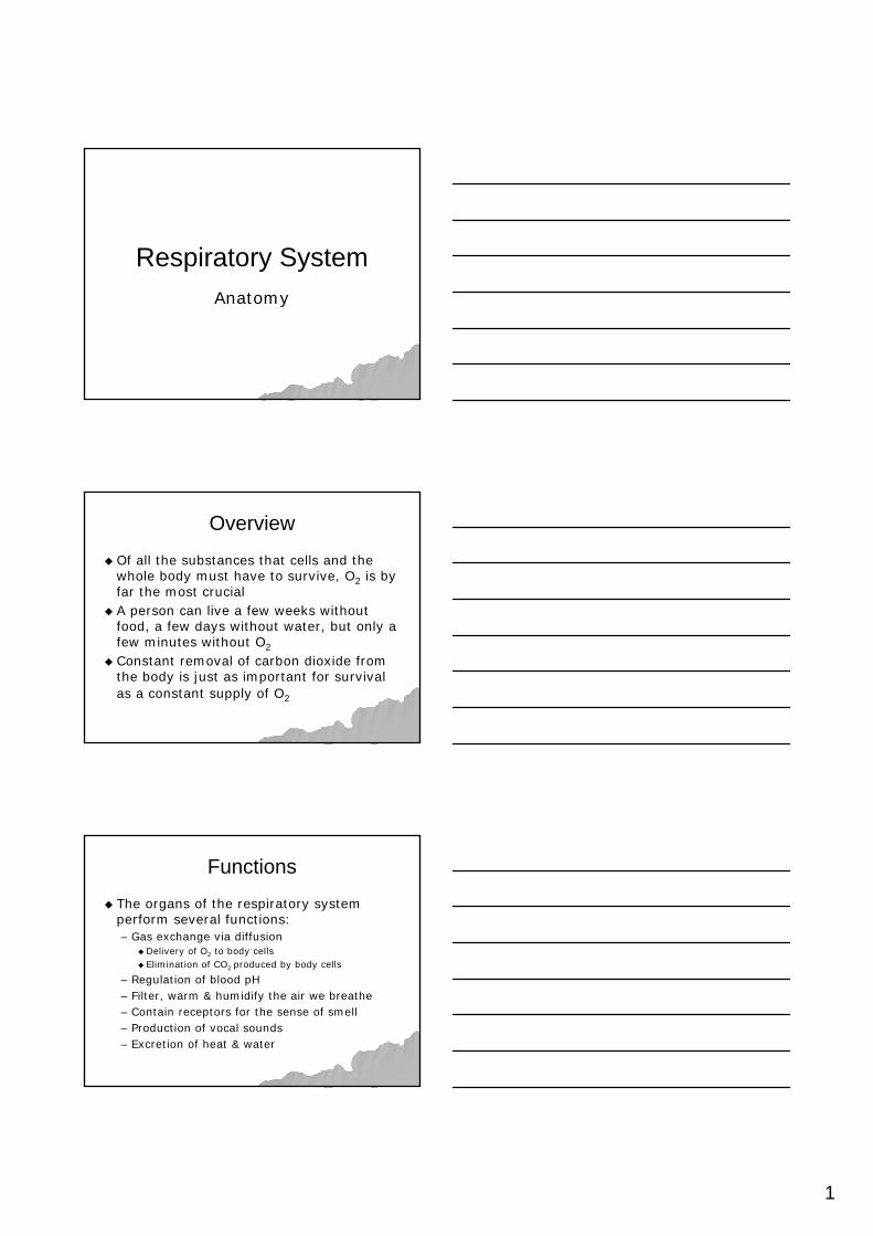
Organs of the respiratory systemOrgans of the respiratory system–– Nose & nasal cavitiesNose & nasal cavities–– PharynxPharynx–– LarynxLarynx–– TracheaTrachea–– BronchiBronchi–– Lungs Lungs –– AlveoliAlveoli
Basic structure is that of a tube with many branches ending Basic structure is that of a tube with many branches ending in millions of extremely tiny, very thinin millions of extremely tiny, very thin--walled sacs called walled sacs called alveolialveoliCConsists of passageways that filter incoming air & carry it onsists of passageways that filter incoming air & carry it into the lungsinto the lungs
3
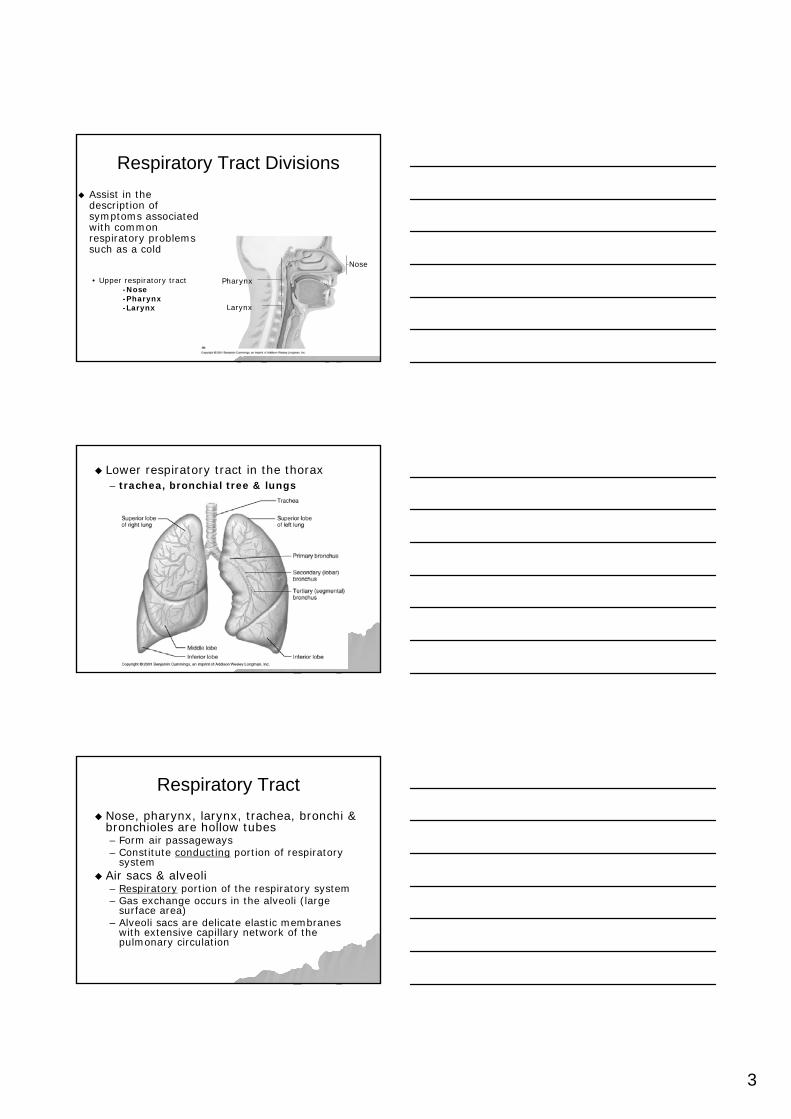
Respiratory Tract DivisionsRespiratory Tract DivisionsAssist in the Assist in the description of description of symptoms associated symptoms associated with common with common respiratory problems respiratory problems such as a coldsuch as a cold
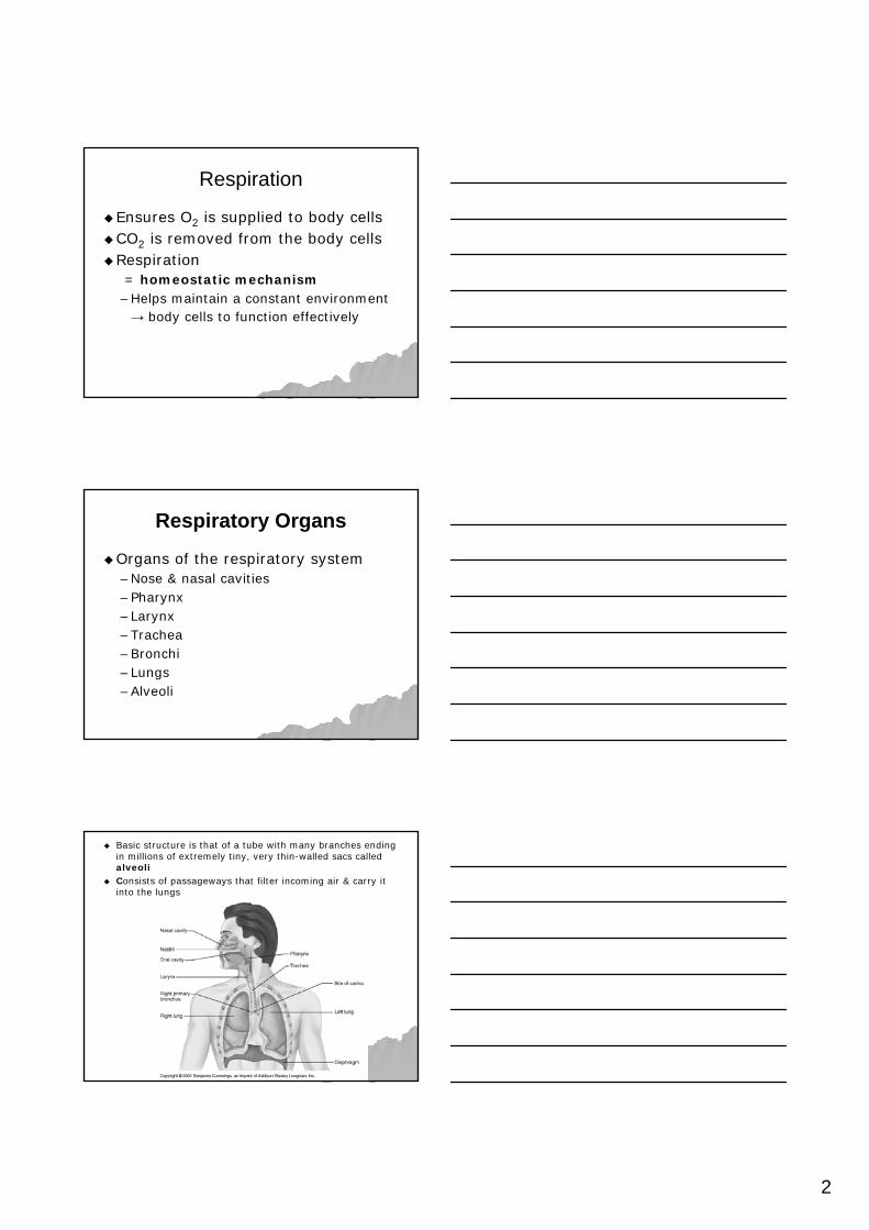
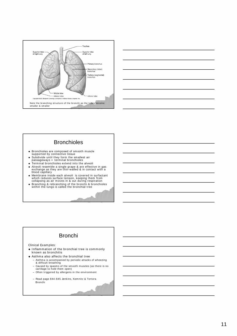
Lower respiratory tract in the thoraxLower respiratory tract in the thorax–– trachea, bronchial tree & lungstrachea, bronchial tree & lungs
Respiratory TractRespiratory TractNose, pharynx, larynx, trachea, bronchi & Nose, pharynx, larynx, trachea, bronchi & bronchioles are hollow tubesbronchioles are hollow tubes–– Form air passagewaysForm air passageways–– Constitute Constitute conductingconducting portion of respiratory portion of respiratory
systemsystemAir sacs & alveoli Air sacs & alveoli –– RespiratoryRespiratory portion of the respiratory systemportion of the respiratory system–– Gas exchange occurs in the alveoli (large Gas exchange occurs in the alveoli (large
surface area) surface area) –– Alveoli sacs are delicate elastic membranes Alveoli sacs are delicate elastic membranes
with extensive capillary network of the with extensive capillary network of the pulmonary circulationpulmonary circulation
4
Anatomy of the Anatomy of the Respiratory SystemRespiratory System
Upper Respiratory TractUpper Respiratory Tract
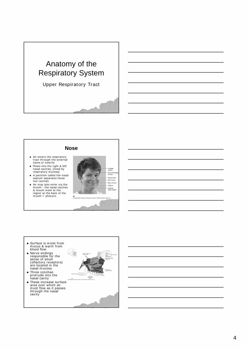
NoseNoseAir enters the respiratory Air enters the respiratory tract through the external tract through the external nares or nostrilsnares or nostrilsFlows into the right & left Flows into the right & left nasal cavities, (lined by nasal cavities, (lined by respiratory mucosa)respiratory mucosa)A partition called the nasal A partition called the nasal septum separates these septum separates these two cavitiestwo cavitiesAir may also enter via the Air may also enter via the mouth mouth -- the nasal cavities the nasal cavities & mouth meet at the & mouth meet at the region at the back of the region at the back of the mouth = pharynxmouth = pharynx
Surface is moist from Surface is moist from mucus & warm from mucus & warm from blood flowblood flowNerve endings Nerve endings responsible for the responsible for the sense of smell sense of smell (olfactory receptors) (olfactory receptors) are located in the are located in the nasal mucosanasal mucosaThree conchae Three conchae protrude into the protrude into the nasal cavitynasal cavityThese increase surface These increase surface area over which air area over which air must flow as it passes must flow as it passes through the nasal through the nasal cavitycavity
5
NoseNose
The structure of the conchae increases the surface area over which inhaled air travels ensuring that it is thoroughly warmed & filtered
NoseNoseBlood vessels in the nasal mucosa cool hot air & Blood vessels in the nasal mucosa cool hot air & warm cold airwarm cold airAir entering the nose is generally contaminated Air entering the nose is generally contaminated with one or more common irritants such as with one or more common irritants such as insects, dust, pollen & bacteriainsects, dust, pollen & bacteriaAir is purified removing almost all contaminants Air is purified removing almost all contaminants before inspired air reaches the lungsbefore inspired air reaches the lungsMucus secreted by mucosa adds moisture to dry Mucus secreted by mucosa adds moisture to dry air while trapping fine dust particles & microair while trapping fine dust particles & micro--organismsorganismsCiliated cells of the mucosa move contaminated Ciliated cells of the mucosa move contaminated mucus into the throat where it is swallowedmucus into the throat where it is swallowed
Clinical Example:Clinical Example:–– Because the mucosa lines the nose, Because the mucosa lines the nose,
sinus infections often develop from sinus infections often develop from colds in which the nasal mucosa is colds in which the nasal mucosa is inflamedinflamed
–– When the nasal cavity is blocked, the air When the nasal cavity is blocked, the air in the sinuses is absorbedin the sinuses is absorbed
–– Sometimes a sinus headache is incurred Sometimes a sinus headache is incurred & localised over the inflamed area& localised over the inflamed area
6
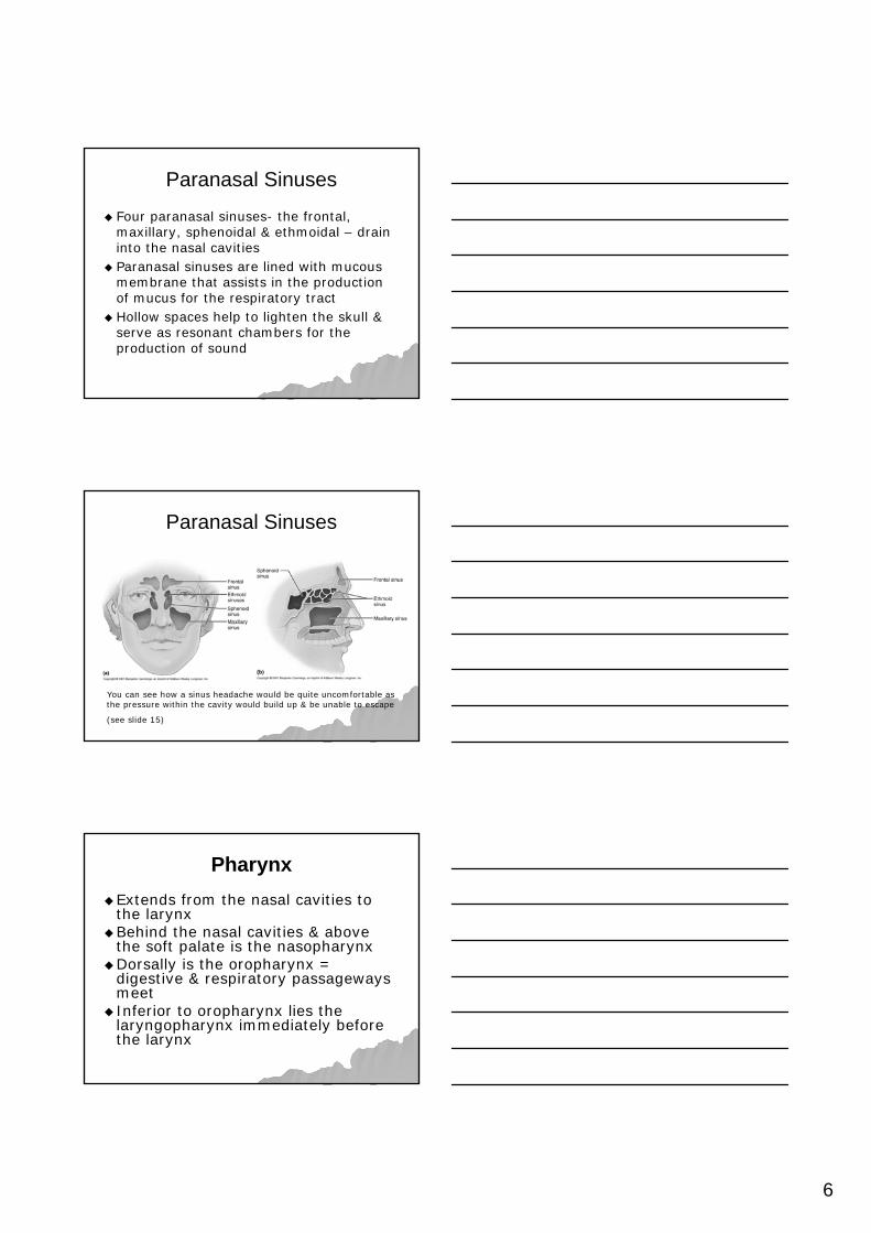
Paranasal SinusesParanasal Sinuses
Four paranasal sinusesFour paranasal sinuses-- the frontal, the frontal, maxillary, sphenoidal & ethmoidal maxillary, sphenoidal & ethmoidal –– drain drain into the nasal cavitiesinto the nasal cavitiesParanasal sinuses are lined with mucous Paranasal sinuses are lined with mucous membrane that assists in the production membrane that assists in the production of mucus for the respiratory tractof mucus for the respiratory tractHollow spaces help to lighten the skull & Hollow spaces help to lighten the skull & serve as resonant chambers for the serve as resonant chambers for the production of soundproduction of sound
Paranasal SinusesParanasal Sinuses
You can see how a sinus headache would be quite uncomfortable asthe pressure within the cavity would build up & be unable to escape
(see slide 15)
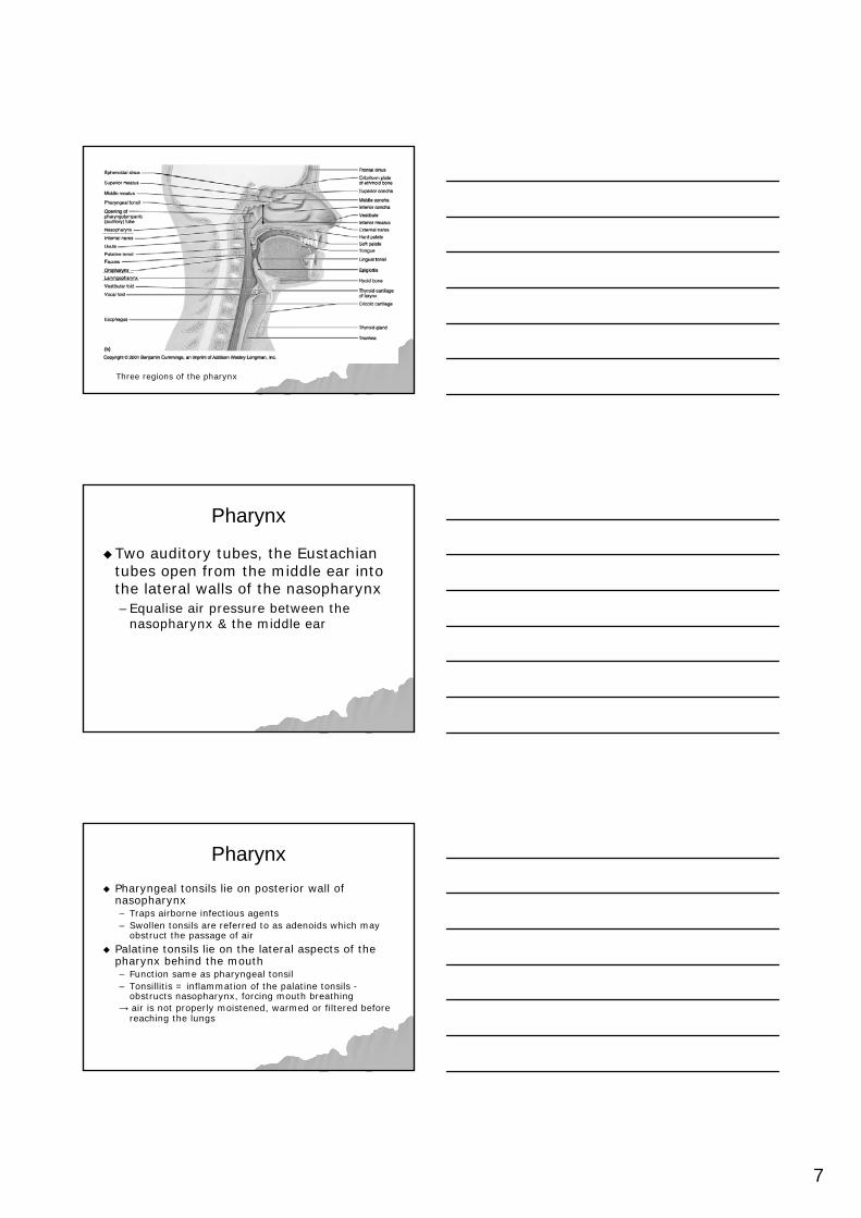
PharynxPharynxExtends from the nasal cavities to Extends from the nasal cavities to the larynxthe larynxBehind the nasal cavities & above Behind the nasal cavities & above the soft palate is the nasopharynxthe soft palate is the nasopharynxDorsally is the oropharynx = Dorsally is the oropharynx = digestive & respiratory passageways digestive & respiratory passageways meet meet Inferior to oropharynx lies the Inferior to oropharynx lies the laryngopharynx immediately before laryngopharynx immediately before the larynxthe larynx
7
Three regions of the pharynx
PharynxPharynx
Two auditory tubes, the Eustachian Two auditory tubes, the Eustachian tubes open from the middle ear into tubes open from the middle ear into the lateral walls of the nasopharynxthe lateral walls of the nasopharynx–– Equalise air pressure between the Equalise air pressure between the
nasopharynx & the middle earnasopharynx & the middle ear
PharynxPharynxPharyngeal tonsils lie on posterior wall of Pharyngeal tonsils lie on posterior wall of nasopharynxnasopharynx–– Traps airborne infectious agentsTraps airborne infectious agents–– Swollen tonsils are referred to as adenoids which may Swollen tonsils are referred to as adenoids which may
obstruct the passage of airobstruct the passage of air
Palatine tonsils lie on the lateral aspects of the Palatine tonsils lie on the lateral aspects of the pharynx behind the mouth pharynx behind the mouth –– Function same as pharyngeal tonsilFunction same as pharyngeal tonsil–– Tonsillitis = inflammation of the palatine tonsils Tonsillitis = inflammation of the palatine tonsils --
obstructs nasopharynx, forcing mouth breathing obstructs nasopharynx, forcing mouth breathing →→ air is not properly moistened, warmed or filtered before air is not properly moistened, warmed or filtered before
reaching the lungsreaching the lungs
8
Question:
How might the position of the tonsils assist in performing their immune function of trapping and destroying pathogens?
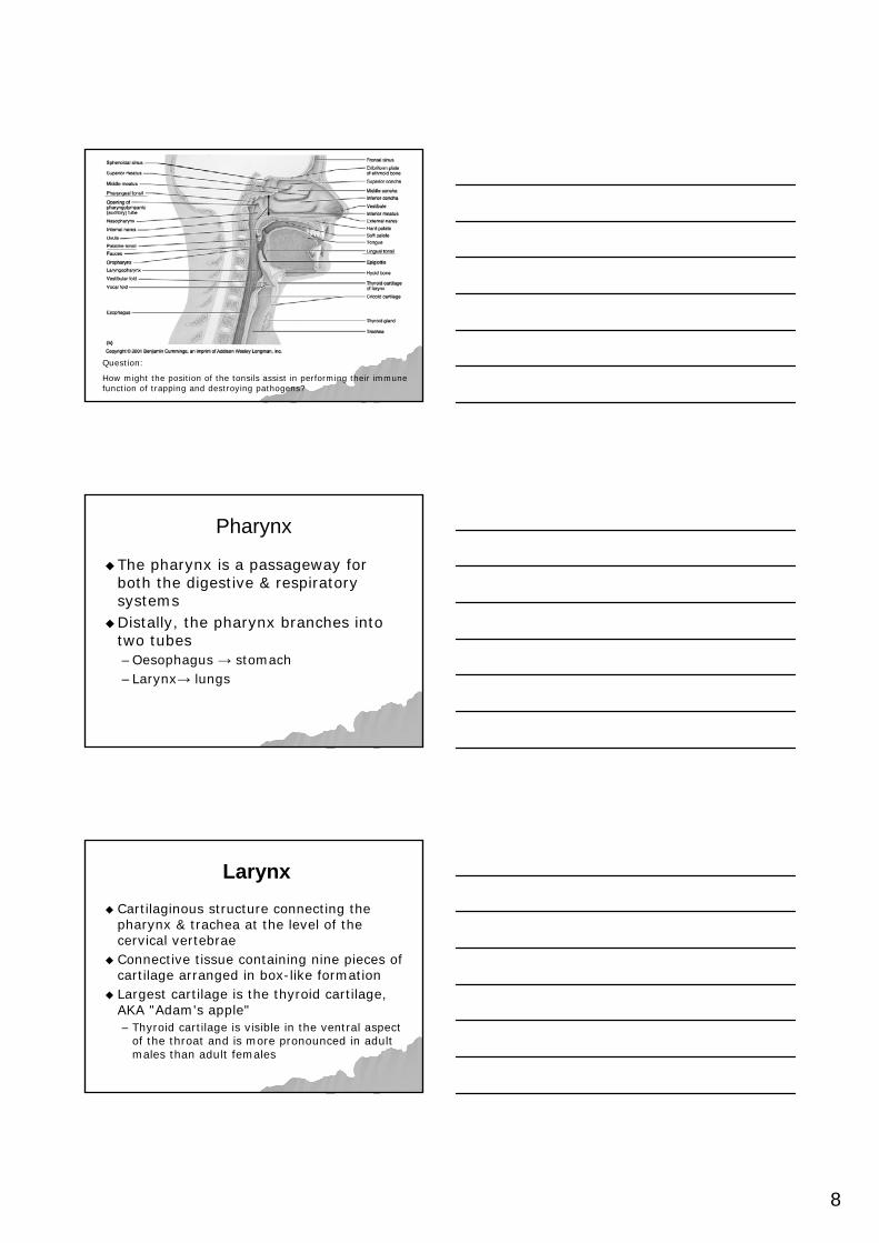
Pharynx Pharynx
The pharynx is a passageway for The pharynx is a passageway for both the digestive & respiratory both the digestive & respiratory systemssystemsDistally, the pharynx branches into Distally, the pharynx branches into two tubes two tubes –– Oesophagus Oesophagus →→ stomachstomach–– LarynxLarynx→→ lungslungs
LarynxLarynxCartilaginous structure connecting the Cartilaginous structure connecting the pharynx & trachea at the level of the pharynx & trachea at the level of the cervical vertebraecervical vertebraeConnective tissue containing nine pieces of Connective tissue containing nine pieces of cartilage arranged in boxcartilage arranged in box--like formationlike formationLargest cartilage is the thyroid cartilage, Largest cartilage is the thyroid cartilage, AKA "Adam's apple" AKA "Adam's apple" –– Thyroid cartilage is visible in the ventral aspect Thyroid cartilage is visible in the ventral aspect
of the throat and is more pronounced in adult of the throat and is more pronounced in adult males than adult femalesmales than adult females
9
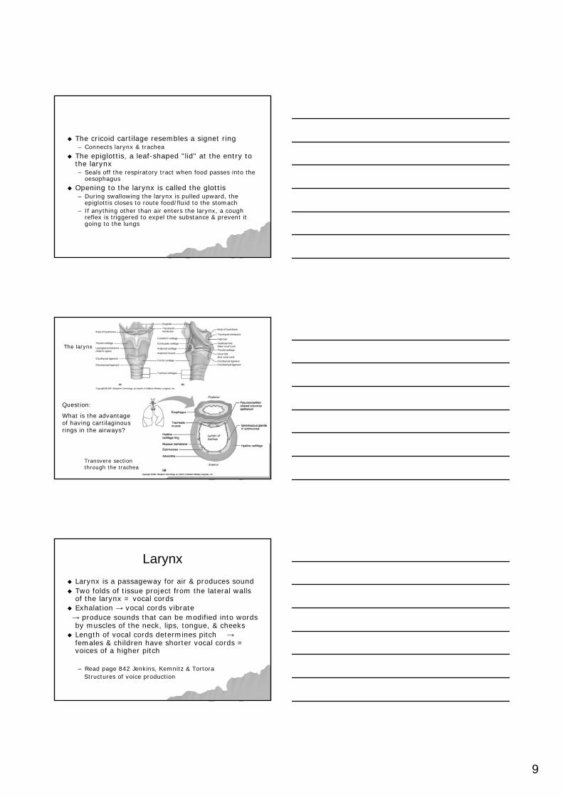
The cricoid cartilage resembles a signet ring The cricoid cartilage resembles a signet ring –– Connects larynx & tracheaConnects larynx & trachea
The epiglottis, a leafThe epiglottis, a leaf--shaped "lid" at the entry to shaped "lid" at the entry to the larynxthe larynx–– Seals off the respiratory tract when food passes into the Seals off the respiratory tract when food passes into the
oesophagusoesophagus
Opening to the larynx is called the glottisOpening to the larynx is called the glottis–– During swallowing the larynx is pulled upward, the During swallowing the larynx is pulled upward, the
epiglottis closes to route food/fluid to the stomachepiglottis closes to route food/fluid to the stomach–– If anything other than air enters the larynx, a cough If anything other than air enters the larynx, a cough
reflex is triggered to expel the substance & prevent it reflex is triggered to expel the substance & prevent it going to the lungsgoing to the lungs
Question:
What is the advantage of having cartilaginous rings in the airways?
The larynx
Transvere section through the trachea
LarynxLarynxLarynx is a passageway for air & produces soundLarynx is a passageway for air & produces soundTwo folds of tissue project from the lateral walls Two folds of tissue project from the lateral walls of the larynx = vocal cordsof the larynx = vocal cordsExhalation Exhalation →→ vocal cords vibratevocal cords vibrate→→ produce sounds that can be modified into words produce sounds that can be modified into words by muscles of the neck, lips, tongue, & cheeksby muscles of the neck, lips, tongue, & cheeksLength of vocal cords determines pitch Length of vocal cords determines pitch →→females & children have shorter vocal cords = females & children have shorter vocal cords = voices of a higher pitchvoices of a higher pitch
–– Read page 842 Jenkins, Kemnitz & TortoraRead page 842 Jenkins, Kemnitz & TortoraStructures of voice productionStructures of voice production
10
TracheaTracheaLarynx opens into a rigid tube = trachea Larynx opens into a rigid tube = trachea Trachea is ~12 to 15cms long in the midline of Trachea is ~12 to 15cms long in the midline of the neckthe neckSupported & held open by a stack of CSupported & held open by a stack of C--shaped shaped rings of cartilage open at the dorsal aspectrings of cartilage open at the dorsal aspectThe area between adjacent cartilages & the tips The area between adjacent cartilages & the tips of cartilage contains connective tissue & smooth of cartilage contains connective tissue & smooth musclemuscleThe trachea is an open passageway for incoming The trachea is an open passageway for incoming & outgoing air& outgoing airCiliated cells filter air before it enters the bronchiCiliated cells filter air before it enters the bronchi
TracheaTracheaBy pushing against your throat about an By pushing against your throat about an inch above the sternum, you can feel the inch above the sternum, you can feel the shape of the tracheashape of the tracheaOnly if you use considerable force can you Only if you use considerable force can you squeeze it closedsqueeze it closedAir has no other way to get to the lungs, & Air has no other way to get to the lungs, & complete tracheal obstruction can squeeze complete tracheal obstruction can squeeze the trachea shut & cause death in a the trachea shut & cause death in a matter of minutesmatter of minutes–– Eg. choking on food, tumour or infection Eg. choking on food, tumour or infection
causing inflammation of the lymph nodes of causing inflammation of the lymph nodes of the neckthe neck
BronchiBronchiThe trachea branches into two primary bronchiThe trachea branches into two primary bronchi–– Same structure as the tracheaSame structure as the trachea–– Right bronchus is slightly larger & more vertical than the Right bronchus is slightly larger & more vertical than the
leftleft
Bronchi become smaller & smaller Bronchi become smaller & smaller →→ secondary secondary bronchi then tertiary bronchibronchi then tertiary bronchiAs they extend further into the lungs diameter is As they extend further into the lungs diameter is reduced to about one millimetrereduced to about one millimetreBronchi are now called bronchiolesBronchi are now called bronchiolesThe amount of cartilage reduces as the tubes The amount of cartilage reduces as the tubes become smaller & smaller disappearing in the become smaller & smaller disappearing in the distal bronchiolesdistal bronchioles
11
Note the branching structure of the bronchi as the tubes become smaller & smaller
BronchiolesBronchiolesBronchioles are composed of smooth muscle Bronchioles are composed of smooth muscle supported by connective tissuesupported by connective tissueSubdivide until they form the smallest air Subdivide until they form the smallest air passageways = terminal bronchiolespassageways = terminal bronchiolesTerminal bronchioles extend into the alveoliTerminal bronchioles extend into the alveoliAlveoli resemble a single grape & are effective in gas Alveoli resemble a single grape & are effective in gas exchange as they are thinexchange as they are thin--walled & in contact with a walled & in contact with a blood capillaryblood capillaryMembrane inside each alveoli is covered in surfactant Membrane inside each alveoli is covered in surfactant which reduces surface tension, keeping them from which reduces surface tension, keeping them from collapsing as air moves in & out during respirationcollapsing as air moves in & out during respirationBranching & rebranching of the bronchi & bronchioles Branching & rebranching of the bronchi & bronchioles within the lungs is called the bronchial treewithin the lungs is called the bronchial tree
Inflammation of the bronchial tree is commonly Inflammation of the bronchial tree is commonly known as bronchitisknown as bronchitisAsthma also affects the bronchial treeAsthma also affects the bronchial tree–– Asthma is accompanied by periodic attacks of wheezing Asthma is accompanied by periodic attacks of wheezing
& difficult breathing& difficult breathing–– Caused by spasms of the smooth muscles (as there is no Caused by spasms of the smooth muscles (as there is no
cartilage to hold them open)cartilage to hold them open)–– Often triggered by allergens in the environmentOften triggered by allergens in the environment
LungsLungsPaired organs occupying most of the space of the Paired organs occupying most of the space of the thoracic cavitythoracic cavityConsist of millions of small, cupConsist of millions of small, cup--shaped out shaped out pockets (sacs) called pockets (sacs) called alveolialveoliRespiratory membranes of alveoli are a thin Respiratory membranes of alveoli are a thin barrier in which gases can pass by diffusion barrier in which gases can pass by diffusion ~ 300 million alveoli in an average adult~ 300 million alveoli in an average adultLungs are separated from one another by a Lungs are separated from one another by a median dividing wallmedian dividing wallCalled the mediastinumCalled the mediastinum–– contains the heart, thymus, oesophagus, large blood contains the heart, thymus, oesophagus, large blood
vessels embedded in connective tissuevessels embedded in connective tissue
LungsLungs
Lungs are conical shaped with Lungs are conical shaped with elastic, spongy texture due to the elastic, spongy texture due to the nature of the alveolinature of the alveoliRight lung is subdivided into three Right lung is subdivided into three lobeslobesLeft lung is subdivided into two lobesLeft lung is subdivided into two lobesEach lobe is divided into smaller Each lobe is divided into smaller lobules, each lobule is serviced by a lobules, each lobule is serviced by a large bronchiolelarge bronchiole
13
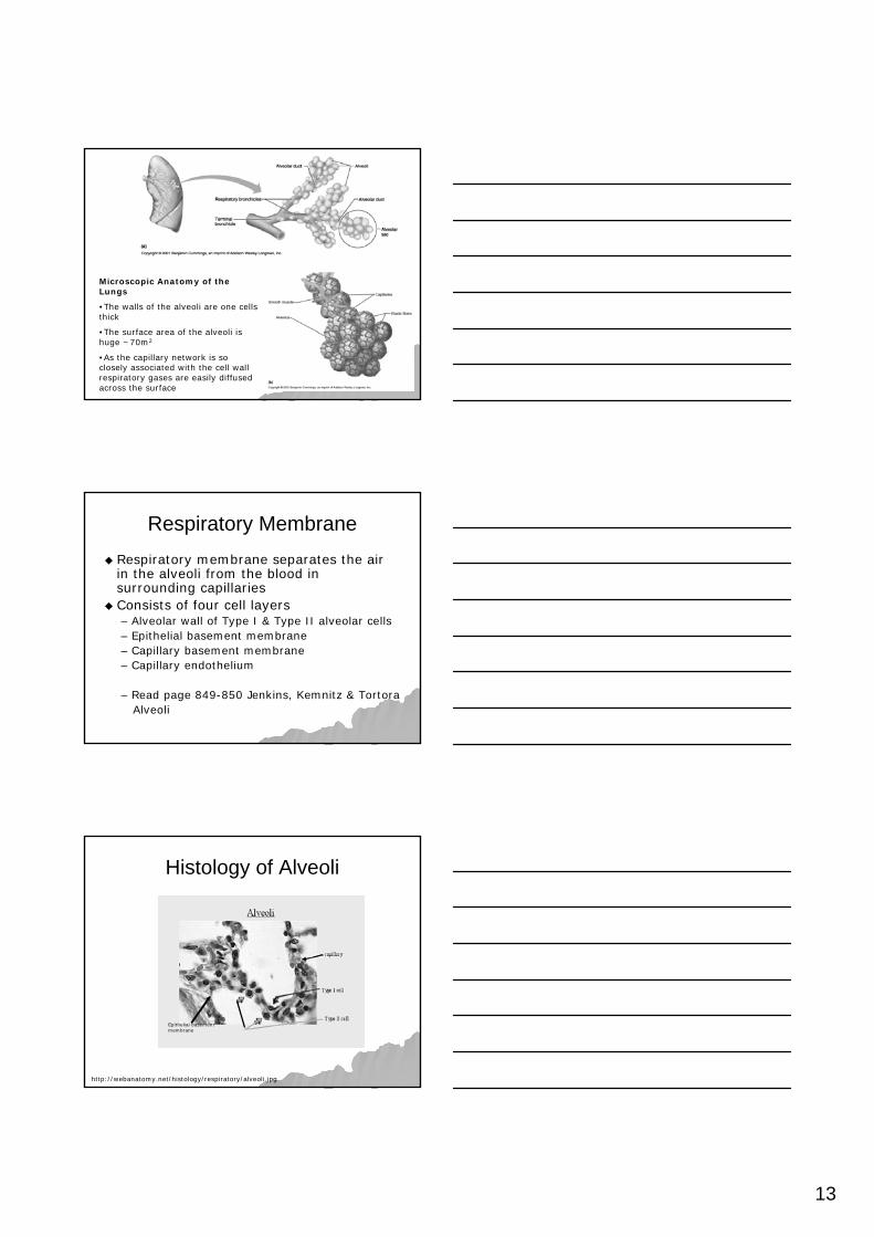
Microscopic Anatomy of the Lungs
•The walls of the alveoli are one cells thick
•The surface area of the alveoli is huge ~70m2
•As the capillary network is so closely associated with the cell wall respiratory gases are easily diffused across the surface
Respiratory MembraneRespiratory MembraneRespiratory membrane separates the air Respiratory membrane separates the air in the alveoli from the blood in in the alveoli from the blood in surrounding capillaries surrounding capillaries Consists of four cell layersConsists of four cell layers–– Alveolar wall of Type I & Type II alveolar cellsAlveolar wall of Type I & Type II alveolar cells–– Epithelial basement membraneEpithelial basement membrane–– Capillary basement membraneCapillary basement membrane–– Capillary endotheliumCapillary endothelium
TwoTwo--layered membrane surrounding each layered membrane surrounding each lunglungInner layer = visceral pleuraInner layer = visceral pleura–– covers the surface of each lungcovers the surface of each lung–– reaches into the fissures between the lobes of reaches into the fissures between the lobes of
the lungthe lung–– encloses the mediastinumencloses the mediastinum
Outer layer = parietal pleuraOuter layer = parietal pleura–– lines the inner surface of the thoracic cavity lines the inner surface of the thoracic cavity
Who remembers Fred Dagg & his song ‘If it weren't for your gumboots’? The pleurisy mentioned in the song is an inflammation of the pleurae. It is a very painful condition as it reduces the ability of the pleural surfaces to move over each other causing rubbing/friction with each breath.
PleuraPleuraVisceral & parietal pleura are continuous Visceral & parietal pleura are continuous with one another where the primary with one another where the primary bronchus, blood vessels & nerves enter bronchus, blood vessels & nerves enter each lungeach lungTwo layers of the pleura form a collapsed Two layers of the pleura form a collapsed sacsacArea within the sac = pleural cavityArea within the sac = pleural cavity–– Fluid in the cavity keeps the twoFluid in the cavity keeps the two--pleural membranes in pleural membranes in
close contact with each other & allows them to glide close contact with each other & allows them to glide smoothly over each othersmoothly over each other
–– Fluid adheres the two layers of the pleura to one anotherFluid adheres the two layers of the pleura to one another
15
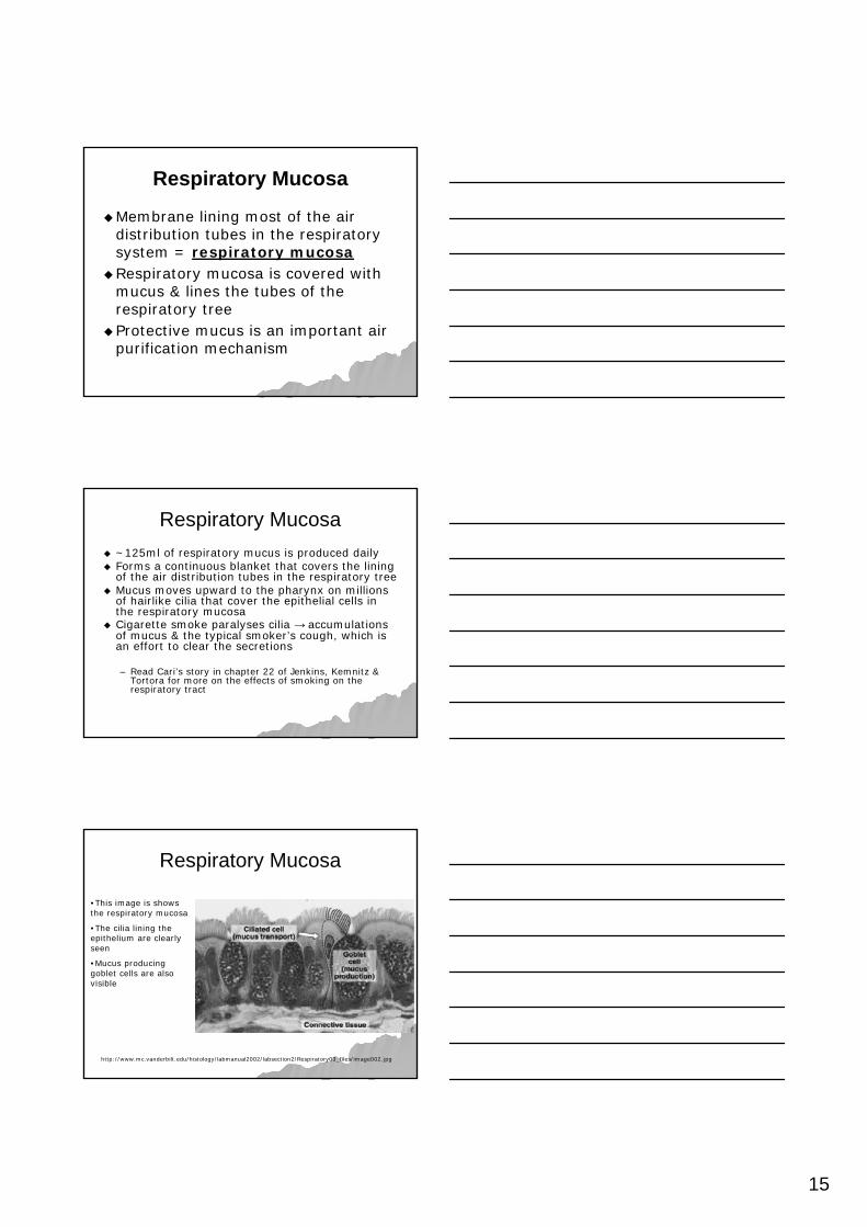
Respiratory MucosaRespiratory Mucosa
Membrane lining most of the air Membrane lining most of the air distribution tubes in the respiratory distribution tubes in the respiratory system = system = respiratory mucosarespiratory mucosaRespiratory mucosa is covered with Respiratory mucosa is covered with mucus & lines the tubes of the mucus & lines the tubes of the respiratory treerespiratory treeProtective mucus is an important air Protective mucus is an important air purification mechanismpurification mechanism
Respiratory MucosaRespiratory Mucosa~125ml of respiratory mucus is produced daily~125ml of respiratory mucus is produced dailyForms a continuous blanket that covers the lining Forms a continuous blanket that covers the lining of the air distribution tubes in the respiratory treeof the air distribution tubes in the respiratory treeMucus moves upward to the pharynx on millions Mucus moves upward to the pharynx on millions of hairlike cilia that cover the epithelial cells in of hairlike cilia that cover the epithelial cells in the respiratory mucosathe respiratory mucosaCigarette smoke paralyses cilia Cigarette smoke paralyses cilia →→ accumulations accumulations of mucus & the typical smokerof mucus & the typical smoker’’s cough, which is s cough, which is an effort to clear the secretionsan effort to clear the secretions
–– Read CariRead Cari’’s story in chapter 22 of Jenkins, Kemnitz & s story in chapter 22 of Jenkins, Kemnitz & Tortora for more on the effects of smoking on the Tortora for more on the effects of smoking on the respiratory tractrespiratory tract