15
Restriction Digestion Restriction Digestion of of Arabidopsis thaliana Arabidopsis thaliana Genomic DNA Genomic DNA
| Date post: | 20-Dec-2015 |
| Category: |
Documents |
| View: | 242 times |
| Download: | 1 times |

Restriction Digestion of Restriction Digestion of Arabidopsis thalianaArabidopsis thaliana
Genomic DNA Genomic DNA
Broad and Long Term ObjectiveBroad and Long Term Objective
To determine the copy number of MybTo determine the copy number of Myb
transcription factor genes in the genome oftranscription factor genes in the genome of
the model plant the model plant Arabidopsis thalianaArabidopsis thaliana

Research PlanResearch PlanIsolate Genomic DNA
Digest Genomic DNA with Various Restriction Enzymes
Agarose Gel Electrophoresis and Southern Transfer
Make Non-Radioactive Myb Probe
Hyribidize Probe to Southern Blot
Washes and Colorimetric Detection
Data Analysis
So
uth
ern
Blo
t
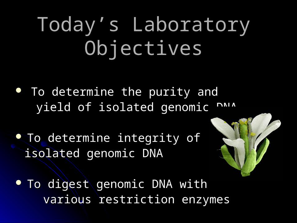
Today’s Laboratory Today’s Laboratory ObjectivesObjectives
To determine the purity and To determine the purity and yield of isolated genomic DNAyield of isolated genomic DNA
To determine integrity ofTo determine integrity of isolated genomic DNAisolated genomic DNA
To digest genomic DNA with To digest genomic DNA with various restriction enzymes various restriction enzymes

Spectrophotometric determination Spectrophotometric determination of DNA concentration/purityof DNA concentration/purity
Nucleic acids absorb light at 260 nmNucleic acids absorb light at 260 nm Proteins absorb light at 280 nmProteins absorb light at 280 nm Purity of Nucleic Acid indicated by APurity of Nucleic Acid indicated by A260260/A/A280 280
Pure DNA APure DNA A260260/A/A280 280 = 1.6-1.8= 1.6-1.8
Estimate of DNA purityEstimate of DNA purity** ==
AA260260/A/A280 280 ratio (1.6-1.8 is optimal)ratio (1.6-1.8 is optimal)DNA yield per gram tissueDNA yield per gram tissue** = =
Total DNA yield/starting weight of Total DNA yield/starting weight of A. A. thalianathaliana tissue tissue
DNA concentration (ng/ul)DNA concentration (ng/ul)** ==
AA260260 (dilution factor) (50 ng/ul) (dilution factor) (50 ng/ul)Total DNA yield (ng)Total DNA yield (ng)** ==
DNA concentration (volume of water, ul)DNA concentration (volume of water, ul)

Theoretical Basis of Agarose Theoretical Basis of Agarose Gel ElectrophoresisGel Electrophoresis
Agarose is a polysaccharide from marine alage that is Agarose is a polysaccharide from marine alage that is used in a matrix to separate DNA moleculesused in a matrix to separate DNA molecules
Because DNA ia a (-) charged molecule when subjected Because DNA ia a (-) charged molecule when subjected to an electric current it will migrate towards a (+) poleto an electric current it will migrate towards a (+) pole

Pouring an Agarose GelPouring an Agarose Gel
1 2 3
4 5 6
7 8 9

Sizing a Piece of DNASizing a Piece of DNA
Size of DNA molecule can be determined by using Size of DNA molecule can be determined by using
standards of known sizestandards of known size**
1.1. A standard curve is made by plotting the size A standard curve is made by plotting the size (in bp) of the standards (Y-axis) against the (in bp) of the standards (Y-axis) against the distance each fragment has migrated from the distance each fragment has migrated from the well (X-axis) using semi-log paperwell (X-axis) using semi-log paper
2. Measure the distance the unknown fragment 2. Measure the distance the unknown fragment migrated from the wellmigrated from the well
3. Determine size of the unknown fragment based 3. Determine size of the unknown fragment based upon the line of best fit by drawing a vertical upon the line of best fit by drawing a vertical line upward from distance migrated and a line upward from distance migrated and a horizontal line across to the y axis. Report the y horizontal line across to the y axis. Report the y value (size).value (size).
http://www.csun.edu/~vceed002/ref/measurement/data/graphpaper/semi_log_numbered.pdf

Assessing the Integrity of DNA Assessing the Integrity of DNA
High Quality Genomic DNA
>95% DNA will be of high molecular weight, migrating as intact band near the top of the gel
Very little evidence of smaller fragments indicated by a smear of many different sized DNA fragments

Restriction EnzymesRestriction Enzymes bacterial proteins that restrict host range for certain bacteriophages by cleaving specific DNA sequences
bacterial “immune system": destroy any "non-self" DNA
Self DNA protected by host proteins that methylate the specific DNA sequences recognized by the restriction enzyme (restriction/modification systems)

Type II Restriction Type II Restriction EnzymesEnzymes
Type II restriction systems: Type II restriction systems: endonuclease and methylase are endonuclease and methylase are separate proteins (binary system)separate proteins (binary system)
Type I, III restriction systems:Type I, III restriction systems:endonuclease and methylase in same endonuclease and methylase in same proteinprotein
Hundreds of type II restriction enzymes Hundreds of type II restriction enzymes have been identifiedhave been identified
Most recognize and cut palindromic Most recognize and cut palindromic sequencessequences
Many leave staggered (sticky) endsMany leave staggered (sticky) ends Important for molecular biologists Important for molecular biologists
because restriction enzymes create because restriction enzymes create unpaired "sticky ends"unpaired "sticky ends" which anneal which anneal with any complementary sequencewith any complementary sequence

Using Restriction EnzymesUsing Restriction Enzymes
The activity of restriction enzymes is dependent upon The activity of restriction enzymes is dependent upon precise environmental conditions:precise environmental conditions:
pHpHTemperatureTemperatureSalt ConcentrationSalt ConcentrationIonsIons
One enzymatic unit (U) is defined as the amount of enzyme One enzymatic unit (U) is defined as the amount of enzyme required to completely digest 1 ug of DNA in 1 hr at 37º C:required to completely digest 1 ug of DNA in 1 hr at 37º C:
3-5 U/ug of genomic DNA 3-5 U/ug of genomic DNA 1 U/ug of plasmid DNA1 U/ug of plasmid DNAStocks typically at 10 U/ulStocks typically at 10 U/ul

Digesting Genomic DNA for Digesting Genomic DNA for Southern BlottingSouthern Blotting
• Restriction sites are located at random in the genome
EcoRI sites Digestion with EcoRI
Mixture of different sized fragments
Separation of fragments by size (electrophoresis)
Myb gene sequence
Hybridization with myb probe
Eco
RI d
iges
ted
undi
gest
ed

Next WeekNext Week
Agarose gel elctrophoresis of Agarose gel elctrophoresis of
digested DNAdigested DNA
Capillary transfer of DNA from the Capillary transfer of DNA from the
gel to a nylon membranegel to a nylon membrane

Common lab report problemsCommon lab report problems- Emiliana huxleyi, Escherichia coli, E. coli not E. hux- Introduction:
Describe experimental details in Materials and MethodsActive voice: “The E. huxleyi cDNA was sequenced, and the sequence
was analyzed using ORF finder, BLASTN,…”- Materials and Methods:
Volumes not necessaryComposition and concentration of solutions (what is glucose buffer?, what
concentration of CaCl2?Justify methods: “In order to precipitate the nucleic acids in solution, 0.6
volumes of isopropanol was added to the supernatant…”List bioinformatics programs and their uses: “Multiple sequence alignments
were performed using ClustalW”- Results
DNA concentration and total yieldCalculation of transformation efficiencies
-Discussion:Do not simply repeat results- analyze your data (what were the expected
results for transformation efficiency of + control, - control,experimental; what did you observe; why?)
Specific questions/experiments for future research (given these results, what is the next step?)


