77 Resuscitative Endovascular Balloon Occlusion of the Aorta (REBOA) Top Stent The Art of EndoVascular hybrid Trauma and bleeding Management Chapter 4 Resuscitative Endovascular Balloon Occlusion of the Aorta (REBOA) Jonny Morrison, Viktor Reva, Lars Lönn, Junichi Matsumoto, Yosouke Matsumara, John Holcomb, Koji Idoguchi, Tal Hörer and Joe DuBose Picture the scene, you are the Trauma Surgeon on-call for your hospital and word has been received that paramedics are minutes away with a pedestrian hit by a car. Nothing about this sounds good – the patient is profoundly hy- potensive and unconscious with a disfigured pelvis. What goes through your mind? Your patient is almost certainly about to bleed to death, but what other injuries does he have – a traumatic brain injury, long bone fractures? Where is the bleeding focus – pelvis, solid abdominal organs, chest, or all of these? What clinical capabilities do you have and do you need to report them now? e patient arrives and looks terrible – pale, clammy, moribund. Your team gets to work – high-flow oxygen by mask, pelvic binder applied, large-bore venous access, bloods away, and O-negative blood transfusion commenced. e primary survey reports no major chest injury on physical examination or x-ray, FAST scan positive, but the pelvic film shows disruption to the left sacro-iliac joint and pubic rami anteriorly. e last blood pressure recorded was 60/40mmHg, despite the first unit of blood already in, and the pelvic binder correctly positioned and secured. Your team looks to you – what is the plan now? Let us pause for a moment and consider the options. Your
Transcript
77
Resuscitative Endovascular Balloon Occlusion of the Aorta (REBOA)
Top Stent The Art of EndoVascular hybrid Trauma and bleeding Management
Chapter 4
Resuscitative Endovascular Balloon Occlusion of the Aorta (REBOA)
Jonny Morrison, Viktor Reva, Lars Lönn, Junichi Matsumoto, Yosouke Matsumara, John Holcomb, Koji Idoguchi, Tal Hörer and Joe DuBose
Picture the scene, you are the Trauma Surgeon on-call for your hospital and word has been received that paramedics are minutes away with a pedestrian hit by a car. Nothing about this sounds good – the patient is profoundly hy-potensive and unconscious with a disfigured pelvis. What goes through your mind?
Your patient is almost certainly about to bleed to death, but what other injuries does he have – a traumatic brain injury, long bone fractures? Where is the bleeding focus – pelvis, solid abdominal organs, chest, or all of these? What clinical capabilities do you have and do you need to report them now?
The patient arrives and looks terrible – pale, clammy, moribund. Your team gets to work – high-flow oxygen by mask, pelvic binder applied, large-bore venous access, bloods away, and O-negative blood transfusion commenced. The primary survey reports no major chest injury on physical examination or x-ray, FAST scan positive, but the pelvic film shows disruption to the left sacro-iliac joint and pubic rami anteriorly. The last blood pressure recorded was 60/40mmHg, despite the first unit of blood already in, and the pelvic binder correctly positioned and secured. Your team looks to you – what is the plan now? Let us pause for a moment and consider the options. Your
78
Resuscitative Endovascular Balloon Occlusion of the Aorta (REBOA)
Top Stent The Art of EndoVascular hybrid Trauma and bleeding Management
patient is bleeding to death from somewhere in his abdomen and/or pelvis. You need to stop this process as quickly as possible. The “ideal” solution is immediate Damage Con-trol Surgery (DCS, e.g., trauma laparotomy and pelvic fixation followed by pre-peritoneal packing) as part of your ongoing damage con-trol resuscitation. This would preferably be done in a hybrid operating room (OR) with endovascular embolization capability. If you are lucky enough to have that facility – use it! However, life is rarely ideal. The OR may be 10 minutes and two elevator rides away. The OR might have another critical case on the table and may need to call in a second team. Your patient may have a difficult airway, and the anesthesiologist needs specialist equip-ment. You do not know if your patient is go-ing to last that long – you need a bridge that will support the patient until you can get to somewhere where you can stop the bleeding.
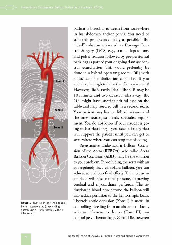
Resuscitative Endovascular Balloon Occlu-sion of the Aorta (REBOA), also called Aorta Balloon Occlusion (ABO), may be the solution to your problem. By occluding the aorta with an appropriately sized compliant balloon, you can achieve several beneficial effects. The increase in afterload will raise central pressure, improving cerebral and myocardium perfusion. The re-duction in blood flow beyond the balloon will also reduce perfusion to the hemorrhagic focus. Thoracic aortic occlusion (Zone I) is useful in controlling bleeding from an abdominal focus, whereas infra-renal occlusion (Zone III) can control pelvic hemorrhage. Zone II lies between
Figure 1: Illustration of Aortic zones. Zone I supra-celiac (descending aorta), Zone II para-viceral, Zone III infra-renal.
Zone I
Zone II
Zone III
79
Resuscitative Endovascular Balloon Occlusion of the Aorta (REBOA)
Top Stent The Art of EndoVascular hybrid Trauma and bleeding Management
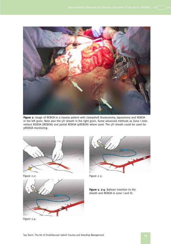
Figure 2: Usage of REBOA in a trauma patient with clampshell thoracotomy, laparotomy and REBOA in the left groin. Note also the 5Fr sheath in the right groin. Some advanced methods as Zone I inter-mittent REBOA (iREBOA) and partial REBOA (pREBOA) where used. The 5Fr sheath could be used for pREBOA monitoring.
Figure 2. 2-4: Balloon insertion to the sheath and REBOA in zone I and III.
Figure 2.2:
Figure 2.4:
Figure 2.3:
80
Resuscitative Endovascular Balloon Occlusion of the Aorta (REBOA)
Top Stent The Art of EndoVascular hybrid Trauma and bleeding Management
these zones and is where the visceral arteries supplying the GI tract, liver and kid-neys originate, which must be considered when doing REBOA.
Although REBOA can be a wonderful tool in the right situation, it is im-portant to note that it might be associated with some potential severe risks. REBOA effectively cuts off the blood supply to the lower body, at the level of either the diaphragm or the pelvis. Every minute, more ischemic “debt” accu-mulates, which will eventually have to be paid back by your patient once the balloon is deflated. Occlusion at or above the visceral arteries (Zone I or II) is tolerated least well, due to the burden of visceral ischemia. In theory, Zone-I occlusion still permits “some” collateral or retrograde perfusion of the ab-dominal viscera, so Zone-II occlusion is discouraged. To mitigate this burden, another option is to use periodic inflation (or intermittent REBOA), which we will discuss later in this section.
Remarks:
» REBOA is a less invasive procedure than thoracotomy, and permits aortic occlusion at different levels and in different ways, depending on clinical need.
» REBOA is a bridge to definitive repair, not a solution!
In general terms, if you do use balloon occlusion, you want to have the shortest time possible. For Zone-I occlusion, under 30 mins is optimal, but over 60 mins is very dangerous, due to ischemic insult and consequent reper-fusion injury. For Zone-III occlusion, up to two-three hours can be tolerated (it has been used for up to 5-6 hours, but this is not recommended), but fewer than two is preferable. Remember, once the balloon is up, it is a race against time to get it down again! If you use Zone-II occlusion, a very short occlusion time of minutes maybe the only way to use it.
Advanced Tips:
» Once familiar with the anatomy of aortic zones, there are several advanced maneu-vers to consider – intermittent and partial inflation. Consider using them if possible.
» REBOA is not an “inflate it and forget it” technique. Inflation may keep your pa-tient alive, but prolonged use beyond what is necessary can cause severe organ damage. It might also migrate!
» Have someone assigned to monitor the balloon and keep its usage limited to the time needed (bridge to surgery). Ask anesthesia to record the inflation time.
81
Resuscitative Endovascular Balloon Occlusion of the Aorta (REBOA)
Top Stent The Art of EndoVascular hybrid Trauma and bleeding Management
Stages of REBOA Deployment
Arterial Access: REBOA is all about access – see Chapter 1&2. An arterial access sheath can also be transduced via the side arm or contralateral CFA to measure invasive blood pressure and provide samples for blood-gas chemistry. Early access in the form of a small sheath can be used for monitoring, with the option of “up-sizing” to a larger access sheath for interventions such as REBOA. Importantly, your access sheath must be of an appropriate size (at least 5Fr) to accommodate whatever you plan to deploy through the sheath. As mentioned above, this is the limiting stage of REBOA, and access in hypo-tensive patients might be challenging!
Advanced Tip:
» In elderly patients with vascular disease, beware of calcified arteries. You can use micro-set to get in, and use ultrasound if possible.
» If you fail, leave the sheath in place for now. Take it out later on, as it might bleed.
» Calling a colleague to assist with REBOA is not a sign of weakness. It is often a sign of sound judgement in challenging cases. Work as a team around the patient with an EVTM mind-set.
» Use both femoral arteries for access, if possible, as the contralateral side can be used for arterial pressure and have a modifying partial REBOA effect. It is easier to insert the sheath before commencing occlusion, so do that expeditiously.
» While you are working to establish femoral artery access for potential REBOA, also consider using the common femoral vein as a site for central venous access and fluid resuscitation – your anesthesiologist will thank you!
Catheters are “rated” according to the sheath size required; e.g., a 14Fr Cook Coda balloon will fit in a 14Fr sheath. Although there are exceptions, like the smaller Coda balloon (30mm, which needs 9Fr). The Medtronic Re-liant balloon catheter can fit in an 11Fr sheath, as can the Boston Scientific Equalizer, but such fittings are “off-label” or outside the “instructions for use” (IFU) for these devices. In addition, when using a narrower sheath for the devices, they become difficult to manipulate, as there is not enough space in the sheath for the device. On occasions, it is useful deliberately to use a larger sheath size to permit easier flushing – a good habit, especially when not anti-coagulating the patient. We should mention that there are some 10Fr REBOA catheters as well as Fogarty balloons in different sizes. The Rescue
82
Resuscitative Endovascular Balloon Occlusion of the Aorta (REBOA)
Top Stent The Art of EndoVascular hybrid Trauma and bleeding Management
Balloon (Tokai, Japan) and ER-REBOA (Pry-time, USA) use a 7Fr introducer and are in early clinical usage; we will discuss them in greater detail below.
The optimal arrangement is to have a “REBOA set” prepared, which includes a sheath, balloon catheter, wire, and a bottle of contrast medium. Some of us have two sets as mentioned before: ACCESS kit and REBOA kit (or a combined set). If not available, then it may be best to postpone this bold maneuver for the next exsanguinating patient. Always have at least 2-3 sets for REBOA, as various items from a set may accidentally fall out of the sterile field during the inevitable con-trolled chaos of serious trauma resuscitation, or the balloon may rupture during insertion. The key elements of access are better outlined in the dedicated “It’s all about vascular access” chapter in this text.
Balloon Selection and Positioning: Your choice of balloon obviously depends upon what you have at hand. The most common types of balloons use a conventional “over-the-wire” system, such as the Cook Coda (14Fr) and the Medtronic Reliant (12Fr) or Equalizer balloon, (14Fr) which are primarily designed for use with aortic stent-grafts. In general, these balloons can inflate to large di-ameters (40 to 46mm), but will fit in any size of “healthy” thoracic and abdominal aorta. As these catheters are designed to be deployed under fluoroscopic guidance, the shafts con-tain no distance markers, which can make
Figure 3 1-3: Some examples of REBOA in place on CT
Figure 3.1
Figure 3.2
Figure 3.3
83
Resuscitative Endovascular Balloon Occlusion of the Aorta (REBOA)
Top Stent The Art of EndoVascular hybrid Trauma and bleeding Management
you a bit nervous if imaging is unavailable. They also do not have clear marking, and a diluted contrast media should preferably be used when you inflate the balloons. In smaller centers where stent-grafting is not a routine procedure, you can find a large angioplas-ty balloon, such as Cordis Maxi LD (12Fr), which can expand to 25mm diameter. The CODA LP balloon is a 9Fr system (30 mm) and can be also used for REBOA. In healthy and young patients, these diameters should be sufficient. Remember that the standard angio-plasty balloon is a high-pressure balloon usu-ally blown up using an inflation device. The device is designed for balloon expansion to reach the desired diameter in the atheroscle-rotic arterial lesions by rupturing the plaques under high pressure. In a normal vessel, it can lead to intimal tear and dissection. This effect should certainly be avoided; hence, if you have only such a high-pressure balloon, in-flate it by hand (it can reach up to 8ATM but not more). While the use of the above-men-tioned balloons in “traumatic hemorrhage” is an extension of their initially conceived usage, several low-profile (7Fr) devices, specifically designed for trauma, are available, and their use is expanding. Low-profile devices have ad-vantages, since they reduce the need for large sheath sizes and their associated complica-tions – plus closure becomes easier. Examples include, as previously mentioned, the Tokai Rescue balloon (RB) and Prytime ER-RE-BOA catheter. The latter is a novel wireless system, which incorporates a wire in the shaft,
Figure 4 1-2: Rescue Balloon by Tokai (With permission).
Figure 5 1-2: ER REBOA by Prytime (With permission).
Figure 4.1
Figure 4.2
Figure 5.1
Figure 5.2
84
Resuscitative Endovascular Balloon Occlusion of the Aorta (REBOA)
Top Stent The Art of EndoVascular hybrid Trauma and bleeding Management
eliminating the need to pre-place the wire. The RB has been in use for some years now in Japan, and also has been used without a wire (but this practice is “off label”/outside the IFU for the device). It has the advantage of possible usage up in the neck vessels and not only the aorta.
The first step is to choose an appropriate wire over which to deploy the balloon. It needs to be stiff enough to support the REBOA catheter, but not so stiff as to risk perforating the patient’s arterial wall. To reduce this risk, a wire should have a tight atraumatic J-tip, as, for example, the Rosen wire. A floppy-tip Amplatz or Lunderquist are other options commonly employed in some centers. The wire also needs to be long enough to permit deployment inside the patient, while having a sufficient length outside on which to mount the REBOA catheter. Finally, it must be of an appropriate diameter to fit the central channel of the catheter; for example, a Coda balloon uses a 0.035inch diameter wire and a Reliant 0.038inch wire, whereas a Rescue balloon uses an 0.025inch wire. Remember, you can use a smaller wire than recommended, but not a larger one!
General Tip:
» Know your equipment before starting to use it!
Introduce the wire into the sheath and advance it slowly. Provided there is no resistance, delivery of the wire can be speeded up. Ideally, the wire should be inserted under fluoroscopic imaging to ensure that it maintains aortic travel and does not enter a side branch. Other options for imaging include the use of plain radiology and ultrasound to assess intraluminal wire position.
It is also helpful to have measured the wire against the patient’s torso in order to get a sense of the length that is required. If you have ultrasonography available in the ER, just place the probe on the abdomen and look at the aorta shadow. Can you see a white line (the wire) in it? Remember that bowel gas and patient body habitus can make seeing the wire in the aorta with ul-trasound a significant challenge. If the images are not clear, do not dwell on getting a perfect picture.
The ideal wire position is in the proximal descending thoracic aorta, which will permit deployment of the balloon catheter in the thoracic aortic or in-fra-renal aorta depending on the desired clinical effect. It is important that the
85
Resuscitative Endovascular Balloon Occlusion of the Aorta (REBOA)
Top Stent The Art of EndoVascular hybrid Trauma and bleeding Management
wire does not migrate proximally too much, as the wire can disrupt the aortic valve, or perforate the left ventricle, or migrate into the carotid or vertebral arteries. Introducing a wire into the arterial system is not a benign procedure. We just mentioned that the wire might perforate other vessels or go directly into the carotid artery, so always use the soft end of the wire first. Once the wire is in situ, the balloon catheter can be delivered over the wire by loading the catheter onto the wire, securing the end (usually done by an assistant) and advancing the catheter into position, using the same method as for the wire. Again, you are using the Seldinger over-the-wire method, as mentioned in the access chapter. If you use the ER-REBOA, no wire is needed and you will push the catheter into the sheath and advance to your target location. That we will discuss now.
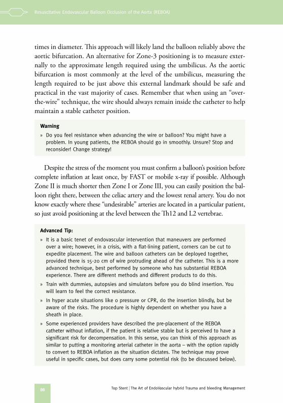
A variety of means adequately to position the REBOA balloon into aortic Zone I have been described and may prove useful. Particularly useful when imaging is not available, you can measure the appropriate distance between the sheath and the patient’s xiphoid process, and then apply a marker (finger or steri-strip) to the shaft of the catheter that represents this distance. In order to position the balloon in aortic Zone III, a rule of “5x6” has been proposed, where you advance approximately 5 cm of a balloon catheter into a sheath 6
Figure 5.3: REBOA insertion in a trauma patient with positive DPL.
86
Resuscitative Endovascular Balloon Occlusion of the Aorta (REBOA)
Top Stent The Art of EndoVascular hybrid Trauma and bleeding Management
times in diameter. This approach will likely land the balloon reliably above the aortic bifurcation. An alternative for Zone-3 positioning is to measure exter-nally to the approximate length required using the umbilicus. As the aortic bifurcation is most commonly at the level of the umbilicus, measuring the length required to be just above this external landmark should be safe and practical in the vast majority of cases. Remember that when using an “over-the-wire” technique, the wire should always remain inside the catheter to help maintain a stable catheter position.
Warning
» Do you feel resistance when advancing the wire or balloon? You might have a problem. In young patients, the REBOA should go in smoothly. Unsure? Stop and reconsider! Change strategy!
Despite the stress of the moment you must confirm a balloon’s position before complete inflation at least once, by FAST or mobile x-ray if possible. Although Zone II is much shorter then Zone I or Zone III, you can easily position the bal-loon right there, between the celiac artery and the lowest renal artery. You do not know exactly where these “undesirable” arteries are located in a particular patient, so just avoid positioning at the level between the Th12 and L2 vertebrae.
Advanced Tip:
» It is a basic tenet of endovascular intervention that maneuvers are performed over a wire; however, in a crisis, with a flat-lining patient, corners can be cut to expedite placement. The wire and balloon catheters can be deployed together, provided there is 15-20 cm of wire protruding ahead of the catheter. This is a more advanced technique, best performed by someone who has substantial REBOA experience. There are different methods and different products to do this.
» Train with dummies, autopsies and simulators before you do blind insertion. You will learn to feel the correct resistance.
» In hyper acute situations like 0 pressure or CPR, do the insertion blindly, but be aware of the risks. The procedure is highly dependent on whether you have a sheath in place.
» Some experienced providers have described the pre-placement of the REBOA catheter without inflation, if the patient is relative stable but is perceived to have a significant risk for decompensation. In this sense, you can think of this approach as similar to putting a monitoring arterial catheter in the aorta – with the option rapidly to convert to REBOA inflation as the situation dictates. The technique may prove useful in specific cases, but does carry some potential risk (to be discussed below).
87
Resuscitative Endovascular Balloon Occlusion of the Aorta (REBOA)
Top Stent The Art of EndoVascular hybrid Trauma and bleeding Management
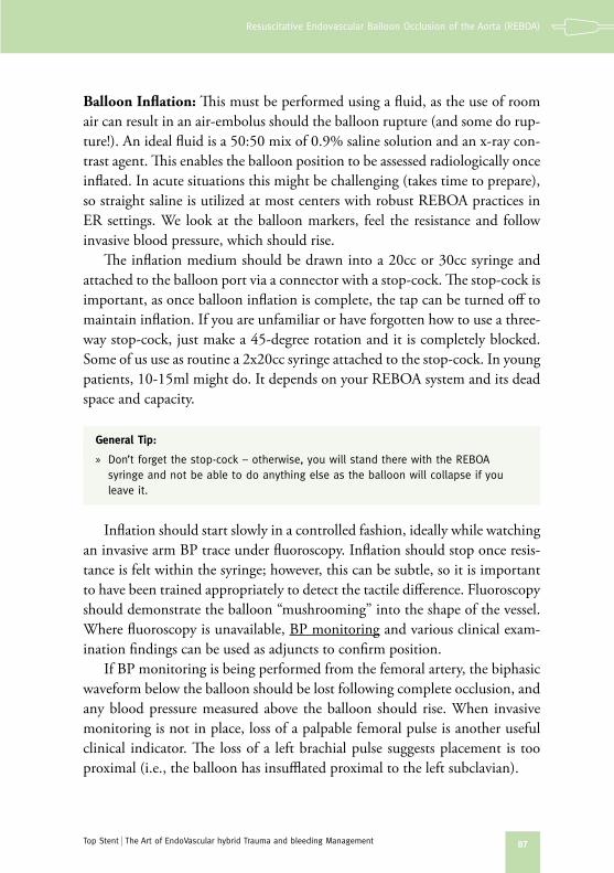
Balloon Inflation: This must be performed using a fluid, as the use of room air can result in an air-embolus should the balloon rupture (and some do rup-ture!). An ideal fluid is a 50:50 mix of 0.9% saline solution and an x-ray con-trast agent. This enables the balloon position to be assessed radiologically once inflated. In acute situations this might be challenging (takes time to prepare), so straight saline is utilized at most centers with robust REBOA practices in ER settings. We look at the balloon markers, feel the resistance and follow invasive blood pressure, which should rise.
The inflation medium should be drawn into a 20cc or 30cc syringe and attached to the balloon port via a connector with a stop-cock. The stop-cock is important, as once balloon inflation is complete, the tap can be turned off to maintain inflation. If you are unfamiliar or have forgotten how to use a three-way stop-cock, just make a 45-degree rotation and it is completely blocked. Some of us use as routine a 2x20cc syringe attached to the stop-cock. In young patients, 10-15ml might do. It depends on your REBOA system and its dead space and capacity.
General Tip:
» Don’t forget the stop-cock – otherwise, you will stand there with the REBOA syringe and not be able to do anything else as the balloon will collapse if you leave it.
Inflation should start slowly in a controlled fashion, ideally while watching an invasive arm BP trace under fluoroscopy. Inflation should stop once resis-tance is felt within the syringe; however, this can be subtle, so it is important to have been trained appropriately to detect the tactile difference. Fluoroscopy should demonstrate the balloon “mushrooming” into the shape of the vessel. Where fluoroscopy is unavailable, BP monitoring and various clinical exam-ination findings can be used as adjuncts to confirm position.
If BP monitoring is being performed from the femoral artery, the biphasic waveform below the balloon should be lost following complete occlusion, and any blood pressure measured above the balloon should rise. When invasive monitoring is not in place, loss of a palpable femoral pulse is another useful clinical indicator. The loss of a left brachial pulse suggests placement is too proximal (i.e., the balloon has insufflated proximal to the left subclavian).
88
Resuscitative Endovascular Balloon Occlusion of the Aorta (REBOA)
Top Stent The Art of EndoVascular hybrid Trauma and bleeding Management
For blind Zone-III occlusion, after in-sertion of at least 30 cm of a catheter into a sheath, inflate a balloon inside the aorta until resistance is felt, withdraw a few centimeters to allow the balloon to move up and down freely, and then pull out the balloon cathe-ter slowly until it presses against the origin of the common iliac artery. You are in the right place now! Just advance the catheter a cou-ple of centimeters back into the aorta, inflate completely, and secure.
Advanced Tip:
» US imaging can also be useful, although body habitus and bowel gas can obscure a good image. For Zone-1 placement, a sub-xiphoid window through the left lobe of the liver can demonstrate the aorta at the level of the diaphragm, and the operator can ob-serve the wire and catheter traveling into the thoracic aorta. For Zone III, a transverse view just above the umbilicus will yield a view of the infra-renal aorta. You might see the bal-loon shadow.
» This technique is highly operator-dependent and should not be undertaken unless you are appropriately trained and experienced. Micro-bubbles or carbon dioxide gas can be used as an inflation medium to improve ultrasound imaging of the balloon, but this depends on the clinical scenario.
Once the balloon is inflated, especially in Zone I, it is imperative that you keep a close eye on the secured catheter as there is a risk of distal migration. Systolic blood pressure above the balloon might suddenly rocket by 50 mm Hg or more. This can push the balloon back,
Figure 6: Holding pREBOA without a stop-cock in a pREBOA case. Note that one hand is on the catheter and sheath together, the other on the syringe, adjusting it to maintain SBP.
Figure 7: Holding a REBOA in place. With built-in stopcock and syringe at-tached. Some of us strongly believe that you should always hold the catheter in your hands, at all times.
89
Resuscitative Endovascular Balloon Occlusion of the Aorta (REBOA)
Top Stent The Art of EndoVascular hybrid Trauma and bleeding Management
bit by bit, especially if a short sheath and/or a soft wire is used. If the balloon is not well secured, in a few seconds the balloon may abut the aortic bifurcation. It is not uncommon to see the shaft of the balloon bend and even dislocate upside-down in the aorta. We would recommend that you or a specifically assigned team member hold the REBOA and sheath and control it at ALL TIMES. After full insufflation of the balloon or during inflation, you can pro-ceed to pREBOA (partial REBOA) as soon as possible, using the proximal systolic blood pressure (above the balloon) as a guide to dictate its effective use. A suggested target systolic blood pressure above the balloon is around 80-90mmHg (maybe a bit higher in cases of suspected brain injury, but this is unknown and based on “gut-feeling”).
General Tip:
» Communicate with your ICU partner, the anesthesiologist and all members of the treatment team during REBOA use – let everyone know when the balloon is in-flated, coming down, going up, etc.
» Hold the REBOA and introducer in your left hand, and the stop-cock/syringe in your right hand to allow total control and modification.
Remember that during transportation eny line can be accidentally withdrawn. Fix the sheath and decide whether to keep holding the REBOA catheter, consid-ering migration and displacement! To avoid this, you should use proper fixing on the REBOA – reliably securing all external parts of the “balloon set”; a silk suture is best for securing both the sheath and the catheter. However, remember to have a spare scalpel and replacement suture at hand in case urgent re-positioning is required. If you can, have one person (or you) holding the REBOA during trans-portation aimed at pREBOA. This will minimize ischemia time.
Balloon Deflation: This can be required for several reasons – to reposition the balloon, to test for bleeding foci inter-operatively, to permit transient reper-fusion, or for final removal. The main rule here is very slow deflation. Don’t panic! Stay calm! The bleeding is controlled and the patient is alive – but fast deflation will inevitably result in circulatory collapse. Withdrawing 1-2cc from the balloon every 30 sec is a reasonable rate of deflation. However, be aware that due to the compliance in the balloon, the last few (2-4) ml will have the greatest effect on the balloon diameter. Do not rush at the end!
90
Resuscitative Endovascular Balloon Occlusion of the Aorta (REBOA)
Top Stent The Art of EndoVascular hybrid Trauma and bleeding Management
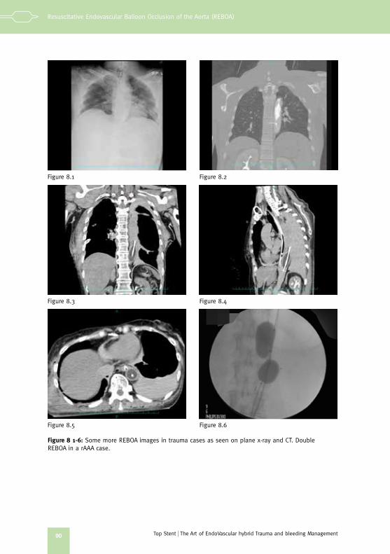
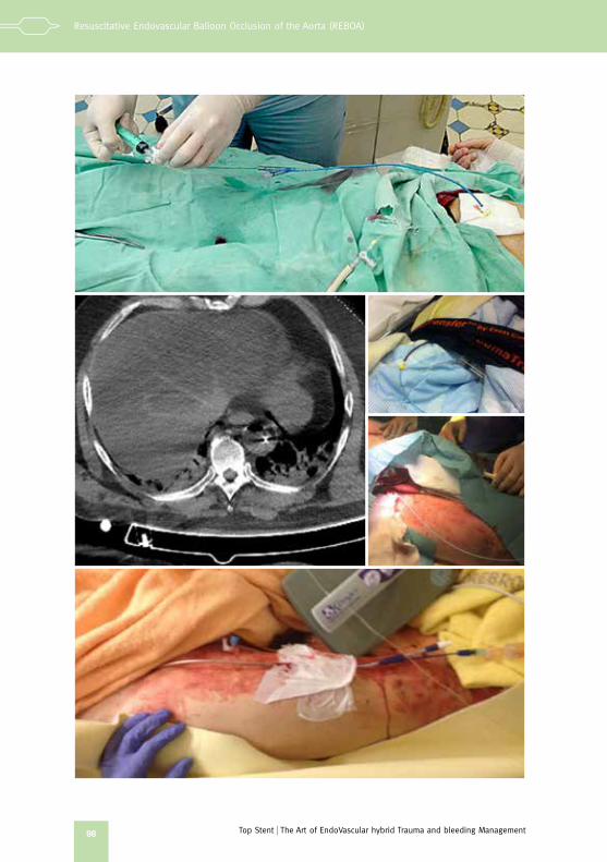
Figure 8 1-6: Some more REBOA images in trauma cases as seen on plane x-ray and CT. Double REBOA in a rAAA case.
Figure 8.1
Figure 8.3
Figure 8.5
Figure 8.2
Figure 8.4
Figure 8.6
91
Resuscitative Endovascular Balloon Occlusion of the Aorta (REBOA)
Top Stent The Art of EndoVascular hybrid Trauma and bleeding Management
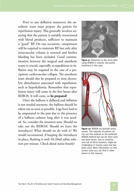
Figure 9: Dissection in the aorta after using REBOA in trauma. Successful treatment in this case.
Figure 10: REBOA via brachial artery access. The majority of authors do not use this method as the preferred REBOA method but can be done (and has been done). Due to anatomy and risk of blind insertion, might be challenging in trauma cases but has been used. More information on this access route you can find in other places in this manual.
Prior to any deflation maneuver, the an-esthetic team must prepare the patient for reperfusion injury. This generally involves en-suring that the patient is suitably resuscitated with blood products, sufficient to maintain a “good” BP. On rare occasions, vasopressors will be required to maintain BP, but only after intravascular volume is restored and further bleeding has been excluded. Good commu-nication between the surgical and anesthetic teams is crucial, especially as expeditious re-in-flation may be required in the case of a pre-cipitous cardiovascular collapse. The anesthetic team should also be prepared to treat electro-lyte disturbances associated with reperfusion, such as hyperkaliemia. Remember that reper-fusion injury will come in the first hours after REBOA. It will come, so be prepared!
Once the balloon is deflated and inflation is not needed anymore, the balloon should be taken out as soon as possible. Legs have had to be amputated in the past due to the presence of a balloon catheter long after it was need-ed. So, consider the situation now. Should we take out the REBOA? Should we leave the introducer? What should we do with it? We would recommend, if keeping the introducer in place, flushing it with 10-20ml saline solu-tion per minute. Check distal status hourly!
92
Resuscitative Endovascular Balloon Occlusion of the Aorta (REBOA)
Top Stent The Art of EndoVascular hybrid Trauma and bleeding Management
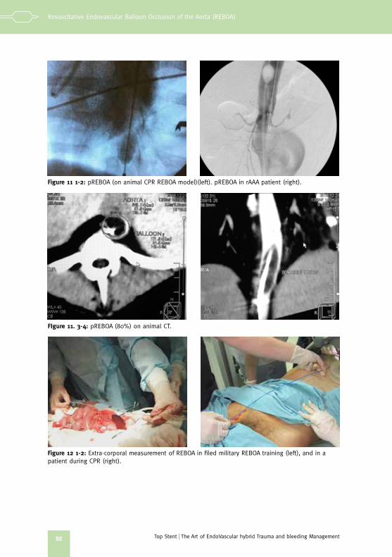
Figure 11 1-2: pREBOA (on animal CPR REBOA model)(left). pREBOA in rAAA patient (right).
FIgure 11. 3-4: pREBOA (80%) on animal CT.
Figure 12 1-2: Extra-corporal measurement of REBOA in filed military REBOA training (left), and in a patient during CPR (right).
93
Resuscitative Endovascular Balloon Occlusion of the Aorta (REBOA)
Top Stent The Art of EndoVascular hybrid Trauma and bleeding Management
Intermittent Occlusion (iREBOA): This is where the balloon is deliberately deflated in order to provide a degree of reperfusion, and to help the surgeon or radiologist locate the focus of bleeding in a controlled manner. The emphasis is on “controlled”, where ideally a hemostatic maneuver is ready to be delivered, and the operator needs the balloon down just to locate the bleeding. Another scenario is when the patient become stable and the REBOA is deflated, but inflated again when the patient becomes unstable during the operative pro-cedure (we have experienced patients with some iREBOAs during surgery).
Figure 13: REBOA in place at the end of surgery (non-trauma). Deflated (dREBOA) at that time.
Advanced Tip:
» Total REBOA (tREBOA) is where the balloon is inflated with out the intention to deflate until hemorrhage control is achieved. There are other options susch as pREBOA or iREBOA, but these can induce cardiovascular instability, so need to be used with caution. Equally, these techniques can help reduce the ischemic insult, so be brave!
94
Resuscitative Endovascular Balloon Occlusion of the Aorta (REBOA)
Top Stent The Art of EndoVascular hybrid Trauma and bleeding Management
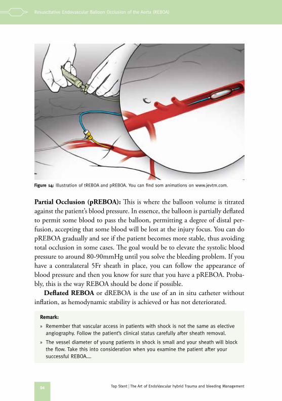
Partial Occlusion (pREBOA): This is where the balloon volume is titrated against the patient’s blood pressure. In essence, the balloon is partially deflated to permit some blood to pass the balloon, permitting a degree of distal per-fusion, accepting that some blood will be lost at the injury focus. You can do pREBOA gradually and see if the patient becomes more stable, thus avoiding total occlusion in some cases. The goal would be to elevate the systolic blood pressure to around 80-90mmHg until you solve the bleeding problem. If you have a contralateral 5Fr sheath in place, you can follow the appearance of blood pressure and then you know for sure that you have a pREBOA. Proba-bly, this is the way REBOA should be done if possible.
Deflated REBOA or dREBOA is the use of an in situ catheter without inflation, as hemodynamic stability is achieved or has not deteriorated.
Remark:
» Remember that vascular access in patients with shock is not the same as elective angiography. Follow the patient’s clinical status carefully after sheath removal.
» The vessel diameter of young patients in shock is small and your sheath will block the flow. Take this into consideration when you examine the patient after your successful REBOA….
Figure 14: Illustration of tREBOA and pREBOA. You can find som animations on www.jevtm.com.
95
Resuscitative Endovascular Balloon Occlusion of the Aorta (REBOA)
Top Stent The Art of EndoVascular hybrid Trauma and bleeding Management
Remark:
» Is the balloon still inflated? Totaly deflated? Remember that you are doing an endovascular procedure and might miss invisible details! You must have balloon control at all times!
Sheath Removal: A large-diameter sheath should be removed after the pro-cedure as it can induce thrombosis and reduce perfusion to the extremities. In a large trauma center you can seek assistance from a vascular surgeon once you have inflated the balloon. The best option for removal of a large sheath (8Fr or greater) is open surgical exploration followed by lateral suture of the femoral artery, or round serosa suture if you are familiar with this. Manual compression is not appropriate in a coagulopathic patient but has been done. You have to know that it will not bleed in the ICU when the patient is warmed up and is covered…
For smaller sheath sizes (7Fr or less), several vascular closure devices are available (e.g., the Abbott Perclose Proglide). At the end of the procedure, the presence of a pedal pulse in the extremity should be confirmed by manual palpation and Doppler ultrasound. US examination might give you some data but you can’t see the flow the whole way. If you are not absolutely certain that distal perfusion is normal, perform angiography or even thrombecto-my directly. Some experienced providers advocate the routine use of extremity angiography after REBOA – particularly soon after your experience with this modality.
Contra-indications
Finally, a word about contra-indications. Be aware that a balloon can also make the situation dramatically worse. The use of REBOA in the setting of chest trauma can theoretically exacerbate bleeding in the chest, neck, upper extremities, and head. It only makes sense if you have a source of arterial bleeding ABOVE the REBOA balloon. Insufflation will increase the pressure and subsequently the speed of hemorrhage. For this reason, known injuries to the heart, aortic arch, as well as to the arterial structures of the neck and lungs, are probably contra-indications for tREBOA. Patients with these injuries might, however, tolerate pREBOA as a lifesaving maneuver in selected situations where other options are not immediately available.
96
Resuscitative Endovascular Balloon Occlusion of the Aorta (REBOA)
Top Stent The Art of EndoVascular hybrid Trauma and bleeding Management
Caution:
» Be aware that REBOA has the potenial to make some injuries worse...
The potential of REBOA dangerously to elevate intracranial pressure in patients with traumatic brain injury is also an important theoretical concern. However, the impact of REBOA use in this setting is largely unknown at pres-ent and remains a matter of active investigation.
A specific catastrophe to be aware of is concerned with the setting of severe chest trauma with multiple-rib fractures (especially, 1st and 2nd ribs), scapula fractures, or a widened mediastinum. REBOA has the potential to convert a stable aortic pseudoaneurysm into an uncontained aortic rupture. For these reasons, bear in mind as a team leader that information is all important. Ob-tain as much knowledge of your patient’s injuries as you can before commit-ting to a major intervention, such as REBOA.
Do not forget the risk of complications, such as femoral and iliac artery dissections (and aorta dissection), thrombus formation (and ischemia), vessel perforation, and bleeding at the insertion site. So, this is a risky business. Be aware!
Remember, REBOA is a “living entity” and NEVER a definitive solution. Respect it and THINK BEFORE USING IT!
Good luck!
97
Resuscitative Endovascular Balloon Occlusion of the Aorta (REBOA)
Top Stent The Art of EndoVascular hybrid Trauma and bleeding Management
REBOA in trauma patients
98
Resuscitative Endovascular Balloon Occlusion of the Aorta (REBOA)
Top Stent The Art of EndoVascular hybrid Trauma and bleeding Management
99
Resuscitative Endovascular Balloon Occlusion of the Aorta (REBOA)
Top Stent The Art of EndoVascular hybrid Trauma and bleeding Management
Notes
100
Resuscitative Endovascular Balloon Occlusion of the Aorta (REBOA)
Top Stent The Art of EndoVascular hybrid Trauma and bleeding Management