To cite: Chan KKW, Tang F, Tham CCY, et al. Retinal vasculature in glaucoma: a review. BMJ Open Ophth 2017;1:e000032. doi:10.1136/bmjophth-2016- 000032 Received 09 August 2016 Revised 13 February 2017 Accepted 20 March 2017 1 Department of Ophthalmology and Visual Sciences, The Chinese University of Hong Kong, Hong Kong, China 2 Department of Ophthalmology and Visual Sciences, Prince of Wales Hospital and Alice Ho Miu Ling Nethersole Hospital, Hong Kong, China Correspondence to Dr Carol Y Cheung, Department of Ophthalmology and Visual Sciences, The Chinese University of Hong Kong Eye Centre, Hong Kong Eye Hospital, 147K Argyle Street, Hong Kong, China; [email protected]Retinal vasculature in glaucoma: a review Karen K W Chan, 1,2 Fangyao Tang, 1 Clement C Y Tham, 1 Alvin L Young, 1,2 Carol Y Cheung 1 ABSTRACT Despite the critical impact of glaucoma on global blindness, its aetiology is not fully characterised. Elevated intraocular pressure is highly associated with glaucomatous optic neuropathy. However, visual field loss still progresses in some patients with normal or even low intraocular pressure. Vascular factors have been suggested to play a role in glaucoma development, based on numerous studies showing associations of glaucoma with blood pressure, ocular perfusion pressure, vasospasm, cardiovascular disease and ocular blood flow. As the retinal vasculature is the only part of the human circulation that readily allows non-invasive visualisation of the microcirculation, a number of quantitative retinal vascular parameters measured from retinal photographs using computer software (eg, calibre, fractal dimension, tortuosity and branching angle) are currently being explored for any association with glaucoma and its progression. Several population-based and clinical studies have reported that changes in retinal vasculature (eg, retinal arteriolar narrowing and decreased fractal dimension) are associated with optic nerve damage and glaucoma, supporting the vascular theory of glaucoma pathogenesis. This review summarises recent findings on the relationships between quantitatively measured structural retinal vascular changes with glaucoma and other markers of optic nerve head damage, including retinal nerve fibre layer thickness. Clinical implications, recent new advances in retinal vascular imaging (eg, optical coherence tomography angiography) and future research directions are also discussed. INTRODUCTION Despite the critical impact of glaucoma on global blindness, its aetiology is not fully characterised. It has been recognised that elevated intraocular pressure (IOP) exerts direct mechanical damage to the optic nerve head (ONH). 1 2 However, among glaucoma patients, only one-third to half have elevated IOP at the initial stages. 3–5 In some, visual field loss continues despite adequate IOP control to normal levels. Consequently, non-IOP-dependent mech- anisms have been proposed. The ‘vascular theory’ of glaucoma hypothesises retinal ganglion cell (RGC) loss as a consequence of insufficient blood supply. 6 7 Vasospasm and autoregulatory dysfunction have been postulated to reduce ocular blood flow. This role is further supported by the association of glaucoma with vascular diseases, such as hypertension and diabetes, 8–10 though discrepancies exist, 11 12 and inclusion as part of the primary vasospastic syndrome following its relationship with Raynaud’s phenomenon, autoimmune diseases and migraine. 13–16 Nevertheless, ongoing discussion over the influence of ocular perfusion pressure (OPP) on glaucoma recognises the inconsistent findings of the influence of diastolic and systolic OPP in the incidence and progression of glaucoma in large epidemiological studies, 5 17–21 which is further complicated by the dynamic relationship between OPP, blood pressure and IOP. 22 Both static and dynamic properties of the retinal microcirculation may be implicated in the vascular phenomenon in glaucoma. Study of the retinal microcirculation is thus made possible by the accessibility of retinal vasculature via non-invasive means. Over the past two decades, semiautomated soft- ware systems have enabled objective and reliable quantification of geometric compo- nents of the retinal vasculature from retinal photography, including retinal vascular calibre, tortuosity, branching angle and fractal dimensions. 23 In effect, multiple studies have linked geometric retinal vascular parameters with vascular diseases including ischaemic heart disease, hyperten- sion, stroke and diabetes. 24–33 In this review, we summarise recent find- ings on the relationships between quantitatively measured structural retinal vascular changes with glaucoma and other markers of ONH damage. We further discuss the recent new advances in retinal vascular imaging (eg, optical coherence tomography angiography) and future research directions. Chan KKW, et al. BMJ Open Ophth 2017;1:e000032. doi:10.1136/bmjophth-2016-000032 1 on 1 June 2018 by guest. Protected by copyright. http://bmjophth.bmj.com/ BMJ Open Ophth: first published as 10.1136/bmjophth-2016-000032 on 11 July 2017. Downloaded from
Transcript
To cite: Chan KKW, Tang F,Tham CCY, et al. Retinalvasculature in glaucoma: areview. BMJ Open Ophth2017;1:e000032.doi:10.1136/bmjophth-2016-000032
Received 09 August 2016Revised 13 February 2017Accepted 20 March 2017
1Department ofOphthalmology and VisualSciences, The ChineseUniversity of Hong Kong,Hong Kong, China2Department ofOphthalmology and VisualSciences, Prince of WalesHospital and Alice Ho MiuLing Nethersole Hospital,Hong Kong, China
Correspondence to
Dr Carol Y Cheung,Department ofOphthalmology and VisualSciences, The ChineseUniversity of Hong Kong EyeCentre, Hong Kong EyeHospital, 147K Argyle Street,Hong Kong, China;[email protected]
Retinal vasculature in glaucoma: areview
Karen K W Chan,1,2 Fangyao Tang,1 Clement C Y Tham,1 Alvin L Young,1,2
Carol Y Cheung1
ABSTRACTDespite the critical impact of glaucoma on globalblindness, its aetiology is not fully characterised.Elevated intraocular pressure is highly associated withglaucomatous optic neuropathy. However, visual fieldloss still progresses in some patients with normal oreven low intraocular pressure. Vascular factors havebeen suggested to play a role in glaucomadevelopment, based on numerous studies showingassociations of glaucoma with blood pressure, ocularperfusion pressure, vasospasm, cardiovascular diseaseand ocular blood flow. As the retinal vasculature is theonly part of the human circulation that readily allowsnon-invasive visualisation of the microcirculation, anumber of quantitative retinal vascular parametersmeasured from retinal photographs using computersoftware (eg, calibre, fractal dimension, tortuosity andbranching angle) are currently being explored for anyassociation with glaucoma and its progression. Severalpopulation-based and clinical studies have reportedthat changes in retinal vasculature (eg, retinal arteriolarnarrowing and decreased fractal dimension) areassociated with optic nerve damage and glaucoma,supporting the vascular theory of glaucomapathogenesis. This review summarises recent findingson the relationships between quantitatively measuredstructural retinal vascular changes with glaucoma andother markers of optic nerve head damage, includingretinal nerve fibre layer thickness. Clinical implications,recent new advances in retinal vascular imaging (eg,optical coherence tomography angiography) and futureresearch directions are also discussed.
INTRODUCTIONDespite the critical impact of glaucoma onglobal blindness, its aetiology is not fullycharacterised. It has been recognised thatelevated intraocular pressure (IOP) exertsdirect mechanical damage to the opticnerve head (ONH).1 2 However, amongglaucoma patients, only one-third to halfhave elevated IOP at the initial stages.3–5 Insome, visual field loss continues despiteadequate IOP control to normal levels.Consequently, non-IOP-dependent mech-
anisms have been proposed. The ‘vasculartheory’ of glaucoma hypothesises retinalganglion cell (RGC) loss as a consequence
of insufficient blood supply.6 7 Vasospasmand autoregulatory dysfunction have beenpostulated to reduce ocular blood flow. Thisrole is further supported by the associationof glaucoma with vascular diseases, such ashypertension and diabetes,8–10 thoughdiscrepancies exist,11 12 and inclusion aspart of the primary vasospastic syndromefollowing its relationship with Raynaud’sphenomenon, autoimmune diseases andmigraine.13–16 Nevertheless, ongoingdiscussion over the influence of ocularperfusion pressure (OPP) on glaucomarecognises the inconsistent findings of theinfluence of diastolic and systolic OPP inthe incidence and progression of glaucomain large epidemiological studies,5 17–21
which is further complicated by thedynamic relationship between OPP, bloodpressure and IOP.22
Both static and dynamic properties of theretinal microcirculation may be implicatedin the vascular phenomenon in glaucoma.Study of the retinal microcirculation is thusmade possible by the accessibility of retinalvasculature via non-invasive means. Overthe past two decades, semiautomated soft-ware systems have enabled objective andreliable quantification of geometric compo-nents of the retinal vasculature from retinalphotography, including retinal vascularcalibre, tortuosity, branching angle andfractal dimensions.23 In effect, multiplestudies have linked geometric retinalvascular parameters with vascular diseasesincluding ischaemic heart disease, hyperten-sion, stroke and diabetes.24–33
In this review, we summarise recent find-ings on the relationships betweenquantitatively measured structural retinalvascular changes with glaucoma and othermarkers of ONH damage. We furtherdiscuss the recent new advances in retinalvascular imaging (eg, optical coherencetomography angiography) and futureresearch directions.
Chan KKW, et al. BMJ Open Ophth 2017;1:e000032. doi:10.1136/bmjophth-2016-000032 1
on 1 June 2018 by guest. Protected by copyright.
http://bmjophth.bm
j.com/
BM
J Open O
phth: first published as 10.1136/bmjophth-2016-000032 on 11 July 2017. D
METHODS AND MATERIALSA comprehensive literature search on PubMed wasperformed for studies published until August 2016with keywords ‘glaucoma’, ‘retinal nerve fibre layerthickness’, ‘geometry’, ‘retinal vascular calibre’, ‘tortu-osity’, ‘branching angle’ and ‘fractal dimensions’.Combinations of these terms were used as well. Searchresults were limited to studies published in English andin human subjects only. Selected papers were thenreviewed thoroughly and evidence was summarised.
OCULAR MICROCIRCULATION IN GLAUCOMAOwing to the increasing recognition of involvement ofvascular phenomena in glaucoma, interest in the pres-ence of retinal microcirculatory changes in glaucomapatients has been raised. Improvement in blood flowand visual field measurements in some eyes followingtreatment with vasodilating calcium channel blockers34
or carbon dioxide inhalation35 present evidence ofvascular autodysregulation. In addition, a recent studyshowed multiple comparable ocular and systemicvascular alterations in the early stages of patients bothwith primary open-angle glaucoma (POAG) andnormal tension glaucoma (NTG), which were not repli-cated in controls.36 The idea that a continuum ofdisturbed circulation exists between the two previously‘distinct’ disease entities is proposed and furtherextends the need for evaluation of vascular properties(eg, ocular blood flow).ONH blood flow is tightly autoregulated to meet
the functional and metabolic demands of the retina,
including RGC. Technological advancements havemade visualisation, direct measurements and quantifi-cation of in- vivo ocular blood flow possible, thougha gold standard that provides all the relevant infor-mation in one reading has yet to be established.Current modes of analysis of this dynamic parameterinclude, but are not limited to, angiography, laserDoppler techniques, Heidelberg Retina Flowmeter,laser speckle phenomenon and retinal vesselanalyser.37 38 MRI can provide not only dynamicblood flow measurement within deep orbital struc-tures but also a non-invasive measurement ofintracranial structures.39 Nevertheless, the vast varietyof instruments create difficulty for data unification,though a consistent demonstration of decreasedaverage blood flow in some glaucoma patients wasfound in the retinal,40 41 ONH42 43 and choroidal44
circulations.Murray’s Principle of Minimum Work established
that the vascular network conforms to an ‘optimally’designed topographical geometry.45 This minimisesshear stress and work across vascular network andallows sufficient blood distribution to tissue with theleast amount of energy. As blood flow is a function ofcardiac output and regulated by relative local resis-tance, deviations to ideal structure and function of themicrocirculation will lead to reduced efficiency andimpaired circulatory transport.46 In view of the chal-lenges in dynamic analysis, interest has turned in thedirection of the vascular network’s static components,
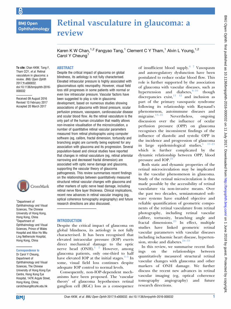
Figure 1 Quantitative measurement of retinal vasculature from retinal fundus photograph using a computer-assisted program
(Singapore I Vessel Assessment (SIVA)).
2 Chan KKW, et al. BMJ Open Ophth 2017;1:e000032. doi:10.1136/bmjophth-2016-000032
Open Access
on 1 June 2018 by guest. Protected by copyright.
http://bmjophth.bm
j.com/
BM
J Open O
phth: first published as 10.1136/bmjophth-2016-000032 on 11 July 2017. D
including its design, since they reflect resistance toocular blood flow and affect function.
QUANTITATIVE MEASUREMENTS OF RETINALVASCULATUREWith the introduction of modern digitalised retinalphotography, semiautomated computer-assistedprogrammes have been developed to objectively andreliably quantify subtle retinal vascular changes fromretinal photographs, with focus on calibre measure-ment. Optimate (Department of Ophthalmology andVisual Science, University of Wisconsin—Madison) andIVAN (Department of Ophthalmology and VisualScience, University of Wisconsin—Madison) softwaresanalyse digitalised retinal photographs and measurethe retinal vessel widths.47 48 With the development ofdigital retinal photography, newer programs such asSingapore I Vessel Assessment (SIVA)25 and VesselAssessment and Measurement Platform for Images ofthe REtina (VAMPIRE)49 softwares have evolved toevaluate novel classes of retinal vascular geometricparameters, including tortuosity, fractal dimension andbranching angle, providing comprehensive assessmentof retinal vasculature (figure 1). Such developmentprovides an accessible, non-invasive model to studycorrelations and consequences of microvasculardysfunction in both systemic and ocular diseases.50–52
For example, systemic review has confirmed that widerretinal venular calibres predict stroke,53 and meta-anal-ysis showed independent associations between widerretinal venules and narrower arterioles with increasedrisk for cardiovascular events in women.54
Retinal vascular calibreRetinal vascular calibre is measured in terms of centralretinal artery equivalent (CRAE), central retinal veinequivalent (CRVE) and arteriovenous ratio (AVR).47 48
CRAE is a summary index reflecting the average widthof retinal arterioles, and CRVE is a summary indexreflecting the average width of retinal venules. It hasbeen recognised that CRAE and CRVE should beanalysed independently, as they reflect distinct systemicvascular disease pathways. AVR is a dimensionless ratiothat is used to compensate for magnification differ-ences and refractive error, and its value is non-specificto changes in arterioles, venules or both.
Retinal vascular tortuosityRetinal vascular tortuosity reflects vessel curvature andis summarised as the ratio between the actual distancea vessel travels from points A to B and the shorteststraight-line distance between points A and B.55 Alarger tortuosity index indicates more curves in aretinal vessel. Retinal vascular tortuosity can becomputed as the integral of the curvature square alongthe path of the vessel, normalised by the total path
length.24 Since this measure is represented as a ratio,its value is dimensionless.56
Retinal vascular bifurcation angleRetinal vascular bifurcation angle is defined as the firstangle subtended between two daughter vessels at avascular junction. Both retinal arteriolar branchingangle and retinal venular branching angle could bederived, and they represent the average branchingangle of arterioles and venules, respectively.57
Fractal dimensionFractal dimension describes how thoroughly a patternfills two-dimensional spaces and represents a ‘global’measure that summarises the whole branching patternof the retinal vascular tree.58 59 It is calculated from askeletonised line tracing using a box-counting method.Larger values indicate a more complex branchingpattern.
RETINAL VASCULAR CHANGES ASSOCIATED WITHGLAUCOMAGeneralised narrowing of the retinal vessels is charac-teristic of advanced glaucomatous optic nervedamage.60 A number of epidemiological studies haveshown association between retinal vascular changes,particularly narrowing in retinal vascular calibre, withglaucoma. Table 1 presents the associations betweenquantitative retinal vascular parameters with glaucomain population-based and hospital-based cross-sectionalstudies.Prior to availability of semiautomated machines,
retinal vessel calibres in eyes with glaucoma wereexplored via manual means.60–62 Evidence ofdecreasing retinal vessel calibre with increasing glau-coma stage was demonstrated, with strongercorrelation for arteries than veins. Quadrants withgreater ONH damage corresponded with narrowerretinal arteries. Regarding this association, two schoolsof explanations have been postulated. On the onehand, RGC loss has been suggested to lead to vasocon-striction as an adjustment to decreased metabolicneeds. This is in line with the observation of retinalarterial narrowings in eyes with non-glaucomatousoptic atrophy.62–64 Alternatively, the underlying patho-logical process leading to RGC loss has been proposedto be related to impaired local autoregulation, vasoac-tive substance leakage and consequentlyvasoconstriction.65 On the molecular level, this issupported by elevated biomarkers of oxidative stress inaqueous humour, serum and trabecular meshworksamples of glaucoma patients.66 67 Reactive free radi-cals scavenge nitrous oxide, an innate vasodilatorsecreted by smooth muscles that alter vascular tone.Short posterior ciliary artery (SPCA), in particular, hasbeen found to exhibit transient vasospasm on radicalexposure in in-vitro models,68 and reduced SPCA
Chan KKW, et al. BMJ Open Ophth 2017;1:e000032. doi:10.1136/bmjophth-2016-000032 3
Open Access
on 1 June 2018 by guest. Protected by copyright.
http://bmjophth.bm
j.com/
BM
J Open O
phth: first published as 10.1136/bmjophth-2016-000032 on 11 July 2017. D
blood flow velocities were associated with glaucomaprogression.69 Altered systemic vasoreactivity withendothelial cell dysfunction was also confirmed in NTGpatients,70 71 while population-based trials havedemonstrated lower diastolic perfusion pressure, ameasure of ocular blood flow, as a significant factor inthe glaucoma incidence.5 17 19 However, objectiveevidence for underlying mechanisms have yet to befurther clarified in the future.Though these studies were limited by use of manual,
subjective methods in measurement of retinal vesseldiameters, their results were consistent with recentfindings employing computer-assisted programs. DeLeon et al investigated intereye differences in retinalvascular calibre in persons with asymmetrical glaucomausing the IVAN system.72 Once again, CRAE andCRVE were narrower for eyes with more severedisease. This relationship held after adjustment forage, gender, vascular risk factors and IOP, suggestingthe difference in calibre to be due to severity discrep-ancy or other unknown factors, instead of systemicvascular diseases. Similarly, using the IVAN system,Yoo et al
73 analysed CRAE of glaucomatous suspectswho showed unilateral glaucomatous conversion andnoted narrower CRAE at baseline and at the point ofglaucoma conversion. Angelica et al
74 dismissed the
usage of retinal vessel calibre as a predictor in glau-coma in a hospital-based cross-sectional study, as nosignificant association could be drawn, though noexplanation was given.Population-based studies have further supported the
above findings. The Blue Mountains Eye Study (BMES)showed that eyes with POAG were 2.7 times morelikely to have generalised retinal arteriolar narrowingthan eyes without glaucoma.75 This remained true afteradjusting for risk factors for glaucoma and is indepen-dent of IOP and OPP. The Singapore Malay Eye Studyfound consistent association of quantitatively measuredretinal vascular calibre with prevalence of glaucomaand larger vertical cup–disc ratio (CDR).76 The BeijingEye Study showed significantly thinner retinal arteriesbut insignificant difference in retinal vein diameters.77
In the Handan Eye Study, both narrower retinal arte-rioles and venules were observed in primary angleclosure glaucoma and POAG than those in normalcontrols, primary angle closure or primary angleclosure suspect,78 suggesting that the narrowing ofretinal vessels resulting from the glaucoma process isirrespective of status of angle closure. More recently,Yoo et al reported similar findings of retinal arteriolarnarrowing in glaucoma, and further found that thediagnostic ability of retinal arteriolar calibre was
Table 1 Continued
Study and
year Study type
Sample
size
Method of
assessment
Changes in parameters in association with glaucoma
comparable to retinal nerve fibre layer (RNFL) thick-ness in detecting OAG, which is an optimisticintroduction to its potential use in clinical settings.79
Nevertheless, the Beaver Dam Eye Study, a Caucasianpopulation-based cohort study, did not find any associ-ations of retinal vascular calibre related to prevalentglaucoma, large cup-to-disk ratio or elevated IOP.80
The authors attributed this deviation of their findingsto the difference in the methodology of selection ofarterioles for evaluation. A number of previous studieshave focused solely on peripapillary vessel calibres62 74
however, Klein et al excluded peripapillaryvessels because of the variability in retinal nerve fibrelayer thickness in this area.Overall, population-based and hospital-based cross-
sectional studies largely supported the association ofnarrower vessel calibre with glaucoma, though indi-vidual studies focused on CRAE alone or only foundsignificant reduction in CRAE and not CRVE.Owing to the relatively new availability of technology
in advanced geometry measurements, only two studieshave evaluated retinal vascular geometric parametersother than calibre size. In a hospital-based study, Cian-caglini81 et al found correlation between ONH damagewith a reduced retinal vascular fractal dimension. TheSingapore Malay Eye Study also had a consistentfinding of lower retinal vascular fractal dimension inglaucoma.82 In this study, Wu et al also evaluated vesseltortuosity and branching angle, and noted significantlysmaller vessel tortuosity and retinal venular branchingangle in eyes with glaucoma. Taken together, thesefindings suggest that circulatory optimality of vessels inglaucoma eyes may be compromised due to provenchanges in the design of the geometrical pattern.However due to the cross-sectional nature of data,information on the temporality of retinal vascularchanges with glaucoma incidence is limited.
RETINAL VASCULAR CHANGES WITH GLAUCOMA-ASSOCI-ATED OUTCOMESReduced RNFL thickness, greater CDR and character-istic visual field defects are hallmarks of glaucomatousoptic neuropathy. Table 2 summarises cross-sectionalstudies that defined the relationship between retinalvascular parameters with these glaucoma-associatedoutcomes.The correlation between narrower retinal vessel
calibre and thinner RNFL thickness has been consis-tent since the 1980s.83–85 Studies analysed includedhospital-based or population-based cross-sectionaldata, measurements carried out by manual means orcomputer programs, and populations of children,adolescents and adults. Although the biological mecha-nisms remain uncertain, these findings support thehypothesis that the loss of RGCs in thinned RNFLlowers metabolic and vascular demands, leading tonarrower vascular calibre as part of an autoregulatoryresponse.65 86–91 This is supported by a similar finding
of decreased vessel diameter in non-glaucomatousoptic neuropathies such as non-arteritic ischaemicoptic neuropathy and descending optic nerveatrophy.63 Regardless, the temporal relationship ofwhether peripapillary vessel narrowing causes damageto the optic nerve, or the reverse, is true, has yet to bedemonstrated definitively.Discrepancy in the strength of association between
arterioles and venules with RNFL was noted. TheSingapore Malay Eye Study noted stronger associationin venules than arterioles,65 87 92 while Kim et al
89 onlyassociated RNFL thickness with arteriolar calibre, butnot venular. The contrasting findings may beexplained by the complex interaction between variousmediators for vasodilatation and vasoconstriction onarterioles and venules. Retinal venular calibre is morestrongly influenced by diabetes mellitus, while arteri-olar calibre is more related to hypertension.89 It hasalso been proposed that narrower venular calibre mayindicate venous congestion and cytotoxic damage, withsubsequent secondary constriction of arteriole.93–96
The different spectrum of baseline systemic diseases instudies may therefore contribute to the discrepancy infindings. Nevertheless, compatible association betweenthinner RNFL thickness with narrowed calibre inhealthy children and adolescents indicate that the rela-tionship in adults with pathological eyes are at least inpart physiological in origin.91
Apoptosis of RGCs lead to increased CDR, which is apathognomonic feature of glaucoma. Studies have beeninconsistent in demonstrating its relationship withvessel calibre.88 90 91 Lim et al
90 described the associa-tion between narrower retinal venular diameter withCDR, which was lacking for arteriolar calibre. This wasattributed to retinal veins’ lower resistance to deforma-tion due to their non-existent tunica media.90
Nevertheless, while increase in CDR is a clinical indi-cator for glaucoma progression, the reliability of CDRto detect glaucoma is limited by the wide variability incup sizes, and interobserver and intraobserver vari-ability. Poor correlation between RGC counts and CDRhas also been demonstrated, suggesting that CDR is aninsensitive method for evaluation of glaucomatousstructural damage.97
Consistency is seen for the correlation between arte-riole calibre with visual field defect. Hall et al
compared calibre in POAG patients with markeddifference in visual field defects between hemifields,and found significant correlation between arteriolarcalibre with visual field defect.92 Similarly, Jonas andNaumann86 correlated visual field defects with botharteriole and venule calibres. Koh et al was the onlystudy that evaluated vessel tortuosity and correlateddecreased tortuosity with a thinner neuroretinal rim,which was more significant in arterioles.98 This was inline with studies that linked straighter retinal vesselswith ischaemic heart disease and higher bloodpressure99.
Chan KKW, et al. BMJ Open Ophth 2017;1:e000032. doi:10.1136/bmjophth-2016-000032 7
Open Access
on 1 June 2018 by guest. Protected by copyright.
http://bmjophth.bm
j.com/
BM
J Open O
phth: first published as 10.1136/bmjophth-2016-000032 on 11 July 2017. D
LONGITUDINAL RELATIONSHIP BETWEEN RETINALVASCULAR CHANGES WITH GLAUCOMAProspective studies provide information on the causa-tive relationship between the parameters in questionand glaucoma. This is relevant in determining whethervascular dysfunction preceded development of glau-coma or is a consequence of optic neuropathyprogression. Table 3 lists longitudinal studies that eval-uated the relationship between vascular geometry withthe incidence or progression of glaucoma.Two studies evaluated glaucoma incidence. In an
urban Caucasian population, 10-year follow-up datafrom the BMES revealed that narrower retinal arte-rioles were associated with higher OAG incidence, andsuggest the potential use of retinal vessel calibre toidentify patients with increased risk for glaucomadevelopment.100 This finding supports previous cross-sectional studies’ concept that vascular changes areinvolved in the early course or pathogenesis of glau-coma. However, the Rotterdam Study, anotherCaucasian population-based study of 6.5 years offollow-up, had contradicting results.101 Both retinalarteriolar and venular baseline diameters were notfound to be associated with incident OAG and incidentoptic disc changes. The discrepancy in findings may be
due to the difference in duration of follow-up andhigher incidence of POAG in BMES. Moreover, due tothe elderly skewed cohort, the Rotterdam Study had asubstantial number of participants (n=838) who passedaway during the follow-up.Progression of glaucoma was evaluated in two
prospective studies. Papastathopoulos and Jonasperformed a minimum 8-month follow-up for a groupof patients with progressive glaucomatous optic nervedamage and noted significant focal narrowing ofretinal arterioles associated with neuroretinal rimloss.64 This was not found in patients with static opticdiscs. Retinal venules were not analysed. Nevertheless,the authors concluded that focal narrowing does notnecessarily involve progression of glaucoma, and is notpathognomonic for any particular subtype.More recently, Lee et al compared 27 eyes with bilat-
eral NTG who showed asymmetrical glaucomaprogression after a mean follow-up of 24.3 months andfound significant narrowing of retinal arteriolar calibrein progressed eyes but not in contralateral stableeyes.102 No correlation was found for retinal venularcalibre, however, they hypothesised this may be due toclinically asymptomatic engorgement of venous bloodflow in glaucoma, together with different regulatory
Table 3 Relationship between vascular geometry with the incidence or progression of glaucoma
Study and year Study type
Follow-
up
duration
Sample
size
Method of
assessment Outcome
Changes in
parameters in
association with
glaucoma
Arteriolar
calibre
Venular
calibre
Lee et al102
(2014)
Hospital-based,
prospective
study
24.3
months
NTG:
27
IVAN Progression
of glaucoma:
27
Reduced Not
significant
Kawasaki et al100
(The Blue
Mountains Eye
Study)
(2013)
Population-
based,
prospective
study
10 years Total:
2417
Optimate Incidence of
glaucoma: 82
Reduced Not
significant
Ikram et al101
(Rotterdam Eye
Study)
(2005)
Population-
based,
prospective
study
6.5
years
Total:
3464
Optimate Incidence of
glaucoma: 74
Not
significant
Not
significant
Papastathopoulos
& Jonas64
(1999)
Hospital-based,
prospective
study
37
months
OHT:
31
POAG:
59
NTG:
22
SOAG:
11
Colour stereo
optic disc
photographs
Progression
of glaucoma:
37
Reduced –
CRAE, central retinal artery equivalent; CRVE, central retinal vein equivalent; NTG, normal tension glaucoma; OHT, ocular hypertension;
mechanisms governing changes in retinal artery andvein diameters.102 No significant intereye differencewas observed in the mean baseline vessel calibrebetween progressed and stable eyes.
DYNAMIC RETINAL VASCULAR CHANGES WITH GLAUCOMAThe vascular theory of glaucoma considers optic nervedamage as a consequence of insufficient blood supplydue to either increased IOP or other dysregulatoryfactors reducing ocular blood flow. Thus apart fromassociating structural vessel properties with glaucoma,functional performance reflects abnormalities anddysregulations in pathogenic eyes. Technologicaladvancements have allowed quantitative evaluation ofocular blood flow and perfusion, and could serve as animaging target for early diagnosis and monitoring ofglaucoma.ONH blood flow could be determined from simulta-
neous measurements of the blood column diameterand the centreline blood speed. Scanning laserDoppler flowmetry with automated perfusion imaginganalysis evaluates frequency shift of perfused vesselsand capillaries. Vessels are identified, segmented, andvelocity then derived from the rate of flow shift. Laserspeckle flowgraphy also calculates the speckle patternthat arises from the scatter of the laser irradiation froman illuminated fundus. Changes in the velocity of theblood flow blur the speckle pattern and the mean blurrate is then derived. Both methods have shownreduced ONH and peripapillary blood flow dynamicsin glaucoma.103–105 Diminished flow in POAG suspecteyes before the development of clinically detectablevisual field loss was confirmed as well.106 107 However,laser Doppler flowmetry only evaluates a small area ofthe retina, while absorbance and reflectance of disctissue limits repeatability of laser speckle flowgraphy.Ocular perfusion, another reflection of ocular blood
flow, can be estimated by retinal arteriovenous passagetime via digital scanning laser fluorescein angiograms.It characterises the passage of blood from the retinalartery, through capillaries, to the retinal vein.Prolonged passage time has been found to be reducedin both NTG and POAG patients,108–110 which wasattributed to reduction of the capillary diameter poten-tially due to vasospasms or arteriosclerosis.Nevertheless, routine usage of fluorescein angiography(FA) is limited by its invasiveness, difficulty in accuratequantification and potential adverse systemic effects.
LIMITATIONS OF THE CURRENT STUDIES AND FUTUREDIRECTIONSDespite the promising potential of retinal vascularimaging in glaucoma, there are still gaps in translatingresearch into clinical practice. A shortcoming is thelack of knowledge about the normative data and refer-ence levels for measurement. The majority of clinicaltrials compared pathological eyes with healthy eyes and
derived conclusions based on comparison. Baselinereference values have yet to be concluded, which isfurther complicated by the influence of systemic,genetic and environmental factors on the variations ofretinal vascular calibre size.111 Widespread implemen-tation is also limited by availability of expertise.Current software is not fully automated and willrequire input from trained technicians to operatestandardised protocols and provide expert manipula-tion and handling of specialised computer software. Inaddition, most studies in the current review did notspecify subtypes in the associations, though the rela-tionship between altered structural parameters withglaucoma held irrespective of IOP or angle closure.This may support the idea that vascular mechanismsunderlie all subtypes of POAG. Further work couldfocus on elucidating differences in vessels in normaland high pressure glaucoma. Another limitation in theevaluation of retinal vascular calibre lies in its multifac-torial influence by other systemic and individualcharacteristics. While most studies have taken intoaccount patients’ age, gender, systemic vasculardiseases and IOP, other variabilities, such as caffeineconsumption and smoking habits, have not beenconsidered.111 112 Hao et al reported significantchanges in individual vessel calibre over a cardiac cyclebut not in vessel calibre summaries (including CRAEand CRVE) and geometric measures, suggesting a mildcorrelation of pulse cycle and vessel diameter that needto be taken into account during sampling.113 In addi-tion, although multiple advanced analytic tools enablequantification of retinal vascular imaging, there aretechnological challenges that may compromise preci-sion. Refractive errors and axial length variabilitiescause discrepancies in magnification, while imagedisplay quality (contrast, brightness and focus) may becompromised by media opacities and pupil size.23 Thelack of automated imaging of retinal vascular also leadsto unavoidable intragrader and intergrader variabilitythat has yet to be refined.114
Future directions include focused analysis of thechronological nature of retinal vessel changes viameans of longitudinal studies so as to better delineatethe chicken–egg relationship between glaucomatouschanges and narrowed vessel calibre. Detailed subtypeanalysis is also warranted to delineate whether thevascular phenomenon is more profound in NTG eyes,as conventionally believed, or actually exists as a spec-trum among all glaucoma subtypes. Effort in clinicalapplication of current data and ease of software use indaily practice should also be explored to close the gapbetween clinical and experimental investigations.
NEW ADVANCES IN RETINAL IMAGINGAdvances in technology have attempted to supplementthe shortcomings of existing instruments. Peripapillarycapillaries have been recognised to be a highly special-ised vasculature that supply the nerve fibre layer,115
Chan KKW, et al. BMJ Open Ophth 2017;1:e000032. doi:10.1136/bmjophth-2016-000032 9
Open Access
on 1 June 2018 by guest. Protected by copyright.
http://bmjophth.bm
j.com/
BM
J Open O
phth: first published as 10.1136/bmjophth-2016-000032 on 11 July 2017. D
and a better understanding of this network may reflectfocal or contiguous disc capillary network defects or actas a supplementary indicator of RGC damage.FA is the gold standard for imaging the capillary
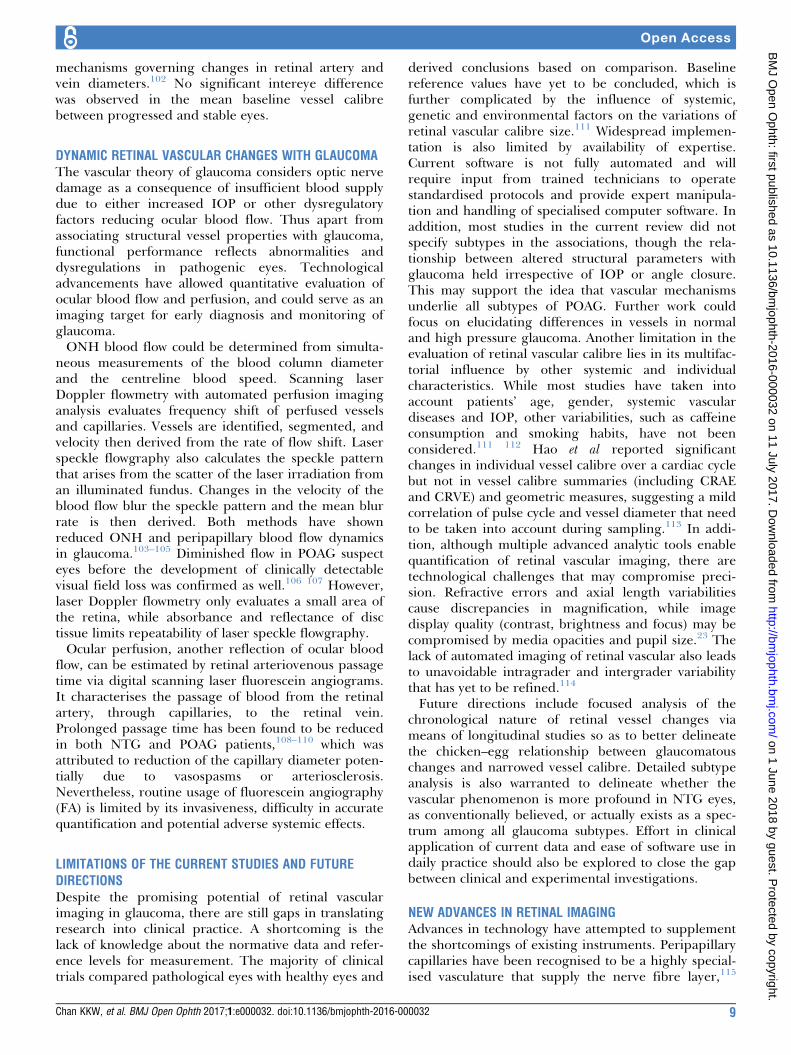
network. However, it is invasive in operation(requiring intravenous injection of fluorescein dye),time consuming, confounded by superimposition ofcapillaries from different retinal layers and onlyoffers two-dimensional image analysis with lack ofquantifiable parameters. All of the shortages abovereduce the clinical utility of FA. Optical coherencetomography—angiography (OCT-A) offers three-dimensional, non-invasive retinal and choroidalmicrocirculation vasculature analysis and blood flowestimation116 117 (figure 2). It is based on mappingerythrocyte movement over time by comparingsequential optical coherence tomography-B scan(OCT-B scan) ultrasounds images at a given cross-section. OCT-A is able to separately detect the super-ficial capillary network in the ganglion cell layer, thedeep capillary network in the outer plexiform layerand choriocapillaris below retinal pigment epitheliumwithout intravenous dye injection, providing depth-resolved visualisation of the retinal and choroidvasculature and blood flow. Moreover, OCT-A cangenerate data on vascular flow to quantify retinal oroptic disc perfusion, independent of time and dyeinjection. As OCT-angiograms are coregistered withOCT-B scans from the same area, it also allows forsimultaneous visualisation of structure and blood flow
for clinical interpretation. Recently, data derivedfrom OCT-A readings have shown that peripapillaryvessel density, peripapillary flow index and optic discperfusion are reduced in glaucomatous eyescompared with aged-matched normal eyes.118–122
These changes correlated to disease severity, struc-tural changes and functional damages, includingRNFL thickness, visual field mean deviation, visualfield pattern SD and visual field index. In addition,OCT-A indices have outperformed RNFL thicknessin having a stronger correlation with visual fieldloss.117 118 123 These findings support the notionthat OCT-A is a promising and useful imagingmodality for evaluating glaucomatous microvasculop-athy, which may allow earlier diagnosis and detectionof nerve fibre functional loss before thinning occurs.Compared with FA, OCT-A also offers superiordetails in analysing radial peripapillary capillaries,which is a unique plexus within the inner nerveRNFL that provides nutritional support to theRGCs.124 Reduction in the network’s density hasbeen strongly correlated with thinner RNFL thicknessand poorer visual field index.125 Compared withvessel measurements based on digital photography,which is more appropriate for large vessels with lesssensitivity, studies that utilised OCT-A allowed moreaccurate measurement of the low velocities of deepplexuses. Furthermore, since OCT-A is a depth-resolved technique, it offers technical advantage inthe growing interest of investigating the deep layer
Figure 2 Assessment of retinal capillary network around optic nerve head using optical coherence tomography angiography
in a normal eye (A–C) and a glaucomatous eye (D–F). Decreased peripapillary capillary density is indicated by blue arrows.
10 Chan KKW, et al. BMJ Open Ophth 2017;1:e000032. doi:10.1136/bmjophth-2016-000032
Open Access
on 1 June 2018 by guest. Protected by copyright.
http://bmjophth.bm
j.com/
BM
J Open O
phth: first published as 10.1136/bmjophth-2016-000032 on 11 July 2017. D
microvasculature. Recently, with OCT-A imaging, Suhet al
126 reported that decreased deep-layer vesseldensity within the parapapillary area, which is down-stream from the SPCA perfused deep ONH, isassociated with lamina cribrosa defect, visual fieldimpairment and RNFL thinning. This finding maysupport the microvascular pathophysiology conceptsof glaucoma, since the superficial and deep retinallayers are perfused individually by the central retinaland short posterior ciliary arteries, respectively.127
However, OCT-A has several limitations. First,limited by the current scanning speed and patientcomfort during the acquisition, a 6�6mm2 area isthe largest scanning field that can be provided bythe most updated OCT-A imaging device. This maybe suboptimal for peripheral retinal vasculature.Second, data on validity of OCT-A assessment, suchas intersection or intrasection reliability, compara-bility with gold standard and correlation with clinicaloutcomes is still scarce. Third, despite modern tech-nology, automated, objective and robust methodsthat have evidence-based proof of accuracy for vascu-lature identification for quantitative assessment ofcapillary perfusion are still lacking.128 129 In addi-tion, image artefacts are common in OCT-A,especially motion and projection artefacts, leading toinaccurate assessment.130 Advanced softwares toneutralise artefacts while maintaining adequate inten-sity and visibility of pathological vascular changes arerequired,125 while media opacities and segmentationerrors should be taken into account as factors thatinfluence OCT-A interpretation.Retinal functional imaging is another method to
obtain blood flow velocity by comparing erythrocytemovement in serial retinal images. Elevated meanretinal blood flow velocity was found in peripapillaryvasculature,131 which may reflect a steal phenomenonwhen retinal vessels experience increased flow followingdecreased retinal capillary perfusion. Increased venousvelocity has been consistently found in eyes withpreperimetric glaucomatous optic neuropathy, whichmay reflect early vascular dysfunction.132 Imagingsystems that employ adaptive optics, such as retinalfundus camera, OCT and scanning laser ophthalmo-scope, have also provided in-vivo, high-resolutionimaging of the vasculature and nerve fibre layer thatovercomes poor lateral resolution in conventionalocular optics,133 and have been shown to be in preciseagreement with histology in primate studies.134
CONCLUSIONThis review offers solid, consistent evidence for proofof concept that structural designs and changes in theretinal vasculature are associated with glaucoma. Mostcross-sectional studies support the association betweennarrowed vessel calibre with glaucoma and glaucoma-associated outcomes, including thinner RNFL,increased CDR and thinner neuroretinal rim area.
Specific vessel patterns, including reduced fractaldimension, tortuosity and branching angle, have alsobeen largely associated with glaucoma in hospital-based and population-based studies, though evidenceis scarce. Further meta-analysis or pooled analysiscould quantitatively evaluate their consistency. Longitu-dinal data bear weight in elucidating the temporalassociation of these findings with the incidence orprogression of glaucoma. However, the small numberof related studies limits the significance of theevidence, particularly when conclusions are in contra-diction. More prospective, long-term follow-up dataare needed.New retinal imaging techniques confirm the patho-
genetic concept of vascular dysregulation inglaucoma eyes, especially with NTG. Clear differ-ences when compared with controls aredemonstrated. Their potential usefulness in the diag-nosis, staging and monitoring of glaucoma isrecognised, and their function as a future imagingtarget should be utilised.
Contributors All authors contributed substantial information or material in thissubmission for publication.
Competing interests None declared.
Provenance and peer review Not commissioned; externally peer reviewed.
Open Access This is an Open Access article distributed in accordance withthe Creative Commons Attribution Non Commercial (CC BY-NC 4.0) license,which permits others to distribute, remix, adapt, build upon this work non-commercially, and license their derivative works on different terms, providedthe original work is properly cited and the use is non-commercial. See: http://creativecommons.org/licenses/by-nc/4.0/
1 Kass MA, Heuer DK, Higginbotham EJ, et al. TheOcular Hypertension Treatment Study: a randomized trialdetermines that topical ocular hypotensive medication delays orprevents the onset of primary open-angle glaucoma. ArchOphthalmol 2002;120:701–13. discussion 829–30.
2 Satilmis M, Org€ul S, Doubler B, et al. Rate of progression ofglaucoma correlates with retrobulbar circulation and intraocularpressure. Am J Ophthalmol 2003;135:664–9.
3 Klein BE, Klein R, Sponsel WE, et al. Prevalence of glaucoma. TheBeaver Dam Eye Study. Ophthalmology 1992;99:1499–504.
4 Gupta N, Weinreb RN. New definitions of glaucoma. Curr OpinOphthalmol 1997;8:38–41.
5 Tielsch JM, Katz J, Singh K, et al. A population-based evaluation ofglaucoma screening: the Baltimore Eye Survey. Am J Epidemiol1991;134:1102–10.
6 Flammer J, Autoregulation MM. A balancing act between supply anddemand. Can J Ophthalmol 2008;43:317–21.
7 Galassi F, Giambene B, Varriale R. Systemic vascular dysregulationand retrobulbar hemodynamics in normal-tension glaucoma. InvestOphthalmol Vis Sci 2011;52:4467–71.
8 Tielsch JM, Katz J, Sommer A, et al. Hypertension, perfusionpressure, and primary open-angle glaucoma. A population-basedassessment. Arch Ophthalmol 1995;113:216–21.
9 Mitchell P, Smith W, Chey T, et al. Open-angle glaucoma anddiabetes: the Blue Mountains Eye Study, Australia. Ophthalmology1997;104:712–8.
10 Wang JJ, Mitchell P, Smith W. Is there an association betweenmigraine headache and open-angle glaucoma? Findings
Chan KKW, et al. BMJ Open Ophth 2017;1:e000032. doi:10.1136/bmjophth-2016-000032 11
Open Access
on 1 June 2018 by guest. Protected by copyright.
http://bmjophth.bm
j.com/
BM
J Open O
phth: first published as 10.1136/bmjophth-2016-000032 on 11 July 2017. D
changes and risk of stroke: the Singapore Malay Eye Study. Stroke
2013;44:2402–8.33 Ong Y-T, De Silva DA, Cheung CY, et al. Microvascular structure
and network in the retina of patients with ischemic stroke. Stroke
2013;44:2121–7.34 Guthauser U, Flammer J, Mahler F. The relationship between digital
and ocular vasospasm. Graefes Arch Clin Exp Ophthalmol
1988;226:224–6.35 Harris A, Sergott RC, Spaeth GL, et al. Color Doppler analysis of
ocular vessel blood velocity in normal-tension glaucoma. Am J
Ophthalmol 1994;118:642–9.
36 Mroczkowska S, Benavente-Perez A, Negi A, et al. Primary open-angle glaucoma vs normal-tension glaucoma: the vascularperspective. JAMA Ophthalmol 2013;131:36–43.
37 Flammer J, Org€ul S, Costa VP, et al. The impact of ocular bloodflow in glaucoma. Prog Retin Eye Res 2002;21:359–93.
38 Flammer J, Org€ul S. Optic nerve blood-flow abnormalities inglaucoma. Prog Retin Eye Res 1998;17:267–89.
39 Golzan SM, Avolio A, Magnussen J, et al. Visualization of orbitalflow by means of phase contrast MRI. 2012 Annual InternationalConference of the IEEE Engineering in Medicine and Biology
Society. 3384–7. 2012. [Epub ahead of print].40 Grunwald JE, Sinclair SH, Riva CE. Autoregulation of the retinal
circulation in response to decrease of intraocular pressure below
normal. Invest Ophthalmol Vis Sci 1982;23:124–7.41 Wolf S, Arend O, Sponsel WE, et al. Retinal hemodynamics using
scanning laser ophthalmoscopy and hemorheology in chronic open-angle glaucoma. Ophthalmology 1993;100:1561–6.
42 Michelson G, Langhans MJ, Groh MJM. Perfusion of thejuxtapapillary retina and the neuroretinal rim area in primary open
angle glaucoma. J Glaucoma 1996;5:91–8.43 Harju M, Vesti E. Blood flow of the optic nerve head and
peripapillary retina in exfoliation syndrome with unilateral Glaucoma
or ocular hypertension. Graefes Arch Clin Exp Ophthalmol2001;239:271–7.
45 Murray CD. The physiological principle of minimum work: I. Thevascular system and the cost of blood volume. Proc Natl Acad SciU S A 1926;12:207–14.
46 Sherman TF. On connecting large vessels to small. The meaning ofMurray’s law. J Gen Physiol 1981;78:431–53.
47 Wong TY, Knudtson MD, Klein R, et al. Computer-assistedmeasurement of retinal vessel diameters in the Beaver Dam EyeStudy: methodology, correlation between eyes, and effect of
refractive errors. Ophthalmology 2004;111:1183–90.48 Hubbard LD, Brothers RJ, King WN, et al. Methods for evaluation of
retinal microvascular abnormalities associated with hypertension/sclerosis in the Atherosclerosis Risk in Communities Study.
Ophthalmology 1999;106:2269–80.49 Perez-Rovira A, MacGillivray T, Trucco E, et al. VAMPIRE: vessel
assessment and measurement platform for images of the REtina.2011 Annual International Conference of the IEEE Engineering in
Medicine and Biology Society. 3391–4. 2011 [Epub aheadof print]
50 Struijker-Boudier HA, Heijnen BF, Liu YP, et al. Phenotyping themicrocirculation. Hypertension 2012;60:523–7.
51 Lehmann MV, Schmieder RE. Remodeling of retinal small arteries inhypertension. Am J Hypertens 2011;24:1267–73.
52 Khavandi K, Arunakirinathan M, Greenstein AS, et al. Retinalarterial hypertrophy: the new LVH? Curr Hypertens Rep
2013;15:244–52.53 McGeechan K, Liew G, Macaskill P, et al. Prediction of incident
stroke events based on retinal vessel caliber: a systematic reviewand individual-participant meta-analysis. Am J Epidemiol
2009;170:1323–32.54 McGeechan K, et al. Meta-analysis: retinal vessel caliber and risk
for coronary heart disease. Ann Intern Med 2009;151:404–13.55 Patton N, Aslam TM, MacGillivray T, et al. Retinal image analysis:
concepts, applications and potential. Prog Retin Eye Res
2006;25:99–127.56 Hart WE, Goldbaum M, Cot�e B, et al. Measurement and
classification of retinal vascular tortuosity. Int J Med Inform 1999;53(2-3):239–52.
57 Zamir M, Medeiros JA, Cunningham TK. Arterial bifurcations in thehuman retina. J Gen Physiol 1979;74:537–48.
58 Liew G, Wang JJ, Cheung N, et al. The retinal vasculature as afractal: methodology, reliability, and relationship to blood pressure.Ophthalmology 2008;115:1951–6.
59 Mainster MA. The fractal properties of retinal vessels: embryologicaland clinical implications. Eye 1990;4:235–41.
60 Jonas JB, Nguyen XN, Naumann GO. Parapapillary retinal vesseldiameter in normal and glaucoma eyes. I. Morphometric data.Invest Ophthalmol Vis Sci 1989;30:1599–603.
61 Rader J, Feuer WJ, Anderson DR. Peripapillary vasoconstriction inthe glaucomas and the anterior ischemic optic neuropathies. Am J
Ophthalmol 1994;117:72–80.62 Rankin SJA, Drance SM. Peripapillary focal retinal arteriolar
narrowing in open angle glaucoma. J Glaucoma 1996;5:22–8.63 Frisen L, Claesson M. Narrowing of the retinal arterioles in
descending optic atrophy. A quantitative clinical study.
Ophthalmology 1984;91:1342–6.
12 Chan KKW, et al. BMJ Open Ophth 2017;1:e000032. doi:10.1136/bmjophth-2016-000032
Open Access
on 1 June 2018 by guest. Protected by copyright.
http://bmjophth.bm
j.com/
BM
J Open O
phth: first published as 10.1136/bmjophth-2016-000032 on 11 July 2017. D
64 Papastathopoulos KI, Jonas JB. Follow up of focal narrowing ofretinal arterioles in glaucoma. Br J Ophthalmol 1999;83:285–9.
65 Zheng Y, Cheung N, Aung T, et al. Relationship of retinal vascularcaliber with retinal nerve fiber layer thickness: the Singapore MalayEye Study. Invest Ophthalmol Vis Sci 2009;50:4091–6.
66 Sacca SC, Pascotto A, Camicione P, et al. Oxidative DNA damagein the human trabecular meshwork: clinical correlation in patientswith primary open-angle glaucoma. Arch Ophthalmol2005;123:458–63.
67 Sorkhabi R, Ghorbanihaghjo A, Javadzadeh A, et al. Oxidative DNAdamage and total antioxidant status in glaucoma patients. Mol Vis2011;17:41–6.
68 Zeitz O, Wagenfeld L, Wirtz N, et al. Influence of oxygen freeradicals on the tone of ciliary arteries: a model of vasospasms ofocular vasculature. Graefes Arch Clin Exp Ophthalmol2007;245:1327–33.
69 Zeitz O, Galambos P, Wagenfeld L, et al. Glaucoma progression isassociated with decreased blood flow velocities in the shortposterior ciliary artery. Br J Ophthalmol 2006;90:1245–8.
70 Buckley C, Hadoke PW, Henry E, et al. Systemic vascularendothelial cell dysfunction in normal pressure glaucoma. Br JOphthalmol 2002;86:227–32.
71 Henry E, Newby DE, Webb DJ, et al. Altered endothelin-1vasoreactivity in patients with untreated normal-pressure glaucoma.Invest Opthalmol Vis Sci 2006;47:2528–32.
72 De Leon JM, Cheung CY, Wong TY, et al. Retinal vascular caliberbetween eyes with asymmetric glaucoma. Graefes Arch Clin ExpOphthalmol 2015;253:583–9.
73 Yoo E, Yoo C, Lee TE, et al. Retinal vessel diameter in bilateralglaucoma suspects: comparison between the eye converted toglaucoma and the contralateral non-converted eye. Graefes ArchClin Exp Ophthalmol 2016;254:1599–608.
74 Angelica MM, Sanseau A, Argento C. Arterial narrowing as apredictive factor in glaucoma. Int Ophthalmol 2001;23:271–4.
75 Mitchell P, Leung H, Wang JJ, et al. Retinal vessel diameter andopen-angle glaucoma: the Blue Mountains Eye Study.Ophthalmology 2005;112:245–50.
76 Amerasinghe N, Aung T, Cheung N, et al. Evidence of retinalvascular narrowing in glaucomatous eyes in an Asian population.Invest Opthalmol Vis Sci 2008;49:5397–402.
77 Wang S, Xu L, Wang Y, et al. Retinal vessel diameter in normal andglaucomatous eyes: the Beijing Eye Study. Clin Exp Ophthalmol2007;35:800–7.
78 Gao J, Liang Y, Wang F, et al. Retinal vessels change in primaryangle-closure glaucoma: the Handan Eye Study. Sci Rep2015;5:9585.
79 Yoo E, Yoo C, Lee B-ram, et al. Diagnostic ability of retinal vesseldiameter measurements in open-angle glaucoma. Invest OpthalmolVis Sci 2015;56:7915–22.
80 Klein R, Klein BE, Tomany SC, et al. The relation of retinalmicrovascular characteristics to age-related eye disease: theBeaver Dam Eye Study. Am J Ophthalmol 2004;137:435–44.
81 Ciancaglini M, Guerra G, Agnifili L, et al. Fractal dimension as a newtool to analyze optic nerve head vasculature in primary open angleglaucoma. In vivo 2015;29:273–9.
82 Wu R, Cheung CY, Saw SM, et al. Retinal vascular geometry andglaucoma: the Singapore Malay Eye Study. Ophthalmology2013;120:77–83.
83 Araie M. Pattern of visual field defects in normal-tension and high-tension glaucoma. Curr Opin Ophthalmol 1995;6:36–45.
84 Bowd C, Zangwill LM, Berry CC, et al. Detecting early glaucoma byassessment of retinal nerve fiber layer thickness and visual function.Invest Ophthalmol Vis Sci 2001;42:1993–2003.
85 Garway-Heath DF, Ruben ST, Viswanathan A, et al. Vertical cup/disc ratio in relation to optic disc size: its value in the assessment ofthe glaucoma suspect. Br J Ophthalmol 1998;82:1118–24.
86 Jonas JB, Naumann GO. Parapapillary retinal vessel diameter innormal and glaucoma eyes. II. Correlations. Invest Ophthalmol VisSci 1989;30:1604–11.
87 Tham YC, Cheng CY, Zheng Y, et al. Relationship between retinalvascular geometry with retinal nerve fiber layer and ganglion cell-inner plexiform layer in nonglaucomatous eyes. Invest OphthalmolVis Sci 2013;54:7309–16.
88 Cheung N, Huynh S, Wang JJ, et al. Relationships of retinal vesseldiameters with optic disc, macular and retinal nerve fiber layerparameters in 6-year-old children. Invest Opthalmol Vis Sci2008;49:2403–8.
89 Kim JM, Sae Kim M, Ju Jang H, et al. The association betweenretinal vessel diameter and retinal nerve fiber layer thickness inasymmetric normal tension glaucoma patients. Invest Opthalmol VisSci 2012;53:5609–14.
90 Lim LS, Saw SM, Cheung N, et al. Relationship of retinal vascularcaliber with optic disc and macular structure. Am J Ophthalmol2009;148:368–75.
91 Samarawickrama C, Huynh SC, Wang JJ, et al. Relationshipbetween retinal structures and retinal vessel caliber in normaladolescents. Investigative Opthalmology & Visual Science2009;50:5619–24.
92 Hall JK, Andrews AP, Walker R, et al. Association of retinal vesselcaliber and visual field defects in glaucoma. Am J Ophthalmol2001;132:855–9.
93 Witt N, Wong TY, Hughes AD, et al. Abnormalities of retinalmicrovascular structure and risk of mortality from ischemic heartdisease and stroke. Hypertension 2006;47:975–81.
94 Wong TY, Klein R, Couper DJ, et al. Retinal microvascularabnormalities and incident stroke: the Atherosclerosis Risk inCommunities Study. The Lancet 2001;358:1134–40.
95 Wong TY, Klein R, Sharrett AR, et al. Retinal arteriolar narrowingand risk of coronary heart disease in men and women. TheAtherosclerosis Risk in Communities Study. JAMA2002;287:1153–9.
96 Wong TY, Klein R, Sharrett AR, et al; ARIC Investigators. Retinalarteriolar narrowing and risk of diabetes mellitus in middle-agedpersons. Jama 2002;287:2528–33.
97 Tatham AJ, Weinreb RN, Zangwill LM, et al. The relationshipbetween cup-to-disc ratio and estimated number of retinal ganglioncells. Invest Opthalmol Vis Sci 2013;54:3205–14.
98 Koh V, Cheung CY, Zheng Y, et al. Relationship of retinal vasculartortuosity with the neuroretinal rim: the Singapore Malay Eye Study.Invest Ophthalmol Vis Sci 2010;51:3736–41.
99 Taarnhøj NC, Munch IC, Sander B, et al. Straight versus tortuousretinal arteries in relation to blood pressure and genetics. Br JOphthalmol 2008;92:1055–60.
100 Kawasaki R, Wang JJ, Rochtchina E, et al. Retinal vessel caliber isassociated with the 10-year incidence of glaucoma: the bluemountains eye study. Ophthalmology 2013;120:84–90.
101 Ikram MK, de Voogd S, Wolfs RC, et al. Retinal vessel diametersand incident open-angle glaucoma and optic disc changes: theRotterdam study. Invest Ophthalmol Vis Sci 2005;46:1182–7.
102 Lee TE, Kim YY, Yoo C. Retinal vessel diameter in normal-tensionglaucoma patients with asymmetric progression. Graefes Arch ClinExp Ophthalmol 2014;252:1795–801.
103 Yokoyama Y, Aizawa N, Chiba N, et al. Significant correlationsbetween optic nerve head microcirculation and visual field defectsand nerve fiber layer loss in glaucoma patients with myopicglaucomatous disk. Clin Ophthalmol 2011;5:1721–7.
104 Tobe LA, Harris A, Hussain RM, et al. The role of retrobulbar andretinal circulation on optic nerve head and retinal nerve fibre layerstructure in patients with open-angle glaucoma over an 18-monthperiod. Br J Ophthalmol 2015;99:609–12.
105 Piltz-Seymour JR. Laser Doppler flowmetry of the optic nerve headin glaucoma. Surv Ophthalmol 1999;43(Suppl 1):S191–S198.
106 Michelson G, Langhans MJ, Harazny J, et al. Visual field defect andperfusion of the juxtapapillary retina and the neuroretinal rim area inprimary open-angle glaucoma. Graefes Arch Clin Exp Ophthalmol1998;236:80–5.
107 Piltz-seymour JR, Grunwald JE, Hariprasad SM, et al. Optic nerveblood flow is diminished in eyes of primary open-angle glaucomasuspects. Am J Ophthalmol 2001;132:63–9.
108 Arend O, Remky A, Plange N, et al. Capillary density and retinaldiameter measurements and their impact on altered retinalcirculation in glaucoma: a digital fluorescein angiographic study. BrJ Ophthalmol 2002;86:429–33.
109 Huber K, Plange N, Remky A, et al. Comparison of colour Dopplerimaging and retinal scanning laser fluorescein angiography inhealthy volunteers and normal pressure glaucoma patients. ActaOphthalmol Scand 2004;82:426–31.
110 Plange N, Kaup M, Remky A, et al. Prolonged retinal arteriovenouspassage time is correlated to ocular perfusion pressure in normaltension glaucoma. Graefes Arch Clin Exp Ophthalmol2008;246:1147–52.
111 Sun C, Wang JJ, Mackey DA, et al. Retinal vascular caliber:systemic, environmental, and genetic associations. SurvOphthalmol 2009;54:74–95.
112 Knudtson MD, Klein BE, Klein R, et al. Variation associated withmeasurement of retinal vessel diameters at different points in thepulse cycle. Br J Ophthalmol 2004;88:57–61.
113 Hao H, Sasongko MB, Wong TY, et al. Does retinal vasculargeometry vary with cardiac cycle? Invest Ophthalmol Vis Sci2012;53:5799–805.
114 Knudtson MD, Lee KE, Hubbard LD, et al. Revised formulas forsummarizing retinal vessel diameters. Curr Eye Res 2003;27:143–9.
Chan KKW, et al. BMJ Open Ophth 2017;1:e000032. doi:10.1136/bmjophth-2016-000032 13
Open Access
on 1 June 2018 by guest. Protected by copyright.
http://bmjophth.bm
j.com/
BM
J Open O
phth: first published as 10.1136/bmjophth-2016-000032 on 11 July 2017. D
115 Alterman M, Henkind P. Radial peripapillary capillaries of the retina.II. Possible role in Bjerrum scotoma. Br J Ophthalmol1968;52:26–31.
116 Spaide RF, Klancnik JM, Cooney MJ. Retinal vascular layersimaged by fluorescein angiography and optical coherencetomography angiography. JAMA Ophthalmol 2015;133:45–50.
117 Jia Y, Wei E, Wang X, et al. Optical coherence tomographyangiography of optic disc perfusion in glaucoma. Ophthalmology2014;121:1322–32.
118 Liu L, Jia Y, Takusagawa HL, et al. Optical coherence tomographyangiography of the peripapillary retina in glaucoma. JAMAOphthalmol 2015;133:1045–52.
119 L�eve�que P-M, Z�eboulon P, Brasnu E, et al. Optic discvascularization in glaucoma: value of spectral-domain opticalcoherence tomography angiography. J Ophthalmol 2016;2016:1–9.
120 Chen CL, Bojikian KD, Gupta D, et al. Optic nerve head perfusion innormal eyes and eyes with glaucoma using optical coherencetomography-based microangiography. Quant Imaging Med Surg2016;6:125–33.
121 Akagi T, Iida Y, Nakanishi H, et al. Microvascular density inglaucomatous eyes with hemifield visual field defects: an opticalcoherence tomography angiography study. Am J Ophthalmol2016;168:237–49.
122 Bojikian KD, Chen CL, Wen JC, et al. Optic disc perfusion inprimary open angle and normal tension glaucoma eyes usingoptical coherence tomography-based microangiography. PLoSOne 2016;11:e0154691.
123 Nilforushan N, Nassiri N, Moghimi S, et al. Structure-functionrelationships between spectral-domain OCT and standardachromatic perimetry. Invest Ophthalmol Vis Sci 2012;53:2740–8.
124 Yu PK, Cringle SJ, Yu DY. Correlation between theradial peripapillary capillaries and the retinal nerve fibrelayer in the normal human retina. Exp Eye Res2014;129:83–92.
125 Mammo Z, Heisler M, Balaratnasingam C, et al. Quantitative opticalcoherence tomography angiography of radial peripapillarycapillaries in glaucoma, glaucoma suspect, and normal eyes. Am JOphthalmol 2016;170:41–9.
126 Suh MH, Zangwill LM, Manalastas PI, et al. Deep retinal layermicrovasculature dropout detected by the optical coherencetomography angiography in glaucoma. Ophthalmology2016;123:2509–18.
127 Onda E, Cioffi GA, Bacon DR, et al. Microvasculature of the humanoptic nerve. Am J Ophthalmol 1995;120:92–102.
128 Kim AY, Chu Z, Shahidzadeh A, et al. Quantifying microvasculardensity and morphology in diabetic retinopathy using Spectral-Domain optical coherence tomography angiography. InvestOphthalmol Vis Sci 2016;57:OCT362–70.
129 Hwang TS, Gao SS, Liu L, et al. Automated quantification ofcapillary nonperfusion using optical coherence tomographyangiography in diabetic retinopathy. JAMA Ophthalmol2016;134:367–73.
131 Jangi AA, Spielberg L, Landa G, et al. Peripapillary blood flowvelocity in glaucoma evaluated by the retinal functional imager(RFI). Investigative ophthalmology & visual science2010;51:2692–92.
132 Burgansky-Eliash Z, Bartov E, Barak A, et al. Blood-Flow velocity inglaucoma patients measured with the retinal function imager. CurrEye Res 2016;41:965–70.
133 Hasegawa T, Ooto S, Takayama K, et al. Cone integrity inglaucoma: an adaptive-optics scanning laser ophthalmoscopystudy. Am J Ophthalmol 2016;171:53–66.