For personal use. Only reproduce with permission from The Lancet. THE LANCET Infectious Diseases Vol 4 January 2004 http://infection.thelancet.com 34 Malignant (necrotising) external otitis is an invasive infection of the external auditory canal. Although elderly patients with diabetes remain the population most commonly affected, immunosuppressed individuals (eg, from HIV infection, chemotherapy, etc) are also susceptible to malignant external otitis. Pseudomonas aerug- inosa is isolated from the aural drainage in more than 90% of cases. The pathophysiology is incompletely understood although aural water exposure (eg, irrigation for cerumen impaction) has been reported as a potential iatrogenic factor. The typical patient presents with exquisitely painful otorrhoea. If untreated, cranial neuropathies (most commonly of the facial nerve) can develop due to subtemporal extension of the infection. The diagnosis of malignant external otitis is based on a combination of clinical findings, an increased erythrocyte sedimentation rate, and radiographic evidence of soft tissue with or without bone erosion in the external canal and infratemporal fossa. Treatment con- sists of prolonged administration (6–8 weeks) of an antipseudomonal agent (typically an orally administered quinolone). With the introduction and widespread use of both oral and topical quinolones, there are reports of less severe presentation of malignant external otitis and even the emergence of ciprofloxacin resistance. Reservation of systemic quinolones for the treatment of invasive ear infections is recommended. Lancet Infect Dis 2004; 4: 34–39 Malignant (necrotising) external otitis was first recognised as a distinct clinical entity by Meltzer in 1959 1 and Chandler presented the first comprehensive description in a case series from 1968 to 1974. 2,3 At that time the disease was uncommon, but by the 1990s several notable developments had occurred. Discrete syndromes were recognised as variants of malignant external otitis including pseudomonas mastoiditis and pseudomonas skull-based osteomyelitis. The pathogenesis was more clearly delineated. 4 A link was established between aural irrigation and malignant external otitis thus implicating a iatrogenic cause in some patients. An increased erythrocyte Review Malignant external otitis JRG and BFB are at the Department of Otolaryngology, VLY is at the Department of Medicine, and BFB is also at the Department of Radiology, of the University of Pittsburgh School of Medicine, Pittsburgh, PA, USA. Correspondence: Dr Victor L Yu, Infectious Disease Section, VA Medical Center, University Drive C, Pittsburgh, PA 15240, USA. Tel +1 412 688 6643; fax +1 412 688 6950; email [email protected]The changing face of malignant (necrotising) external otitis: clinical, radiological, and anatomic correlations Jennifer Rubin Grandis, Barton F Branstetter IV, and Victor L Yu Parotid gland Foramen magnum Styloid mastoid foramen Hypoglossal canal Foramen magnum Styloid process Temporal muscle Fissures of Santorini Mastoid air cells Cartilaginous osseous junction Meninges Brain Mandible Foramen magnum Occipital condyle Temporal bone Mandibular fossa Tympanic membrane Styloid process Glossopharyngeal nerve Facial nerve Hypoglossal canal Figure 1. Anatomical sites of infection in malignant external otitis. The infection starts in the external ear canal, crosses the osseous-cartilaginous junction, and invades the temporal bone and temporomandibular joint. Contiguous spread to the meninges, brain, sinuses, and parotid can rarely occur (reprinted with permission from reference 4). Complete Table of Contents Subscription Information for
Transcript
For personal use. Only reproduce with permission from The Lancet.
THE LANCET Infectious Diseases Vol 4 January 2004 http://infection.thelancet.com34
Malignant (necrotising) external otitisis an invasive infection of the externalauditory canal. Although elderlypatients with diabetes remain thepopulation most commonly affected,immunosuppressed individuals (eg,from HIV infection, chemotherapy,etc) are also susceptible to malignantexternal otitis. Pseudomonas aerug-inosa is isolated from the auraldrainage in more than 90% of cases.The pathophysiology is incompletelyunderstood although aural waterexposure (eg, irrigation for cerumenimpaction) has been reported as apotential iatrogenic factor. The typicalpatient presents with exquisitelypainful otorrhoea. If untreated, cranialneuropathies (most commonly of thefacial nerve) can develop due tosubtemporal extension of theinfection. The diagnosis of malignantexternal otitis is based on acombination of clinical findings, anincreased erythrocyte sedimentationrate, and radiographic evidence ofsoft tissue with or without boneerosion in the external canal andinfratemporal fossa. Treatment con-sists of prolonged administration (6–8weeks) of an antipseudomonal agent(typically an orally administeredquinolone). With the introduction andwidespread use of both oral andtopical quinolones, there are reportsof less severe presentation ofmalignant external otitis and even the emergence ofciprofloxacin resistance. Reservation of systemic quinolonesfor the treatment of invasive ear infections is recommended.
Lancet Infect Dis 2004; 4: 34–39
Malignant (necrotising) external otitis was first recognised as adistinct clinical entity by Meltzer in 19591 and Chandlerpresented the first comprehensive description in a case seriesfrom 1968 to 1974.2,3 At that time the disease was uncommon,but by the 1990s several notable developments had occurred.Discrete syndromes were recognised as variants of malignant
external otitis including pseudomonas mastoiditis andpseudomonas skull-based osteomyelitis. The pathogenesis wasmore clearly delineated.4 A link was established between auralirrigation and malignant external otitis thus implicating aiatrogenic cause in some patients. An increased erythrocyte
Review Malignant external otitis
JRG and BFB are at the Department of Otolaryngology, VLY is atthe Department of Medicine, and BFB is also at the Department ofRadiology, of the University of Pittsburgh School of Medicine,Pittsburgh, PA, USA.
Correspondence: Dr Victor L Yu, Infectious Disease Section, VAMedical Center, University Drive C, Pittsburgh, PA 15240, USA. Tel +1 412 688 6643; fax +1 412 688 6950; email [email protected]
The changing face of malignant (necrotising)external otitis: clinical, radiological, and anatomiccorrelations
Jennifer Rubin Grandis, Barton F Branstetter IV, and Victor L Yu
Parotid gland
Foramen magnum
Styloidmastoidforamen
Hypoglossal canal
Foramen magnum
Styloid process
Temporalmuscle
Fissures of Santorini
Mastoid air cells
Cartilaginousosseousjunction
Meninges
Brain
MandibleForamen magnum
Occipitalcondyle
Temporal bone
Mandibular fossa
Tympanic membraneStyloid process
Glossopharyngeal nerveFacial nerve
Hypoglossalcanal
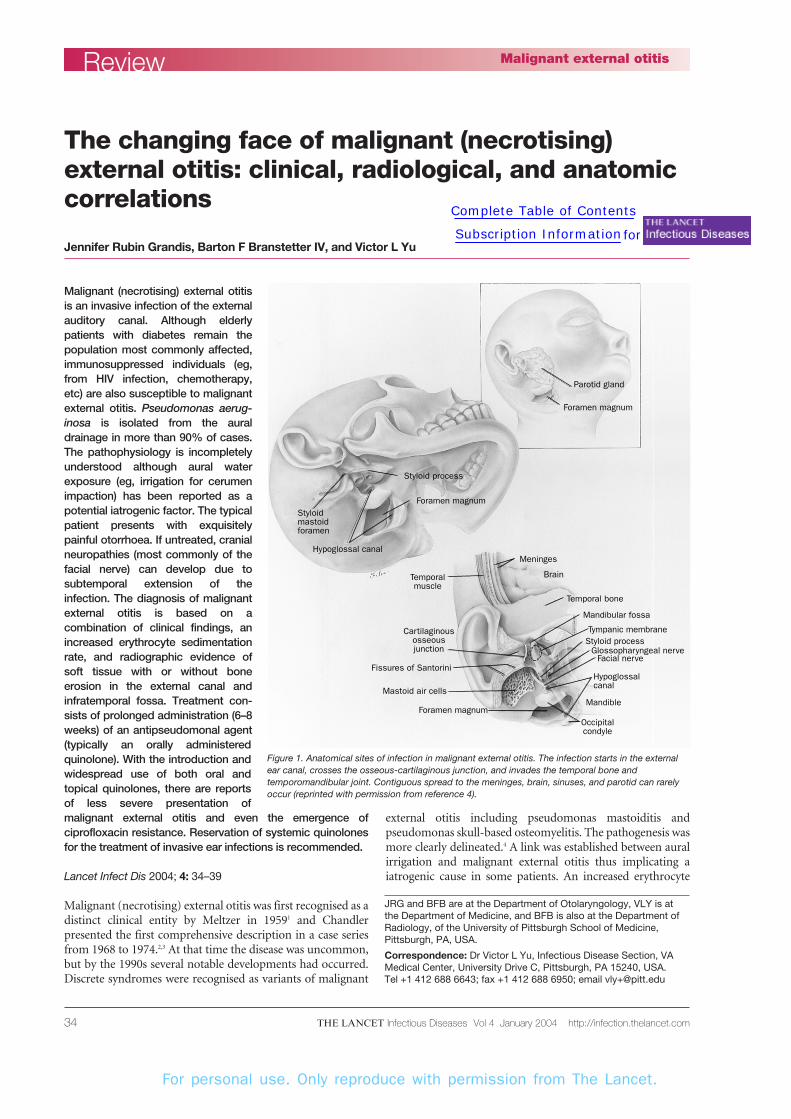
Figure 1. Anatomical sites of infection in malignant external otitis. The infection starts in the external ear canal, crosses the osseous-cartilaginous junction, and invades the temporal bone andtemporomandibular joint. Contiguous spread to the meninges, brain, sinuses, and parotid can rarelyoccur (reprinted with permission from reference 4).
For personal use. Only reproduce with permission from The Lancet.
THE LANCET Infectious Diseases Vol 4 January 2004 http://infection.thelancet.com 35
sedimentation rate (ESR) was identi-fied as a helpful tool in screening forthis syndrome and monitoringresponse to therapy.4,5 Computedtomography (CT) and magnetic-resonance imaging (MRI) scans wereshown to be useful for diagnosis andfor assessment of treatment.
Ciprofloxacin, an antipseudo-monal antibiotic that could be admin-istered orally, became available in the 1990s and ultimately sup-planted intravenous therapy withantipseudomonal �-lactam agentsand aminoglycosides. Unfortunately,treatment with oral quinolones hasbeen threatened since their use hasbecome widespread and, in ouropinion, indiscriminate. In this reviewwe summarise the major changes inaetiology and epidemiology of thisevolving disease and comment onnewer approaches to its management.
PathophysiologyMalignant external otitis is an invasiveinfection of the external auditory canal and skull base (figure 1) that typically arises in the elderlypatient with diabetes mellitus. Most cases (86–90%) have beenreported in diabetic patients.Pseudomonas aeruginosa is nearlyalways the causative organism (>98%of cases),4 although the administration of topical antibioticsbefore culture often precludes isolation of the pathogen. Sinceboth ageing and diabetes mellitus are associated withabnormalities of small blood vessels, it has been postulated thatmicroangiopathy in the ear canal predisposes elderly diabeticpatients to malignant external otitis.2,4 Although no directrelation has been delineated between the degree of glucoseintolerance and disease susceptibility,4 an increased pH indiabetic cerumen has been reported, which may contribute tothe development of malignant external otitis.6 Increasing lifeexpectancy and obesity may lead to a growing incidence ofdiabetes mellitus and hence, malignant external otitis.
EpidemiologyThe epidemiology of malignant external otitis has changed inthe past 10 years. Although it is difficult to document precisely,it seems that this syndrome has been more frequentlydiagnosed as the index of suspicion for malignant externalotitis has increased for generalist physicians. Although rare,paediatric cases are also being seen. By contrast with adults,children are more likely to be immunocompromised on thebasis of malignancy and malnutrition. Although no deathshave been reported to our knowledge, children tend to bemore toxic with their illness, as illustrated by the developmentof fever, leucocytosis, and P aeruginosa bacteraemia.4,7
Malignant external otitis is now being reported in patientswith AIDS.8–14 AIDS patients who develop malignant externalotitis tend to be younger than the typical elderly patient withthis invasive ear infection, and most are not diabetic. Inaddition, Aspergillus fumigatus has been isolated in AIDSpatients as well as P aeruginosa. Most patients have been curedwith systemic antipseudomonal or antifungal regimens.Although malignant external otitis seems to be uncommon inAIDS patients, the diagnosis should be considered in anypatient who presents with painful otorrhoea that isunresponsive to treatment regimens for simple external otitis(ie, topical antibiotics and local debridement).
Malignant external otitis is caused by P aeruginosa innearly all cases. When more than one organism is recoveredon culture, the other isolates tend to represent normal skinflora. It is important to isolate the organism from the eardrainage before instituting therapy. If P aeruginosa has neverbeen isolated from the otorrhoea, then a biopsy of the bone forculture is indicated to eliminate the possibility of malignancyand establish the necessity for long-term antibiotic therapy. P aeruginosa is a Gram-negative bacterium that is ubiquitousin water.15,16 The recovery of Pseudomonas spp on culture isindicative of infection since pseudomonas are not a comp-onent of normal ear canal flora. Exposure to water colonisedwith pseudomonas has been shown to cause simple external
ReviewMalignant external otitis
Temporal muscle
Externalacousticmeatus
Bonyexternalacousticmeatus
Temporal bone
Tympanicmembrane
Cartilage ofexternalacousticmeatus
Parotidgland
Mastoidprocess
Facialnerve
Styloidprocess
Glossopharyngealnerve
Fissures ofSantorini
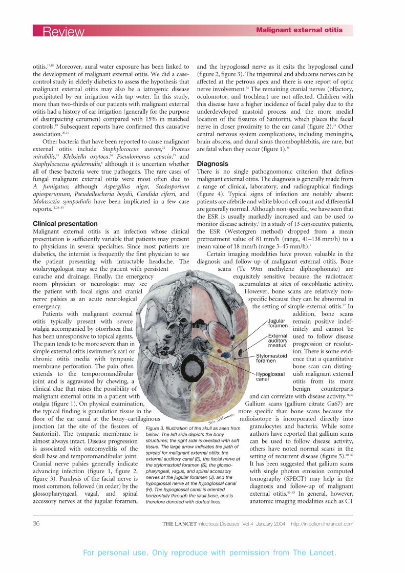
Figure 2. Infection enters the mastoid and skull base through the fissures of Santorini. The mostfrequent sites of cranial nerve involvement are the facial nerve as it exits through the stylomastoidforamen, the glossopharyngeal, the vagus, and the accessory nerves as they exit through the jugularforamen, and the hypoglossal nerve as it passes along the hypoglossal canal (reprinted withpermission from reference 34).
For personal use. Only reproduce with permission from The Lancet.
THE LANCET Infectious Diseases Vol 4 January 2004 http://infection.thelancet.com36
otitis.17,18 Moreover, aural water exposure has been linked tothe development of malignant external otitis. We did a case-control study in elderly diabetics to assess the hypothesis thatmalignant external otitis may also be a iatrogenic diseaseprecipitated by ear irrigation with tap water. In this study,more than two-thirds of our patients with malignant externalotitis had a history of ear irrigation (generally for the purposeof disimpacting cerumen) compared with 15% in matchedcontrols.19 Subsequent reports have confirmed this causativeassociation.20,21
Other bacteria that have been reported to cause malignantexternal otitis include Staphylococcus aureus,22 Proteusmirabilis,23 Klebsiella oxytoca,24 Pseudomonas cepacia,25 andStaphylococcus epidermidis,6 although it is uncertain whetherall of these bacteria were true pathogens. The rare cases offungal malignant external otitis were most often due to A fumigatus; although Aspergillus niger, Scedosporiumapiosperumum, Pseudallescheria boydii, Candida ciferri, andMalassezia sympodialis have been implicated in a few casereports.11,26–33
Clinical presentationMalignant external otitis is an infection whose clinicalpresentation is sufficiently variable that patients may presentto physicians in several specialties. Since most patients arediabetics, the internist is frequently the first physician to seethe patient presenting with intractable headache. Theotolaryngologist may see the patient with persistentearache and drainage. Finally, the emergencyroom physician or neurologist may see the patient with focal signs and cranialnerve palsies as an acute neurologicalemergency.
Patients with malignant externalotitis typically present with severeotalgia accompanied by otorrhoea thathas been unresponsive to topical agents.The pain tends to be more severe than insimple external otitis (swimmer’s ear) orchronic otitis media with tympanicmembrane perforation. The pain oftenextends to the temporomandibularjoint and is aggravated by chewing, aclinical clue that raises the possibility ofmalignant external otitis in a patient withotalgia (figure 1). On physical examination,the typical finding is granulation tissue in thefloor of the ear canal at the bony–cartilaginousjunction (at the site of the fissures ofSantorini). The tympanic membrane isalmost always intact. Disease progressionis associated with osteomyelitis of theskull base and temporomandibular joint.Cranial nerve palsies generally indicateadvancing infection (figure 1, figure 2,figure 3). Paralysis of the facial nerve ismost common, followed (in order) by theglossopharyngeal, vagal, and spinalaccessory nerves at the jugular foramen,
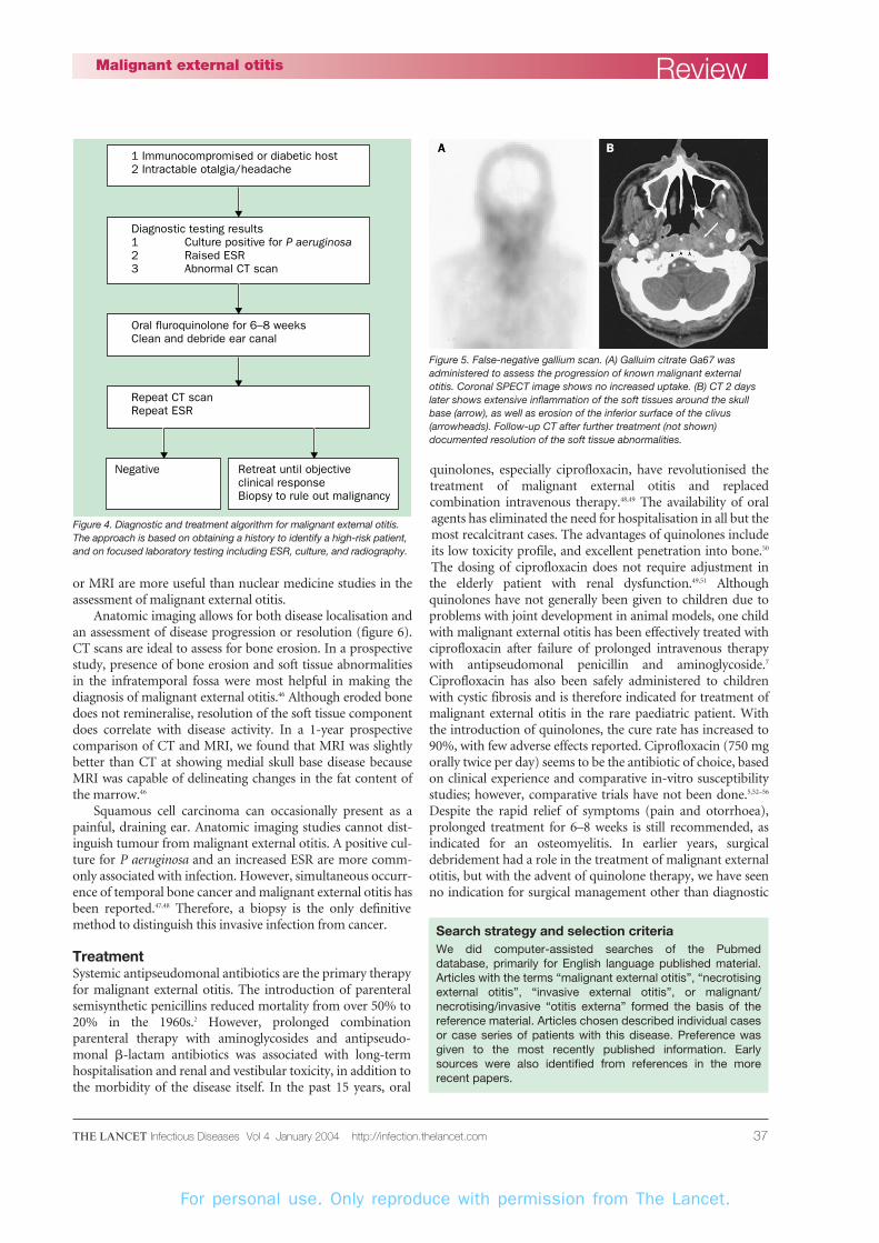
and the hypoglossal nerve as it exits the hypoglossal canal(figure 2, figure 3). The trigeminal and abducens nerves can beaffected at the petrous apex and there is one report of opticnerve involvement.34 The remaining cranial nerves (olfactory,oculomotor, and trochlear) are not affected. Children withthis disease have a higher incidence of facial palsy due to theunderdeveloped mastoid process and the more mediallocation of the fissures of Santorini, which places the facialnerve in closer proximity to the ear canal (figure 2).35 Othercentral nervous system complications, including meningitis,brain abscess, and dural sinus thrombophlebitis, are rare, butare fatal when they occur (figure 1).36
DiagnosisThere is no single pathognomonic criterion that definesmalignant external otitis. The diagnosis is generally made froma range of clinical, laboratory, and radiographical findings(figure 4). Typical signs of infection are notably absent:patients are afebrile and white blood cell count and differentialare generally normal. Although non-specific, we have seen thatthe ESR is usually markedly increased and can be used tomonitor disease activity.4 In a study of 13 consecutive patients,the ESR (Westergren method) dropped from a meanpretreatment value of 81 mm/h (range, 41–138 mm/h) to amean value of 18 mm/h (range 3–45 mm/h).5
Certain imaging modalities have proven valuable in thediagnosis and follow-up of malignant external otitis. Bone
scans (Tc 99m methylene diphosphonate) areexquisitely sensitive because the radiotracer
accumulates at sites of osteoblastic activity.However, bone scans are relatively non-
specific because they can be abnormal inthe setting of simple external otitis.37 In
addition, bone scansremain positive indef-initely and cannot beused to follow diseaseprogression or resolut-ion. There is some evid-ence that a quantitativebone scan can disting-uish malignant externalotitis from its morebenign counterparts
and can correlate with disease activity.38,39
Gallium scans (gallium citrate Ga67) aremore specific than bone scans because theradioisotope is incorporated directly into
granulocytes and bacteria. While someauthors have reported that gallium scanscan be used to follow disease activity,others have noted normal scans in thesetting of recurrent disease (figure 5).40–42
It has been suggested that gallium scanswith single photon emission computedtomography (SPECT) may help in thediagnosis and follow-up of malignantexternal otitis.43–45 In general, however,anatomic imaging modalities such as CT
Review Malignant external otitis
Jugularforamen
Stylomastoidforamen
Hypoglossalcanal
Externalauditorymeatus
Figure 3. Illustration of the skull as seen frombelow. The left side depicts the bonystructures; the right side is overlaid with softtissue. The large arrow indicates the path ofspread for malignant external otitis: theexternal auditory canal (E), the facial nerve atthe stylomastoid foramen (S), the glosso-pharyngeal, vagus, and spinal accessorynerves at the jugular foramen (J), and thehypoglossal nerve at the hypoglossal canal(H). The hypoglossal canal is orientedhorizontally through the skull base, and istherefore denoted with dotted lines.
For personal use. Only reproduce with permission from The Lancet.
THE LANCET Infectious Diseases Vol 4 January 2004 http://infection.thelancet.com 37
or MRI are more useful than nuclear medicine studies in theassessment of malignant external otitis.
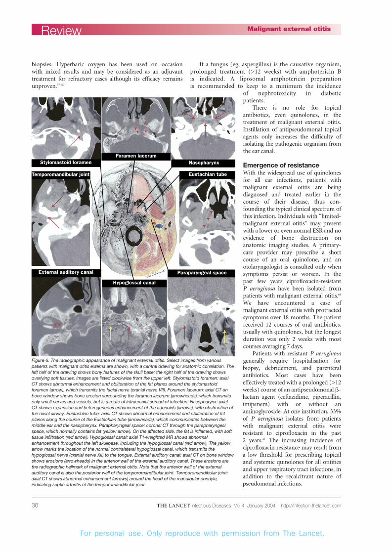
Anatomic imaging allows for both disease localisation andan assessment of disease progression or resolution (figure 6).CT scans are ideal to assess for bone erosion. In a prospectivestudy, presence of bone erosion and soft tissue abnormalitiesin the infratemporal fossa were most helpful in making thediagnosis of malignant external otitis.46 Although eroded bonedoes not remineralise, resolution of the soft tissue componentdoes correlate with disease activity. In a 1-year prospectivecomparison of CT and MRI, we found that MRI was slightlybetter than CT at showing medial skull base disease becauseMRI was capable of delineating changes in the fat content ofthe marrow.46
Squamous cell carcinoma can occasionally present as apainful, draining ear. Anatomic imaging studies cannot dist-inguish tumour from malignant external otitis. A positive cul-ture for P aeruginosa and an increased ESR are more comm-only associated with infection. However, simultaneous occurr-ence of temporal bone cancer and malignant external otitis hasbeen reported.47,48 Therefore, a biopsy is the only definitivemethod to distinguish this invasive infection from cancer.
Treatment Systemic antipseudomonal antibiotics are the primary therapyfor malignant external otitis. The introduction of parenteralsemisynthetic penicillins reduced mortality from over 50% to20% in the 1960s.2 However, prolonged combinationparenteral therapy with aminoglycosides and antipseudo-monal �-lactam antibiotics was associated with long-termhospitalisation and renal and vestibular toxicity, in addition tothe morbidity of the disease itself. In the past 15 years, oral
quinolones, especially ciprofloxacin, have revolutionised thetreatment of malignant external otitis and replacedcombination intravenous therapy.48,49 The availability of oralagents has eliminated the need for hospitalisation in all but themost recalcitrant cases. The advantages of quinolones includeits low toxicity profile, and excellent penetration into bone.50
The dosing of ciprofloxacin does not require adjustment inthe elderly patient with renal dysfunction.49,51 Althoughquinolones have not generally been given to children due toproblems with joint development in animal models, one childwith malignant external otitis has been effectively treated withciprofloxacin after failure of prolonged intravenous therapywith antipseudomonal penicillin and aminoglycoside.7
Ciprofloxacin has also been safely administered to childrenwith cystic fibrosis and is therefore indicated for treatment ofmalignant external otitis in the rare paediatric patient. Withthe introduction of quinolones, the cure rate has increased to90%, with few adverse effects reported. Ciprofloxacin (750 mgorally twice per day) seems to be the antibiotic of choice, basedon clinical experience and comparative in-vitro susceptibilitystudies; however, comparative trials have not been done.5,52–56
Despite the rapid relief of symptoms (pain and otorrhoea),prolonged treatment for 6–8 weeks is still recommended, asindicated for an osteomyelitis. In earlier years, surgicaldebridement had a role in the treatment of malignant externalotitis, but with the advent of quinolone therapy, we have seenno indication for surgical management other than diagnostic
ReviewMalignant external otitis
1 Immunocompromised or diabetic host2 Intractable otalgia/headache
Repeat CT scanRepeat ESR
Negative Retreat until objectiveclinical responseBiopsy to rule out malignancy
Oral fluroquinolone for 6–8 weeksClean and debride ear canal
Diagnostic testing results1 Culture positive for P aeruginosa2 Raised ESR3 Abnormal CT scan
Figure 4. Diagnostic and treatment algorithm for malignant external otitis.The approach is based on obtaining a history to identify a high-risk patient,and on focused laboratory testing including ESR, culture, and radiography.
Figure 5. False-negative gallium scan. (A) Galluim citrate Ga67 wasadministered to assess the progression of known malignant externalotitis. Coronal SPECT image shows no increased uptake. (B) CT 2 dayslater shows extensive inflammation of the soft tissues around the skullbase (arrow), as well as erosion of the inferior surface of the clivus(arrowheads). Follow-up CT after further treatment (not shown)documented resolution of the soft tissue abnormalities.
Search strategy and selection criteriaWe did computer-assisted searches of the Pubmeddatabase, primarily for English language published material.Articles with the terms “malignant external otitis”, “necrotisingexternal otitis”, “invasive external otitis”, or malignant/necrotising/invasive “otitis externa” formed the basis of thereference material. Articles chosen described individual casesor case series of patients with this disease. Preference wasgiven to the most recently published information. Earlysources were also identified from references in the morerecent papers.
For personal use. Only reproduce with permission from The Lancet.
THE LANCET Infectious Diseases Vol 4 January 2004 http://infection.thelancet.com38
biopsies. Hyperbaric oxygen has been used on occasion with mixed results and may be considered as an adjuvanttreatment for refractory cases although its efficacy remainsunproven.57–60
If a fungus (eg, aspergillus) is the causative organism,prolonged treatment (>12 weeks) with amphotericin B is indicated. A liposomal amphotericin preparation is recommended to keep to a minimum the incidence
of nephrotoxicity in diabetic patients.
There is no role for topicalantibiotics, even quinolones, in thetreatment of malignant external otitis.Instillation of antipseudomonal topicalagents only increases the difficulty ofisolating the pathogenic organism fromthe ear canal.
Emergence of resistanceWith the widespread use of quinolonesfor all ear infections, patients withmalignant external otitis are beingdiagnosed and treated earlier in thecourse of their disease, thus con-founding the typical clinical spectrum ofthis infection. Individuals with “limited-malignant external otitis” may presentwith a lower or even normal ESR and noevidence of bone destruction onanatomic imaging studies. A primary-care provider may prescribe a shortcourse of an oral quinolone, and anotolaryngologist is consulted only whensymptoms persist or worsen. In the past few years ciprofloxacin-resistant P aeruginosa have been isolated frompatients with malignant external otitis.61
We have encountered a case ofmalignant external otitis with protractedsymptoms over 18 months. The patientreceived 12 courses of oral antibiotics,usually with quinolones, but the longestduration was only 2 weeks with mostcourses averaging 7 days.
Patients with resistant P aeruginosagenerally require hospitalisation forbiopsy, debridement, and parenteralantibiotics. Most cases have beeneffectively treated with a prolonged (>12weeks) course of an antipseudomonal �-lactam agent (ceftazidime, piperacillin,imipenem) with or without anaminoglycoside. At one institution, 33%of P aeruginosa isolates from patientswith malignant external otitis wereresistant to ciprofloxacin in the past 2 years.61 The increasing incidence ofciprofloxacin resistance may result froma low threshold for prescribing topicaland systemic quinolones for all otititiesand upper respiratory tract infections, inaddition to the recalcitrant nature ofpseudomonal infections.
Review Malignant external otitis
Stylomastoid foramenForamen lacerum
Nasopharynx
Eustachian tube
Paraparyngeal space
Temporomandibular joint
External auditory canal
Hypoglossal canal
Figure 6. The radiographic appearance of malignant external otitis. Select images from variouspatients with malignant otitis externa are shown, with a central drawing for anatomic correlation. Theleft half of the drawing shows bony features of the skull base; the right half of the drawing showsoverlying soft tissues. Images are listed clockwise from the upper left. Stylomastoid foramen: axialCT shows abnormal enhancement and obliteration of the fat planes around the stylomastoidforamen (arrow), which transmits the facial nerve (cranial nerve VII). Foramen lacerum: axial CT onbone window shows bone erosion surrounding the foramen lacerum (arrowheads), which transmitsonly small nerves and vessels, but is a route of intracranial spread of infection. Nasopharynx: axialCT shows expansion and heterogeneous enhancement of the adenoids (arrows), with obstruction ofthe nasal airway. Eustachian tube: axial CT shows abnormal enhancement and obliteration of fatplanes along the course of the Eustachian tube (arrowheads), which communicates between themiddle ear and the nasopharynx. Parapharyngeal space: coronal CT through the parapharyngealspace, which normally contains fat (yellow arrow). On the affected side, the fat is inflamed, with softtissue infiltration (red arrow). Hypoglossal canal: axial T1-weighted MR shows abnormalenhancement throughout the left skullbase, including the hypoglossal canal (red arrow). The yellowarrow marks the location of the normal contralateral hypoglossal canal, which transmits thehypoglossal nerve (cranial nerve XII) to the tongue. External auditory canal: axial CT on bone windowshows erosions (arrowheads) in the anterior wall of the external auditory canal. These erosions arethe radiographic hallmark of malignant external otitis. Note that the anterior wall of the externalauditory canal is also the posterior wall of the temporomandibular joint. Temporomandibular joint:axial CT shows abnormal enhancement (arrows) around the head of the mandibular condyle,indicating septic arthritis of the temporomandibular joint.
For personal use. Only reproduce with permission from The Lancet.
THE LANCET Infectious Diseases Vol 4 January 2004 http://infection.thelancet.com 39
ConclusionMalignant external otitis remains a serious invasive infectiondespite the recent success of quinolones antibiotics. It isimportant to maintain a high index of suspicion when thetypical host (an elderly diabetic or immunocompromisedpatient) presents with otalgia or intractable headache that isdisproportionate to findings on physical examination. Ahistory of ear irrigation should be sought because of the notedassociation between malignant external otitis and waterexposure.19,20 The diagnosis of malignant external otitis is basedon physical examination findings, raised ESR, and CTevidence of bone erosion and infiltrated infratemporal softtissues. CT remains the most appropriate initial imaging
method because it addresses many differential considerationsin the external auditory canal. MRI is preferred whendocumenting progression or resolution of soft tissue infection and osteomyelitis. Quinolone antibiotics, especiallyciprofloxacin, have emerged as the treatment of choice formalignant external otitis. However, the emergence ofciprofloxacin resistance in P aeruginosa is becoming a majorproblem. Given the plethora of oral antibacterial agentsavailable for the more common otitides, especially otitismedia, we recommend that quinolones should be reservedonly for the treatment of invasive ear infections.
Conflicts of interestWe have no conflicts of interest.
ReviewMalignant external otitis
References1 Meltzer PE, Kelemen G. Pyocutaneous osteomyelitis
of the temporal bone, madible, and zygoma.Laryngoscope 1959; 169: 1300–16.
2 Chandler JR. Malignant external otitis. Laryngoscope1968; 78: 1257–94.
3 Chandler JR. Malignant external otitis and facialparalysis. Otolaryngol Clin North Am 1974; 7: 375–83.
4 Rubin J, Yu VL. Malignant external otitis: insights intopathogenesis, clinical manifestations, diagnosis, andtherapy. Am J Med 1988; 85: 391–98.
5 Rubin J, Stoehr G, Yu VL, Muder RR, Matador A,Kamerer DB. Efficacy of oral ciprofloxacin plus rifampin for treatment of malignant externalotitis. Arch Otolaryngol Head Neck Surg 1989; 115:1063–69.
6 Barrow HN, Levenson MJ. Necrotizing “malignant”external otitis caused by Staphylococcus epidermidis.Arch Otolaryngol Head Neck Surg 1992; 118: 94–96.
7 Paul AC, Justus A, Balraj A, Job A, Kirubakaran CP.Malignant otitis externa in an infant with selective IgAdeficiency: a case report. Int J Pediatr Otorhinolaryngol2001; 60: 141–45.
9 Weinroth SE, Schessel D, Tuazon CU. Malignant otitisexterna in AIDS patients: case report and review of theliterature. Ear Nose Throat J 1994; 73: 772–74, 777–78.
10 Reiss P, Hadderingh R, Schot LJ, Danner SA. Invasiveexternal otitis caused by Aspergillus fumigatus in twopatients with AIDS. AIDS 1991; 5: 605–06.
11 Munoz A, Martinez-Chamorro E. Necrotizingexternal otitis caused by Aspergillus fumigatus:computed tomography and high resolution magneticresonance imaging in an AIDS patient. J Laryngol Otol1998; 112: 98–102.
12 McElroy EA Jr, Marks GL. Fatal necrotizing otitisexterna in a patient with AIDS. Rev Infect Dis 1991; 13:1246–47.
13 Kielhofner M, Atmar RL, Hamill RJ, Musher DM.Life-threatening Pseudomonas aeruginosa infections inpatients with human immunodeficiency virusinfection. Clin Infect Dis 1992; 14: 403–11.
14 Rivas Lacarte MP, Pumarola Segura F. Malignantotitis externa and HIV antibodies. A case report. AnOtorrinolaringol Ibero Am 1990; 17: 505–12.
15 Favero MS, Carson LA, Bond WW, Petersen NJ.Pseudomonas aeruginosa: growth in distilled waterfrom hospitals. Science 1971; 173: 836–38.
16 Yoshpe-Purer Y, Golderman S. Occurrence ofStaphylococcus aureus and Pseudomonas aeruginosa inIsraeli coastal water. Appl Environ Microbiol 1987; 53:1138–41.
17 Wright DN, Alexander JM. Effect of water on thebacterial flora of swimmers’ ears. Arch Otolaryngol1974; 99: 15–18.
18 Seyfried PL, Cook RJ. Otitis externa infections relatedto Pseudomonas aeruginosa levels in five Ontario lakes.Can J Public Health 1984; 75: 83–91.
19 Rubin J, Yu VL, Kamerer DB, Wagener M. Auralirrigation with water: a potential pathogenicmechanism for inducing malignant external otitis?Ann Otol Rhinol Laryngol 1990; 99: 117–19.
20 Ford GR, Courteney-Harris RG. Another hazard ofear syringing: malignant external otitis. J Laryngol Otol1990; 104: 709–10.
21 Zikk D, Rapoport Y, Himelfarb MZ. Invasiveexternal otitis after removal of impacted cerumen byirrigation. N Engl J Med 1991; 325: 969–70.
22 Bayardelle P, Jolivet-Granger M, Larochelle D.Staphylococcal malignant external otitis. Can Med Assoc J 1982; 126: 155–56.
24 Garcia Rodriguez JA, Montes Martinez I, GomezGonzalez JL, Ramos Macias A, Lopez Alburquerque T.A case of malignant external otitis involving Klebsiellaoxytoca. Eur J Clin Microbiol Infect Dis 1992; 11: 75–77.
25 Dettelbach MA, Hirsch BE, Weissman JL.Pseudomonas cepacia of the temporal bone: malignantexternal otitis in a patient with cystic fibrosis.Otolaryngol Head Neck Surg 1994; 111: 528–32.
26 Cunningham M, Yu VL, Turner J, Curtin H.Necrotizing otitis externa due to Aspergillus in animmunocompetent patient. Arch Otolaryngol HeadNeck Surg 1988; 114: 554–56.
27 Petrak RM, Pottage JC, Levin S. Invasive externalotitis caused by Aspergillus fumigatus in animmunocompromised patient. J Infect Dis 1985; 151:196.
28 Lyos AT, Malpica A, Estrada R, Katz CD, Jenkins HA.Invasive aspergillosis of the temporal bone: an unusualmanifestation of acquired immunodeficiencysyndrome. Am J Otolaryngol 1993; 14: 444–48.
29 Gordon G, Giddings NA. Invasive otitis externa due toAspergillus species: case report and review. Clin InfectDis 1994; 19: 866–70.
30 Harley WB, Dummer JS, Anderson TL, Goodman S.Malignant external otitis due to Aspergillus flavus withfulminant dissemination to the lungs. Clin Infect Dis1995; 20: 1052–54.
37 Levin WJ, Shary JH 3rd, Nichols LT, Lucente FE. Bonescanning in severe external otitis. Laryngoscope 1986;96: 1193–95.
38 Uri N, Gips S, Front A, Meyer SW, Hardoff R.Quantitative bone and 67Ga scintigraphy in thedifferentiation of necrotizing external otitis fromsevere external otitis. Arch Otolaryngol Head Neck Surg1991; 117: 623–26.
39 Stokkel MP, Takes RP, van Eck-Smit BL, Baatenburgde Jong RJ. The value of quantitative gallium-67single-photon emission tomography in the clinicalmanagement of malignant external otitis. Eur J NuclMed 1997; 24: 1429–32.
40 Ostfeld E, Aviel A, Pelet D. Malignant external otitis:The diagnostic value of bone scintigraphy.Laryngoscope 1981; 91: 960–64.
41 Garty I, Rosen G, Holdstein Y. The radionuclidediagnosis, evaluation and follow-up of malignantexternal otitis (MEO). The value of immediate bloodpool scanning. J Laryngol Otol 1985; 99: 109–15.
42 Gherini SG, Brackmann DE, Bradley WG. Magneticresonance imaging and computerized tomography inmalignant external otitis. Laryngoscope 1986; 96:542–48.
43 Stokkel MP, Boot CN, van Eck-Smit BL. SPECTgallium scintigraphy in malignant external otitis:initial staging and follow-up. Case reports.Laryngoscope 1996; 106: 338–40.
44 Paramsothy M, Khanijow V, Ong TO. Use of gallium-67 in the assessment of response to antibiotic therapyin malignant otitis externa—a case report. SingaporeMed J 1997; 38: 347–49.
45 Amorosa L, Modugno GC, Pirodda A. Malignantexternal otitis: review and personal experience. ActaOtolaryngol Suppl 1996; 521: 3–16.
46 Grandis JR, Curtin HD, Yu VL. Necrotizing(malignant) external otitis: prospective comparison ofCT and MR imaging in diagnosis and follow-up.Radiology 1995; 196: 499–504.
47 Mattucci KF, Setzen M, Galantich P. Necrotizing otitisexterna occurring concurrently with epidermoidcarcinoma. Laryngoscope 1986; 96: 264–66.
48 Grandis JR, Hirsch BE, Yu VL. Simultaneouspresentation of malignant external otitis and temporalbone cancer. Arch Otolaryngol Head Neck Surg 1993;119: 687–89.
49 Wiseman LR, Balfour JA. Ciprofloxacin. A review ofits pharmacological profile and therapeutic use in theelderly. Drugs Aging 1994; 4: 145–73.
50 Barza M. Pharmacokinetics and efficacy of the new quinolones in infections of the eye, ear, nose, and throat. Rev Infect Dis 1988; 10 (suppl 1):S241–7.
51 Hirata CA, Guay DR, Awni WM, Stein DJ, Peterson PK. Steady-state pharmacokinetics of intravenous and oral ciprofloxacin in elderlypatients. Antimicrob Agents Chemother 1989; 33:1927–31.
52 Sade J, Lang R, Goshen S, Kitzes-Cohen R.Ciprofloxacin treatment of malignant external otitis.Am J Med 1989; 87: 138S–141S.
53 Joachims HZ, Danino J, Raz R. Malignant externalotitis: treatment with fluoroquinolones. Am J Otolaryngol 1988; 9: 102–05.
54 Leggett JM, Prendergast K. Malignant external otitis:the use of oral ciprofloxacin. J Laryngol Otol 1988;102: 53–54.
55 Hickey SA, Ford GR, O’Connor AF, Eykyn SJ,Sonksen PH. Treating malignant otitis with oralciprofloxacin. Bmj 1989; 299: 550–51.
56 Levenson MJ, Parisier SC, Dolitsky J, Bindra G.Ciprofloxacin: drug of choice in the treatment ofmalignant external otitis (MEO). Laryngoscope 1991;101: 821–24.
57 Lucente FE, Parisier SC, Som PM, Arnold LM.Malignant external otitis: a dangerous misnomer?Otolaryngol Head Neck Surg 1982; 90: 266–69.
58 Mader JT, Love JT. Malignant external otitis. Curewith adjunctive hyperbaric oxygen therapy. ArchOtolaryngol 1982; 108: 38–40.
59 Lucente FE, Parisier SC, Som PM. Complications ofthe treatment of malignant external otitis.Laryngoscope 1983; 93: 279–81.
60 Davis JC, Gates GA, Lerner C, Davis MG Jr, Mader JT,Dinesman A. Adjuvant hyperbaric oxygen inmalignant external otitis. Arch Otolaryngol Head NeckSurg 1992; 118: 89–93.
61 Berenholz L, Katzenell U, Harell M. Evolving resistantpseudomonas to ciprofloxacin in malignant otitisexterna. Laryngoscope 2002; 112: 1619–22.