Rift Valley Fever Virus L Protein Forms a Biologically Active Oligomer�
Aya Zamoto-Niikura,1§ Kaori Terasaki,1 Tetsuro Ikegami,1,2,3,4
C. J. Peters,1,2,3,4 and Shinji Makino1,3,4*Departments of Microbiology and Immunology1 and Pathology,2 the Sealy Center for Vaccine Development,3 and the Center for
Biodefense and Emerging Infectious Diseases,4 the University of Texas Medical Branch at Galveston, Galveston, Texas 77555
Received 25 June 2009/Accepted 27 September 2009
Rift Valley fever virus (RVFV) (genus Phlebovirus, family Bunyaviridae) causes mosquito-borne epidemicdiseases in humans and livestock. The virus carries three RNA segments, L, M, and S, of negative or ambisensepolarity. L protein, an RNA-dependent RNA polymerase, encoded in the L segment, and N protein, encoded inthe S segment, exert viral RNA replication and transcription. Coexpression of N, hemagglutinin (HA)-taggedL, and viral minigenome resulted in minigenome replication and transcription, a finding that demonstratedHA-tagged L was biologically active. Likewise L tagged with green fluorescent protein (GFP) was biologicallycompetent. Coimmunoprecipitation analysis using extracts from cells coexpressing HA-tagged L and GFP-tagged L showed the formation of an L oligomer. Bimolecular fluorescence complementation analysis andcoimmunoprecipitation studies demonstrated the formation of an intermolecular L-L interaction through itsN-terminal and C-terminal regions and also suggested an intramolecular association between the N-terminaland C-terminal regions of L protein. A biologically inactive L mutant, in which the conserved signature SDDmotif was replaced by the amino acid residues GNN, exhibited a dominant negative phenotype when coex-pressed with wild-type L in the minigenome assay system. Expression of this mutant L also inhibited viral geneexpression in virus-infected cells. These data provided compelling evidence for the importance of oligomer-ization of RVFV L protein for its polymerase activity.
Rift Valley Fever virus (RVFV), which belongs to the genusPhlebovirus in the family Bunyaviridae, is endemic in sub-Saharan African countries and causes large outbreaks in en-demic areas and countries outside of the endemic area, includ-ing Egypt, Saudi Arabia, and Yemen (2). RVFV is transmittedby mosquitoes, and the virus infection causes a high rate ofabortions in pregnant ruminants and acute lethal hepatitis innewborn lambs (26). Most human patients show an acute fe-brile myalgic syndrome, with a small minority of patients, per-haps �1%, experiencing severe hemorrhagic fever or enceph-alitis. Also, some patients show retinal vasculitis, which resultsin partial blindness for an undefined period (22).
RVFV has three single-stranded genomic RNA segments,designated L, M, and S. The L and M RNA segments are ofnegative polarity. The L segment contains a 6,279-nucleotide-long open reading frame (ORF) encoding L protein and a viralRNA-dependent RNA polymerase (RdRp), while the M seg-ment has a single ORF encoding NSm protein, a 78-kDa pro-tein, and two major viral envelope proteins, Gn and Gc. The Ssegment is of ambisense polarity and encodes NSs protein andN protein; the former and the latter are encoded in anti-viral-sense RNA and in viral-sense RNA, respectively. In bunyavi-ruses, both L protein and N protein are needed for viral RNAreplication and viral mRNA synthesis, the latter of which usesa host mRNA-derived cap structure as a primer (3, 16, 29). N
protein encapsidates viral RNAs to form helical nucleocapsids,which serve as a template for viral RNA synthesis (21, 23).
RVFV L protein plays a central role in viral RNA synthesis,yet very little is known about how L protein exerts its functions;the properties of viral replication complexes, structures of viralRNA polymerases, and host factors needed for viral RNAsynthesis have been characterized poorly for bunyaviruses. Paststudies of other negative-stranded RNA viruses, including Sen-dai virus (36), parainfluenza virus 3 (37), and measles virus (4),all of which belong to the paramyxoviruses, and lymphocyticchoriomeningitis virus (LCMV) (32), an arenavirus, suggestedthat their L proteins function as an oligomer to exert the RdRpfunction. In addition, the influenza A virus RdRp heterotrimer(18, 19) has been reported to form oligomers. Currently, it isunclear whether the L proteins of bunyaviruses also form anoligomer and exhibit RdRp activities in infected cells. Wepresent here that RVFV L protein forms an oligomer. Wefurther identified L protein regions that were involved in Loligomerization and explored the biological importance of theL protein oligomer for viral RNA synthesis.
MATERIALS AND METHODS
Cells and viruses. BHK/T7-9 cells stably expressing T7 RNA polymerase (15)and Vero E6 cells were maintained as described previously (14). rMP12-rLuc(14), an RVFV MP-12 strain carrying the Renilla luciferase (rLuc) gene in placeof the NSs gene in the S segment, was used.
Plasmids. Plasmids encoding the mutant L protein were generated by modi-fication of pT7-IRES-vL encoding the MP-12 L protein (14). pT7-IRES-HA-Land pT7-IRES-GFP-L were constructed by adding a hemagglutinin (HA) tagsequence (TAC CCC TAC GAC GTG CCC GAC TAC GCC) and by insertinga cassette of the green fluorescent protein (GFP) gene and 10-alanine linkersequence (5) between the encephalomyocarditis virus internal ribosome entrysite (IRES) and the L gene ORF of pT7-IRES-vL, respectively. pT7-IRES-GFPwas constructed by replacing the L gene of pT7-IRES-vL with a GFP gene. Thenucleotide residues TCA GAT GAT encoding SDD at amino acids 1132 to 1134
* Corresponding author. Mailing address: Department of Microbi-ology and Immunology, University of Texas Medical Branch,Galveston, TX 77555-1019. Phone: (409) 772-2323. Fax: (409) 772-5065. E-mail: [email protected].
§ Present address: Division of Experimental Animal Research, Na-tional Institute of Infectious Diseases, 4-7-1 Gakuen, Musahi-mu-rayama, Tokyo 208-0011, Japan.
in the L gene of pT7-IRES-GFP-L were replaced with GGA AAT AAT encod-ing GNN, resulting in pT7-IRES-GFP-L(GNN). Unique restriction enzyme sites(see Fig. 5A) were used to make pT7-IRES-vL-derived plasmids, each of whichhad a deletion at different sites of the L protein. A modified version of yellowfluorescent protein (YFP), Venus (25), was synthesized (Bio Basic) and insertedupstream of the L gene ORF, as described for pT7-IRES-GFP-L. A series ofplasmids were constructed by adding the sequences encoding amino acid resi-dues 1 to 172 of Venus (12) (VN) or 155 to 239 of Venus (VC) with a linker(GGGGS)3 sequence (34) to the N and/or C terminus of the L gene of pT7-IRES-vL. We also constructed a plasmid encoding VN-tubulin, a fusion proteinconsisting of N-terminal VN, a linker (GGGGS)3 sequence, and an alpha-tubulingene.
Minigenome assay. Subconfluent BHK/T7-9 cell monolayers in a 12-well platewere cotransfected with 0.5 �g of pT7-IRES-vL, 1 �g of pT7-IRES-vN encodingN protein (14), and 1 �g of pPro-T7-M-rLuc(�) encoding a viral-sense RVFVminigenome carrying an rLuc gene (13). The total amount of plasmid wasadjusted to 2.5 �g by using empty-vector pT7-IRES (13). In some experiments,pT7-IRES-vL was substituted with another plasmid encoding a mutant L gene.Different amounts of plasmids encoding the L protein or those encoding mutantL proteins were also used as indicated below. Cell extracts were prepared formeasuring rLuc activities and Northern blot analysis at 36 h posttransfection. ForrLuc activities, cells were lysed with 1 ml of lysis buffer supplied by the manu-facturer, and luminescences were measured according to the manufacturer’sprotocol (Promega). Statistical analysis was performed for three independentexperiments to obtain mean and standard deviation. For Northern blot analysis,total intracellular RNA was extracted by using Trizol (Invitrogen) according tothe manufacturer’s instruction.
Coimmunoprecipitation analysis. BHK/T7-9 cells in a six-well plate werecotransfected with 2 �g of plasmid pT7-IRES-HA-L and 2 �g of plasmid pT7-IRES-GFP-L. After washing the cells with cold phosphate-buffered saline twiceat 24 h posttransfection, cell extracts were prepared by lysing cells on ice for 20min in 300 �l of TN buffer (50 mM Tris-HCl, pH 8.0, and 150 mM NaCl)containing 1% NP-40 and protease inhibitor (complete mini EDTA free;Roche). Lysates were centrifuged at 10,000 � g for 10 min at 4°C to removenuclei and large debris. Then the lysates were incubated with 25 �l of anti-GFPmicrobeads (Miltenyi Biotec) for 2 h on ice with occasional mixing, and immu-noprecipitation was performed according to the manufacturer’s protocol, withadditional extensive washing. For immunoprecipitation with anti-HA antibody,precleared lysate was incubated with 40 �l (bed volume of 20 �l) of mouseanti-HA monoclonal antibody-conjugated agarose (Sigma) overnight with rota-tion. The agarose was washed five times with 1 ml of cold TN buffer containing1% NP-40 and protease inhibitor and boiled with sodium dodecyl sulfate (SDS)sample buffer for 5 min. The supernatant of the agarose was subjected toSDS-polyacrylamide gel electrophoresis (PAGE) and subsequent Western blotanalysis.
Western blot analysis. Proteins that were separated by 7.5% or 12% SDS-PAGE were blotted on a polyvinylidene difluoride membrane (Immobilon-P;Millipore) with CAPS [3-(cyclohexylamino)-1-propanesulfonic acid] buffer (10mM CAPS, pH 11.0; 10% methanol). Membranes were blocked with TBSTbuffer (10 mM Tris-HCl, pH 7.6; 130 mM NaCl; and 0.1% Tween 20) containing1% bovine serum albumin (Santa Cruz) at room temperature for more than 1 h.Anti-GFP rabbit polyclonal antibody (Santa Cruz); anti-HA rabbit polyclonalantibody (Santa Cruz); anti-L 434 rabbit polyclonal antibody, which was pro-duced by immunizing rabbits with a bacterially expressed fragment of the Lprotein corresponding to the amino acid positions 1 to 434; or anti-actin goatpolyclonal antibody (Santa Cruz) was added in TBST buffer containing 1%bovine serum albumin and incubated with membrane at 4°C overnight. Themembrane was washed with TBST buffer and incubated with horseradish per-oxidase-conjugated, anti-rabbit antibody (Santa Cruz) for 45 min at room tem-perature. After the membranes were washed, they were developed with an ECLdetection kit (GE Healthcare), according to the manufacturer’s instructions.Light emission was detected by exposure to Blue autoradiography film (ISCBioexpress).
Northern blot analysis. Intracellular RNAs were dissolved in RNase-freedistilled water, and 4 �g RNA was incubated in denaturing buffer, which in-cluded RBS (20 mM MOPS [morpholinepropanesulfonic acid], 1 mM EDTA,and 5 mM sodium acetate), 50% formamide, and 20% formaldehyde, for 10 minat 68°C. RNAs were separated on 1.5% agarose gels containing RBS and 18%formaldehyde and transferred onto a nylon membrane (Roche). Northern blotanalysis was performed with strand-specific RNA probes for a sense and anti-sense rLuc gene (13) by using a DIG system (Roche) according to the manu-facturer’s protocol. rRNAs (28S and 18S) were detected by staining agarose gelswith ethidium bromide.
Virus infection. BHK/T7-9 cells in a 12-well plate were cotransfected with 1.0�g of pT7-IRES-vN and 0.5 �g of one of the following plasmids: pT7-IRES-L,pT7-IRES-L(GNN), or pT7-IRES. At 24 h posttransfection, cells were infectedwith rMP12-rLuc (14) at a multiplicity of infection (MOI) of 5. Samples wereprepared for an rLuc assay at the indicated hours postinfection (p.i.).
Bimolecular fluorescence complementation (BiFC) analysis. BHK/T7-9 cells ina two-well chambered cover glass were transfected with plasmid encoding a fusionprotein of VN and L proteins, a fusion protein of VC and L proteins, or an L proteincarrying the N-terminal Venus protein (Venus-L) as controls. In another negativecontrol, cells were cotransfected with plasmid encoding VN-tubulin and plasmidencoding the L protein carrying the N-terminal VC (VC-L). In the experimentalgroup, cells were transfected with 1.0 �g of plasmid encoding a fusion protein and1.0 �g of an empty vector, pT7-IRES, or cotransfected with 1.0 �g of two plasmidsas indicated in the legend to Fig. 6. In all cases, the total amount of plasmids wasadjusted to 2.0 �g. At 12 h posttransfection, live cells were visualized by the use ofa Zeiss LSM 510 UV META laser-scanning confocal microscope with a YFP-specific filter. In a separate experiment, cells were harvested with SDS sample bufferand then subjected to SDS-PAGE and Western blot analysis.
RESULTS
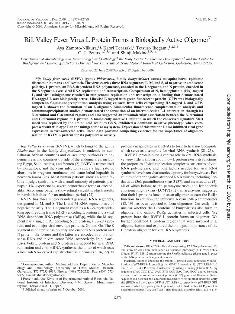
RVFV L proteins carrying an N-terminal GFP tag or HA tagare biologically active. We generated two L protein expressionplasmids, pT7-IRES-HA-L encoding the L protein tagged withan HA epitope at the N terminus and pT7-IRES-GFP-L en-coding the L protein tagged with GFP at the N terminus.BHK/T7-9 cells stably expressing T7 polymerase (15) wereindependently transfected with pT7-IRES-HA-L, pT7-IRES-GFP-L, and the parental plasmid pT7-IRES-vL encoding thewild-type (wt) L protein (14) or mock transfected. Westernblot analysis of cell extracts obtained at 24 h posttransfectionshowed an efficient expression of GFP-L, HA-L, and wt Lproteins; due to an addition of a 27-kDa GFP to the L protein,GFP-L showed a slower migration than HA-L and wt L in thegels (Fig. 1A). No signal was detected in mock-transfectedcells.
To test whether HA-L and GFP-L are biologically active, weperformed a minigenome assay, in which expressed L and Nproteins drove RNA replication and transcription of the ex-pressed viral-sense minigenome RNA carrying a reporter genein BHK/T7-9 cells; all L genes, the N gene, and the mini-genome were cloned in T7 polymerase-driven expression vec-tors (13, 14). BHK/T7-9 cells were cotransfected with pPro-T7-M-rLuc(�) encoding viral-sense M RNA carrying the rLucgene in the place of the M gene ORF (13), pT7-IRES-vNencoding the N protein (14), and one of the L expressionplasmids, pT7-IRES-GFP-L, pT7-IRES-HA-L, or pT7-IRES-vL. As a negative control, cells were cotransfected with pPro-T7-M-rLuc(�), pT7-IRES-vN, and an empty plasmid, pT7-IRES. At 36 h posttransfection, intracellular RNAs wereextracted and subjected to Northern blot analysis. Consistentwith our previous study (13), a strand-specific RNA probe thatwas designed to bind to the rLuc gene of the expressed viral-sense minigenome detected two RNA signals; one was primarytranscripts, which did not undergo hepatitis delta virus ri-bozyme-mediated cleavage of nascent T7 RNA transcriptsfrom pPro-T7-M-rLuc(�), and the other was the expected sizeof the minigenome RNA (Fig. 1B, top). Northern blot analysiswith another strand-specific probe that binds to antiviral senseminigenome RNA showed that wt L, GFP-L, and HA-L wereall biologically active to support minigenome RNA replicationand transcription (Fig. 1B, middle). The production of com-parable levels of rLuc protein, as determined by measuring
rLuc activities in extracts from cells expressing wt L, GFP-L,and HA-L further confirmed that all three L proteins werebiologically competent (Fig. 1C).
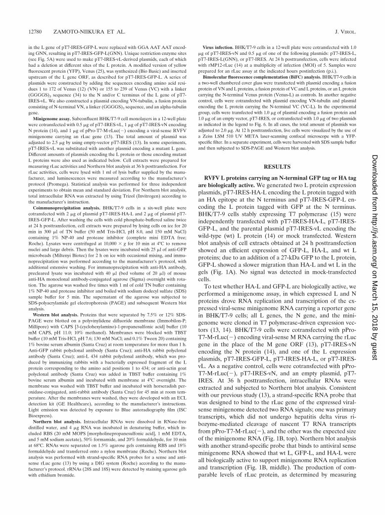
Interaction of GFP-L and HA-L in expressed cells. Wetested whether HA-L interacted with GFP-L in coexpressedcells. BHK/T7-9 cells were independently transfected withpT7-IRES-HA-L, pT7-IRES-GFP-L, and pT7-IRES-GFP ex-pressing GFP or cotransfected with pT7-IRES-GFP-L and
pT7-IRES-HA-L or pT7-IRES-HA-L and pT7-IRES-GFP; 4�g of plasmid was used for expression of a single protein, while2 �g of each plasmid was used for coexpression. Western blotanalysis of the cell extracts using anti-HA and anti-GFP anti-bodies showed an efficient accumulation of HA-L, GFP-L, andGFP in the cells expressing a single protein, while the amountsof HA-L and GFP-L in the cells coexpressing both proteinsand that of HA-L in the cells coexpressing HA-L and GFPwere less prominent, probably partly due to the use of reducedamounts of each plasmid in coexpressing cells (Fig. 2, top).Anti-HA antibody coimmunoprecipitated GFP-L, but notGFP, along with HA-L (Fig. 2, middle). A reciprocal experi-ment, in which cell extracts were immunoprecipitated withanti-GFP antibody, also showed coprecipitation of HA-L withGFP-L but not with GFP (Fig. 2, bottom). These data dem-onstrated that HA-L interacted with the L protein region ofthe GFP-L protein in coexpressed cells.
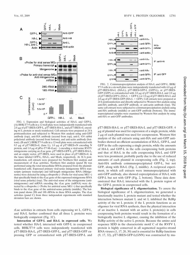
Biological significance of L oligomerization. To assess thebiological significance of L oligomerization, we generated afunctionally inactive L protein mutant and tested whether theinteraction between mutant L and wt L inhibited the RdRpactivity of the wt L protein; if the L protein functions as anoligomer for viral RNA synthesis, then the physical interactionof an inactive L mutant with an active L protein(s) in cellscoexpressing both proteins would result in the formation of abiologically inactive L oligomer, causing the inhibition of theRdRp activity of the active L protein. Because the amino acidsequence SDD in the characteristic motif C of the RVFV Lprotein is highly conserved in all segmented negative-strandRNA viruses (1, 17, 20, 30) and is essential for RdRp functions(9, 17, 32), pT7-IRES-GFP-L(GNN) was constructed by mu-
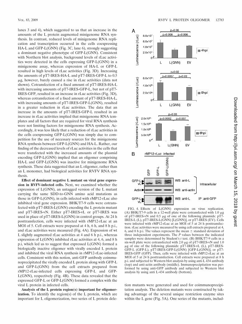
FIG. 1. Expression and biological activities of HA-L and GFP-L.(A) BHK/T7-9 cells in a 12-well plate were independently transfected with2.0 �g of pT7-IRES-GFP-L, pT7-IRES-HA-L, and pT7-IRES-vL encod-ing wt L protein or mock transfected. Cell extracts were prepared at 24 hposttransfection and subjected to Western blot analysis using anti-GFPantibody (top), anti-HA antibody (second from top), anti-L 434 rabbitpolyclonal antibody (second from bottom), and anti-actin antibody (bot-tom). (B and C) BHK/T7-9 cells in a 12-well plate were cotransfected with0.5 �g of pT7-IRES-vL (lane L), 1.0 �g of pT7-IRES-vN encoding Nprotein, and 1.0 �g of pPro-T7-M-rLuc(�) encoding a viral-sense RVFVminigenome carrying an rLuc gene. pT7-IRES-GFP-L, pT7-IRES-HA-L,and an empty vector, pT7-IRES, were used in place of pT7-IRES-vL inthe lanes labeled GFP-L, HA-L, and Mock, respectively. At 36 h post-transfection, cell extracts were prepared for Northern blot analysis andmeasurement of rLuc activities. Northern blot analysis (panel B) wasperformed using the total intracellular RNAs prepared from the plasmid-transfected cells. Expressed primary viral-sense minigenome RNA tran-scripts (primary transcript) and full-length minigenome RNA (Minige-nome) were detected by using a riboprobe (�Probe for viral sense MG�)that specifically binds to the rLuc gene of the expressed minigenome RNA(viral sense polarity) (top). The anti-viral sense of the minigenome (anti-minigenome) and mRNA encoding the rLuc gene (mRNA) were de-tected by a riboprobe (�Probe for antiviral sense MG�) that specificallybinds to the rLuc gene of the antiviral-sense polarity (middle). The bot-tom panel shows 28S and 18S rRNAs in these RNA samples. The rLucactivities (panel C) from three independent experiments with standarddeviation bars are shown.
FIG. 2. Coimmunoprecipitation analysis of HA-L and GFP-L. BHK/T7-9 cells in a six-well plate were independently transfected with 4.0 �g ofpT7-IRES-HA-L (HA-L), pT7-IRES-GFP-L (GFP-L), or pT7-IRES-GFP (GFP) or cotransfected with 2.0 �g of pT7-IRES-HA-L and 2.0 �gof pT7-IRES-GFP-L (HA-L � GFP-L) or 2.0 �g of pT7-IRES-HA-L and2.0 �g of pT7-IRES-GFP (HA-L � GFP). Cell extracts were prepared at24 h posttransfection and directly subjected to Western blot analysis usinganti-HA antibody, anti-GFP antibody, or anti-actin antibody (top). Thesame cell extracts were subjected to coimmunoprecipitation analysis usinganti-HA antibody (middle) or anti-GFP antibody (bottom). The immu-noprecipitated samples were examined by Western blot analysis by usinganti-HA or anti-GFP antibodies.
tating the amino acids SDD, at the positions 1132 to 1134 ofthe L protein region in pT7-IRES-GFP-L, to GNN. To knowwhether GFP-L(GNN) interacted with a biologically active Lprotein, coimmunoprecipitation analysis was performed usingextracts of cells coexpressing GFP-L(GNN) and HA-L (Fig.3A). Expressed GFP-L(GNN) bound to coexpressed HA-L(Fig. 3A, bottom), an observation demonstrating that the in-troduced mutation did not prevent L oligomerization. Thecoexpression of GFP-L(GNN) with N protein and minigenomeRNA did not induce minigenome RNA replication and tran-
scription (Fig. 3C, lane 3), regardless of efficient GFP-L(GNN)expression (Fig. 3B, lane 3), demonstrating that GFP-L(GNN)was biologically inactive for RNA synthesis. To determinewhether GFP-L(GNN) exerted a dominant negative effect onthe RdRp activity of a biologically active L, the minigenomeassays were performed in cells coexpressing HA-L and GFP-L(GNN). As controls for the assay, GFP-L and GFP werecoexpressed with HA-L. The abundance of minigenome RNAand mRNA was higher in the cells coexpressing GFP-L andHA-L than in those coexpressing HA-L and GFP (Fig. 3C,
FIG. 3. Characterization of a dominant negative mutant of L protein, GFP-L(GNN). (A) BHK/T7-9 cells in a six-well plate were cotransfectedwith 2.0 �g of pT7-IRES-HA-L and 2.0 �g of one of the following plasmids: pT7-IRES-GFP-L (GFP-L), pT7-IRES-GFP-L(GNN) encodingGFP-L(GNN), or pT7-IRES-GFP (GFP). Cell extracts were prepared at 24 h posttransfection and directly subjected to Western blot analysis(Intracellular) by using anti-GFP antibody, anti-HA antibody, or anti-actin antibody. The same cell extracts were subjected to coimmunoprecipi-tation analysis using anti-GFP antibody (IP: anti-GFP), and the immunoprecipitated proteins were detected by anti-GFP antibody or anti-HAantibody by Western blot analysis. (B) BHK/T7-9 cells in a 12-well plate were cotransfected with 1.0 �g of pT7-IRES-vN, 1.0 �g of pPro-T7-M-rLuc(�), and 0.5 �g of one of the following plasmids: pT7-IRES-HA-L (lane 1); pT7-IRES-GFP-L (lane 2); pT7-IRES-GFP-L(GNN) (lane 3);a mixture of 0.25 �g of pT7-IRES-HA-L and 0.25 �g of pT7-IRES-GFP (lane 4); a mixture of 0.25 �g of pT7-IRES-HA-L and 0.25 �g ofpT7-IRES-GFP-L (lane 5); or a mixture of 0.25 �g of pT7-IRES-HA-L and 0.25 �g of pT7-IRES-GFP-L(GNN) (lane 6). Cell extracts wereprepared for Western blot analysis at 36 h posttransfection. Anti-GFP antibody (top two rows), anti-HA antibody (third row from top), anti-L 434rabbit polyclonal antibody (fourth row from top), and anti-actin antibody (bottom) were used for Western blot analysis. (C) Cells were transfectedand prepared as described in the legend for panel B, and intracellular RNAs were subjected to Northern blot analysis as described in the legendfor Fig. 1B. (D) BHK/T7-9 cells in a 12-well plate were cotransfected with 1 �g of pT7-IRES-vN, 1 �g of pPro-T7-M-rLuc(�), and one plasmidor two plasmids shown in the diagram at indicated amounts. Cell extracts were prepared at 36 h posttransfection, and rLuc activities weremeasured. The values represent the mean � standard deviation of three independent experiments. The P values between the indicated sampleswere determined by Student’s t test.
lanes 3 and 4), which suggested to us that an increase in theamounts of the L protein augmented minigenome RNA syn-thesis. In contrast, reduced levels of minigenome RNA repli-cation and transcription occurred in the cells coexpressingHA-L and GFP-L(GNN) (Fig. 3C, lane 6), strongly suggestinga dominant negative phenotype of GFP-L(GNN). Consistentwith Northern blot analysis, background levels of rLuc activi-ties were detected in the cells expressing GFP-L(GNN) in aminigenome assay, whereas expression of HA-L or GFP-Lresulted in high levels of rLuc activities (Fig. 3D). Increasingthe amounts of pT7-IRES-HA-L and pT7-IRES-GFP-L to 0.5�g, however, barely caused a rise in rLuc activities (data notshown). Cotransfection of a fixed amount of pT7-IRES-HA-Lwith increasing amounts of pT7-IRES-GFP-L, but not of pT7-IRES-GFP, resulted in an increase in rLuc activities (Fig. 3D),whereas cotransfection of a fixed amount of pT7-IRES-HA-L,with increasing amounts of pT7-IRES-GFP-L(GNN), resultedin a greater reduction in rLuc activities. The data that anincrease in the amounts of pT7-IRES-GFP-L resulted in anincrease in rLuc activities implied that minigenome RNA tem-plates and all factors that are required for viral RNA synthesiswere not limiting factors for minigenome RNA synthesis. Ac-cordingly, it was less likely that a reduction of rLuc activities inthe cells coexpressing GFP-L(GNN) was simply due to com-petition for the use of necessary sources for the minigenomeRNA synthesis between GFP-L(GNN) and HA-L. Rather, ourfinding of the decreased levels of rLuc activities in the cells thatwere transfected with the increased amounts of the plasmidencoding GFP-L(GNN) implied that an oligomer comprisingHA-L and GFP-L(GNN) was inactive for minigenome RNAsynthesis. These data suggested that an L oligomer, rather thanan L monomer, had biological activities for RVFV RNA syn-thesis.
Effect of dominant negative L mutant on viral gene expres-sion in RVFV-infected cells. Next, we examined whether theexpression of L(GNN), an untagged version of the L mutantcarrying the same SDD-to-GNN amino acid mutations asthose in GFP-L(GNN), in cells infected with rMP12-rLuc alsoinhibited viral gene expression. BHK/T7-9 cells were cotrans-fected with pT7-IRES-L(GNN) encoding the L protein mutantand pT7-IRES-vN. Either pT7-IRES-vL or pT7-IRES wasused in place of pT7-IRES-L(GNN) in control groups. At 24 hposttransfection, cells were infected with rMP12-rLuc at anMOI of 5. Cell extracts were prepared at 4 h, 6 h, and 8 h p.i.,and rLuc activities were measured (Fig. 4A). Expression of wtL slightly augmented rLuc activities at 4 and 6 h p.i., whereasexpression of L(GNN) inhibited rLuc activities at 4, 6, and 8 hp.i, which led us to suggest that expressed L(GNN) formed abiologically inactive oligomer with virally encoded L proteinand inhibited the viral RNA synthesis in rMP12-rLuc-infectedcells. Consistent with this notion, anti-GFP antibody coimmu-noprecipitated the virally encoded L protein along with GFP-Land GFP-L(GNN) from the cell extracts prepared fromrMP12-rLuc-infected cells expressing GFP-L and GFP-L(GNN), respectively (Fig. 4B). These data revealed that theexpressed GFP-L or GFP-L(GNN) formed a complex with theviral L protein in infected cells.
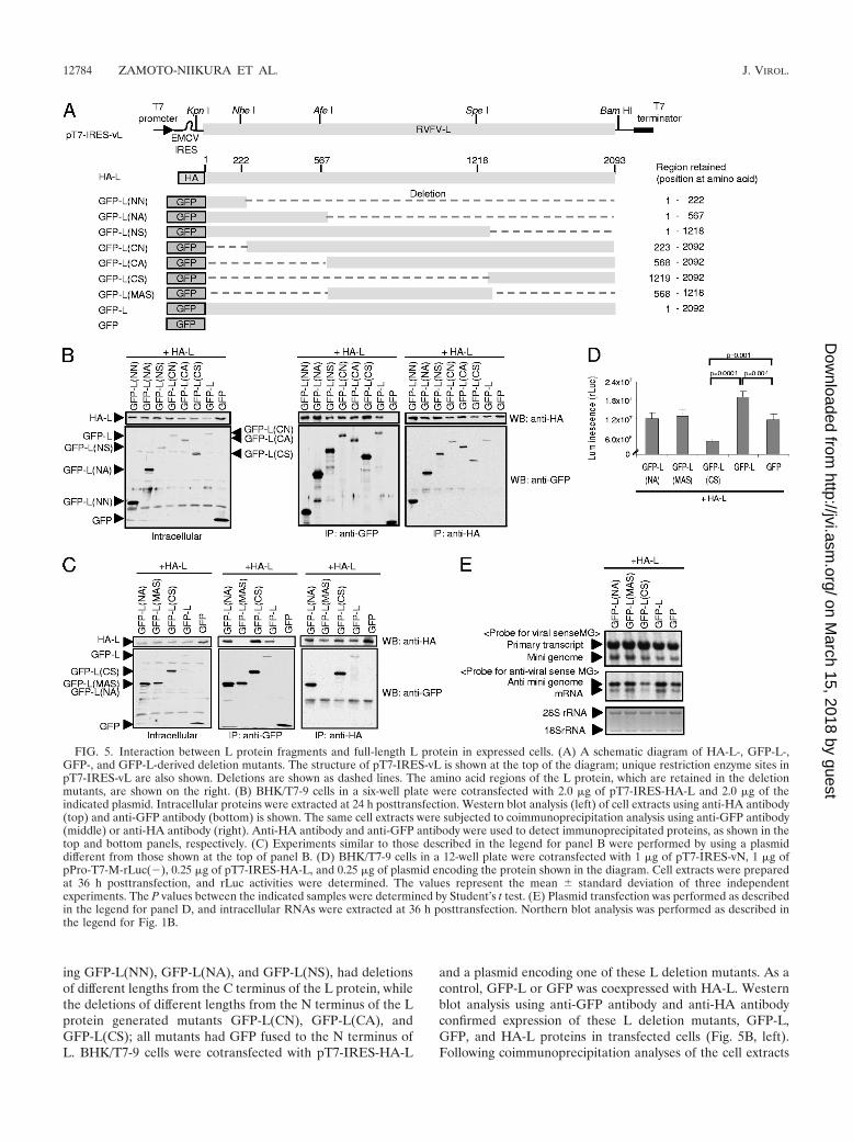
Analysis of the L protein region(s) important for oligomer-ization. To identify the region(s) of the L protein, which areimportant for L oligomerization, two series of L protein dele-
tion mutants were generated and used for coimmunoprecipi-tation analysis. The deletion mutants were constructed by tak-ing advantage of the several unique restriction enzyme siteswithin the L gene (Fig. 5A). One series of the mutants, includ-
FIG. 4. Effects of L(GNN) expression on virus replication.(A) BHK/T7-9 cells in a 12-well plate were cotransfected with 1.0 �gof pT7-IRES-vN and 0.5 �g of one of the following plasmids: pT7-IRES-L (L), pT7-IRES-L(GNN) [L(GNN)], or pT7-IRES (EV). Cellswere infected with rMP12-rLuc at an MOI of 5 at 24 h posttransfec-tion. rLuc activities were measured by using cell extracts prepared at 4,6, and 8 h p.i. The values represent the mean � standard deviation ofthree independent experiments. The P values between the indicatedsamples were determined by Student’s t test. (B) BHK/T7-9 cells in asix-well plate were cotransfected with 2.0 �g of pT7-IRES-vN and 1.0�g of one of the following plasmids: pT7-IRES-vL (L), pT7-IRES-GFP-L (GFP-L), pT7-IRES-GFP-L(GNN) [GFP-L(GNN)], or pT7-IRES-GFP (GFP). Then, cells were infected with rMP12-rLuc at anMOI of 5 at 24 h posttransfection. Cell extracts were prepared at 8 hp.i. and subjected to Western blot analysis by using anti-L 434 antibody(top) and anti-actin antibody (middle). Immunoprecipitation was per-formed by using anti-GFP antibody and subjected to Western blotanalysis by using anti L-434 antibody (bottom).
ing GFP-L(NN), GFP-L(NA), and GFP-L(NS), had deletionsof different lengths from the C terminus of the L protein, whilethe deletions of different lengths from the N terminus of the Lprotein generated mutants GFP-L(CN), GFP-L(CA), andGFP-L(CS); all mutants had GFP fused to the N terminus ofL. BHK/T7-9 cells were cotransfected with pT7-IRES-HA-L
and a plasmid encoding one of these L deletion mutants. As acontrol, GFP-L or GFP was coexpressed with HA-L. Westernblot analysis using anti-GFP antibody and anti-HA antibodyconfirmed expression of these L deletion mutants, GFP-L,GFP, and HA-L proteins in transfected cells (Fig. 5B, left).Following coimmunoprecipitation analyses of the cell extracts
FIG. 5. Interaction between L protein fragments and full-length L protein in expressed cells. (A) A schematic diagram of HA-L-, GFP-L-,GFP-, and GFP-L-derived deletion mutants. The structure of pT7-IRES-vL is shown at the top of the diagram; unique restriction enzyme sites inpT7-IRES-vL are also shown. Deletions are shown as dashed lines. The amino acid regions of the L protein, which are retained in the deletionmutants, are shown on the right. (B) BHK/T7-9 cells in a six-well plate were cotransfected with 2.0 �g of pT7-IRES-HA-L and 2.0 �g of theindicated plasmid. Intracellular proteins were extracted at 24 h posttransfection. Western blot analysis (left) of cell extracts using anti-HA antibody(top) and anti-GFP antibody (bottom) is shown. The same cell extracts were subjected to coimmunoprecipitation analysis using anti-GFP antibody(middle) or anti-HA antibody (right). Anti-HA antibody and anti-GFP antibody were used to detect immunoprecipitated proteins, as shown in thetop and bottom panels, respectively. (C) Experiments similar to those described in the legend for panel B were performed by using a plasmiddifferent from those shown at the top of panel B. (D) BHK/T7-9 cells in a 12-well plate were cotransfected with 1 �g of pT7-IRES-vN, 1 �g ofpPro-T7-M-rLuc(�), 0.25 �g of pT7-IRES-HA-L, and 0.25 �g of plasmid encoding the protein shown in the diagram. Cell extracts were preparedat 36 h posttransfection, and rLuc activities were determined. The values represent the mean � standard deviation of three independentexperiments. The P values between the indicated samples were determined by Student’s t test. (E) Plasmid transfection was performed as describedin the legend for panel D, and intracellular RNAs were extracted at 36 h posttransfection. Northern blot analysis was performed as described inthe legend for Fig. 1B.
by the use of anti-HA antibody or anti-GFP antibody, all Ldeletion mutants were found to have interacted with coex-pressed HA-L protein (Fig. 5B, middle and right). These datademonstrated that both the N-terminal and C-terminal regionsof the L protein bound to the full-length L protein.
To know whether the middle region of the L protein wasinvolved in L protein oligomerization, we constructed plasmidencoding GFP-L(MAS), a fusion protein consisting of N-ter-minal GFP and a C-terminal L protein fragment correspond-ing to amino acids from positions 568 to 1218 (Fig. 5A), andcoexpressed it with HA-L; the size of GFP-L(MAS) was com-parable to that of GFP-L(NA) or GFP-L(CS) (Fig. 5C, left).Anti-GFP antibody failed to coimmunoprecipitate HA-L alongwith GFP-L(MAS) from the cell extracts coexpressing HA-Land GFP-L(MAS) (Fig. 5C, middle). We also did not detect aninteraction between GFP-L(MAS) and HA-L in a reciprocalexperiment, in which anti-HA antibody was used for coimmu-noprecipitation (Fig. 5C, right). These data demonstrated thatthe N- and C-terminal regions, but not the middle region, ofthe L protein interacted with the full-length L protein.
We next tested effects of the expression of these L frag-ments, including GFP-L(NA), GFP-L(MAS), and GFL-L(CS),in the minigenome assay, wherein expression of GFP-L or GFPserved as a control. Consistent with the data shown in Fig. 3,the coexpression of HA-L with GFP-L resulted in higher rLucactivities and rLuc mRNA abundance than did the coexpres-sion of HA-L and GFP (Fig. 5D and E). We did not seereductions in rLuc activities and rLuc mRNA abundance in thecells coexpressing GFP-L(NA) and HA-L and in those coex-pressing GFP-L(MAS) and HA-L. In contrast, GFP-L(CS)expression inhibited HA-L-mediated minigenome RNA syn-thesis and rLuc activities (Fig. 5D and E). These data suggestthat the binding of the C-terminal fragment of the L protein,but not the N-terminal fragment of L protein, to HA-L inhib-ited HA-L protein-mediated minigenome RNA synthesis.
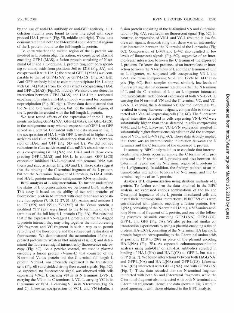
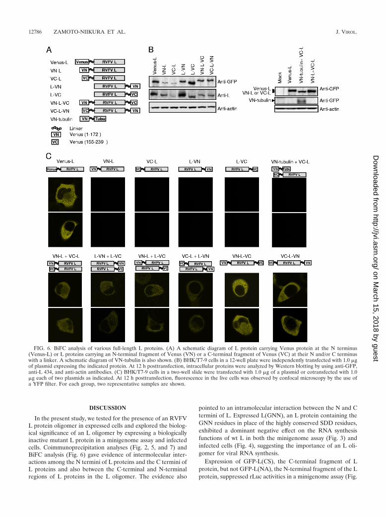
BiFC analysis of L oligomerization. To further understandthe status of L oligomerization, we performed BiFC analysis.This assay is based on the ability of two split proteins offluorescence protein to interact with each other and reconsti-tute fluorophore (7, 10, 12, 27, 31, 35). Amino acid residues 1to 172 (VN) and 155 to 239 (VC) of the Venus protein, amodified YFP (25), were fused to the N terminus or the Cterminus of the full-length L protein (Fig. 6A). We reasonedthat if the expressed VN-tagged L protein and the VC-taggedL protein interact, they may bring together the nonfluorescingVN fragment and VC fragment in such a way as to permitrefolding of the fluorophore and the subsequent restoration offluorescence. We demonstrated the accumulation of the ex-pressed proteins by Western blot analysis (Fig. 6B) and deter-mined the fluorescent signal intensities by fluorescence micros-copy (Fig. 6C). As a positive control, we used a plasmidencoding a fusion protein (Venus-L) that consisted of theN-terminal Venus protein and the C-terminal full-length Lprotein. Venus-L was efficiently expressed in the transfectedcells (Fig. 6B) and yielded strong fluorescent signals (Fig. 6C).As expected, no fluorescence signal was observed with cellsexpressing VN-L, L carrying VN in its N terminus; L-VN, Lcarrying the VN in its C terminus; L-VC, L carrying VC in itsC terminus; or VC-L, L carrying VC in its N terminus (Fig. 6Aand C). Likewise, coexpression of VC-L and VN-tubulin, a
fusion protein consisting of the N-terminal VN and C-terminaltubulin (Fig. 6A), resulted in no fluorescent signal (Fig. 6C). Incontrast, coexpression of VN-L and VC-L resulted in low flu-orescent signals, demonstrating that there was an intermolec-ular interaction between the N termini of the L proteins (Fig.6C). Coexpression of L-VN and L-VC also resulted in lowlevels of fluorescent signals (Fig. 6C), suggestive of an inter-molecular interaction between the C termini of the expressedL proteins. To know the presence of an intermolecular inter-action between the N terminus of L and the C terminus of L inan L oligomer, we subjected cells coexpressing VN-L andL-VC and those coexpressing VC-L and L-VN to BiFC anal-ysis (Fig. 6C). Both samples showed similarly low levels offluorescent signals that demonstrated to us that the N terminusof L and the C terminus of L in an L oligomer interactedintermolecularly. Independent expression of both VN-L-VC, Lcarrying the N-terminal VN and the C-terminal VC, and VC-L-VN, L carrying the N-terminal VC and the C-terminal VL,resulted in strong fluorescent signals, comparable to those de-tected with Venus-L-expressing cells (Fig. 6C). The fluorescentsignal intensities detected in cells expressing VN-L-VC weresubstantially higher than those detected in cells coexpressingVN-L and L-VC. Likewise, VC-L-VN expression resulted insubstantially higher fluorescence signals than did the coexpres-sion of VC-L and L-VN (Fig. 6C). These data strongly impliedthat there was an intramolecular interaction between the Nterminus and the C terminus of the expressed L protein.
In summary, BiFC analysis led us to conclude that intermo-lecular interactions occurred among the C termini of L pro-teins and the N termini of L proteins and also between theC-terminal region and the N-terminal region of L proteins inthe L oligomer(s). In addition, the findings pointed to an in-tramolecular interaction between the N-terminal and the C-terminal regions of an L protein.
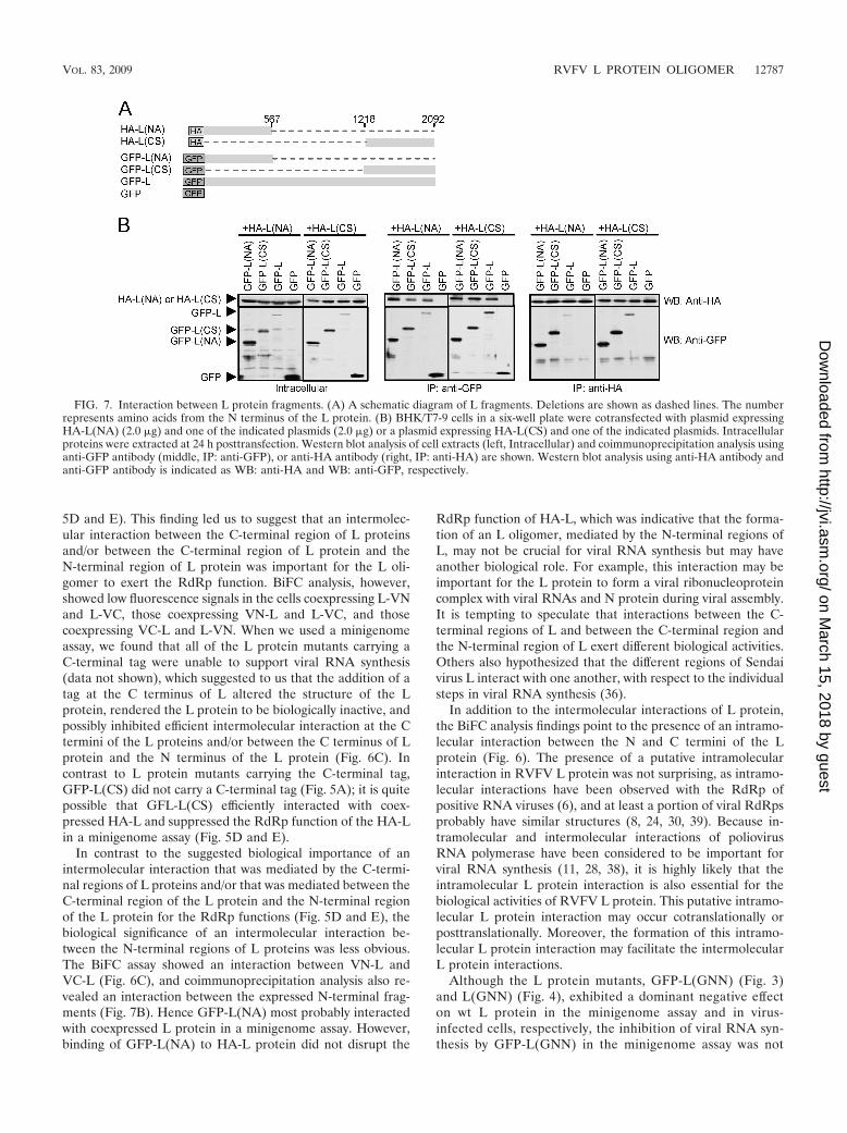
Analysis of L oligomerization using deletion mutants of Lprotein. To further confirm the data obtained in the BiFCanalysis, we expressed various combinations of the N- andC-terminal fragments of the L protein in cultured cells andtested their intermolecular interactions. BHK/T7-9 cells werecotransfected with plasmid encoding a fusion protein, HA-L(NA), consisting of the N-terminal HA tag, a 567-amino-acid-long N-terminal fragment of L protein, and one of the follow-ing plasmids: plasmids encoding GFP-L(NA), GFP-L(CS),GFP-L, and GFP (Fig. 7A). We also performed similar co-transfection experiments by using a plasmid encoding a fusionprotein, HA-L(CS), consisting of the N-terminal HA tag and Lprotein fragment corresponding to the C-terminal amino acidsat positions 1219 to 2092 in place of the plasmid encodingHA-L(NA) (Fig. 7B). As expected, coimmunoprecipitationanalyses using anti-GFP or anti-HA antibodies resulted inbinding of HA-L(NA) and HA-L(CS) to GFP-L, but not toGFP (Fig. 7). We found interactions between both HA-L(NA)and GFP-L(NA) and HA-L(NA) and GFP-L(CS). Likewise,HA-L(CS) interacted with GFP-L(NA) and with GFP-L(CS)(Fig. 7). These data revealed that the N-terminal fragmentinteracted with both N- and C-terminal fragments, while theC-terminal fragment also interacted with both N-terminal andC-terminal fragments. Hence, the data shown in Fig. 7 were ingood agreement with those obtained in the BiFC analysis.
In the present study, we tested for the presence of an RVFVL protein oligomer in expressed cells and explored the biolog-ical significance of an L oligomer by expressing a biologicallyinactive mutant L protein in a minigenome assay and infectedcells. Coimmunoprecipitation analyses (Fig. 2, 5, and 7) andBiFC analysis (Fig. 6) gave evidence of intermolecular inter-actions among the N termini of L proteins and the C termini ofL proteins and also between the C-terminal and N-terminalregions of L proteins in the L oligomer. The evidence also
pointed to an intramolecular interaction between the N and Ctermini of L. Expressed L(GNN), an L protein containing theGNN residues in place of the highly conserved SDD residues,exhibited a dominant negative effect on the RNA synthesisfunctions of wt L in both the minigenome assay (Fig. 3) andinfected cells (Fig. 4), suggesting the importance of an L oli-gomer for viral RNA synthesis.
Expression of GFP-L(CS), the C-terminal fragment of Lprotein, but not GFP-L(NA), the N-terminal fragment of the Lprotein, suppressed rLuc activities in a minigenome assay (Fig.
FIG. 6. BiFC analysis of various full-length L proteins. (A) A schematic diagram of L protein carrying Venus protein at the N terminus(Venus-L) or L proteins carrying an N-terminal fragment of Venus (VN) or a C-terminal fragment of Venus (VC) at their N and/or C terminuswith a linker. A schematic diagram of VN-tubulin is also shown. (B) BHK/T7-9 cells in a 12-well plate were independently transfected with 1.0 �gof plasmid expressing the indicated protein. At 12 h posttransfection, intracellular proteins were analyzed by Western blotting by using anti-GFP,anti-L 434, and anti-actin antibodies. (C) BHK/T7-9 cells in a two-well slide were transfected with 1.0 �g of a plasmid or cotransfected with 1.0�g each of two plasmids as indicated. At 12 h posttransfection, fluorescence in the live cells was observed by confocal microscopy by the use ofa YFP filter. For each group, two representative samples are shown.
5D and E). This finding led us to suggest that an intermolec-ular interaction between the C-terminal region of L proteinsand/or between the C-terminal region of L protein and theN-terminal region of L protein was important for the L oli-gomer to exert the RdRp function. BiFC analysis, however,showed low fluorescence signals in the cells coexpressing L-VNand L-VC, those coexpressing VN-L and L-VC, and thosecoexpressing VC-L and L-VN. When we used a minigenomeassay, we found that all of the L protein mutants carrying aC-terminal tag were unable to support viral RNA synthesis(data not shown), which suggested to us that the addition of atag at the C terminus of L altered the structure of the Lprotein, rendered the L protein to be biologically inactive, andpossibly inhibited efficient intermolecular interaction at the Ctermini of the L proteins and/or between the C terminus of Lprotein and the N terminus of the L protein (Fig. 6C). Incontrast to L protein mutants carrying the C-terminal tag,GFP-L(CS) did not carry a C-terminal tag (Fig. 5A); it is quitepossible that GFL-L(CS) efficiently interacted with coex-pressed HA-L and suppressed the RdRp function of the HA-Lin a minigenome assay (Fig. 5D and E).
In contrast to the suggested biological importance of anintermolecular interaction that was mediated by the C-termi-nal regions of L proteins and/or that was mediated between theC-terminal region of the L protein and the N-terminal regionof the L protein for the RdRp functions (Fig. 5D and E), thebiological significance of an intermolecular interaction be-tween the N-terminal regions of L proteins was less obvious.The BiFC assay showed an interaction between VN-L andVC-L (Fig. 6C), and coimmunoprecipitation analysis also re-vealed an interaction between the expressed N-terminal frag-ments (Fig. 7B). Hence GFP-L(NA) most probably interactedwith coexpressed L protein in a minigenome assay. However,binding of GFP-L(NA) to HA-L protein did not disrupt the
RdRp function of HA-L, which was indicative that the forma-tion of an L oligomer, mediated by the N-terminal regions ofL, may not be crucial for viral RNA synthesis but may haveanother biological role. For example, this interaction may beimportant for the L protein to form a viral ribonucleoproteincomplex with viral RNAs and N protein during viral assembly.It is tempting to speculate that interactions between the C-terminal regions of L and between the C-terminal region andthe N-terminal region of L exert different biological activities.Others also hypothesized that the different regions of Sendaivirus L interact with one another, with respect to the individualsteps in viral RNA synthesis (36).
In addition to the intermolecular interactions of L protein,the BiFC analysis findings point to the presence of an intramo-lecular interaction between the N and C termini of the Lprotein (Fig. 6). The presence of a putative intramolecularinteraction in RVFV L protein was not surprising, as intramo-lecular interactions have been observed with the RdRp ofpositive RNA viruses (6), and at least a portion of viral RdRpsprobably have similar structures (8, 24, 30, 39). Because in-tramolecular and intermolecular interactions of poliovirusRNA polymerase have been considered to be important forviral RNA synthesis (11, 28, 38), it is highly likely that theintramolecular L protein interaction is also essential for thebiological activities of RVFV L protein. This putative intramo-lecular L protein interaction may occur cotranslationally orposttranslationally. Moreover, the formation of this intramo-lecular L protein interaction may facilitate the intermolecularL protein interactions.
Although the L protein mutants, GFP-L(GNN) (Fig. 3)and L(GNN) (Fig. 4), exhibited a dominant negative effecton wt L protein in the minigenome assay and in virus-infected cells, respectively, the inhibition of viral RNA syn-thesis by GFP-L(GNN) in the minigenome assay was not
FIG. 7. Interaction between L protein fragments. (A) A schematic diagram of L fragments. Deletions are shown as dashed lines. The numberrepresents amino acids from the N terminus of the L protein. (B) BHK/T7-9 cells in a six-well plate were cotransfected with plasmid expressingHA-L(NA) (2.0 �g) and one of the indicated plasmids (2.0 �g) or a plasmid expressing HA-L(CS) and one of the indicated plasmids. Intracellularproteins were extracted at 24 h posttransfection. Western blot analysis of cell extracts (left, Intracellular) and coimmunoprecipitation analysis usinganti-GFP antibody (middle, IP: anti-GFP), or anti-HA antibody (right, IP: anti-HA) are shown. Western blot analysis using anti-HA antibody andanti-GFP antibody is indicated as WB: anti-HA and WB: anti-GFP, respectively.
very strong; the rLuc activity in the cells coexpressing HA-Land GFP-L(GNN) was roughly one-fourth of the rLuc ac-tivity detected with the cells coexpressing HA-L and GFP-L,suggesting an approximately 75% reduction in the rLuc ac-tivity by the coexpression of GFP-L(GNN) (Fig. 3D). Incontrast, the coexpression of the same level of wt LCMV Lprotein and its dominant negative mutant in a similar mini-genome assay resulted in a 20- to 30-fold reduction in mini-genome RNA synthesis (32). These data suggest that thestatus of a biologically active L oligomer probably differedbetween RVFV and LCMV. The finding that a dominantnegative mutant of LCMV L protein efficiently suppressedminigenome RNA synthesis suggested to us that a biologi-cally active LCMV L oligomer carries many L proteins andthat inclusion of one molecule of a dominant negative mu-tant in an L oligomer renders the L oligomer biologicallyincompetent. In contrast, a modest inhibition of minig-enome RNA synthesis by a dominant negative mutant ofRVFV L protein implied to us that a biologically active Loligomer included a small number of L proteins. Furtherstudies are required to determine the stoichiometry of the Lprotein oligomer that exerted the RdRp activity.
HA-L and GFP-L were biologically active to supportminigenome RNA synthesis, demonstrating that the addi-tion of the N-terminal tag did not suppress the RdRp func-tion. However, we were unable to rescue an RVFV mutantvirus carrying L RNA encoding HA-L (data not shown),which suggested to us that adding an HA tag sequence at the5 end of the L gene ORF was not tolerated at a certainstep(s) in RVFV replication. In this regard, it is worthnoting that Shi and Elliott reported the generation of re-combinant Bunyamwera orthobunyaviruses expressing a V5epitope tag within the L gene ORF but not at the termini ofthe L gene (33). Because various reagents that are suitablefor purification of HA-tagged or GFP-tagged proteins areavailable, expressed HA-L or GFP-L may be useful for iden-tifying host factors that associate with the RVFV L proteinand characterizing viral replication/transcription complexes.
ACKNOWLEDGMENTS
We thank Krishna Narayanan for discussion and critical reading ofthe manuscript and Thomas Albrecht for supporting our experimentsvia confocal microscopy.
This work was supported by grants from NIAID to S.M. and C.J.P.through the Western Regional Center of Excellence for Biodefenseand Emerging Infectious Diseases Research, NIH grant number U54AI057156, and by NIH-NIAID-DMID-02-24 Collaborative Grant onEmerging Viral Diseases. K.T. was supported by the James W.McLaughlin fellowship fund.
REFERENCES
1. Aquino, V. H., M. L. Moreli, and L. T. Moraes Figueiredo. 2003. Analysis oforopouche virus L protein amino acid sequence showed the presence of anadditional conserved region that could harbour an important role for thepolymerase activity. Arch. Virol. 148:19–28.
2. Bird, B. H., T. G. Ksiazek, S. T. Nichol, and N. J. Maclachlan. 2009. RiftValley fever virus. J. Am. Vet. Med. Assoc. 234:883–893.
3. Bishop, D. H., M. E. Gay, and Y. Matsuoko. 1983. Nonviral heterogeneoussequences are present at the 5 ends of one species of snowshoe harebunyavirus S complementary RNA. Nucleic Acids Res. 11:6409–6418.
4. Cevik, B., D. E. Holmes, E. Vrotsos, J. A. Feller, S. Smallwood, and S. A.Moyer. 2004. The phosphoprotein (P) and L binding sites reside in theN-terminus of the L subunit of the measles virus RNA polymerase. Virology327:297–306.
5. Doyle, T., and D. Botstein. 1996. Movement of yeast cortical actin cytoskel-eton visualized in vivo. Proc. Natl. Acad. Sci. USA 93:3886–3891.
6. Ferrer-Orta, C., A. Arias, C. Escarmis, and N. Verdaguer. 2006. A compar-ison of viral RNA-dependent RNA polymerases. Curr. Opin. Struct. Biol.16:27–34.
7. Frieman, M., B. Yount, M. Heise, S. A. Kopecky-Bromberg, P. Palese, andR. S. Baric. 2007. Severe acute respiratory syndrome coronavirus ORF6antagonizes STAT1 function by sequestering nuclear import factors onthe rough endoplasmic reticulum/Golgi membrane. J. Virol. 81:9812–9824.
8. Hansen, J. L., A. M. Long, and S. C. Schultz. 1997. Structure of the RNA-dependent RNA polymerase of poliovirus. Structure 5:1109–1122.
9. Hass, M., M. Lelke, C. Busch, B. Becker-Ziaja, and S. Gunther. 2008.Mutational evidence for a structural model of the Lassa virus RNA poly-merase domain and identification of two residues, Gly1394 and Asp1395,that are critical for transcription but not replication of the genome. J. Virol.82:10207–10217.
10. Hemerka, J. N., D. Wang, Y. Weng, W. Lu, R. S. Kaushik, J. Jin, A. F.Harmon, and F. Li. 2009. Detection and characterization of influenza A virusPA-PB2 interaction through a bimolecular fluorescence complementationassay. J. Virol. 83:3944–3955.
11. Hobson, S. D., E. S. Rosenblum, O. C. Richards, K. Richmond, K. Kirkeg-aard, and S. C. Schultz. 2001. Oligomeric structures of poliovirus polymer-ase are important for function. EMBO J. 20:1153–1163.
12. Hu, C. D., and T. K. Kerppola. 2003. Simultaneous visualization of multipleprotein interactions in living cells using multicolor fluorescence complemen-tation analysis. Nat. Biotechnol. 21:539–545.
13. Ikegami, T., C. J. Peters, and S. Makino. 2005. Rift Valley fever virusnonstructural protein NSs promotes viral RNA replication and transcriptionin a minigenome system. J. Virol. 79:5606–5615.
14. Ikegami, T., S. Won, C. J. Peters, and S. Makino. 2006. Rescue ofinfectious Rift Valley fever virus entirely from cDNA, analysis of viruslacking the NSs gene, and expression of a foreign gene. J. Virol. 80:2933–2940.
15. Ito, N., M. Takayama-Ito, K. Yamada, J. Hosokawa, M. Sugiyama, and N.Minamoto. 2003. Improved recovery of rabies virus from cloned cDNAusing a vaccinia virus-free reverse genetics system. Microbiol. Immunol.47:613–617.
16. Jin, H., and R. M. Elliott. 1993. Characterization of Bunyamwera virus SRNA that is transcribed and replicated by the L protein expressed fromrecombinant vaccinia virus. J. Virol. 67:1396–1404.
17. Jin, H., and R. M. Elliott. 1992. Mutagenesis of the L protein encoded byBunyamwera virus and production of monospecific antibodies. J. Gen. Virol.73:2235–2244.
18. Jorba, N., E. Area, and J. Ortin. 2008. Oligomerization of the influenza viruspolymerase complex in vivo. J. Gen. Virol. 89:520–524.
19. Jorba, N., R. Coloma, and J. Ortin. 2009. Genetic trans-complementationestablishes a new model for influenza virus RNA transcription and replica-tion. PLoS Pathog. 5:e1000462.
20. Kinsella, E., S. G. Martin, A. Grolla, M. Czub, H. Feldmann, and R. Flick.2004. Sequence determination of the Crimean-Congo hemorrhagic fevervirus L segment. Virology 321:23–28.
21. Le May, N., N. Gauliard, A. Billecocq, and M. Bouloy. 2005. The N terminusof Rift Valley fever virus nucleoprotein is essential for dimerization. J. Virol.79:11974–11980.
22. Meegan, J. M., and C. J. Peters. 1989. Rift Valley fever. CRC Press, BocaRaton, FL.
23. Mohl, B. P., and J. N. Barr. 2009. Investigating the specificity and stoichi-ometry of RNA binding by the nucleocapsid protein of Bunyamwera virus.RNA 15:391–399.
24. Muller, R., O. Poch, M. Delarue, D. H. Bishop, and M. Bouloy. 1994. RiftValley fever virus L segment: correction of the sequence and possible func-tional role of newly identified regions conserved in RNA-dependent poly-merases. J. Gen. Virol. 75:1345–1352.
25. Nagai, T., K. Ibata, E. S. Park, M. Kubota, K. Mikoshiba, and A. Miyawaki.2002. A variant of yellow fluorescent protein with fast and efficient matura-tion for cell-biological applications. Nat. Biotechnol. 20:87–90.
26. Nichol, S. T. 2001. Bunyaviruses, 4th ed. Lippincott-Raven, Philadelphia,PA.
27. Nyfeler, B., S. W. Michnick, and H. P. Hauri. 2005. Capturing proteininteractions in the secretory pathway of living cells. Proc. Natl. Acad. Sci.USA 102:6350–6355.
28. Pata, J. D., S. C. Schultz, and K. Kirkegaard. 1995. Functional oligomer-ization of poliovirus RNA-dependent RNA polymerase. RNA 1:466–477.
29. Patterson, J. L., B. Holloway, and D. Kolakofsky. 1984. La Crosse virionscontain a primer-stimulated RNA polymerase and a methylated cap-depen-dent endonuclease. J. Virol. 52:215–222.
30. Poch, O., I. Sauvaget, M. Delarue, and N. Tordo. 1989. Identification of fourconserved motifs among the RNA-dependent polymerase encoding ele-ments. EMBO J. 8:3867–3874.
31. Remy, I., A. Montmarquette, and S. W. Michnick. 2004. PKB/Akt modulatesTGF-beta signalling through a direct interaction with Smad3. Nat. Cell Biol.6:358–365.
32. Sanchez, A. B., and J. C. de la Torre. 2005. Genetic and biochemical
evidence for an oligomeric structure of the functional L polymerase of theprototypic arenavirus lymphocytic choriomeningitis virus. J. Virol. 79:7262–7268.
33. Shi, X., and R. M. Elliott. 2009. Generation and analysis of recombinantBunyamwera orthobunyaviruses expressing V5 epitope-tagged L proteins.J. Gen. Virol. 90:297–306.
34. Shimozono, S., and A. Miyawaki. 2008. Engineering FRET constructs usingCFP and YFP. Methods Cell Biol. 85:381–393.
35. Shyu, Y. J., C. D. Suarez, and C. D. Hu. 2008. Visualization of AP-1 NF-Bternary complexes in living cells by using a BiFC-based FRET. Proc. Natl.Acad. Sci. USA 105:151–156.
36. Smallwood, S., B. Cevik, and S. A. Moyer. 2002. Intragenic complementationand oligomerization of the L subunit of the Sendai virus RNA polymerase.Virology 304:235–245.
37. Smallwood, S., and S. A. Moyer. 2004. The L polymerase protein of parain-fluenza virus 3 forms an oligomer and can interact with the heterologousSendai virus L, P and C proteins. Virology 318:439–450.
38. Thompson, A. A., R. A. Albertini, and O. B. Peersen. 2007. Stabilization ofpoliovirus polymerase by NTP binding and fingers-thumb interactions. J.Mol. Biol. 366:1459–1474.
39. Vieth, S., A. E. Torda, M. Asper, H. Schmitz, and S. Gunther. 2004. Se-quence analysis of L RNA of Lassa virus. Virology 318:153–168.