39
Visual Evoked Potentials Elayna Rubens, MD Assistant Professor of Neurology Weill Cornell Medical College Memorial Sloan Kettering Cancer Center
Visual Evoked Potentials
Elayna Rubens, MD
Assistant Professor of Neurology
Weill Cornell Medical College
Memorial Sloan Kettering Cancer Center

Outline• Visual Pathway Anatomy
• Basic VEP principles
-VEP Definition
-Types of VEPs
-Waveforms and generators
• VEP Techniques
-Patient/Testing Conditions
-Stimulation Parameters
-Recording Montage
• Interpretation
-Evaluation of the P100
-Variables affecting the P100
• Example VEPs
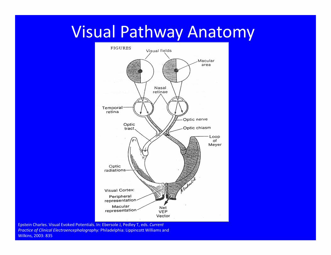
Visual Pathway Anatomy
Epstein Charles. Visual Evoked Potentials. In: Ebersole J, Pedley T, eds. Current
Practice of Clinical Electroencephalography: Philadelphia: Lippincott Williams and
Wilkins, 2003: 835
VEP
• Definition: An electrophysiologic response
time locked to a visual stimulus
• VEPs can be categorized by stimulus
characteristics:
1. Stimulus type: patterned (usually checkerboard) vs.
unpatterned (flash).
2. Field stimulated: monocular full field vs. hemi-field
3. Stimulus Frequency: transient VEPs vs. steady state VEPs
• Clinical use: most often used to evaluate optic
nerve function, but can detect abnormalities at
any point in the visual pathway

Neural Generators of the VEP• P100- Generators within occipital cortex (striate and extrastriate
cortex)
• Pattern VEP is dominated by central (macular) vision serving the central 8-10 degrees of the visual field
• N100- separate generator in the frontal region
P100
• A middle latency, near field potential
• It is the most consistent component of the
VEP and thus used for interpretation
• Assesses the conduction of neuronal activity
from the retina to the occipital cortex
• Typically maximal amplitude is in the mid-
occipital region, but can be displaced above or
below (normal variant)
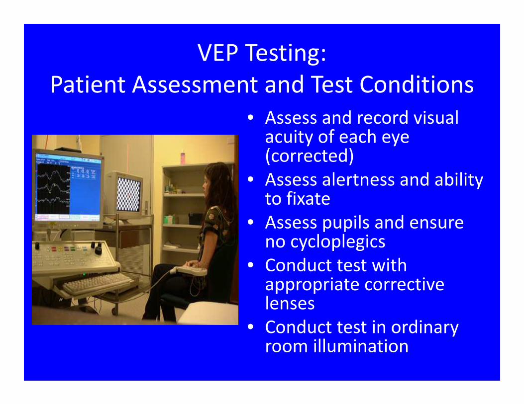
VEP Testing:
Patient Assessment and Test Conditions
• Assess and record visual acuity of each eye (corrected)
• Assess alertness and ability to fixate
• Assess pupils and ensure no cycloplegics
• Conduct test with appropriate corrective lenses
• Conduct test in ordinary room illumination

VEP Testing:
Stimulation Parameters
• Pattern Reversal
Full field - Better for detecting lesions anterior to the chiasm
Hemi-field - Used for detecting lesions posterior to the chiasm (Limited utility overall)
• Flash
Use if subject unable to fixate or has very poor visual acuity
Responses are complex and variable
Interpretation largely limited to “all or none”

Stimulus Parameters:
Pattern ReversalCheck Size 30 min checks
can use 15’ and 60’ as needed
Visual Angle=arctan(width/distance)
Intensity Photopic
Contrast 50-100%
Difference in luminance between bright and dim portions of pattern
Lmax-Lmin*100/(Lmax+Lmin)
Luminance MUST KEEP CONSTANT
Distance >70 cm from screen
Reversal Rate < 4 rev/ second

Effect of Check Size• Checks too small
False positives due to refractive error
• Checks too largeDecreased sensitivity
Antagonistic effects of peripheral/foveal responses
• Using multiple check sizes can be helpfulIf visual acuity is 20/50 or better:
use 30 min and 15min checks
If visual acuity is <20/50:
use 30 min and 120 min checks (+/- flash)

Recording Parameters
Passband 1-100 Hz
Sweep 250msec
500msec (flash)
Number averaged 100-200
Replications at least 2
Sampling Rate >2000/s
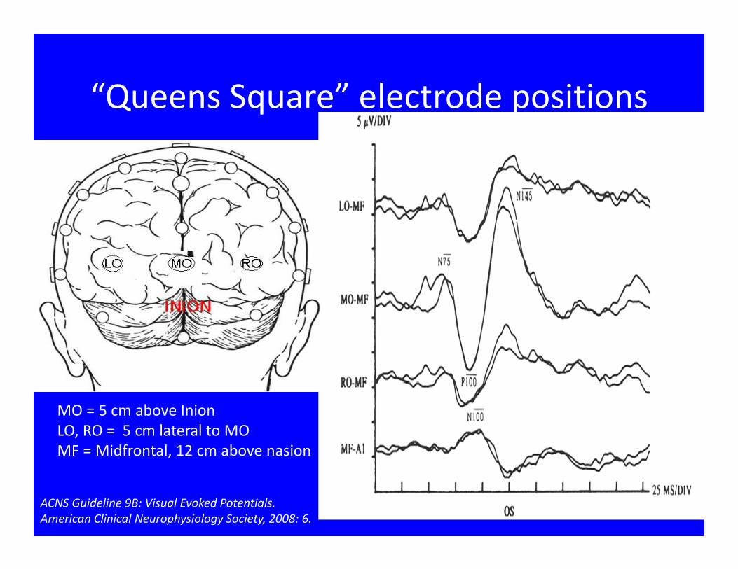
“Queens Square” electrode positions
MO = 5 cm above Inion
LO, RO = 5 cm lateral to MO
MF = Midfrontal, 12 cm above nasion
ACNS Guideline 9B: Visual Evoked Potentials.
American Clinical Neurophysiology Society, 2008: 6.

Midline Montage
Fz – M1
MPz– M1
Oz – M1
Oz – Fz
20 msec/div 3 uv/div

Single Channel
Oz-Cz
25 msec/div 2 uv/div
Patient Factors affecting VEPs
• Visual Acuity (ability to resolve pattern stimulus)
• Visual Field defect
• Ocular Factors
• Cooperation: lack of focus/fixation
• Pupil Size
• Age
• Gender

Interpretation
• Identify major waveform components: N75, P100, N145
• Measure the P100 latency for each eye
• Calculate the latency difference between eyes: interocular latency difference
• Measure the mid occipital P100 amplitude for each eye: peak to peak (N75-P100) or (P100-N145)
• Calculate the interocular amplitude ratio
• Evaluate the topographic distribution of the P100. If using lateral electrodes, is P100 laterally displaced? If so, do hemi field stim.
Interpretation
Major Criteria for abnormality:
• P100 absolute latency prolongation
• P100 interocular latency difference
• Absent waveform (using analysis times as long as
500ms and multiple recording sites)
Minor criteria for abnormality:
• P100 interocular amplitude difference (>2.5:1)
• Abnormal topography
• Abnormal waveform morphology (if monocular)

Interpretation: Localization
Asymmetric Abnormality = anterior to
chiasm (optic nerve or ocular)
Bilateral Abnormality = non localizing
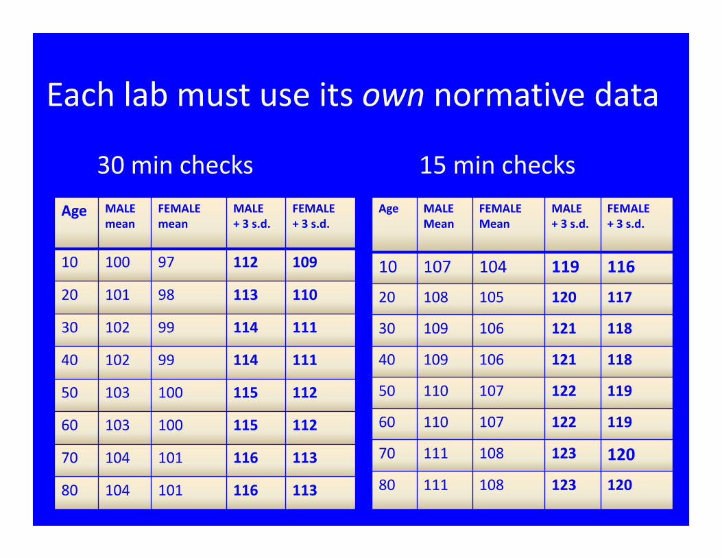
Each lab must use its own normative data
30 min checks 15 min checks
Age MALE
mean
FEMALE
mean
MALE
+ 3 s.d.
FEMALE
+ 3 s.d.
10 100 97 112 109
20 101 98 113 110
30 102 99 114 111
40 102 99 114 111
50 103 100 115 112
60 103 100 115 112
70 104 101 116 113
80 104 101 116 113
Age MALE
Mean
FEMALE
Mean
MALE
+ 3 s.d.
FEMALE
+ 3 s.d.
10 107 104 119 116
20 108 105 120 117
30 109 106 121 118
40 109 106 121 118
50 110 107 122 119
60 110 107 122 119
70 111 108 123 120
80 111 108 123 120

20 msec/div 3 uv/div
46 year-old man with episodes of “visual spots”
OS OD
Fz-A1
Mpz-A1
Oz-A1
Oz-Fz
20 msec/div 7 uv/div
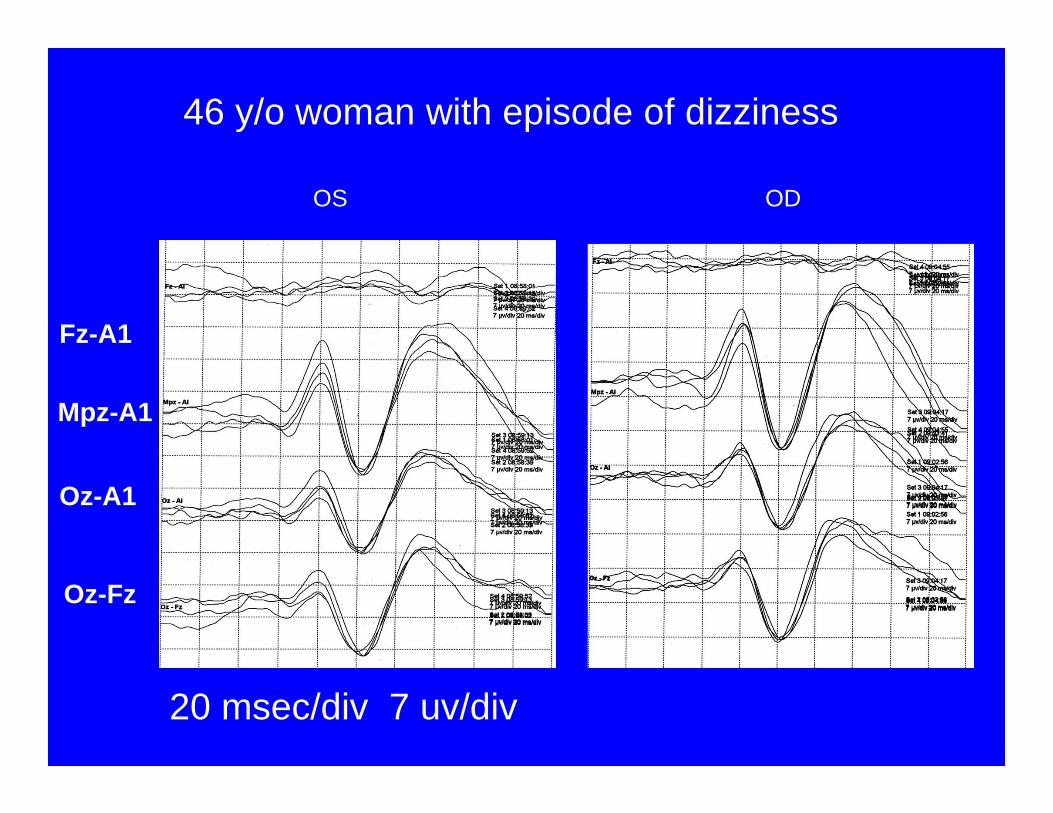
46 y/o woman with episode of dizziness
OS OD
Fz-A1
Mpz-A1
Oz-A1
Oz-Fz

Oz-Cz
Oz-Pz
ODOS
5 µv/div
25ms
52 year-old man with headache and visual disturbance
25 msec/div 5 uv/div

Oz-Cz
Oz-Pz
ODOS
5 µv/div
25ms
52 year-old man with headache and visual disturbance
25 msec/div 5 uv/div

20 msec/div 3 uv/div
78 y/o woman with visual complaints
OS OD
Fz-A1
Mpz-A1
Oz-A1
Oz-Fz
20 msec/div 3 uv/div
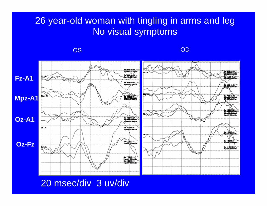
26 year-old woman with tingling in arms and legNo visual symptoms
OS OD
Fz-A1
Mpz-A1
Oz-A1
Oz-Fz
20 msec/div 3 uv/div
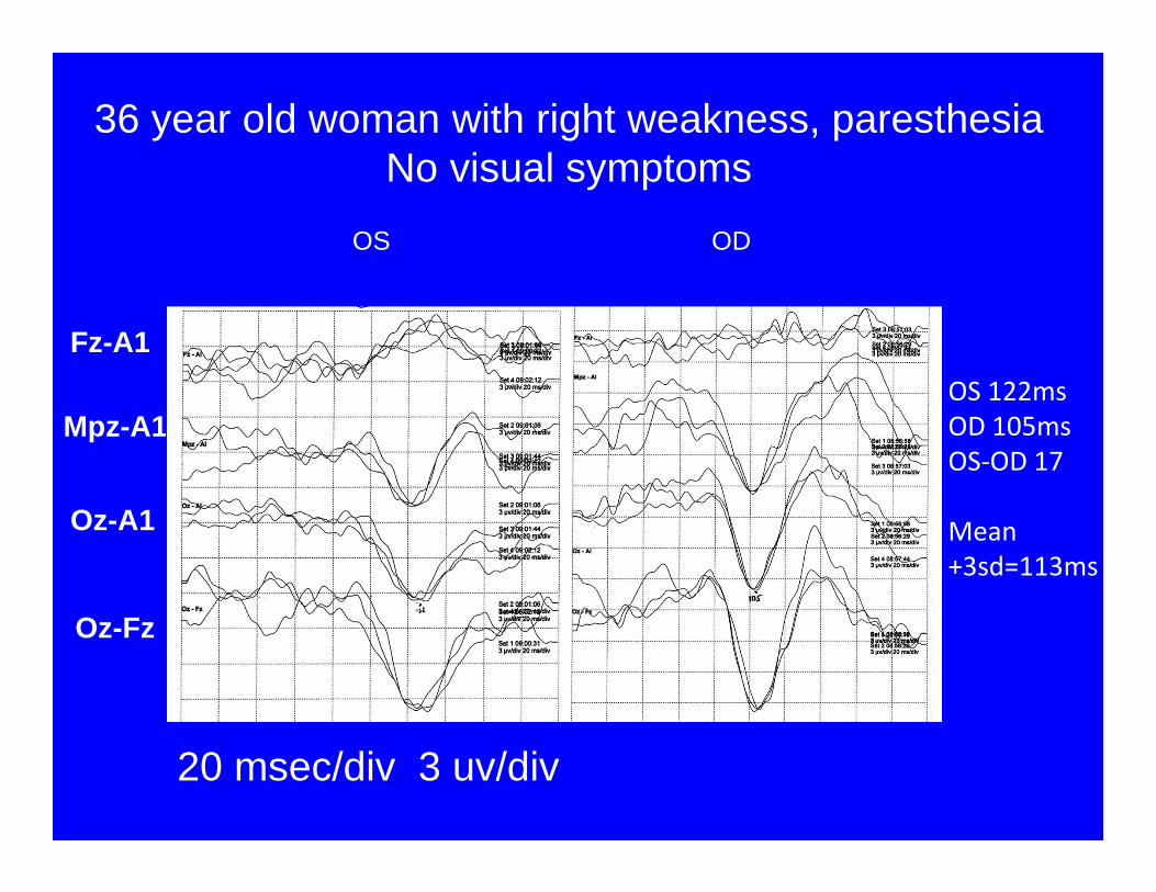
36 year old woman with right weakness, paresthesiaNo visual symptoms
OS OD
Fz-A1
Mpz-A1
Oz-A1
Oz-Fz
OS 122ms
OD 105ms
OS-OD 17
Mean
+3sd=113ms
3μv/div
20ms/div
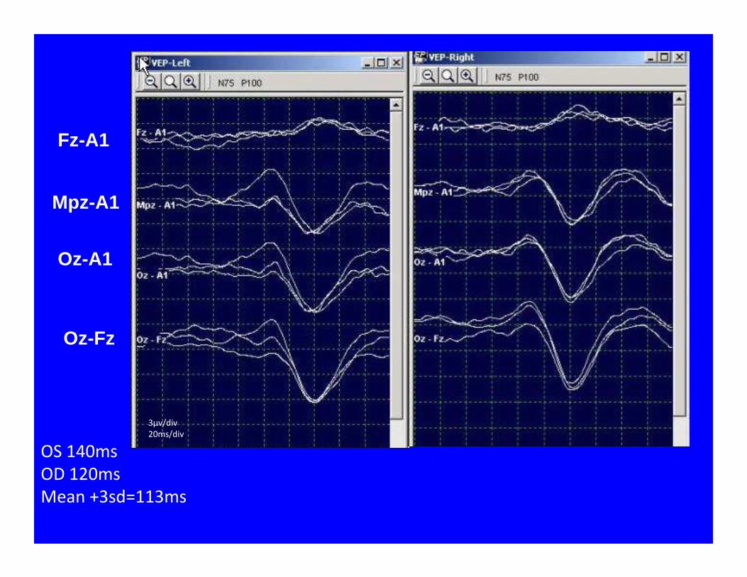
Fz-A1
Mpz-A1
Oz-A1
Oz-Fz
OS 140ms
OD 120ms
Mean +3sd=113ms
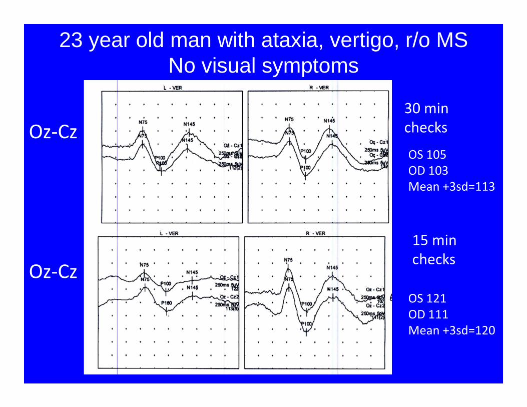
Oz-Cz
Oz-Cz
25ms/div
2µv/div
25ms/div
2µv/div
30 min
checks
15 min
checks
OS 105
OD 103
Mean +3sd=113
OS 121
OD 111
Mean +3sd=120
23 year old man with ataxia, vertigo, r/o MSNo visual symptoms

13 year-old with left eye pain and blurred visionAcuity OS 20/80 OD 20/20
Oz-Cz
Oz-Cz
25ms/div
2µv/div
25ms/div
2µv/div
30 min
checks
Flash
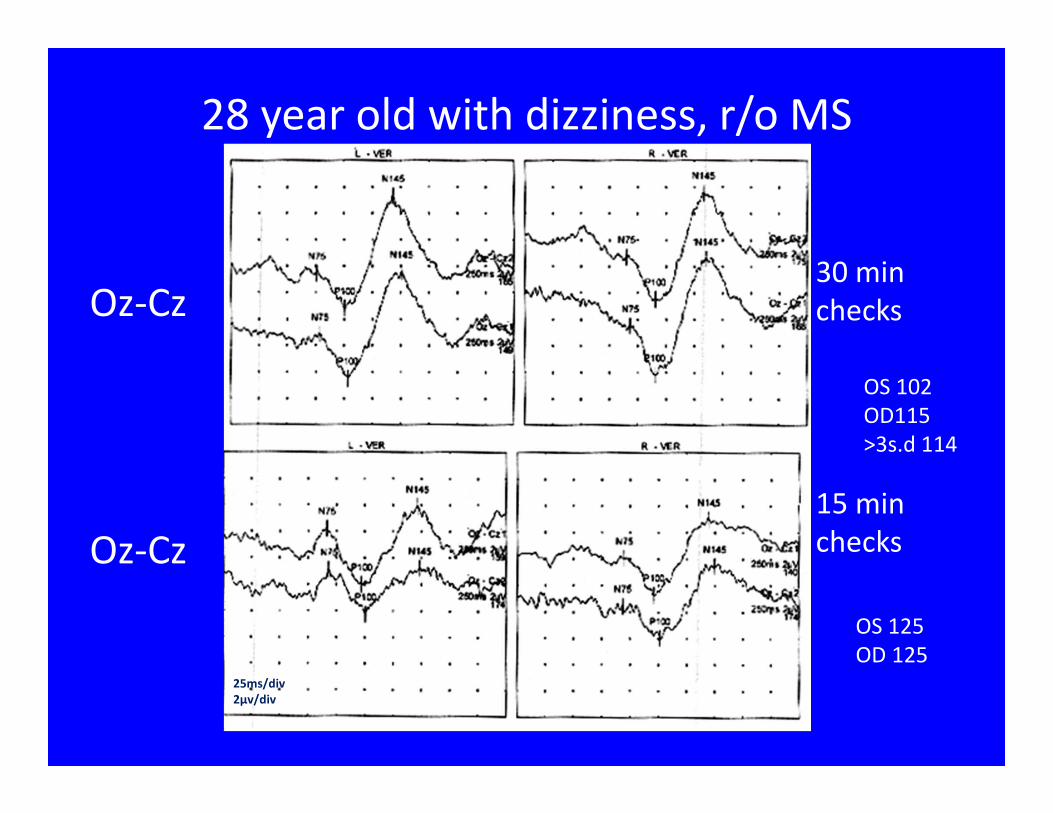
Oz-Cz
Oz-Cz
30 min
checks
15 min
checks
25ms/div
2µv/div
28 year old with dizziness, r/o MS
OS 102
OD115
>3s.d 114
OS 125
OD 125

20 msec/div 3 uv/div
48 y/o cocaine abuser, dysarthria, blurred vision
OS OD
Fz-A1
Mpz-A1
Oz-A1
Oz-Fz

11 month-old with head trauma
Oz-Cz Flash
25ms/div
2µv/div

Oz-Cz
25ms/div
2µv/div
OS 120 ms
OD 143 ms
OS-OD 23
Flash
35 y/o man with MS

Retrochiasmatic Pathology:
Hemifield stimulation Technique
• Imaging modalities have replaced this technique
• Technically difficult. Even small eye movement (one degree!) can lead to large contamination of hemifield responses.
• Arises from projections/activation of the peripheral visual field rather than just the area of the macula
• -larger check sizes
• -lateral recording electrodes: LT and RT

Hemifield Stimulation
Recall that LEFT hemifield stimulationprojects to the LEFT occiput!!!
Epstein Charles. Visual Evoked Potentials. In: Ebersole J, Pedley T, eds. Current
Practice of Clinical Electroencephalography: Philadelphia: Lippincott Williams and
Wilkins, 2003: 835

ACNS Guideline 9B: Visual Evoked Potentials.
American Clinical Neurophysiology Society,
2008: 10.
P100 response over ipsilateral occipital temporal leadsN105 over contralateral occipital temporal leads
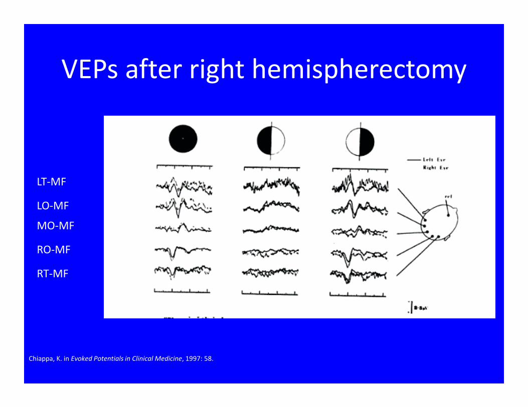
RT-MF
RO-MF
MO-MF
LO-MF
LT-MF
Chiappa, K. in Evoked Potentials in Clinical Medicine, 1997: 58.
VEPs after right hemispherectomy

Summary
• Full Field Pattern VEPs reliably assess the pre-
chiasmal visual pathway, but can also detect
lesions elsewhere.
• Responses may be affected by a variety of
patient factors and test conditions
• Evaluation of the P100 latency must be based
on laboratory specific normative data.
• Other stimulation techniques (flash and
hemifield) can provide additional information,
though their utility is more limited.


