Page 1
| 1
UNIVERSIDADE DE SÃO PAULO
FACULDADE DE FILOSOFIA, CIÊNCIAS E LETRAS DE RIBEIRÃO PRETO
DEPARTAMENTO DE PSICOLOGIA
PROGRAMA DE PÓS-GRADUAÇÃO EM PSICOBIOLOGIA
RUI DE MORAES JR.
Laterality and processing time-course of spatial frequencies on face encoding
Ribeirão Preto
2016
Page 2
| 2
RUI DE MORAES JR.
Laterality and processing time-course of spatial frequencies on face encoding
Tese apresentada à Faculdade de Filosofia, Ciências
e Letras de Ribeirão Preto da Universidade de São
Paulo, como parte das exigências para obtenção do
título de Doutor.
Área de concentração: Psicobiologia
Orientador: prof. Dr. Sérgio Sheiji Fukusima
Ribeirão Preto
2016
Page 3
| 3
Autorizo a reprodução e/ou divulgação total ou parcial da presente obra, por qualquer meio,
convencional ou eletrônico, para fins de pesquisa e estudo, desde que citada a fonte.
FICHA CATALOGRÁFICA
de Moraes, Rui, Jr.
Lateralidade e curso temporal do processamento de frequências espaciais
na codificação de faces. Ribeirão Preto, 2016.
85 p. : il.; 30 cm.
Tese de Doutorado apresentada à faculdade de Filosofia, Ciências e
Letras de Ribeirão Preto/USP – Departamento de Psicologia. Área de
concentração: Psicobiologia.
Orientador: Fukusima, Sérgio Sheiji
Versão do título para o inglês: Laterality and processing time-course of
spatial frequencies on face encoding
1. Percepção de faces. 2. Frequência espacial. 3. Especialização
hemisférica. 4. Coarse-to-fine.
Page 4
| 4
FOLHA DE APROVAÇÃO
Rui de Moraes Jr.
Lateralidade e curso temporal do processamento de frequências espaciais na codificação de
faces
Tese apresentada à Faculdade de Filosofia, Ciências
e Letras de Ribeirão Preto da Universidade de São
Paulo, como parte das exigências para obtenção do
título de Doutor. Área de concentração:
Psicobiologia.
Aprovado em: 01 / 02 / 2016
Banca examinadora:
Prof. Dr. Sérgio Sheiji Fukusima (Orientador)
FFCLRP - Universidade de São Paulo Assinatura: _______________________
Prof. Dr. Nelson Torro Alves
CCHLA - Universidade Federal da Paraíba Assinatura: _______________________
Prof. Dr. Cesar Alexis Galera
FFCLRP - Universidade de São Paulo Assinatura: _______________________
Prof. Dr. Paulo Sérgio Boggio
CCBS - Universidade Presbiteriana Mackenzie Assinatura: _______________________
Prof. Dr. José Aparecido da Silva
FFCLRP - Universidade de São Paulo Assinatura: _______________________
Page 5
| 5
Aos meus pais, Ana e Rui
À minha companheira, Sarah
Page 6
| 6
AGRADECIMENTOS
Gostaria de agradecer ao professor Sérgio Fukusima, pela formação acadêmica que
recebi em seu laboratório e pela liberdade que me deu enquanto aluno de pós-graduação.
Certa vez, em uma dessas conversas triviais de corredor, ele comentou que não existe ex-
orientador, ao se referir respeitosamente ao professor José Aparecido. Eu concordo com você,
Sérgio. Durante os anos em que fui integrante do Laboratório de Percepção e Psicofísica
também fui agraciado com a companhia dos meus colegas: Gabriel, Linita, Lívia e Patrícia.
Em especial, agradeço a amizade importada do Triângulo Mineiro do Bruno e do Léo.
Ainda, obrigado à Renata e ao Igor, pela presteza nos serviços acadêmicos e técnicos
durante o doutorado. De modo específico, sou grato à Valérie Goffaux por disponibilizar os
códigos para filtrar os estímulos do Estudo 1, ao André Cravo e ao Yossi Zana pela
disponibilidade em discutir o trabalho, ao Rafael Vasques pelo suporte computacional, e ao
Mikael Cavallet e à Louise Kauffmann por revisarem o Estudo 2.
Gratidão à FAPESP e ao CNPq, pelos financiamentos concedidos, sem os quais esta
empreitada não se viabilizaria. Também sou agradecido a todos aqueles que se voluntariaram,
seja por amizade, curiosidade ou boa vontade, a participar dos experimentos.
Também não posso deixar de agradecer àqueles que me deram um lar e uma família
nos anos de Ribeirão Preto. Fantin, serei sempre agradecido por sua disposição na cozinha.
Hugo, espero que tenha mudado aquela música feliz do seu alarme matinal.
Durante todo o terceiro ano de doutorado estive no laboratório do Dr. Jocelyn Faubert,
em Montreal no Canadá. Foi uma experiência única e lembranças vívidas desta época ainda
insistem em estourar na minha cabeça constantemente. Sou muito grato à orientação do
professor Faubert. É inspirador o contato com alguém que ao mesmo tempo é excelente
pesquisador, empreendedor arrojado e talentoso para motivar sua equipe. Recebi ajuda de
muitas pessoas em seu laboratório. Obrigado à Isabelle pelos assuntos acadêmicos (e
eventuais traduções para o francês) e Vadim pelo suporte computacional (não é todos os dias
que se tem um programador russo à sua volta). Gracias a los amigos Eduardo e Rafael, pela
calorosa amizade e aprendizado. Obrigado aos alunos da École d'optometrie pela convivência
e amizade: Bruno, Eugenie, Jimmy, Kash, Robyn e Thomas. Merci à tous! Thanks to
everyone!
Na volta ao Brasil, no fim do doutoramento, escrevi as últimas linhas desta tese em
São José dos Campos. Do alto do décimo primeiro andar, com a Mantiqueira pintada no
horizonte e sob o silêncio da minha escrivaninha, lembrava com uma nostalgia prematura o
Page 7
| 7
ambiente agitado e divertido daquela fazenda de café que teve a pretensão de se tornar uma
universidade. Agradeço aos colegas de pós-graduação pela rica convivência compartilhada:
Betão, Dudu, Everton, Gi, Mariana, Nayanne, Pedro, Rafael, Ricardo, Regiane, Thiago, e
Vinny.
Eu também sou muito grato ao apoio familiar que tive. Agradeço sobretudo aos meus
pais, Ana e Rui, pelo carinho e exemplo desde sempre, e por terem me apoiado nas minhas
escolhas profissionais. Ainda, sou grato por ter meus irmãos, Bruno e Lucas, como parceiros
de vida. Sinto muito a falta do convívio diário com vocês. Por último, Sarah, obrigado por me
tirar do computador quando precisava, por me esperar no saguão do aeroporto depois de um
ano no exterior, por dividir o mesmo teto e por ter deixado o meu sorriso mais frouxo desde
aquela festa na Pereira Lago.
Page 8
| 8
RESUMO
de Moraes, R., Jr. (2016). Lateralidade e curso temporal do processamento de frequências
espaciais na codificação de faces. Tese de Doutorado, Faculdade de Filosofia,
Ciências e Letras de Ribeirão Preto, Universidade de São Paulo, Ribeirão Preto.
O sinal de entrada na retina é decomposto em termos de frequência espacial (FE), variações
periódicas de luminância ao longo do espaço. Existe vasta literatura sobre o processamento de
FE no córtex visual primário. No entanto, não se sabe ao certo como esta informação sensorial
básica é processada e integrada numa visão de alto nível. Esta tese aborda este tema ao
investigar lateralidade cerebral, tempo de processamento e contexto cognitivo em três
diferentes seções com objetivos específicos. Estas seções investigaram comportamentalmente
visão de alto nível tendo a face humana como estímulo, dado sua relevância biológica e
social. Na primeira seção (Theoretical Review), uma revisão apresenta estudos clínicos e
neuropsicológicos que mostram áreas cerebrais envolvidas na percepção de faces e como os
hemisférios esquerdo e direito realizam um processamento holístico e analítico baseado em
informações de FEs. A especialização hemisférica de FE no reconhecimento de faces é então
revisada e discutida. Concluiu-se que assimetrias sensoriais podem ser a base para assimetrias
cognitivas de alta ordem. Ademais, foi destacado a influência do tempo de processamento. Na
segunda seção (Study 1), foi investigado por método psicofísico a lateralidade de baixas e
altas FEs no reconhecimento de faces em diferentes tempos de exposição. Faces com
filtragem de FE foram apresentadas em campo visual dividido em alta e baixa restrição
temporal em duas tarefas: reconhecimento facial (Experimento 1) e reconhecimento do sexo
facial (Experimento 2). No Experimento 1, informações faciais de baixas e altas FEs foram
mais eficientemente processadas no hemisfério direito e esquerdo, respectivamente, sem
efeito do tempo de exposição das faces. Os resultados do Experimento 2 mostraram uma
assimetria do hemisfério direito para baixas FEs em baixa restrição temporal. Conclui-se que
o processamento de altas e baixas FEs é lateralizado nos hemisférios cerebrais no
reconhecimento de faces. No entanto, a contribuição de altas e baixas FEs é dependente da
tarefa e do tempo de exposição. Na terceira seção (Study 2) foi investigado qual estratégia
temporal, coarse-to-fine (de baixas para altas FEs) ou fine-to-coarse, cada hemisfério cerebral
utiliza para integrar informação de FE de faces humanas numa tarefa de categorização facial
homem-mulher. Sequências dinâmicas breves coarse-to-fine e fine-to-coarse de faces foram
apresentadas no campo visual esquerdo, direito e central. Os resultados do tempo de resposta
e do score de eficiência invertida mostraram uma prevalência geral de um processamento
coarse-to-fine, independente do campo visual de apresentação. Ainda, os dados da taxa de
erro ressaltam o processamento coarse-to-fine realizado pelo hemisfério direito. No geral, esta
tese fornece insights sobre assimetria cerebral funcional, integração de alto nível e curso
temporal do processamento de FEs, principalmente para aqueles interessados na percepção de
faces. Também foi mostrado que operações lateralizadas, tarefa-dependente e coarse-to-fine
podem coexistir e interagir no cérebro para processar informação de FE.
Palavras-chave: Percepção de faces. Frequência espacial. Especialização hemisférica. Coarse-
to-fine.
Page 9
| 9
ABSTRACT
de Moraes, R., Jr. (2016). Laterality and processing time-course of spatial frequencies on face
encoding. Tese de Doutorado, Faculdade de Filosofia, Ciências e Letras de Ribeirão
Preto, Universidade de São Paulo, Ribeirão Preto.
Retinal input is decomposed in terms of spatial frequency (SF), i.e., periodic variations of
luminance through space. There is extensive literature on the processing of SF in the primary
visual cortex. However, it is still unclear how SF information is processed and integrated in
high-level vision. This thesis addressed this issue in terms of laterality effects, processing
time-course, and the cognitive context in three different sections with specific purposes.
These sections behaviorally tackle high-level vision using human faces as stimuli due to their
biological and social relevance. In the first section (Theoretical Review) a literature review
presented clinical and neurophysiological studies that show brain areas that are involved in
face perception and how the right and left hemispheres perform holistic and analytic
processing, depending on SF information. The SF hemispheric specialization in face
recognition is then reviewed and discussed. Our conclusion is that functional sensorial
asymmetries may be the basis for high-level cognitive asymmetries. In addition, we
highlighted the role of the processing time. In the second section (Study 1), we
psychophysically investigated laterality of low and high SF in face recognition at different
exposure times. The SF filtered faces were presented in a divided visual field at high and low
temporal constraint in two tasks: face recognition (Experiment 1) and face gender recognition
(Experiment 2). In Experiment 1, low and high SF facial information were more efficiently
processed in the right and in the left hemisphere, respectively, with no effect of exposure
time. In Experiment 2, results showed a right hemisphere asymmetry for low SF faces at low
temporal constraint. We concluded that the processing of low and high SF is lateralized in the
brain hemispheres for face recognition. However, low and high SF contribution is dependent
on the task and the exposure time. In the third section (Study 2), we aimed to investigate
which temporal strategy, i.e., coarse-to-fine (from low to high SF) or fine-to-course, each
brain hemisphere performs to integrate SF information of human faces in a male-female
categorization task. Coarse-to-fine and fine-to-course brief dynamic sequences of faces were
presented in the left, right and central visual field. Results of the correct response time and the
inverse efficiency score showed an overall advantage of coarse-to-fine processing,
irrespective of the visual field of presentation. Data of the error rate also highlights the role of
the right hemisphere in the coarse-to-fine processing. All in all, this thesis provided some
insights on functional brain asymmetry, high-level integration, and processing time-course of
SF information, mainly for those interested in face perception. It was also shown that
lateralized, diagnostic-oriented, and coarse-to-fine operations may coexist and interact in the
human brain to process SF information.
Keywords: Face perception. Spatial frequency. Hemispheric specialization. Coarse-to-fine.
Page 10
| 10
LIST OF FIGURES AND TABLES
Figure 1.1. The upper left image (a) illustrates the idea of spatial frequency; adapted from:
webvision.med.utah.edu. The upper right image (b) shows a complex visual scene that was
filtered to preserve low (c) and high spatial frequencies (d); original photo: Ricardo Feres
(Jalapão State Park, TO, Brazil) .............................................................................................. 15
Table 2.1. Studies on the hemispheric specialization of spatial frequency in facial perception
tasks ......................................................................................................................................... 27
Figure 3.1. Examples of stimuli used in the experiment along with their respective masks and
spatial frequency cutoffs in cycles per image (cpi) and cycles per degree of visual angle (cpd).
Legend: BSF - broadband spatial frequencies; LSF - low spatial frequencies; HSF - high
spatial frequencies ................................................................................................................... 37
Figure 3.2. Illustration of one trial in Experiment 1 and 2. Each trial began after a key was
pressed. In Experiment 1 this triggered the target face presentation. A fixation screen followed
the target. Subsequently, the probe face was presented and immediately backward masked. At
the mask offset, participants must respond if target and probe faces were from the same
person. In Experiment 2 the initial screen was followed by a fixation screen. Then, the
experiment displayed the stimulus, which was backward masked. At the mask offset,
participants must respond if the face was male. In both experiments the response was given by
pressing yes/no buttons. The stimulus in Experiment 2 and the probe face in Experiment 1
were presented: (a) in the right or left visual hemifield; (b) in high, low or broad spatial
frequencies; (c) at high or low temporal constraint ................................................................ 38
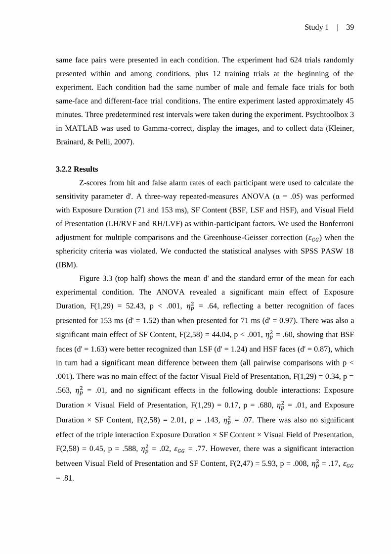
Figure 3.3. Average d' for faces presented in broadband (BSF), high (HSF) and low spatial
frequencies (LSF) in the left hemisphere/right visual field (LH/RVF) and in the right
hemisphere/left visual field (RH/LVF). The faces were presented at high (left half) and low
temporal constraint (right half) in Experiment 1 (top half) and Experiment 2 (bottom half).
Error bars indicate the standard error of the mean .................................................................. 40
Figure 4.1. Example of the six spatial-frequency filtered versions that originated from a full-
bandwidth image along with the information of their central frequency measured in
cycles/image width (cpi) and cycles/degree of visual angle (cpd). One stimulus consisted of a
brief succession of the filtered faces going from lower (left side) to higher (right side) spatial
frequencies in the coarse-to-fine condition, or in the opposite direction going from higher to
lower spatial frequencies in the fine-to-coarse condition ....................................................... 57
Figure 4.2. Illustration of one trial. A fixation screen was followed by the stimulus, a coarse-
to-fine or fine-to-coarse dynamic sequence that was presented in the left, right or central
visual field and immediately backward masked. At the mask offset, participants must
categorize the face as male or female as fast and as accurately as possible ........................... 58
Figure 4.3. Averages of error rate (a), correct response time (b) and inverse efficiency score
(c) for dynamic coarse-to-fine (CtF) and fine-to-coarse (FtC) sequences of faces presented in
the left (LVF), central (CVF) and right visual field (RVF). Error bars indicate the standard
error of the mean ..................................................................................................................... 60
Page 11
| 11
LIST OF ABBREVIATIONS
ANOVA. analysis of variance
BSF. broadband spatial frequencies
cpd. cycles per degree (of visual angle)
cpf. cycles per face (width)
cpi. cycles per image (width)
CRT. cathode ray tube (computer monitor)
CtF. coarse-to-fine
CVF. central visual field
ER. error rate (%)
ERP. event-related potential
FFA. fusiform face area
fMRI. functional magnetic resonance imaging
HSF. high spatial frequencies
Hz. hertz (cycles per second)
IES. inverse efficiency score
LED. light-emitting diode (computer monitor)
LH. left hemisphere
LSF. low spatial frequencies
LVF. left visual field
ms. millisecond
OFA. occipital face area
RH. right hemisphere
RT. response time
RVF. right visual field
SD. standard deviation (of the mean)
SE. standard error (of the mean)
SF. spatial frequency
TMS. transcranial magnetic stimulation
Page 12
| 12
TABLE OF CONTENTS
1. INTRODUCTION ................................................................................................ 13
2. THEORETICAL REVIEW ................................................................................. 17
Hemispheric specialization in face recognition: From spatial frequencies to
holistic/analytic cognitive processing
2.1 Introduction ...................................................................................................... 19
2.2 Face recognition ............................................................................................... 19
2.3 Hemispheric specialization and the neural substrates of analytic and holistic
face processing ....................................................................................................... 20
2.4 Hemispheric specialization of spatial frequencies in face recognition ............ 24
2.5 Final considerations .......................................................................................... 29
3. STUDY 1 ............................................................................................................... 32
Task and exposure time modulate laterality of spatial frequency for faces
3.1 Introduction ...................................................................................................... 34
3.2 Experiment 1 .................................................................................................... 35
3.2.1 Method ....................................................................................................... 36
3.2.2 Results ....................................................................................................... 39
3.2.3 Discussion ................................................................................................. 41
3.3 Experiment 2 .................................................................................................... 42
3.3.1 Method ....................................................................................................... 43
3.3.2 Results ....................................................................................................... 44
3.3.3 Discussion ................................................................................................. 45
3.4 General discussion ........................................................................................... 47
4. STUDY 2 ............................................................................................................... 51
Behavioral evidence for a predominant and non-lateralized coarse-to-fine
encoding for faces
4.1 Introduction ...................................................................................................... 53
4.2 Method .............................................................................................................. 56
4.3 Results .............................................................................................................. 59
4.4 Discussion ........................................................................................................ 61
5. CONCLUDING REMARKS ............................................................................... 65
REFERENCES ..................................................................................................... 69
SUPPLEMENT ..................................................................................................... 81
Ethics committee approval letter - Universidade de São Paulo ............................. 82
Ethics committee approval letter - Université de Montréal .................................... 83
Funding during PhD ............................................................................................... 84
Publications during PhD ......................................................................................... 85
Page 13
Introduction | 13
1. Introduction
Page 14
Introduction | 14
In my biased standpoint as a young experimental psychologist, the visual system is
one of the most impressive gadgets forged during the history of human evolution. Consider
(in case you have not yet done so): just a small range of the electromagnetic spectrum is
coded by the human eye, i.e., visible light. Depending on the light intensity, our eye's colored
ring called the iris modulates the pupil size. Light enters through this aperture and reaches the
retina. The small image reflected on this two-dimensional plan is upside down and partially
occluded by a blind spot. Retinal specialized cells analyze and transform electromagnetic
energy waves into discrete on-off electrochemical energy. The signal travels through different
neural routes at different speeds and only after sequential complex computations the visual
information is integrated in cortical association areas. Almost instantaneously and with no
awareness of this process, the output is the perception of an organized world in depth, color,
contrast, and movement!
Visual perception seems complex, and it is indeed. For centuries scientists from
different fields have been exploring the visual system. One way they try to reveal the
complexity of vision is by exploring the fundamentals and basic aspects. It is known that in
the first stages of vision the retinal input is decomposed into two main dimensions: orientation
and spatial frequency. This thesis followed this path and investigated the latter: spatial
frequency processing.
Spatial frequency is defined as periodic variations of luminance through space. It is
measured in cycles of alternating light and dark areas in a given unit of distance. A cycle
consists of one dark and one light area adjacent in space. In visual perception it is measured in
cycles per degree of visual angle. The greater the amount of luminance alternation in a given
space, the more detailed the perception is (Figure 1a). Therefore, high spatial frequencies
convey information of small details and sharp edges of the visual scene. Conversely, low
spatial frequencies convey coarse information of the visual scene (see Figure 1b, 1c and 1d).
There is extensive literature specifying the role of specialized cells in the primary visual
cortex that respond to different spatial frequency bandwidths (De Valois, Albrecht, & Thorell,
1982; Hubel & Wiesel, 1968; Poggio, 1972). However, it is still unclear how spatial
frequency information is processed and integrated in high-level vision.
This thesis addressed high-level vision in one of the most important visual stimuli: the
human face. The human face is a ubiquitous visual stimulus for everyone throughout the
whole lifespan. It is the most expressive part of the human body, being essential for
interpersonal relations and to express biological signals. As a result, we have developed
extremely efficient strategies to extract and encode facial information. In addition and more
Page 15
Introduction | 15
related to the thesis purpose, previous works suggest that face perception is more sensitive to
spatial frequency information than other types of complex stimuli (Collin, Liu, Troje,
McMullen, & Chaudhuri, 2004; Goffaux, Gauthier, & Rossion, 2003; Yue, Tjan, &
Biederman, 2006).
Figure 1.1: The upper left image (a) illustrates the idea of spatial frequency; adapted from:
webvision.med.utah.edu. The upper right image (b) shows a complex visual scene that was filtered to preserve
low (c) and high spatial frequencies (d); original photo: Ricardo Feres (Jalapão State Park, TO, Brazil).
Influential theoretical frameworks and assumptions on the processing of spatial
frequency information based the questions in this thesis to investigate face encoding. Two of
them were markedly important: hemispheric specialization of spatial frequencies and coarse-
to-fine hypotheses. The first states that the right hemisphere is more efficient in processing
low spatial frequencies and the left hemisphere is more efficient in processing high spatial
frequencies (Sergent, 1982). The latter assumes that there is a precedence of low over high
spatial frequencies in the processing time-course of the visual input (Hegdé, 2008; Schyns &
Oliva, 1994). Although both of them are supported by many investigations, it is unclear how
they relate.
Page 16
Introduction | 16
This central gap guided the course of this thesis and provoked some questions. In the
Theoretical Review section, I explored the literature to investigate if there is a relation
between lateralized holistic/analitic processing and low/high spatial frequencies on face
processing. Could cognitive brain asymmetries be an extension of sensorial lower-level brain
asymmetries? And what is the role of the processing time? In Study 1, I wondered if there are
hemispheric differences in the perception of low and high spatial frequency facial information
at high and low temporal constraint. Could a right-hemisphere asymmetry for low spatial
frequencies at high temporal constraint and a left-hemisphere asymmetry for high spatial
frequencies at low temporal constraint conciliate the coarse-to-fine and the hemispheric
specialization hypotheses? Moreover, could the cognitive context interfere in this process? In
Study 2, I wondered if the left hemisphere could reverse the default coarse-to-fine processing.
Since the left hemisphere is assumed to be more efficient in processing high spatial
frequencies, could spatial frequencies be integrated in a fine-to-coarse fashion?
In spite of a central topic of investigation, the sections of this thesis are independent
studies. Each one raised specific questions, literature, method, and highlighted different
aspects of the problem. Besides facilitating the publication in scientific journals, this thesis
format provides more flexibility than the more traditional formats with restrictive scope. In
addition, this format also enables a straight-to-the-point reading for those pursuing specific
information. A concluding section summarizes the main findings and contribution of this
thesis and shows how the sections are connected and how they were conceived.
Page 17
Theoretical review | 17
2. Theoretical review
Page 18
Theoretical review | 18
Hemispheric specialization in face recognition: From spatial frequencies to
holistic/analytic cognitive processing1
We present clinical and neurophysiological studies that show brain areas that are involved in
face perception and how the right and left hemispheres perform holistic and analytic
processing, depending on spatial frequency information. The hemispheric specialization of
spatial frequency in face recognition is then reviewed and discussed. The limitations of
previous work and suggestions for further investigations are discussed. Our conclusion is that
functional sensorial asymmetries may be the basis for high-level cognitive asymmetries.
1 This section was published in Psychology & Neuroscience journal. Reference: de Moraes, R., Jr., Sousa, B. M.,
& Fukusima, S. S. (2014). Hemispheric specialization in face recognition: From spatial frequencies to
holistic/analytic cognitive processing. Psychology and Neuroscience, 7(4), 503–511.
doi:10.3922/j.psns.2014.4.09
Page 19
Theoretical review | 19
2.1 Introduction
There is multidisciplinary interest in the study of the human face because of its
evolutionary and social relevance. Research on face recognition focuses on complex cognitive
processes, practical applications, clinical studies, and even computational simulations and
biometric models. Understanding basic sensorial and perceptual operations that are performed
by the human visual system to process and recognize faces is important. In this paper, we
review the literature on how lateralized high-level cognitive strategies are supported by the
processing of elementary sensorial information. In particular, we seek to clarify holistic and
analytic processing in face recognition based on spatial frequency information and how the
brain hemispheres process different bandwidths of spatial frequency.
We first review basic information about face recognition. We then present clinical and
neuroimaging studies that show the brain areas that are involved in face perception and how
the right and left hemispheres perform different kinds of processing. The relationship between
holistic/analytic processing and low/high spatial frequency information is established, and the
hemispheric specialization of spatial frequency in face recognition is reviewed and discussed.
2.2 Face Recognition
Humans are experts in face recognition. We can recognize minimal variations in facial
features, even at a distance and under low light conditions, different haircuts, and different
angles. Recognition happens automatically in less than 1 second, without posing cognitive
load (Maurer et al., 2007). Face recognition is fast and accurate. Adults are capable of
recognizing familiar faces with an accuracy greater than 90%, even if some faces have not
been seen for 50 years (Carbon, 2003).
The human face is an important source of information and communication and has
several aspects, including ethnicity, age, gender, attractiveness, emotion, and health condition.
Thus, the face is the most expressive part of the body (Chellappa, Wilson, & Sirohey, 1995).
Faces provide several social features that can be detected by other individuals and are
essential for interpersonal relationships. To a large extent, social interaction is facilitated by
the rapid processing of face recognition, which is linked to our biological necessity of
identifying who is approaching and what kind of greetings or emotional signs an individual
presents.
Page 20
Theoretical review | 20
During the evolutionary process, primates that had a cortical area and specific
processing devoted to face perception were better adapted and favored by natural selection
(Carmel & Bentin, 2002; Chellappa et al., 1995). Details about this perceptual process,
however, remain unclear. There are two theories on the origin of face recognition.
The expertise hypothesis supports the view that face recognition is a generic ability
that is similar to the processing of other classes of stimuli, and faces represent a special case
because of experience and the need to discriminate at the individual level. This implies that
the same processing mechanism may apply to any kind of visual object (Gauthier & Tarr,
1997; Meadows, 1974). The domain-specific hypothesis states that face recognition is a
specific process that is devoted only to this type of stimulus. The origin of this processing
mechanism remains unclear, but it possibly has innate factors or requires experience during a
critical developmental period (Robbins & McKone, 2007; Yovel & Kanwisher, 2004).
Apart from the uncertainty of the origin of facial processing, the idea that faces
involve holistic processing is consolidated in the literature. Faces have a peculiar
organization, and their elements are organized to allow global perception as a gestalt
combination between specific features. Even slight changes in these elements allow
distinguishing between individuals. Converging evidence shows that facial patterns are
processed holistically, which is different from other types of stimuli (Cheung, Richler,
Palmeri, & Gauthier, 2008). This would be related to the processing style of the right
hemisphere (Ellis, 1983; Springer & Deutsch, 1993). This hypothesis has been supported by
research on hemispheric dominance and brain asymmetry in face perception and the
processing modality observed in each hemisphere.
2.3 Hemispheric specialization and the neural substrates of analytic and holistic face
processing
In the 1960s, research on patients with brain injury showed that the majority of
individuals with prosopagnosia had lesions in the right hemisphere. In the following years,
Levy, Trevarthen, and Sperry (1972) reported similar results in patients who had undergone
commissurotomy: a strong asymmetry in facial recognition in favor of the right hemisphere,
whereas the left hemisphere was capable of recognizing familiar faces but had serious
difficulties processing unfamiliar faces as a whole. Moreover, other advantages of the right
hemisphere over the left hemisphere were observed, especially in processing speed, accuracy
Page 21
Theoretical review | 21
in identifying faces, access to long-term memory, and the reception and storage of facial
information (Chellappa et al., 1995; Curyto, 2000; Gazzaniga, 2000).
The superior performance of the right hemisphere in face recognition stems from its
expertise in coding and processing synthetic and holistic visuospatial stimuli and configural
information2 (Rhodes, 1993; Springer & Deutsch, 1993). In particular, it processes non-
verbal, simultaneous, analogical, gestalt, synthetic, and intuitive information. Conversely, the
left hemisphere has processing mechanisms that are suitable for verbal, sequential, temporal,
digital, logical, analytical, and rational information (Springer & Deutsch, 1993).
Human faces activate specific regions of the human brain, which has been consistently
reported in electroencephalography and neuroimaging studies and case reports on patients
with prosopagnosia (Goffaux, Peters, Haubrechts, Schiltz, Jansma, & Goebel, 2011; Rossion
et al., 2000). Many studies that compared face and object discrimination showed that faces
produced bilateral activation in medial portions of the fusiform gyrus, with more activity in
the right hemisphere. These results are consistent with cases of prosopagnosia caused by
bilateral lesions in the occipitotemporal cortex and unilateral lesions in the right fusiform
gyrus (Rossion et al., 2000).
The region associated with face recognition comprises the ventromedial surface of the
temporal and occipital lobes in the mediolateral fusiform gyrus, known as the Fusiform Face
Area (FFA). Activity in this area varies according to the attention directed toward the stimuli,
showing that it is not exclusively triggered by the face itself (Sergent, Ohta, & MacDonald,
1992; Kanwisher, McDermott, & Chun, 1997).
The middle fusiform gyrus is activated in both hemispheres, with higher activation in
the right side. The posterior fusiform gyrus is activated only in the right hemisphere when
attention is focused on facial patterns. The brain area located in the inferior temporal gyrus,
known as the Facial Occipital Area, is more activated by faces than by objects, again with
more activation in the right side (Rossion, Caldara, Seghier, Schuller, Lazeyras, & Mayer,
2003). Additionally, selective activity in the superior temporal sulcus and inferior occipital
gyrus was reported, but these observations are not consistent (Haxby, Ungerleider, Clark,
Schouten, Hoffman, & Martin, 1999; Rossion et al., 2000; Rossion et al., 2003).
2 The term configural has been applied to describe phenomena that involve the perception of relations between
facial features. Configural processing may be divided into three types: (1) first-order relations regarding the
facial pattern with two eyes, one mouth, and one nose, (2) holistic processing, which is the perception of the face
as a gestalt, and (3) second-order relations in the perception of distances between features. However, no
consensus on this term has been reached. Some researchers adopt the three types, and others adopt only one
(Maurer, Le Grand, & Mondloch, 2002). In this review, the terms configural, holistic, and global are
synonymous.
Page 22
Theoretical review | 22
The involvement of the left hemisphere in face recognition is still a matter of debate.
Some researchers argue that the fusiform area of the right hemisphere is responsible for face
recognition, whereas the equivalent area of the left hemisphere performs general object
recognition. However, the total disruption of face processing has been suggested to be caused
by bilateral lesions, whereas unilateral damage causes only selective impairments (Boeri &
Salmaggi, 1994). Furthermore, considerable evidence indicates that both hemispheres are
involved in the recognition of facial patterns, but they perform different roles. According to
this point of view, the right hemisphere processes faces in an integrative and comprehensive
manner, whereas the left hemisphere is responsible for facial features.
The idea of hemispheric specialization that associates the right hemisphere with
holistic processing and the left hemisphere with analytical processing is supported by some
studies. Faces that are presented upright or with differing spaces among facial elements favor
configural processing in the left visual field (projecting to the right hemisphere; see the
divided visual field method in Bourne, 2006) and are perceived more quickly and accurately
than when presented in the right visual field (projecting to the left hemisphere; Cattaneo,
Renzi, Bona, Merabet, Carbon, & Vecchi, 2014; Ramon & Rossion, 2012; Rhodes, 1993).
When faces are presented inverted (upside-down) or modified, inducing the processing of
individual features in a divided visual field, the advantage of the right hemisphere is
eliminated or reduced because of the interruption of holistic coding (Hillger & Koenig, 1991;
Leehey, Carey, Diamond, & Cahn, 1978; Rhodes, 1993). The lateralized repetition-priming
paradigm was tested by Bourne, Vladeaunu, and Hole (2009) using blurred faces and
displaced facial features. The results supported the role of both hemispheres. Configurally
degraded faces produced negative and positive priming in the left and right visual hemifields,
respectively, and featurally degraded faces produced the opposite effect. In two event-related
potential (ERP) studies, upright and inverted faces activated the right and left hemispheres
with more intensity, respectively (McCarthy, Puce, Belger, & Allison, 1999; Rossion et al.,
1999). In another ERP study, faces were altered by either moving or replacing facial features,
inducing configural and featural processing, and the same results were obtained (Scott, &
Nelson, 2006). A positron emission tomography study also supported the involvement of both
hemispheres in face processing. Rossion et al. (2000) observed a decrease in face-specific
activity in the FFA of the right hemisphere when attention was focused on facial components.
In contrast, activity increased in the equivalent area of the left hemisphere. However,
evidence argues against differential holistic/analytic processing in the FFA (Yovel &
Kanwisher, 2004). Additionally, other cortical areas are necessary and recruited for facial
Page 23
Theoretical review | 23
identification (Avidan, Hasson, Malach, & Bermann, 2005; Haxby, Ungerleider, Clark,
Schouten, Hoffman, & Martin, 2001). Functional magnetic resonance imaging (fMRI)
allowed the mapping of non-overlapping neural areas and networks for configural and featural
processing when participants judged spaced-feature faces and altered-feature faces (Maurer et
al., 2007). The results showed no differences between featural and configural processing in
the FFA, supporting the findings of Yovel and Kanwisher (2004). The spacing condition more
robustly activated an area of the fusiform gyrus adjacent to the FFA (slightly superior and
posterior to it) and areas of the frontal and inferior parietal cortices in the right hemisphere,
whereas the featural condition activated the middle prefrontal areas of the left hemisphere.
However, ERP and fMRI data only correlate alterations in brain activation caused by visual
stimulus manipulation. Renzi et al. (2013) performed a transcranial magnetic stimulation
(TMS) study. This technique allows the modulation of brain activity in a controlled task and
establishes cause-effect relationships. The TMS was delivered in cortical areas based on the
study by Maurer et al. (2007). The results showed that TMS disrupted holistic and analytic
processing over the right inferior and left middle frontal gyri, respectively. These summarized
behavioral and neurophysiological studies provide strong evidence of a dissociation between
holistic and analytic processes in face perception mediated by separate and lateralized
networks in the human cortex.
In the facial processing literature, the holistic/global model has received much
attention in the last three decades (Goffaux & Rossion, 2006). The majority of the results
regarding the activation, reaction time, and hit rate advantage of global processing and the
right hemisphere may be attributable to its mode of operation. Lux et al. (2004) suggested that
global processing is the automatic default setting of visual attention and requires less
activation than local processing, which requires attentional control. The local analysis of
stimuli is not natural because of two conflicts that occur: (1) the default processing of global
information and (2) the tendency to focus on items of interest. Thus, the global system is more
frequently used, but both types of processing are fundamental to this task (Casey & Newell,
2007).
In short, the perception and recognition of faces have two different processing
systems. The global/holistic system utilizes a type of processing that is mainly performed by
the right hemisphere, in which features interact in an integrated fashion. The local/analytical
system, in contrast, specializes in feature processing and is mainly performed by the left
hemisphere. Behavioral and neurophysiological evidence suggests that human face processing
requires both featural and configural processing (Goffaux, Hault, Michel, Vuong, & Rossion,
Page 24
Theoretical review | 24
2005).
2.4 Hemispheric specialization of spatial frequencies in face recognition
Configural, global, or holistic perception, as opposed to featural, local, or analytical
perception, involves high-level cognitive operations that depend on low-level perceptual
processing (Hills & Lewis, 2009). The analysis of spatial frequencies (i.e., variations in
luminance across space) is one of the first processes that occur during the encoding of visual
information. This may play an important role in hemispheric asymmetry (Yamaguchi,
Yamagata, & Kobayashi, 2000) and face perception (Goffaux et al., 2005).
Accumulating evidence indicates that the visual system has specific filters for different
bandwidths of spatial frequency (Campbell & Robson, 1968). These filters decompose the
visual scene in the retina, initiating highly complex perceptual and cognitive functions. Cells
of the visual system that are sensitive to high spatial frequencies process sharp borders with
high variations in luminance. Thus, discrete and detailed facial features are perceived, which
is the basis of analytical operations. Cells that are sensitive to low spatial frequencies process
coarse signals in regions of low variations in luminance, forming the basis of holistic
operations (Goffaux et al., 2005; Livingstone & Hubel, 1988). Therefore, different
bandwidths of spatial frequency encode different aspects of visual objects. With regard to the
face, a given bandwidth of the spectrum can affect its perception and recognition, given that
face perception relies on both configural and featural processing (Goffaux et al., 2005;
Sergent, 1996). Additionally, behavioral and neuroimaging data indicate that face processing
is more sensitive to spatial frequency information than to other visual stimuli (Collin, Liu,
Troje, McMullen, & Chaudhuri, 2004; Yue, Tjan, & Biederman, 2006).
According to the idea that low spatial frequencies underlie holistic operations and that
high spatial frequencies underlie analytical operations and considering that holistic and
analytical operations are better performed by the right and left hemispheres, respectively,
Sergent (1982) postulated the hypothesis of the hemispheric specialization of spatial
frequencies. This hypothesis states that the left hemisphere is more sensitive to high spatial
frequencies, whereas the right hemisphere is more sensitive to low spatial frequencies. The
hemispheric specialization of cognitive functions is suggested to derive from differences in
low-level resolution capacity between the brain hemispheres. Thus, the competence of each
hemisphere in visual tasks depends on its sensorial resolution in information processing. This
Page 25
Theoretical review | 25
hypothesis was further supported by psychophysical (Kitterle, Christman, & Conesa, 1993),
electrophysiological (Reinvang, Magnussen, & Greenlee, 2002), clinical (dos Santos,
Andrade, & Fernández-Calvo, 2013), and neuroimaging (Peyrin, Baciu, Segebarth, &
Marendaz, 2004) studies using basic stimuli such as sinusoidal gratings (Proverbio, Zani, &
Avella, 1997) or stimuli with ecological value such as landscapes (Peyrin, Chauvin, Chokron,
& Marendaz, 2003).
Considering that the brain has a specialized system for face recognition, remaining
unclear is whether faces are differentially encoded in the brain hemispheres based on spatial
frequency. Some studies were conducted to explore this issue (Table 1). According to our
bibliographic search, the first attempt to address this issue was made by Keegan, Whitman,
and Tanenhaus (1981; as cited in Keenan, Whitman, & Pepe, 1989, and Whitman & Keegan,
1991). This paper was presented to the International Neuropsychological Society and
describes a task of matching faces in high and low spatial frequencies in a divided visual field.
The results revealed that performance was better for faces with a low spatial frequency in the
left visual hemifield.
In a subsequent study, Moscovitch and Radzins (1987) investigated the effects of
different types of backward masking in the recognition of previously memorized lateralized
faces. They analyzed the interstimulus interval, which is the critical time gap between the
mask and the target to achieve a given criterion of performance in target recognition. In
Experiment 2 in their study, the masking comprised dot clusters in different spatial
frequencies. This was an indirect method of investigation that was supported by empirical
evidence (Legge, 1978), based on the assumption that the target stimulus is strictly masked by
the spatial frequencies that are present in the mask. The results did not support the hypothesis
of the hemispheric specialization of spatial frequencies. According to the authors, the results
could have reflected two biases: (1) the narrow band of spatial frequency covered by the
masks (.5, 3, 8, and 24 cycles per degree [cpd] of visual angle) and (2) the masks’ higher
intensity compared with the target stimuli.
Taking these factors into consideration, Keenan et al. (1989) also proposed a face
recognition task with spatial frequency masking and a divided visual field. They used a
tachistoscope to present faces for 10 ms that were masked by square-wave gratings of 1, 24,
and 48 cpd. The subjects were asked to choose which of five stimuli was the target. As a
measure of performance, however, they used the percentage of judgment errors, and the
results supported the hypothesis of hemispheric specialization.
At the time that these studies were conducted, the technology could not handle the
Page 26
Theoretical review | 26
spatial frequency spectrum in a simple manner, and the early studies had methodological
difficulties and employed indirect techniques. Sergent was the first researcher to use Fourier
transform for the digital filtering of images (Sergent, 1985a, 1987). In Experiment 1, Sergent
(1985a) found lower response times when faces were presented in high resolution (high
luminance variation) for 100 ms in the right visual field in a verbal identification and manual
categorization task that used members of the subject’s department as the facial stimuli. In
Experiment 2, the same faces were presented using two types of band-pass filters. When the
high-pass filter (0-32 cpd) was used, the faces were better recognized by the right visual field,
as in Experiment 1. When the low-pass filter (0-2 cpd) was used, the results were reversed in
both tasks, in addition to a manual male/female categorization task. These results support the
hypothesis of the hemispheric specialization of spatial frequency.
In a subsequent study, Sergent (1987) presented lateralized faces for 40 or 180 ms in a
male/female categorization task using band-pass (0-32 cpd), low-pass (0-2 cpd), and coarsely
quantized (4 blocks per cpd) filters. Regardless of the filter, the response latency was shorter
for faces that were presented in the left visual field in the faster-presentation condition (40
ms). However, in the longer-presentation condition, band-pass faces were better processed
when presented in the right visual field, and no performance differences between visual
hemifields were observed for low-pass faces. Later studies showed that broad band-pass
filtering, such as the 0-32 cpd filter used by Sergent, is not an appropriate technique to
investigate sensitivity to high spatial frequencies and featural processing performed in facial
recognition. The optimal range for face recognition is 8-16 cycles per face [cpf]. The filter
comprises the best band for face recognition, consisting of both coarse and fine visual cues
(Morrison & Schyns, 2001; Parker & Costen, 1999). Therefore, the psychophysical studies
show that the visual system processes faces more quickly with the full spectrum of spatial
frequency or 8-16 cpf compared with high-pass or low-pass filters outside this range (Goffaux
et al., 2011; Perílla-Rodríguez, de Moraes, & Fukusima, 2013). The band-pass filter that
Sergent (1987) used may have indicated the general ability to recognize faces in each
hemisphere. By increasing the exposure time, the analytical process that is best performed by
the left hemisphere was improved, which had an advantage in the condition with the higher
exposure time (i.e., 180 ms). Global processing in the right hemisphere is stronger in early
stages of perception (Ramon & Rossion, 2012).
Page 27
Theoretical review | 27
Table 2.1. Studies on the hemispheric specialization of spatial frequency in facial perception tasks.
Reference Type of
study* Task Dependent
variable
Results
Keegan et al.
(1981) Behavioral Matching task of faces
in high and low spatial
frequencies
** Partially supported the
hypothesis of hemispheric
specialization Performance was better for faces
in low spatial frequency in the
left visual hemifield Moscovitch and Radzins (1987)
Behavioral Backward masking of dot clusters in different
spatial frequencies
(Experiment 2)
Interstimulus interval
Did not support the hypothesis of hemispheric specialization
Keenan et al. (1989)
Behavioral Backward masking of square-wave gratings.
Error percentage
Supported the hypothesis of hemispheric specialization
Sergent (1985a) Behavioral Verbal identification
and manual
categorization (male/female and
members of the
subject’s department) of
low-pass-filtered and band-pass-filtered faces
Response time
and error
percentage
Supported the hypothesis of
hemispheric specialization
Sergent (1987) Behavioral Male/female
categorization task of
band-pass-filtered, low-pass-filtered, and
coarsely quantized faces
Response time
and error
percentage
Partially supported the
hypothesis of hemispheric
specialization Band-pass-filtered faces were
better processed when presented
in the right visual field in the
longer-presentation condition Whitman &
Keegan (1991)
Behavioral Same-different
judgments of pairs of
spatial frequency-
filtered faces presented in the same hemifield
Response time
and error
percentage
Partially supported the
hypothesis of hemispheric
specialization
Presentation in the right hemifield produced more errors
that were greater for faces in low
spatial frequency
In the left hemifield, faster
response times and lower error
rates were observed for faces
presented in low spatial
frequency Goffaux et al.
(2011)
Neuroimaging
(fMRI)
Phase categorization
(intact vs. scrambled) of
high, intermediate, or
low spatial frequencies
FFA
activation,
response time
and d’
Did not support the hypothesis of
hemispheric specialization
Perílla-Rodríguez
et al. (2013)
Behavioral Learning phase of
unfiltered faces
followed by a
recognition test of unfiltered and spatially
filtered faces
Response time
and SDT
indexes
extracted from receiver
operating
characteristic
curves
Partially supported the
hypothesis of hemispheric
specialization
Performance was better when high spatial frequency-filtered
faces were presented in the right
visual field, whereas low spatial
frequency-filtered faces were better recognized than high-pass-
filtered faces when
presented in the left visual hemifield
* All of the behavioral experiments listed above implemented the divided visual field technique to investigate hemispheric specialization. ** Information not available because we did not have access to the original study.
Page 28
Theoretical review | 28
Whitman and Keegan (1991) also conducted a study that was not based on indirect
methods. Additionally, low spatial frequencies were extracted from the original set of images
to achieve high-pass-filtered faces. Pairs of faces were filtered to preserve low or high spatial
frequencies, and the faces were presented for 200 ms in the left or right visual hemifields. The
participants were required to perform same-different judgments. The results partially
supported the hypothesis of the hemispheric specialization of spatial frequency. Presentation
in the right hemifield produced more errors, and this difference was greater for faces in a low
spatial frequency. In the left hemifield, faster response times and lower error rates were
observed for faces presented in a low spatial frequency.
Our literature review only found psychophysical studies that investigated the
relationship between hemispheric specialization and spatial frequency using faces as stimuli.
In a neuroimaging study, Goffaux et al. (2011) observed the activation of brain areas that are
sensitive to facial patterns. The participants performed a behavioral task to categorize the
phase of the stimuli (i.e., intact vs. scrambled), in which high, intermediate, or low spatial
frequencies were presented. In both hemispheres, the FFA showed a coarse-to-fine pattern of
activation for spatial frequency but in different time intervals. No evidence of hemispheric
asymmetry was observed, as proposed by Sergent (1982). However, this work supported the
idea that spatial frequency processing is dynamic and time-dependent, and the results showed
that only around 300 ms low and high spatial frequencies are equally processed in both the
right and left FFA.
Taking this into account, Perilla-Rodríguez et al. (2013) conducted a study of face
recognition in high and low spatial frequencies of unfiltered faces previously memorized. The
faces were presented lateralized for 300 ms using an adaptation of the divided visual field
method. The data were analyzed by the confidence rating method of Signal Detection Theory.
Similar to other previous studies, the hypothesis of the hemispheric specialization of spatial
frequency was partially supported. Low spatial frequency-filtered faces were better
recognized than the high-pass faces when presented in the left visual hemifield. Significant
differences between brain hemispheres were found only for high spatial frequencies. Again,
the higher exposure time may likely be involved in the high frequencies advantage of face
recognition in the right visual field.
In short, the first studies performed in the 1980s had limitations because they used
indirect methods (Keenan et al., 1987; Moscovitch & Radizins, 1987) or performed
inadequate digital filtering that did not maximize the difference between high and low spatial
frequencies (Sergent, 1985a, 1987; Whitman & Keegan, 1991). This scenario was improved
Page 29
Theoretical review | 29
with the computer revolution and the popularization of algorithms, such as fast Fourier
transform, that were incorporated in new studies (Perilla-Rodríguez et al., 2013; Whitman &
Keegan, 1991). Processing time was suggested to play a key role in the occurrence of this
perceptual phenomenon. Therefore, the question that best addresses this issue is not whether
there is hemispheric specialization for spatial frequency in face perception. Instead, we should
ask what are the temporal and spatial dynamics in the brain hemispheres. This point of view is
consistent with trends in cognitive neuroscience that focus on spatial-temporal relations of
distributed networks in the cortex (Nicolelis, 2010). A recent fMRI study contributed to this
topic (Goffaux et al., 2011). This technique has spatial precision but does not have good
temporal resolution. Thus, electrophysiological as well as optical imaging investigations
would be interesting for such a topic (for a review of human electrophysiology in face
perception, see Rossion, 2014).
2.5 Final considerations
Face perception and recognition have been widely studied in the past decades. The
present article is important because we review the basic operations of the human visual
system in the processing of facial patterns and how the brain hemispheres differentially
contribute to this process. The models of hemispheric specialization of the sensorial system
may be a basis for broader cognitive models (or models of cognition) and may help better
understand the basis of mental functioning (Christman, 1997). We conclude that functional
asymmetries are not restricted to high-level processes.
Notably, however, the brain hemispheres may differ in the modality and efficiency of
certain operations, but the differences are restricted to controlled conditions in laboratory
studies. In activities in everyday life, the brain hemispheres constantly interact via the corpus
callosum as a harmonic behavioral unity (Hellige, 1993; Sergent, 1995).
We report a functional asymmetry in the processing of spatial frequency information
in face recognition tasks. Some of the studies reviewed herein, however, did not support the
hypothesis of hemispheric specialization (for review, see Grabowska & Nowicka, 1996).
Behavioral experiments may be more influenced by methodological procedures than by
hemispheric specialization per se (Sergent & Bindra, 1981; Sergent, 1985a, 1987, 1995).
Similarly, many variables are at stake in the lateralization of specific processes, such as
stimulus exposure time, eccentricity in the visual field, experiment duration, and hormonal
Page 30
Theoretical review | 30
variations (Bourne, 2006; Hausmann, Becker, Gather, & Güntürkün, 2002; Sergent, 1987).
The task’s demands and experimental design might influence such variables, thus producing
conflicting results.
Also worth noting is the interchannel inhibition of spatial frequencies. Given the
relative frequency between the components of a complex stimulus, low frequencies may
inhibit the high frequencies and vice versa (Gilbert & Wiesel, 1990). Thus, when one
component of spatial frequency is isolated in a single-component stimulus (e.g., sinusoidal
gratings), it may be processed differently than a compound stimulus (e.g., faces; Christman,
1997).
Two studies that used basic stimuli argue that the sensitivity to different bandwidths is
retinotopically mapped in the visual cortex and do not support the hemispheric specialization
hypothesis. In an ERP study (Boeschoten, Kemner, Kenemans, & Engeland, 2005) and visual
evoked potential study (Kenemans, Baas, Mangun, Lijffijt, & Verbaten, 2000), the processing
of spatial frequency occurred medially for local and high spatial frequency information and
laterally for global and low spatial frequency information. In a similar study, Sasaki et al.
(2001) mapped sensitivity attention areas for local and global characteristics and spatial
frequency in the occipital cortex using fMRI. The attention to local features activated the
foveal representation in the cortex where the sensitivity was higher for high spatial
frequencies. When global attention was required, an increase in low spatial frequency
sensitivity occurred in more peripheral areas. Maps of attention and spatial frequency were
symmetrical, bilateral, and retinotopically marked. As another conflicting result, the right
hemisphere was suggested to be more sensitive than the left hemisphere to process any spatial
frequency (Rebaï, Bernard, Lannou, & Jouen, 1998; Rebaï, Bagot, & Viggiano, 1993). In a
recent fMRI study that performed a different data analysis, participants performed a
categorization of spatially filtered natural scenes, and spatial frequency processing in the
occipital cortex was mapped retinotopically and lateralized (Musel et al., 2013).
Two studies do not corroborate the hypothesis proposed by Sergent (1982) in our
review of studies that investigated the hemispheric specialization of spatial frequencies in face
perception tasks. The first study, Moscovitch and Radzins (1987), reported problems with the
intensity and bands of the masks. These issues were addressed in a later study that
corroborated the hypothesis of hemispheric specialization (Keenan et al., 1989). The second
study, Goffaux et al. (2011), had no direct purpose of investigating hemispheric differences.
The low temporal resolution of fMRI may not have been sufficiently sensitive to capture
asymmetries that occur more intensely under conditions of high temporal constraints (Blanca,
Page 31
Theoretical review | 31
Zalabardo, Gari-Criado, & Siles, 1994; Peyrin, Mermillod, Chokron, & Marendaz, 2006b).
Another explanation is that asymmetry may occur in other cortical areas that were not
scanned (Maurer et al., 2007; Renzi, Schiavi, Carbon, Vecchi, Silvanto, & Cattaneo, 2013).
Finally, we did not perform a systematic review. Thus, the article selection and
discussion of the studies herein may be biased, albeit unintentionally. Future systematic
reviews on face recognition should address issues not discussed in this paper. The facial
expressions of emotions, for example, influence both the sensitivity of spatial frequencies
(Comfort, Wang, Benton, & Zana, 2013) and hemispheric specialization (Torro-Alves,
Fukusima, & Aznar-Casanova, 2008). Additionally, the perception of facial expressions
recruits different processing that involves other structures and networks than those used for
facial recognition (Vuilleumier, Armony, Driver, & Dolan, 2003). Because of the complexity
of this issue and given that it was beyond the scope of this article, facial expressions were not
addressed and would require another extensive review. Processing time is also another factor
that influences both spatial frequency (Goffaux et al., 2011) and hemispheric specialization
(Sergent, 1987). In our literature review, only behavioral studies were found, revealing the
need to address the issue of specialization using other clinical, neuroimaging, and
electrophysiological techniques. Moreover, the importance of spatial-temporal relations of
distributed networks in the cortex was addressed instead of functional asymmetries per se that
are highly dependent on input factors. We expect that future studies might provide a better
understanding of this issue.
Page 32
Study 1 | 32
3. Study 1
Page 33
Study 1 | 33
Task and exposure time modulate laterality of spatial frequency for faces3
The current paper psychophysically investigated laterality of low (LSF) and high spatial
frequencies (HSF) in face recognition at different exposure times. Spatial frequency filtered
faces were presented in a divided visual field at high and low temporal constraint in two tasks:
face recognition (Experiment 1) and face gender recognition (Experiment 2). Both
experiments showed a general primacy in the recognition of LSF over HSF faces. In
Experiment 1, LSF and HSF facial information were more efficiently processed in the right
and in the left hemisphere, respectively, and exposure time had no effect. In Experiment 2,
results showed a right hemisphere asymmetry for LSF faces at low temporal constraint. We
concluded that the spatial frequency processing is lateralized in the brain hemispheres for face
recognition. However, LSF and HSF contribution is dependent on the task and exposure time.
3 Part of this section was presented as poster at the Vision Sciences Society Meeting (Saint Pete Beach, FL,
USA) and the abstract was published in the Journal of Vision (September 2015, Vol.15, 682.
doi:10.1167/15.12.682).
Page 34
Study 1 | 34
3.1 Introduction
The human face provides much biological and social information and it is the most
expressive part of the body. We are experts, being fast and accurate in recognizing faces
because of their social and evolutionary relevance. Therefore, there is an effort to understand
the basis of human face perception. Studies have been exploring the role of low-level visual
information of faces, especially the spatial frequency (SF) content, i.e., periodic variations of
luminance through space. Different SF ranges convey different types of facial information.
Low spatial frequencies (LSF) represent large-scale variations of luminance change and
convey coarse facial information, such as the metric distance between the eyes. High spatial
frequencies (HSF) represent narrow-scale variations of luminance change and convey fine
facial information, such as the shape of the mouth.
Some factors influence the extraction of SF in visual perception. Many studies have
shown that the processing of SF is time- and hemisphere-dependent. Regarding the processing
time-course, the visual system does not extract the spectrum of SF at once. Instead, visual
perception is dynamic and progressively integrates different SF ranges. The LSF conveyed by
fast magnocellular pathways are extracted in the early stages of visual processing, initiating
the visual scene analysis. This low-pass scenario is then detailed by local information of HSF
conveyed by slower parvocellular pathways (Bullier, 2001; Hegdé, 2008). Neurological and
behavioral evidence of such coarse-to-fine processing has been found in a wide variety of
visual stimuli: sinusoidal gratings (Breitmeyer, 1975), hierarchical forms (Navon, 1977),
hybrid images (Schyns & Oliva, 1994), natural scenes (Peyrin et al., 2010) and human faces
(Goffaux et al., 2011). In addition to the evidence that the processing of SF changes over
time, studies show that SF bands are processed differently in the brain hemispheres. Sergent
(1982) postulated the hypothesis of SF hemispheric specialization, which states that the right
hemisphere (RH) is predominantly involved in LSF processing, while the left hemisphere
(LH) would be more sensitive to HSF processing. This hypothesis was also supported by
studies using different types of stimuli: sinusoidal gratings (Proverbio, Zani, & Avella, 1997),
natural scenes (Peyrin, Chauvin, Chokron, & Marendaz, 2003) and human faces (Keenan,
Whitman, & Pepe, 1989). Additionally, the cognitive context also modulates the extraction of
SF in visual perception. Even considering conditions of same visual stimulation, the visual
system tunes to the input information that carries the most useful cues, associated with a
particular SF range, in a given cognitive task (Goffaux, Jemel, Jacques, Rossion, & Schyns,
2003; Schyns & Oliva, 1999).
Page 35
Study 1 | 35
Despite literature support on coarse-to-fine and hemispheric specialization hypotheses,
it is unclear how they relate and, to our knowledge, no psychophysical study considered both
issues to assay the role of LSF and HSF in face recognition. One way to investigate stimulus
processing time, and hence the coarse-to-fine assumption, is by manipulating its exposure
duration. The observer's performance when perceiving a stimulus in a given exposure
duration is related to the stimulus processing time, especially if backward masked (Enns & Di
Lollo, 2000; Keysers & Perrett, 2002). This method has been used in SF sensitivity and face
perception research (e.g., Goffaux et al., 2011; Schyns & Oliva, 1994). Regarding
hemispheric specialization, a classical technique to behaviorally access laterality effects is the
divided visual field method (Bourne, 2006). The anatomical structure of the visual system
validates this method, since the RH initially processes a stimulus presented in the left visual
field (LVF), and the LH initially processes a stimulus presented in the right visual field
(RVF). Many investigations on SF processing and face recognition also implemented this
method (e.g., Cattaneo et al., 2014; Peyrin et al., 2006a).
The main purpose of the current paper was to examine hemispheric differences in the
perception of LSF and HSF facial information by manipulating the stimulus presentation
time. Faces containing LSF, HSF, and broadband spatial frequencies (BSF) were presented in
the LH/RVF and RH/LVF at high and low temporal constraint. As a general assumption
based on coarse-to-fine and SF hemispheric specialization framework, we expected that at
high temporal constraint the coarse LSF information would be more efficiently processed
when presented in the RH/LVF, and at low temporal constraint the detailing from HSF would
favor recognition in the LH/RVF. Since the cognitive context is another factor that modulates
SF extraction from the visual input, we addressed this issue in two tasks: face recognition
(Experiment 1) and face gender recognition (Experiment 2). Our results indicated that the task
and exposure time can influence the laterality of SF in the visual processing of human faces.
3.2 Experiment 1
In Experiment 1 participants performed a matching task of SF filtered faces presented
in a divided visual field at high and low temporal constraints. We investigated if the stimulus
presentation time affects SF sensitivity in the brain hemispheres in face recognition.
Page 36
Study 1 | 36
3.2.1 Method
Participants. Thirty students (15 females) from the University of São Paulo
participated in the study (mean age = 25 years, SD = 4.4 years). The selected volunteers were:
(a) over 18 years old; (b) with normal or corrected-to-normal visual acuity, as assessed by a
Snellen chart, and free from ocular diseases; (c) without neurological disease history; and (d)
right-handed evaluated by the Edinburgh Inventory (Oldfield, 1971; mean score = 82.7, SD =
20.3). All participants read and signed the Statement of Consent approved by the local
Research Ethics Committee.
Stimuli. Fifty-two frontal images of Caucasian and pardo faces (26 female) with
neutral expression of emotion were extracted from the face database of Mendes, Arrais, &
Fukusima (2008). Using Photoshop 7.0 (Adobe), striking facial attributes (wrinkles,
blemishes, pimples, beard) were attenuated and external features (hair, ears, neck) were
removed using an oval surrounding frame. The oval frame surround within the quadrant in
which the stimulus was inserted (256 x 256 pixels, the equivalent of 5.8 × 5.8 degrees of
visual angle) was filled with uniform medium gray. The faces were observed at 4 × 5.8
degrees of visual angle from the observer on a screen also filled with uniform medium gray
on a 19" CRT monitor.
The filtering process was performed using MATLAB 7.9.0 (MathWorks) as
implemented by Goffaux et al. (2011). The quadrants were multiplied by Gaussian bandpass
filters in the frequency domain. One filter preserved a wide range of the visual spectrum,
which generated BSF faces [0-130 cycles per image (cpi), the equivalent of 0 to 22.34 cycles
per degree of visual angle (cpd)]. Another filter preserved only LSF (0-10 cpi; 0-1.68 cpd),
and the last just HSF (30-130 cpi; 5.06-22.34 cpd). Before and after spatial filtering the
luminance of the image set was normalized to global luminance equal to zero and the root
mean square SD of the contrast equal to one. The optimal bandwidth for face recognition did
not overlap with the bandwidths containing LSF and HSF used in our study, so we could
maximize the differences between them (Gao & Maurer, 2011). In addition, the SF bands that
were preserved in the filters used in this experiment were based on the configural processing
dependent on LSF, as well as the featural processing dependent on HSF for face perception
(Goffaux, Hault, Michel, Vuong, & Rossion, 2005). Figure 3.1 (bottom half) shows examples
of the stimuli used.
Page 37
Study 1 | 37
Figure 3.1. Examples of stimuli used in the experiment along with their respective masks and spatial frequency
cutoffs in cycles per image (cpi) and cycles per degree of visual angle (cpd). Legend: BSF - broadband spatial
frequencies; LSF - low spatial frequencies; HSF - high spatial frequencies.
Procedure and experimental design. The experimental procedure was performed in an
individual and single session, in a dark and adapted room, in front of the computer using a
chin and forehead rest. Instructions were given by the researcher and the computer screen and
emphasized the importance of fixating at the central fixation point during the stimulus
presentation.
Each trial began with an initial screen to ensure that the participant was prepared for
the trial. The next screen was displayed only after a white key was pressed (variable inter-trial
interval in self-paced trials). Then a BSF target face was presented centralized for 1200 ms. It
was followed by a fixation point presented in the center of the screen for 500 ms, that in the
last 150 ms changed its color and shape to direct the participant's attention to it and to warn
that the probe face was about to be presented (warning cue). The probe face was presented
lateralized in the RVF or LVF, with the face’s inner edge at 2.5 degrees of visual angle from
the fixation point, according to the divided visual field technique (Bourne, 2006).
Furthermore, it was presented in BSF, HSF or LSF for 6 or 13 frames, approximately 71 and
153 ms, respectively (refresh rate set at 85 Hz). The exposure times were based on a previous
study that found a coarse-to-fine pattern for faces (Goffaux et al., 2011). The opposite
hemifield was filled by a Gaussian noise mask at the same size and eccentricity as the
stimulus and presented during the same time as the probe. This procedure improves fixation
Page 38
Study 1 | 38
control over trials by avoiding that attention driven to a unilaterally presented stimulus
initiates a saccade toward it (Carpenter, 1988). Immediately subsequent, the same Gaussian
noise was applied in both hemifields for 200 ms to eliminate any persisting retinal image of
the stimulus and to limit processing time (Enns & Di Lollo, 2000; Keysers & Perrett, 2002).
To maximize this effect we built a noise mask adjusted for intermediate frequencies for each
SF filter by varying the pixel cluster size: LSF mask (64 × 64 pixels, i.e., 4 cpi in a 256 × 256
pixel image), BSF mask (16 × 16 pixels; i.e., 16 cpi) and HSF mask (4 × 4 pixels; i.e., 64 cpi).
In every trial the experiment computed a new mask with rearranged clusters (see the top half
of Figure 3.1 for examples of the masks). At the mask offset, participants must respond if
target and probe faces were from the same person. The answer was given by pressing a "yes"
green button or a "no" red button. When the response was given, the initial rest screen was
presented again, starting the subsequent trial. Figure 3.2 (top half) illustrates a trial.
Figure 3.2. Illustration of one trial in Experiment 1 and 2. Each trial began after a key was pressed. In
Experiment 1 this triggered the target face presentation. A fixation screen followed the target. Subsequently, the
probe face was presented and immediately backward masked. At the mask offset, participants must respond if
target and probe faces were from the same person. In Experiment 2 the initial screen was followed by a fixation
screen. Then, the experiment displayed the stimulus, which was backward masked. At the mask offset,
participants must respond if the face was male. In both experiments the response was given by pressing yes/no
buttons. The stimulus in Experiment 2 and the probe face in Experiment 1 were presented: (a) in the right or left
visual hemifield; (b) in high, low or broad spatial frequencies; (c) at high or low temporal constraint.
The design "2 Exposure Duration × 2 Visual Field of Presentation × 3 SF Content"
had 52 trials per experimental condition, 26 same-face trials and 26 different-face trials. The
Page 39
Study 1 | 39
same face pairs were presented in each condition. The experiment had 624 trials randomly
presented within and among conditions, plus 12 training trials at the beginning of the
experiment. Each condition had the same number of male and female face trials for both
same-face and different-face trial conditions. The entire experiment lasted approximately 45
minutes. Three predetermined rest intervals were taken during the experiment. Psychtoolbox 3
in MATLAB was used to Gamma-correct, display the images, and to collect data (Kleiner,
Brainard, & Pelli, 2007).
3.2.2 Results
Z-scores from hit and false alarm rates of each participant were used to calculate the
sensitivity parameter d'. A three-way repeated-measures ANOVA (α = .05) was performed
with Exposure Duration (71 and 153 ms), SF Content (BSF, LSF and HSF), and Visual Field
of Presentation (LH/RVF and RH/LVF) as within-participant factors. We used the Bonferroni
adjustment for multiple comparisons and the Greenhouse-Geisser correction ( ) when the
sphericity criteria was violated. We conducted the statistical analyses with SPSS PASW 18
(IBM).
Figure 3.3 (top half) shows the mean d' and the standard error of the mean for each
experimental condition. The ANOVA revealed a significant main effect of Exposure
Duration, F(1,29) = 52.43, p < .001, = .64, reflecting a better recognition of faces
presented for 153 ms (d' = 1.52) than when presented for 71 ms (d' = 0.97). There was also a
significant main effect of SF Content, F(2,58) = 44.04, p < .001, = .60, showing that BSF
faces (d' = 1.63) were better recognized than LSF (d' = 1.24) and HSF faces (d' = 0.87), which
in turn had a significant mean difference between them (all pairwise comparisons with p <
.001). There was no main effect of the factor Visual Field of Presentation, F(1,29) = 0.34, p =
.563, = .01, and no significant effects in the following double interactions: Exposure
Duration × Visual Field of Presentation, F(1,29) = 0.17, p = .680, = .01, and Exposure
Duration × SF Content, F(2,58) = 2.01, p = .143, = .07. There was also no significant
effect of the triple interaction Exposure Duration × SF Content × Visual Field of Presentation,
F(2,58) = 0.45, p = .588, = .02, = .77. However, there was a significant interaction
between Visual Field of Presentation and SF Content, F(2,47) = 5.93, p = .008, = .17,
= .81.
Page 40
Study 1 | 40
Figure 3.3. Average d' for faces presented in broadband (BSF), high (HSF) and low spatial frequencies (LSF) in
the left hemisphere/right visual field (LH/RVF) and in the right hemisphere/left visual field (RH/LVF). The
faces were presented at high (left half) and low temporal constraint (right half) in Experiment 1 (top half) and
Experiment 2 (bottom half). Error bars indicate the standard error of the mean.
In order to reveal the source of such interaction, we carried out Bonferroni's post hoc
tests to analyze differences among SF conditions in the LH/RVF and in the RH/LVF when the
Exposure Duration factor was not taken into account. In the LH/RVF, observers had a better
performance in recognizing BSF faces (d' = 1.70) than LSF (d' = 1.38; p < .001) and HSF
faces (d' = 0.95; p < .001), and LSF and HSF d' means also had a significant difference (p =
.049). These results regarding the LH/RVF follow the same pattern of those shown in the
main effect of the SF Content factor. However, when presentation occurred in the RH/LVF,
BSF (d' = 1.56) and LSF faces (d' = 1.34) were recognized with similar efficiency by the
visual system (p = .111), and more efficiently than HSF faces (d' = 0.78; both comparisons
with p < .001). We also carried out post hoc tests (Bonferroni) to analyze differences between
the visual hemifields for each SF condition. No difference was observed between the RH/LVF
and the LH/RVF in recognizing BSF faces (p = .160). Notably, there was a difference at the
limit of significance for LSF faces, favoring the recognition in the RH/LVF when compared
Page 41
Study 1 | 41
to the LH/RVF (p = .050). The recognition of HSF faces also supports a functional
asymmetry of SF: participants were more efficient at recognizing faces in HSF presented in
the LH/RVF than in the RH/LVF (p = .035).
3.2.3 Discussion
We aimed to investigate how the brain hemispheres use LSF and HSF information
over time at early stages of visual processing in a face recognition task. Thus, we ran a
matching task of SF filtered faces in a divided visual field at high and low temporal
constraint. The exposure time had no effect on the results of this first experiment, which did
not confirm our initial hypothesis where we had stated a LSF-RH asymmetry at high temporal
constraint and a HSF-LH asymmetry at low temporal constraint. However, our results support
the literature on face perception and functional asymmetry of SF. The analysis of d' suggests
that LSF information was more important in recognizing faces when the hemifield of
presentation is not considered. In addition, LSF and HSF facial information were more
efficiently processed in the RH and in the LH, respectively.
Considering the general advantage of LSF over HSF, studies indicate that LSF are
more important than HSF for face perception. In a previous event-related potential (ERP)
study, participants were asked to categorize the orientation (upright or inverted) of faces and
cars in BSF, LSF and HSF. Results showed that the face-specific N170 marker to LSF faces
was larger than to LSF cars, and the inversion delayed its latency for LSF faces, but not for
LSF cars. No amplitude difference was observed between HSF faces and cars, nor latency
delay in the inverted HSF faces (Goffaux, Gauthier, & Rossion, 2003). More directly related
to our task, some evidence supports that LSF are more important to face recognition than
HSF. The study of Deruelle and Fagot (2005) performed a matching task in which a target
face was followed by two probe faces (Experiment 1). There were two types of trials. In the
high-pass or low-pass trials, the SF filtered target face was derived from one of the two
unfiltered probe faces. In the hybrid trials, the two probe faces produced the hybrid target
face. The analyses of error rates in the high- and low-pass trials and analyses of response
choices in the hybrid trials showed that participants relied primarily on LSF. In addition, our
results also showed that participants were more efficient in recognizing BSF faces than HSF
and LSF faces. The BSF filter we used contains intermediate SF that comprises the optimum
range for face recognition, which convey coarse and fine facial information cues (Morrison &
Schyns, 2001; Parker & Costen, 1999).
Page 42
Study 1 | 42
Regarding hemispheric differences, our results showed that the sensitivity to SF bands
is hemispheric-dependent. LSF facial information is better processed in the RH and HSF
information is better processed in the LH. Previous works using spatial filtered stimuli
support the SF hemispheric specialization hypothesis (Coubard et al., 2011; Peyrin, Baciu,
Segebarth, & Marendaz, 2004; Peyrin et al., 2003). When it comes to face perception, a
previous study performed three tasks: identification, categorization of gender, and
categorization of membership of the subject's department (Sergent, 1985b). The faces were
broad-pass and low-pass filtered and presented lateralized for 100 ms (Experiment 2). The
face identification and member categorization tasks yielded a LH-asymmetry for broad-pass
faces, and the three tasks yielded a RH-asymmetry for LSF. The broad-pass filter containing
high luminance variation (0-32 cpd) was used to access a HSF preference. However, even
considering the technological difficulties to process the stimuli at the time the study was
carried out, we must report that the broad-pass filter used comprises both coarse and fine cues
for face recognition. The unidirectional RH asymmetry for LSF found in the male-female
categorization is in line with our second experiment, and we will discuss this point later.
Another behavioral study also supports a differential processing of SF between the brain
hemispheres. Perilla-Rodríguez, de Moraes and Fukusima (2013) presented memorized and
distractor faces in a divided visual field in LSF, HSF, and unfiltered versions. Signal detection
parameters showed that LSF faces were better recognized than HSF faces in the RH/LVF, and
that HSF faces were better recognized by the LH/RVF.
Based on our results we can assume that LSF carry more diagnostic cues important for
face recognition than HSF. Furthermore, fine gradients of luminance variation of the facial
pattern are more efficiently analyzed in the LH, while a coarse resolution analysis is more
efficiently performed by the RH.
3.3 Experiment 2
Studies show that that the processing of facial SF information is modulated by the
task. In Experiment 2 we investigated if SF asymmetry effects and its timing are affected by
the task. The same type of stimuli (i.e., neutral faces) and response modality (i.e., yes-no) of
Experiment 1 were used in a face gender recognition task. In addition, we increased the
experimental control by monitoring the participant's gaze location using an eye tracker.
Page 43
Study 1 | 43
3.3.1 Method
Participants. Thirty students (18 females) from the University of Montreal
participated in the study (mean age = 26 years, SD = 5.2 years). Laterality was assessed by the
Edinburgh Inventory (mean score = 81.3, SD = 22.4). We followed the same ethical and
methodological criteria of Experiment 1.
Stimuli and apparatus. Fifty-two frontal images of Caucasian faces (26 female) with
neutral expression of emotion extracted from the Karolinska Directed Emotional Faces
database (Lundqvist, Flykt, & Öhman, 1998) were used in Experiment 2, since this set is
more suitable for the Canadian sample. The image treatment and spatial filtering was carried
out just as in Experiment 1, as well as the presentation, in which faces were observed at 4 ×
5.8 degrees of visual angle from the observer on a 23" LED monitor.
FaceLAB 5 (Seeing Machines) monitored the fixation locations at a sampling rate of
60 Hz with an accuracy error ranging from 0.5 to 1 degree of visual angle. The eye-tracking
device comprises two infrared cameras, one infrared light and EyeWorks software (Eye
Tracking Inc.). In-house code written in MATLAB registered temporal markers to analyze the
gaze location between stimuli onset and offset.
Procedure and experimental design. The experimental procedure was performed in an
individual and single session in a dark room, in front of the computer using a chin and
forehead rest. The eye tracker was calibrated for each participant using a standard 9-point
grid. Instructions were given by the researcher and the computer screen and emphasized the
importance of fixating at the central fixation point during the stimulus presentation.
Each trial started with a press on a white key on the initial screen and triggered a
1200 ms presentation of the fixation point, which changed its color and shape in the last 250
ms. Then, a face was presented in the LVF or RVF, with the face’s inner edge at 3 degrees of
visual angle from the fixation point. The face was presented in BSF, LSF or HSF during 4 or
10 frames, approximately 67 and 167 ms, respectively (refresh rate set at 60 Hz). The
opposite hemifield was filled by a Gaussian noise mask at the same size and exposure
duration of the stimulus. We varied the size of the mask's pixel cluster for each SF condition
as in Experiment 1. Subsequently, the same Gaussian noise was applied for 200 ms as
backward masking in both hemifields. At the mask offset, participants must respond if the
face was male. The answer was given by pressing a "yes" green button or a "no" red button.
Page 44
Study 1 | 44
When the response was given, the initial rest screen was presented again, starting the
subsequent trial. Figure 3.2 (bottom half) illustrates a trial. The choice of male faces as
"signal" and female faces as "noise" was arbitrary, and we did not counterbalance the female
faces as signal across participants to avoid differences in sensitivity and response criteria. We
also preferred a yes-no signal detection task rather than a categorization task, the latter being
more common in the literature. The use of d' as performance parameter could allow us a better
comparison with the results of Experiment 1 and avoid any possible bias resulting from the
response modality.
All the stimuli set were presented in each condition. Thus, the design "2 Exposure
Duration × 2 Visual Field of Presentation × 3 SF Content" had 52 trials per experimental
condition, totaling 624 trials, randomly presented within and among conditions, plus 12
training trials at the beginning of the experiment. The entire experiment lasted approximately
45 minutes and three predetermined rest intervals were taken. Psychtoolbox 3 in MATLAB
displayed the images and collected the data.
3.3.2 Results
An offline analysis eliminated trials in which the participants switch the gaze to the
left or to the right 1.5 degrees of visual angle away from the central fixation point during
stimulus presentation. One participant had more than 30% of the trials invalidated and was
excluded from the sample. Altogether, 8.38% of the trials were excluded for inaccurate gaze.
Likewise in Experiment 1, we calculated the sensitivity parameter d'. Two cases were
excluded because they were outside the boundaries of 3.5 SD units within their experimental
condition and because each isolated case was responsible for significant effects. Both cases
were replaced by the mean of the condition. Statistical analyses were performed exactly as in
Experiment 1.
Figure 3.3 (bottom half) shows the mean d' and the standard error of the mean for each
experimental condition. The ANOVA revealed a significant main effect of Exposure
Duration, F(1,28) = 133.81, p < .001, = .83, which showed that increasing the exposure
time from 67 ms (d' = 0.96) to 167 ms (d' = 1.54) resulted in greater d'. There was also a
significant main effect of SF Content, F(2,56) = 53.95, p < .001, = .66. As in Experiment
1, BSF (d' = 1.69) faces were better recognized than LSF (d' = 1.17) and HSF faces (d' =
0.88), which in turn had a significant mean difference between them (all pairwise
comparisons with p ≤ .001). There was no main effect of the factor Visual Field of
Page 45
Study 1 | 45
Presentation, F(1,28) = 0.66, p = .425, = .02. All double interactions were also non-
significant: Exposure Duration × Visual Field of Presentation, F(1,28) = 0.09, p = .766, <
.01; Exposure Duration × SF Content: F(2,56) = 0.58, p = .563, = .02; Visual Field of
Presentation × SF Content: F(2,56) = 1.11, p = .338, = .04. However, there was a
significant triple interaction: Exposure Duration × Visual Field of Presentation × SF Content,
F(2,56) = 5.74, p = .005, = .17.
In order to specify the dynamics of hemispheric differences as function of exposure
duration, we divided the ANOVA in the two Exposure Duration conditions. Results showed a
significant Visual Field of Presentation × SF Content interaction for the 167 ms condition,
F(2,56) = 5.01, p = .010, = .15, but not for the 67 ms condition, F(2,56) = 1.85, p = .167,
= .06. Subsequently, we examined differences among SF conditions in the LH/RVF and in
the RH/LVF for the 167 ms condition. Pairwise comparisons (Bonferroni corrected) showed
that observers were more efficient at recognizing BSF (d' = 2.09) than LSF (d' = 1.26; p <
.001) and HSF (d' = 1.22; p < .001) in the LH/RVF. Likewise, they were also more efficient at
recognizing BSF (d' = 1.94) than LSF (d' = 1.58; p = .043) and HSF (d' = 1.15; p < .001) in
the RH/LVF. On the other hand, LSF faces were more efficiently recognized than HSF faces
in the RH/LVF (p = .013), but not in the LH/RVF (p > .999), revealing a RH-LSF asymmetry.
We also performed Bonferroni's post hoc tests to analyze differences between the visual
hemifields for each SF condition. There was no difference between the RH/LVF and the
LH/RVF when recognizing BSF (p = .280) and HSF faces (p = .419). However, the
recognition of faces in LSF was more efficient in the RH/LVF than when presented in the
LH/RVF (p = .021), supporting the RH-LSF asymmetry for faces in the 167 ms condition.
3.3.3 Discussion
In Experiment 2 we aimed to investigate how the brain hemispheres use SF
information over time at early stages of visual processing in a male-female facial recognition
task. We used similar stimuli and response modality of Experiment 1 to investigate if the task
can modulate laterality and temporal processing of SF in face encoding. Our results showed a
better general sensitivity for LSF, a RH asymmetry for LSF faces at low temporal constraint,
and no HSF preferences. Results suggest that gender facial information is more efficiently
driven by LSF in the RH.
Previous experiments support the notion that gender facial information is mostly
conveyed by LSF cues. A behavioral study investigated perception of identity, gender and
Page 46
Study 1 | 46
emotion in adults and children using LSF-HSF hybrid faces. In one session of Experiment 2,
the participants were asked to categorize the gender of the face displayed in the center of the
screen for 400 ms (for children) or 100 ms (for adults). The number of low-pass choices
showed a LSF bias for children and adults (Deruelle & Fagot, 2005). However no bias for SF
was found in the task of gender categorization using hybrid faces conducted by Schyns and
Oliva (1999). More behavioral evidence comes from a study with SF filtered faces, rather
than hybrids. The faces were displayed until response or up to 2000 ms. In the gender
categorization task of expressive faces (Experiment 1) and neutral faces (Experiment 3), HSF
faces had a greater error rate than LSF faces, although the response latencies to LSF faces
were slower than HSF faces (Aguado, Serrano-Pedraza, Rodríguez, & Román, 2010). In an
ERP study, participants performed a gender and a familiarity task by responding male/female
and familiar/unfamiliar after a training phase. The N170 face-sensitive ERP showed larger
amplitude in the gender task compared to the familiarity task for LSF faces only. The gender
task showed difference of amplitude between LSF and HSF faces. In addition, the behavioral
data showed less accurate and slower responses for HSF than LSF faces in the gender
categorization task (Goffaux et al., 2003).
Besides showing a LSF primacy for processing gender information of the face,
Experiment 2 also highlights the role of the RH supporting a RH asymmetry for LSF, as was
expected when considering the hypothesis of SF hemispheric specialization (Sergent, 1982).
Our results are in accordance with a previous work. This study presented lateralized broad-
pass and low-pass filtered faces for 100 ms in three tasks: identification, membership (of the
subject's department) categorization, and male-female categorization. Unlike identification
and membership categorization, which yielded double asymmetry for SF as described in the
discussion section of Experiment 1, the gender categorization only showed a RH asymmetry
for LSF (Sergent, 1985b). This may reflect an absence of an HSF lateralized process for
gender recognition, since HSF do not seem to contain the most diagnostic cues for such a
task.
We were expecting to observe this RH-LSF asymmetry in the high temporal constraint
condition, since coarse information is conveyed faster than fine information. In the study of
Aguado et al. (2010) reported above, responses to LSF faces had longer latencies in the male-
female categorization tasks, although LSF faces yielded less errors. The authors proposed an
interpretation based on speed-accuracy trade-off. In short, an efficiency in processing LSF
faces was not followed by: (a) faster processing reflected by the response time, in Aguado et
al. (2010); and (b) better sensitivity in the RH in the experimental condition of high temporal
Page 47
Study 1 | 47
constraint, in our Experiment 2. Here we propose an alternative interpretation for this issue
other than speed accuracy trade-off. New evidence suggests that the initial LSF input rapidly
reaches high-order areas which send feedback to low-level areas to modulate visual
processing (see Kauffmann, Ramanoël, & Peyrin, 2014). It is possible that task demands may
modulate this rapid top-down analysis of LSF and influence the subsequent SF processing.
We will return to and explore this hypothesis in the general discussion.
Based on our results, we can propose that HSF are not as critical as LSF to recognize
the gender of a face. Futhermore, the RH seems to play a major role in this task. Finally, top-
down processes may modulate the asymmetrical sensitivity of LSF in the RH and its
occurrence in later stages of visual processing.
3.4 General discussion
Here we investigated how the visual system processes LSF and HSF in the brain
hemispheres at high and low temporal constraints in a face recognition matching task
(Experiment 1) and in a male-female recognition task (Experiment 2). Results of Experiment
1 showed that LSF and HSF facial information are more efficiently processed in the RH and
LH, respectively, for face recognition, with no effect of the exposure time. Results of
Experiment 2 showed a clear RH asymmetry for recognizing the gender of the face when
presented in LSF in the low temporal constraint condition. Our results show that SF bands are
processed differently by the brain hemispheres, and that the task and the presentation time
influence SF hemispheric specialization.
Behavioral studies show that not only the visual input properties, but also the task
modulates LSF and HSF processing of facial information (for examples of task and stimulus
modulating SF processing, see: Awasthi, Sowman, Friedman, & Williams, 2013; Rotshtein,
Schofield, Funes, & Humphreys, 2010; Schyns & Oliva, 1999). In this paper we go further,
addressing the role of the task in the processing time-course and in asymmetry effects. When
comparing Experiments 1 and 2, we observed different patterns of functional asymmetry, and
in Experiment 2 the stimulus presentation time influenced the emergence of the asymmetry
effect. Considering that both experiments used neutral faces and yes-no responses, we
attribute different results as a function of the task the participants performed.
Our results provide evidence that visual perception is flexible even for highly expertise
stimuli such as faces, adapting its spatial and temporal processing to demands of the cognitive
Page 48
Study 1 | 48
context. Cognitive top-down factors may modulate the extraction of SF content in face
perception, selecting the most important information for a given high-level process.
Therefore, our data argue against the cognitive impenetrability hypothesis, which states that
there are no cognitive influences over perceptual processes (Pylyshyn, 1999). Our
contribution to this issue is showing that not only SF sensitivity is modulated by the cognitive
context, but also functional asymmetries and their timing. However, the direction of the
functional asymmetry seems to be unidirectional, i.e., when information selection favors LSF,
the processing is carried out mostly by the RH, as occurs to the LH when information
selection favors HSF.
Recent studies investigated the neural basis of this top-down modulation of SF
extraction from visual input. Besides underlying face perception, LSF may play an important
and general role in the beginning of visual analysis guiding SF processing. Evidence from
primate neurophysiological recordings (Hupé et al., 2001), EEG (Peyrin et al., 2010),
neuroimaging (Bar et al., 2006) and dynamic causal modeling (Kveraga, Boshyan, & Bar,
2007) showed that LSF information conveyed by magnocellular pathways rapidly reaches
high-order areas: dorsal cortical stream (Bullier, 2001), orbitofrontal cortex (Bar et al., 2006),
and frontal and temporo-parietal areas (Peyrin et al., 2010). These areas perform a coarse
initial analysis that triggers top-down processes and then send information back to refine HSF
analysis. The feedback projection site is a matter of debate: fusiform gyrus in Bar et al. (2006)
and occipital cortex in Peyrin et al. (2010). Top-down parsing may influence SF bandwidths
precedence and lateralization, and may overcome default cognitive operations, such as coarse-
to-fine processing. This model offers an explanation for our experiments, which had different
asymmetry effects and timing, in spite of the same visual stimulation.
Although we still have no answers for many questions regarding neural circuits,
connectivity, and temporal dynamics of this rapid top-down LSF-based facilitation, efforts are
being made to characterize this process in scene perception (see Kauffmann et al., 2014). An
equal effort to characterize this process in face perception would be interesting since the
human face: (a) is strongly mediated by top-down processes; (b) is strongly dependent on
LSF, mainly in the RH; and (c) has delimited face-sensitive cortical areas, which enables to
better define the connectivity among related areas. In addition, configural and featural
manipulation of faces activate independent networks in the RH and in the LH, respectively,
that include frontal and parietal cortical areas, as shown by functional magnetic ressonance
(fMRI) and transcranial magnetic stimulation (TMS) investigations (Maurer et al., 2007;
Page 49
Study 1 | 49
Renzi et al., 2013). Evidence shows that configural and featural high-level cognitive
operations are based on LSF and HSF information (we will discuss this point shortly).
Several studies related facial recognition with SF sensitivity, temporal processing,
hemispheric specialization or task influence. Nonetheless, to our knowledge, this is the first
study that put together all these variables and hence gives better comprehension of the
interactions among them. Furthermore, unlike previous behavioral studies that used classical
performance indexes such as accuracy and error rate, we used the d' from Signal Detection
Theory as the sensitivity parameter. Signal detection measures are uncontaminated by
response bias, and therefore variability in measured sensitivity is reduced since there is no
variability due to changes in decision criteria (Pastore & Scheirer, 1974; Stanislaw &
Todorov, 1999).
Additionally, the present work may be of value for researchers interested in high-level
integration of information. Influential models assume that visual perception starts with SF
analysis (Bar, 2003; Bullier, 2001; Hegdé, 2008; Schyns & Oliva, 1994). There is extensive
literature on the processing of SF from specialized cells in the retina to the primary visual
cortex (De Valois, Albrecht, & Thorell, 1982; Livingstone & Hubel, 1988). However, it is not
clear how information is integrated in higher-order cognitive representations (Goffaux et al.,
2011). Here we offer some insights of how cognitive representations of the human face, a
highly expertise and complex stimulus, use SF information in the brain hemispheres
depending on factors such as the task and exposure time.
Furthermore, it is possible that the low-level asymmetry we found may be the basis for
the high-level cognitive operations carried out in face processing (see de Moraes, Sousa, &
Fukusima, 2014). Behavioral (Bourne, Vladeanu, & Hole, 2009), ERP (Scott & Nelson,
2006), fMRI (Maurer et al., 2007) and TMS (Renzi et al., 2013) data provide evidence for
configural and featural processing in the RH and LH, respectively. Some studies argue that
configural and featural face processing rely on LSF and HSF information, respectively
(Collishaw & Hole, 2000; Valérie Goffaux et al., 2005; Maurer et al., 2007; Scott & Nelson,
2006). However, this assumption is not a consensus and the literature reports contradictory
results (Boutet, Collin, & Faubert, 2003; Collin, Rainville, Watier, & Boutet, 2014; Watier,
Collin, & Boutet, 2010). Collin et al. (2014) argue that inconsistent results might be due to an
interdependence of configural and featural strategies, which possibly rely on the same SF
range. They also pointed out that if both processes are distinct, previous studies used
approaches that did not directly access the problem.
Page 50
Study 1 | 50
To summarize, in the two experiments we reported in this paper we investigated how
the brain hemispheres process LSF and HSF at high and low temporal constraints in a face
recognition task (Experiment 1) and in a face gender recognition task (Experiment 2). We
initially established a general hypothesis based on coarse-to-fine and hemispheric
specialization assumptions: a RH asymmetry for LSF at high temporal constraint and a LH
asymmetry for HSF at low temporal constraint. The results did not confirm this initial
hypothesis, but interesting interactions emerged from the data. Both experiments showed a
general primacy in the recognition of LSF over HSF faces, which indicate that LSF bands
convey more diagnostic cues in the tasks we carried out. In Experiment 1, LSF and HSF facial
information were more efficiently processed in the RH and in the LH, respectively, although
exposure time had no effect. In Experiment 2, the results showed a RH asymmetry for LSF
faces at low temporal constraint. We concluded that the LSF and HSF processing are
lateralized in the RH and LH, respectively, for face recognition. However, their contribution
is dependent on the task and the exposure time.
Page 51
Study 2 | 51
4. Study 2
Page 52
Study 2 | 52
Behavioral evidence for a predominant and non-lateralized coarse-to-fine encoding for
faces4
Influential models on visual perception assume that there is a precedence of low over high
spatial frequencies (SF) in the processing time-course of the visual input, i.e., a coarse-to-fine
encoding (CtF). Additionally, hemispheric asymmetries for strategies of SF processing have
been shown. A CtF processing would be favored in the right hemisphere, whereas the reverse
fine-to-coarse (FtC) processing would be favored in the left hemisphere. In the current paper
we aimed to behaviorally investigate which temporal strategy, i.e., CtF or FtC, each brain
hemisphere performs to integrate SF information of human faces. To address this issue we
conducted a male-female categorization task using the divided visual field paradigm; CtF and
FtC brief dynamic sequences of faces were presented in the left, right, and central visual field.
Results of the correct response time and the inverse efficiency score showed an overall
advantage of CtF processing for face categorization, irrespective of the visual field of
presentation. Error rate data also highlights the role of the right hemisphere in CtF processing.
Here we provide evidence at the behavioral level for a general and non-lateralized precedence
of the default CtF strategy, carried out by the visual system to encode faces, a complex
stimulus with ecological value.
4 The experiment of this section was presented in poster format and awarded with an honorable mention at the
"X Reunião Anual do Programa de Pós-Graduação em Psicobiologia e VII Reunião Anual do INeC" (Ribeirão
Preto, SP, Brazil) in September 2015.
Page 53
Study 2 | 53
4.1 Introduction
The human face is a special class of visual stimulus due to its biological and social
relevance. Evolutionary and environmental pressures pushed the visual system to develop
efficient strategies to encode facial information. Thus, there is a whole field devoted to
understand how visual information is processed and integrated during face perception. From a
neurobiological point of view, neurophysiological data indicate that cells of the primary visual
cortex decompose the visual input mainly in terms of spatial frequencies (SF; i.e., periodic
variations of luminance through space) and orientations. More and more complex
computations are subsequently performed by higher-level areas along the ventral visual
stream, until visual information is integrated to yield high-level face representations in
inferior temporal cortices (De Valois, Albrecht, & Thorell, 1982; Hubel & Wiesel, 1968;
Poggio, 1972). However, it is not clear how exactly sensorial low-level information such as
SF is integrated in high-order cognitive representations during face perception.
Influential and recent models of visual perception assume that the visual system
integrates SF information following a predominantly coarse-to-fine (CtF) processing strategy
(Bar, 2003, 2007; Bar et al., 2006; Bullier, 2001; Hegdé, 2008; Kauffmann, Ramanoël, &
Peyrin, 2014; Marr, 1982; Schyns & Oliva, 1994). According to these models, low SF, which
convey coarse information about a visual stimulus, are rapidly processed via fast
magnocellular pathways enabling a coarse parsing of the visual input. This initial low-pass
analysis is subsequently refined by the extraction of detailed information contained in high
SF, conveyed more slowly through parvocellular pathways. Evidence of such CtF processing
has been found in studies using a wide variety of visual stimuli: sinusoidal gratings
(Breitmeyer, 1975), hierarchical forms (Navon, 1977), hybrid images (Schyns & Oliva,
1994), natural scenes (Kauffmann, Chauvin, Guyader, & Peyrin, 2015; Musel, Chauvin,
Guyader, Chokron, & Peyrin, 2012) and human faces (Goffaux et al., 2011; Halit, de Haan,
Schyns, & Johnson, 2006; Vlamings, Goffaux, & Kemner, 2009). Therefore, the literature
supports the CtF strategy as a general and default integration mode performed by the visual
system to process SF information.
However, many studies showed that the strategy of SF processing is flexible and that a
CtF processing strategy can be reversed in favor of a fine-to-coarse (FtC) strategy, depending
on factors such as task constraints (e.g., Schyns & Oliva, 1994; Oliva & Schyns, 1997;
Schyns & Oliva, 1999) and also hemispheric specialization. For example, Peyrin et al. (2005)
conducted an fMRI study to investigate how the brain hemispheres integrate SF information
Page 54
Study 2 | 54
during scene perception. Pairs of scene images were presented in succession for 100 ms and
separated by a time gap of 400 ms. The pairs of images were presented either in a CtF (a low-
pass scene followed by a high-pass scene) or FtC sequence (a high-pass scene followed by a
low-pass scene). For fMRI data analysis, the authors used a direct inter-hemispheric
comparison method to address hemispheric asymmetries during CtF and FtC scene
processing. This method enables the assessment of whether activity in regions of one
hemisphere differ from activity in homologous regions of the opposite hemisphere according
to the experimental conditions. Results showed that CtF sequences elicited greater activation
within the right than left occipito-temporal cortex whereas FtC sequences elicited greater
activation in the left than right occipito-temporal cortex. These results thus suggested a right-
hemispheric predominance for CtF processing, and a left-hemispheric predominance for FtC
processing.
These results are supported by other studies suggesting a functional brain asymmetry
to process SF information. Sergent (1982) first proposed that the right hemisphere is more
efficient in processing low SF, whereas the left hemisphere is more efficient in processing
high SF. A large empirical framework further supported this hypothesis (e.g., dos Santos,
Andrade, & Fernandez-Calvo, 2013; Musel et al., 2013; Reinvang, Magnussen, & Greenlee,
2002). Interestingly, past studies also showed SF asymmetry for face processing (de Moraes,
Sousa, & Fukusima, 2014; Perilla-Rodríguez, de Moraes, & Fukusima, 2013; Sergent,
1985b). Overall, previous studies on SF processing and hemispheric functional asymmetries
led us to wonder about the temporal integration of SF to encode human faces in the brain
hemispheres.
Despite the general assumption that SF processing is time-dependent, most of the past
studies on SF processing during face perception used long stimuli exposure duration or did
not directly address the CtF hypothesis (Eger, Schyns, & Kleinschmidt, 2004; Gauthier,
Curby, Skudlarski, & Epstein, 2005; Iidaka, Yamashita, Kashikura, & Yonekura, 2004;
Vuilleumier, Armony, Driver, & Dolan, 2003). Some insights about this issue were provided
in a recent fMRI study using a slow event-related design conducted by Goffaux et al. (2011).
In this study, participants viewed intact- or scrambled-phase versions of filtered faces
containing low, middle or high SF presented for 75, 150 or 300 ms and subsequently masked.
They performed an intact-scrambled manual categorization task. Face-preferring areas
(bilateral Fusiform Face Area - FFA, superior temporal sulci, anterior inferotemporal cortex
and right occipital face area) were identified with an independent functional localizer. Data
showed that the bilateral FFA, superior temporal sulci and anterior inferotemporal cortex
Page 55
Study 2 | 55
robustly responded more strongly to low SF at 75 ms. This response to low SF decayed with
face exposure duration, mostly at the 150 ms condition. Conversely, at the 150 ms or 300 ms
condition (depending on the face-sensitive site) response to high SF increased. These results
therefore suggested that a CtF integration of SF information was favored within face-selective
cortex. Importantly for our purpose, the results did not reveal any hemispheric asymmetry in
the strategy of SF processing for face encoding within these regions. However, these authors
did not directly compare activity in homologous face-selective regions of each hemisphere
according to the experimental conditions, as performed in the study of Peyrin et al. (2005). It
is therefore not guaranteed that both hemispheres process SF information in a predominantly
CtF fashion during face processing.
The present study aimed to examine at the behavioral level the strategy of SF
processing favored during face categorization, and explicitly considered potential hemispheric
asymmetries in this process. A behavioral experiment is interesting since the relatively low
temporal resolution (i.e., sampling rate) of fMRI may impair investigations on the processing
time-course of rapid sensorial and cognitive operations (Amaro & Barker, 2006). In addition,
neurophysiological data not always represents the behavioral output.
In order to investigate strategies of SF processing during face encoding, we used
sequences of filtered faces adapted from previous studies on scene perception (Musel et al.,
2012; Kauffmann et al., 2015). These sequences mimic the default CtF or the reverse FtC
sequence of SF processing, in order to impose either of these two processing strategies, while
participants performed a categorization task. In order to address hemispheric asymmetries,
these stimuli were presented in a divided visual field. This classic experimental paradigm
enables behavioral investigations on functional brain asymmetry. It consists of brief
lateralized presentations of the stimulus. The anatomical structure of the visual system
validates the approach, since the right hemisphere initially processes a stimulus projected in
the left visual field (LVF), and the left hemisphere initially processes a stimulus projected in
the right visual field (RVF) (see Bourne, 2006).
We used the divided visual field method to present SF filtered faces in CtF and FtC
brief sequences in the LVF, RVF and central visual field (CVF). The participants performed a
male-female categorization task while response latency and accuracy were recorded. Our task
aimed to behaviorally tackle high-level vision. The male-female categorization is a real-
world-based task with ecological value and it is more complex than the "intact vs. scrambled"
task in Goffaux et al. (2011; although it was well-suited for the fMRI design). The displayed
Page 56
Study 2 | 56
sequences, besides simulating the visual system operation, samples more SF than most
studies, which generally use a single filter for each extreme in the SF domain.
Based on previous data suggesting a predominant CtF processing strategy for face
perception (Goffaux et al., 2011), we expected that stimuli depicting a CtF sequence would be
categorized faster overall than those depicting a FtC sequence in a face categorization task.
Furthermore, if the strategy of SF processing for face encoding varies according to the brain
hemisphere predominantly involved to perform the task, we would expect better performances
to categorize CtF and FtC sequences presented in the LVF and in the RVF, respectively.
4.2 Method
Participants
Thirty-two right-handed students with normal or corrected-to-normal vision were
recruited at the University of São Paulo. Two students failed the acuity test as assessed by a
Snellen chart (visual acuity below 6/7.5) and another was considered ambidextrous when
evaluated by the Edinburgh Inventory (Oldfield, 1971). Thus, 29 subjects (16 females) took
part in the study (mean age: 22.9, SD = 4.6; mean score in Edinburg Inventory: 84.7, SD =
18.8). None of them had neurological or ocular disorders. All participants read and signed the
Statement of Consent approved by the local Research Ethics Committee.
Stimuli
Forty images of faces (half females) posing in frontal view and with neutral expression
were extracted from the Karolinska Directed Emotional Faces database (Lundqvist, Flykt, &
Öhman, 1998). Striking facial attributes (e.g., wrinkles, blemishes, pimples, beard) were
attenuated using Photoshop 7.0 (Adobe). The faces were gray-scale transformed and inserted
into a 256 × 256 pixel-size quadrant. Viewed at 85 cm, the images subtended a visual angle of
5.8 × 5.8 degrees and the faces themselves encompassed about 4.0 degrees of visual angle. An
egg-shaped mask in uniform medium gray superimposed external features (e.g., hair, ears,
neck).
Page 57
Study 2 | 57
Figure 4.1. Example of the six spatial-frequency filtered versions that originated from a full-bandwidth image
along with the information of their central frequency measured in cycles/image width (cpi) and cycles/degree of
visual angle (cpd). One stimulus consisted of a brief succession of the filtered faces going from lower (left side)
to higher (right side) spatial frequencies in the coarse-to-fine condition, or in the opposite direction going from
higher to lower spatial frequencies in the fine-to-coarse condition.
The filtering process was performed using MATLAB 7.9.0 (MathWorks) as
implemented by Kauffmann et al. (2015). Each image was filtered by six Gaussian band-pass
filters with central frequencies set at 4, 5.6, 8, 12 17 and 24 cycles per image (cpi) and SD of
4.3 cpi, which corresponds to 0.7, 1, 1.4, 2, 2.9 and 4.1 cycles per degree (cpd) and SD of 0.7
cpd. We removed SF information below 1.2 cpi (0.2 cpd) and normalized the luminance (i.e.,
mean luminance of 128 on a 256 gray-level scale) among the filtered faces (see Kauffmann et
al., 2015 for more details on the filtering procedure).
We used the filtered versions of the stimuli to create brief dynamic sequences. The six
filtered versions of each face were assembled to create a sequence. They were presented in
succession going from lower to higher SF (CtF sequences) or vice-versa (FtC sequences).
Thus, only the order of presentation differentiated CtF from FtC sequences. The sequences
lasted 141 ms and each one of the six filtered faces was displayed for an average time of 23.5
ms (refresh rate set at 85 Hz) on a 19" CRT monitor. Figure 4.1 shows examples of the stimuli
used.
Procedure and experimental design
The experimental procedure was performed in an individual and single session in a
dark and adapted room. Instructions were given by the researcher and the computer screen
Page 58
Study 2 | 58
and emphasized the importance of fixating at the central fixation point during the stimulus
presentation.
Each trial began with the presentation of a central fixation point for 500 ms in order to
drive the participant's gaze to the screen center. It was immediately followed by a dynamic
sequence lasting 141 ms. The sequences could be presented either in the LVF, RVF or central
visual field (CVF), in a CtF or FtC succession. When the stimulus was presented lateralized,
the face's inner edge was 2.5 degrees of visual angle distant from the fixation point, which
was still displayed on the screen. In addition, the opposite hemifield was filled by a mask (1/f
noise) at the same size and eccentricity of the stimulus and presented for the same time
period. This procedure improves fixation control over trials by avoiding that attention driven
to a unilaterally presented stimulus initiates a saccade toward it (Carpenter, 1988). Next, the
same mask covered the stimuli area for 35 ms to prevent retinal persistence. At the mask
offset, participants had to categorize the face as male or female as fast and as accurately as
possible. Figure 4.2 illustrates one trial. The answer was given by pressing a blue or red
button with the index or middle finger, depending on the response category (button positions
were counterbalanced across participants). An RB-730 response pad (Cedrus) registered the
response and its latency during a maximum time gap of 1500 ms. The following trial started
just after the response.
Figure 4.2. Illustration of one trial. A fixation screen was followed by the stimulus, a coarse-to-fine or fine-to-
coarse dynamic sequence that was presented in the left, right or central visual field and immediately backward
masked. At the mask offset, participants must categorize the face as male or female as fast and as accurately as
possible.
All faces from the original stimuli set were presented twice in each condition. Thus,
the design "3 Visual Field × 2 Sequence" had 80 trials per experimental condition, totaling
480 trials, randomly presented within and among conditions, plus 36 training trials at the
beginning of the experiment. Stimuli used for training were not part of the experiment. The
Page 59
Study 2 | 59
entire experiment lasted approximately 15 min. E-prime 2.0 (Psychology Software Tools)
displayed the stimuli and collected the data.
4.3 Results
We first analyzed the error rate (ER) and the response time (RT) for categorizing the
faces. For the RT analysis only correct responses and latencies inside the boundaries of two
SD units of the average correct RT in each condition for each participant were considered.
This procedure excluded 19.89% trials for judgment errors and omissions and 3.67% for
extreme values, totaling 23.56% of the overall trials. A two-way repeated-measures ANOVA
(α = .05) was performed with Visual Field (LVF, RVF and CVF) and Sequence (CtF and FtC)
as within-participant factors for both ER and RT. We used the Bonferroni adjustment for
multiple comparisons. Statistical analyses were conducted with SPSS PASW 18 (IBM).
Figures 4.3a and 4.3b show the mean ER and the mean RT along standard errors of the mean
for each experimental condition.
The ANOVA performed on ER revealed a significant main effect of Visual Field,
F(2,56) = 62.33, p < .001, = .69. Participants made less errors when the faces were
presented in the CVF (mean ± SE: 12.63 ± 1.05%) compared to lateralized presentations
(LVF: 23.43 ± 1.60% and RVF: 22.59 ± 1.64%; p < .001 in both comparisons). There was no
main effect of Sequence, F(1,28) = 1.01, p = .324, = .04. However, there was an
interaction between Visual Field and Sequence, F(2,56) = 3.49, p = .037, = .11. Further
comparisons revealed that when stimuli were presented in the LVF, participants were more
accurate in categorizing faces in a CtF than FtC sequence (CtF: 22.03 ± 1.77%; FtC; 24.83 ±
1.64%; p = .027), whereas no difference was found between CtF and FtC sequences when
stimuli were displayed in the RVF (CtF: 22.37 ± 1.66%; FtC: 22.80 ± 1.82%; p = .711) or in
the CVF (CtF: 13.32 ± 1.09%; FtC: 11.94 ± 1.20%; p = .154).
The ANOVA performed on RT also revealed a significant main effect of Visual Field,
F(2,56) = 23.39, p < .001, = .46. Participants categorized the faces more rapidly when they
were presented in the CVF (405 ms ± 15 ms) than in the LVF (439 ± 17 ms) and RVF (439 ±
18 ms; p < .001 in both comparisons). There was a marginal main effect of Sequence, F(1,28)
= 3.89, p = .058, = .12, favoring categorization of CtF (425 ± 16 ms) over FtC (430 ± 17
Page 60
Study 2 | 60
ms) sequences. The interaction between Visual Field and Sequence was not significant,
F(2,56) = 0.01, p = .991, < .01.
Figure 4.3. Averages of error rate (a), correct response time (b) and inverse efficiency score (c) for dynamic
coarse-to-fine (CtF) and fine-to-coarse (FtC) sequences of faces presented in the left (LVF), central (CVF) and
right visual field (RVF). Error bars indicate the standard error of the mean.
We also analyzed the inverse efficiency score (IES), proposed by Townsend & Ashby
(1978). The IES combines the ER and the RT in a single dependent variable. Besides
summarizing behavioral findings, the IES circumvent speed-accuracy trade-offs and
individual differences in strategy (e.g., one is concerned with just speed or accuracy). The IES
is computed individually per condition as follows: IES = RT / (1 - ER), and is expressed in
Page 61
Study 2 | 61
ms. Figure 4.3c shows the mean IES and the standard error of the mean for each experimental
condition.
The IES received the same statistical treatment as the ER and RT. The ANOVA
performed on IES revealed a significant main effect of Visual Field, F(2,56) = 67.91, p <
.001, = .71. As expected, the efficiency in categorizing faces was greater in the CVF
(CVF: 464 ± 16 ms; LVF: 577 ± 22 ms; RVF: 571 ± 24 ms; both comparisons with p < .001).
The main effect of Sequence reached significance, F(1,28) = 4.23, p = .049, = .13, showing
that face categorization was more efficient in CtF (531 ± 20 ms) than FtC (544 ± 20 ms)
sequences. The interaction between Visual Field and Sequence was not significant, F(2,56) =
1.98, p = .148, = .07 (Greenhouse-Geisser corrected, ε = .80).
In short, the analysis of ER showed an interaction: when presentation occurred in the
LVF, categorizations of CtF sequences were more accurate than categorization of FtC
sequences, whereas there was no difference between categorization of CtF and FtC sequences
when presented in the CVF or RVF. Results regarding the RT in the LVF showed the same
pattern, but with no significant difference, as it can be seen by the flattened bars in Figure
4.3b. Both IES and RT analysis evidenced no interaction between factors Sequence and
Visual Field. However, IES and RT showed a significant effect and a strong trend toward
significance, respectively, for the main factor Sequence. Thus, results point toward the same
direction: an overall advantage for CtF processing irrespective of the brain hemispheres.
4.4 Discussion
The present behavioral study aimed to provide supplementary arguments in favor of a
predominantly CtF processing strategy carried out by the visual system to encode faces. We
additionally examined whether this predominant CtF processing strategy could be reversed in
favor of a FtC processing strategy depending on the brain hemisphere mainly used to perform
the task. For this purpose we implemented the divided visual field method. Our results as
measured by RT and IES showed an overall better efficiency in categorizing faces (male vs.
female categorization) in a CtF than in a reverse FtC sequence, regardless of the visual field
of presentation.
First, this overall CtF advantage suggests that the visual system initially extracts low
SF conveyed by fast magnocellular pathways and builds a coarse face representation. This
Page 62
Study 2 | 62
representation is later on refined by high SF conveyed by slower parvocellular pathways.
These data are consistent with a previous fMRI study conducted by Goffaux et al. (2011)
wherein most face-sensitive sites produced a CtF activation in the brain hemispheres.
Therefore, the above-mentioned study and our data agree that temporal integration of SF for
face encoding is primarily unidirectional and stable across the brain hemispheres. Therefore,
there is no evidence of an inversion in the temporal processing of SF in the left hemisphere as
was found by Peyrin et al. (2005). In that study, FtC sequences of scenes revealed greater
activation within the left occipito-temporal cortex; the right side revealed the default CtF
preference.
However, RT and IES had modest mean differences between CtF and FtC conditions
of the factor Sequence, at 5 ms and 13 ms respectively. Latency differences regarding manual
responses in perceptual and cognitive tasks tend to be higher than the obtained values.
Furthermore, the RT analysis reached a borderline significance trend, p = .058, and the IES p-
value is just below the traditional significance threshold of 5%, p = .049, when comparing the
general difference between CtF and FtC of the main factor Sequence. This is possibly related
to the task's high difficulty level, as shown by the ER, and consequent additional computation
due to decision-making processes. Nevertheless, four reasons make us confident that the
output of the main analysis reflects the nature of the visual system: a default integration of SF
in a CtF fashion. First, data collection was well conducted and the use of a response box
ensured precision in timing and hence avoided random effects caused by the buffer of
conventional keyboards. Second, a reliable post-hoc test (Bonferroni) was used, which
controls familywise error. Third, a considerable effect size (partial eta-squared) was reported
according Cohen's rules of thumb (Cohen, 1988) for the main factor Sequence for both RT
and IES analyzes, .12 and .13, respectively. Fourth and lastly, as stated in the introductory
section, previous studies suggest a general primacy of the CtF integration order for SF
processing. Therefore we could have conducted planned comparisons by preregistering a
hypothesis. Since data corroborated the initial hypothesis, a greater statistical significance
could be achieved in this one-tailed procedure. However, we opted for the traditional two-
tailed post-hoc test approach. All in all, the predominant CtF advantage observed in the
present study appears to be a robust effect.
As a secondary result, the ER data showed that when presentation occurred in the
LVF, participants were more accurate into categorizing CtF sequences. It suggests that the
right hemisphere predominantly performs this strategy. This CtF advantage might be related
to the holistic processing performed by the right hemisphere to encode faces (Jacques &
Page 63
Study 2 | 63
Rossion, 2015; Maurer et al., 2007; Renzi et al., 2013; Rossion et al., 2000). Holistic
processing emerges very early during face processing (Ramon & Rossion, 2012; Richler,
Mack, Gauthier, & Palmeri, 2009) and relies on low SF (Collishaw & Hole, 2000; Goffaux,
Hault, Michel, Vuong, & Rossion, 2005; Goffaux & Rossion, 2006; Goffaux, 2009, however,
see Collin, Rainville, Watier, & Boutet, 2014). Therefore, taking together the results of RT,
IES and ER, we can assume that both brain hemispheres preferentially integrate SF
information in a CtF order, and that this process may be more pronounced in the right
hemisphere.
Outside the scope of face perception, the present study supports influential and general
CtF models on visual perception (e.g., Bar, 2003; Bullier, 2001; Marr, 1982). Here we
provide a behavioral contribution to the field issuing laterality effects. In addition, we used a
complex stimulus, the human face. Besides its biological and social relevance, the human face
advantageously seems to be more sensitive to SF than other visual stimuli. Selective
extraction of SF facial information impairs the execution of specific tasks more markedly than
for most visual stimuli. For example, low SF are essential for configural representation, facial
identity relies on intermediate SF, and perception of local elements is based on high SF
(Collin, Liu, Troje, McMullen, & Chaudhuri, 2004; Goffaux, Gauthier, & Rossion, 2003;
Yue, Tjan, & Biederman, 2006).
However, our assumptions are restricted to a single task: a male-female categorization.
An alternative to the fixed and unidirectional CtF model, the diagnostic approach, states that
differences in the task or in the stimulus drive the selection of specific SF ranges by the visual
system. The diagnostic approach assumes a flexible usage of different spatial scales even in
early stages of visual processing, since they carry different diagnostic cues (Morrison &
Schyns, 2001; Schyns & Oliva, 1997, 1999). Previous studies have shown that a male-female
categorization relies more on low than high SF (Deruelle & Fagot, 2005; Goffaux, Jemel,
Jacques, Rossion, & Schyns, 2003). In fact, many face-encoding tasks show this preference,
since low SF has a general primacy over high SF in face encoding (Goffaux, Gauthier, et al.,
2003; Goffaux & Rossion, 2006). Nonetheless, the literature reports face categorization tasks
wherein a bias of high or middle SF was found (Gao & Maurer, 2011; Schyns & Oliva, 1999;
Vuilleumier et al., 2003). Therefore, it would be of great value testing the visual stimulation
we implemented here, brief dynamic sequences in CtF and FtC order, in categorization tasks
other than the male-female we used. Likewise, this visual stimulation design could be used to
investigate high-order face-processing paradigms, e.g., composite face effect and face
inversion effect. Probably the CtF and the diagnostic approaches might coexist and
Page 64
Study 2 | 64
simultaneously operate in the visual system. It is unclear how they relate though. We expect
that future studies bring a better understanding on this issue.
To summarize, here we aimed to behaviorally investigate which temporal strategy, i.e.,
CtF or FtC, each brain hemisphere performs to integrate SF information of human faces. To
address this issue we conducted a male-female categorization task in a divided visual field.
The CtF and FtC brief sequences of faces were presented in the LVF, RVF and CVF. The ER
results showed that categorizations in the LVF were more accurate in CtF sequences. It
suggests that the right hemisphere integrates information more efficiently in a CtF strategy.
However, the right-hemisphere asymmetry in accuracy was not followed by a right-
hemisphere asymmetry in response speed. In order to control speed-accuracy trade-off we
analyzed the IES. The RT and IES results point toward the same direction: a small but
significant primacy (borderline trend toward significance, in the case of RT) of CtF over FtC
processing regardless of the visual field of presentation. In short, our results of the RT and
IES suggest that there is an overall precedence of a CtF temporal integration of SF
information for human faces regardless of the brain hemisphere. In addition, results of the ER
also suggest a better efficiency of CtF processing in the right hemisphere.
The current work provides new evidence on the precedence of low over high SF in the
processing time-course of human faces in both brain hemispheres. Our study gives behavioral
support to a previous neuroimaging investigation. We used an interesting experimental design
that simulates the default CtF processing and its FtC alternate mode in a divided visual field.
It seems that face encoding is more stable across brain hemispheres and does not switch the
order of SF integration, unlike other-stimuli processing does, such as in landscapes of natural
scenes that might perform FtC processing in the left hemisphere. Beyond face perception,
influential models assume that the CtF strategy is the default mode of the visual system.
Therefore, the current study adds evidence at the behavioral level using a complex stimuli
with ecological value. Future studies should be aware of the effects of the task's difficulty
level when measuring RT. Furthermore, it would be of great value testing this experimental
design using different face-processing tasks (e.g., recognition, identification, categorization of
facial expression) and paradigms (e.g., composite face effect, face inversion effect).
Page 65
Concluding remarks | 65
5. Concluding remarks
Page 66
Concluding remarks | 66
This concluding section summarizes the main findings and contribution of this thesis.
This section also shows how the chapters are connected and how they were conceived.
Finally, we pointed out unsolved questions in the field.
In the first section, where the main theoretical framework of this work's research topic
was presented, we discussed studies that corroborate a functional brain asymmetry to process
spatial frequency information of human faces. A rich and recent literature on laterality of
high-level face encoding was described, and then linked to lower-level asymmetry of spatial
frequencies. Furthermore, we presented limitations of previous studies and highlighted the
influence of the stimulus processing time.
This factor, the processing time, in addition to the task influence, led us to design
Study 1. This study investigated laterality of low and high spatial frequencies in face
recognition at different exposure times and in two different tasks. Besides the assumption of
spatial frequency hemispheric specialization, we based our hypotheses and data interpretation
in the light of the coarse-to-fine and the diagnostic approach frameworks. The first states a
temporal precedence of low over high spatial frequencies processing. The second assumes a
flexible usage of different spatial scales depending on the task. The d' data showed that the
processing of low and high spatial frequencies are respectively lateralized in the right and left
hemisphere for face recognition. However, their contribution is dependent on the task and the
processing time-course. Study 1 provided insights on the mutual influence of spatial
frequency sensitivity, temporal processing, hemispheric specialization, and task influence on
face encoding.
The experimental design of Study 1 was sensitive to assay laterality effects. However,
since speed processing varies in the population, fixed exposure constraints may impair
observation of the processing time-course in perceptual and cognitive operations. We were
also interested to verify if the left hemisphere could invert the default coarse-to-fine encoding
for faces. Thus, we implemented a visual stimulation that simulated and imposed coarse-to-
fine and fine-to-coarse processing in a face categorization task. The response time and the
inverse efficiency score showed an overall coarse-to-fine advantage, irrespective of the brain
hemisphere. Data support the notion that face encoding is more stable across brain
hemispheres and does not switch the order of spatial frequency integration, unlike scene
encoding might do in the left hemisphere.
After summarizing the studies, some words must be said specifically regarding the
theoretical background that based our hypotheses. The experimental studies carried out for
this doctoral thesis agree with the large amount of literature that supports an overall primacy
Page 67
Concluding remarks | 67
of low over high spatial frequencies for face processing. The low spatial frequencies may play
an important role in configural face encoding. Study 1 supported two main assumptions
regarding visual cognition. On the one hand, results supported the diagnostic approach
proposed by Philippe Schyns and Aude Oliva, since spatial frequency sensitivity was
modulated by the task. On the other hand, results supported the hypothesis of hemispheric
specialization proposed by Justine Sergent: when information selection favored low spatial
frequencies, the processing was carried out mostly by the right hemisphere, as occurred to the
left hemisphere when information selection favored high spatial frequencies. Although we
considered the coarse-to-fine framework, a coarse-to-fine pattern was not observed in Study
1. But when we ran Study 2 implementing a different design and collecting a different
response variable, the results showed an overall and non-lateralized coarse-to-fine advantage
for face categorization.
Taking into account our data and the theoretical frameworks we have just mentioned,
i.e., hemispheric specialization of spatial frequencies, the diagnostic approach, and the coarse-
to-fine temporal integration, at least two points must be raised. First and well known to
experimental psychologists, factors such as study design, visual stimulation, experimental
paradigm, and other methodological variables may be critical to the behavioral output. Second
and more important for future research, many sensorial and cognitive operations work in
parallel very efficiently without posing cognitive load. Therefore, lateralized, diagnostic-
oriented, and coarse-to-fine operations may coexist in the human brain to process spatial
frequency information for human faces, and for other types of visual stimuli as well. The
work of Musel et al. (2013), that investigated categorization of spatial frequency filtered
scenes, illustrates this possibility. Although the idea of retinotopic5 and lateralized spatial
frequency processing seems conflicting, they were observed in the same fMRI dataset by
performing different analyses. Revealing how different operations of information processing
work in parallel is a big challenge to understand the machinery of the human brain. Many
questions still remain unsolved. How these different operations interact? In what conditions
one type of operation overlaps another type? Are there specific brain networks or sensitive
cortical sites for different operations?
This thesis provides a humble contribution to fields of visual cognition interested in
functional brain asymmetry, high-level integration, and processing time-course of spatial
frequency information, mainly for those interested in face perception. As can be seen, there is
5 Cortical cellular organization that follows the retina mapping, which is respectively more sensitive to high and
low spatial frequency bandwidths in medial and lateral areas.
Page 68
Concluding remarks | 68
still a long highway of research to go follow. What we have offered here is just an invitation
to hit the road!
Page 69
References | 69
References6
6 APA style, American Psychological Association.
Page 70
References | 70
Aguado, L., Serrano-Pedraza, I., Rodríguez, S., & Román, F. J. (2010). Effects of spatial
frequency content on classification of face gender and expression. The Spanish
Journal of Psychology, 13(02), 525–537.
Amaro, E., & Barker, G. J. (2006). Study design in fMRI: Basic principles. Brain and
Cognition, 60(3), 220–232.
Avidan, G., Hasson, U., Malach, R., & Berhmann, M. (2005). Detailed exploration of face-
related processing in congenital prosopagnosia: 2. Functional neuroimaging results.
Journal of Cognitive Neuroscience, 17, 1150–1167.
Awasthi, B., Sowman, P. F., Friedman, J., & Williams, M. A. (2013). Distinct spatial scale
sensitivities for early categorization of faces and places: Neuromagnetic and
behavioral findings. Frontiers in Human Neuroscience, 7(March), 91.
Bar, M. (2003). A cortical mechanism for triggering top-down facilitation in visual object
recognition. Journal of Cognitive Neuroscience, 15(4), 600–609.
Bar, M. (2007). The proactive brain: Using analogies and associations to generate predictions.
Trends in Cognitive Sciences, 11(7), 280–289.
Bar, M., Kassam, K. S., Ghuman, A. S., Boshyan, J., Schmid, A. M., Dale, A. M., … Halgren,
E. (2006). Top-down facilitation of visual recognition. Proceedings of the National
Academy of Sciences of the United States of America, 103(36), 449–454.
Boeri, R., & Salmaggi, A. (1994). Prosopagnosia: Commentary. Current Opinion in
Neurology, 7(1), 61–64.
Blanca, M. J., Zalabardo, C., Gari-Criado, F., & Siles, R. (1994). Hemispheric differences in
global and local processing dependent on exposure duration. Neuropsychologia, 32,
1343–1351.
Boeschoten, M. A., Kemner, C., Kenemans, J. L., & Engeland, H.V. (2005). The relationship
between local and global processing and the processing of high and low spatial
frequencies studied by event-related potentials and source modeling. Cognitive Brain
Research, 24(2), 228–236.
Bourne, V. J. (2006). The divided visual field paradigm: Methodological considerations.
Laterality, 11(4), 373–393.
Bourne, V. J., Vladeanu, M., & Hole, G. J. (2009). Lateralised repetition priming for
featurally and configurally manipulated familiar faces: Evidence for differentially
lateralised processing mechanisms. Laterality, 14(3), 287–299.
Boutet, I., Collin, C., & Faubert, J. (2003). Configural face encoding and spatial frequency
information. Perception & Psychophysics, 65(7), 1078–1093.
Page 71
References | 71
Breitmeyer, B. G. (1975). Simple reaction time as a measure of the temporal response
properties of transient and sustained channels. Vision Research, 15, 1411–1412.
Bullier, J. (2001). Integrated model of visual processing. Brain Research Reviews, 36(2–3),
96–107.
Campbell, F.W., & Robson, J. G. (1968). Application of Fourier analysis to the visibility of
gratings. Journal of Physiology, 197, 551–566.
Carbon, C. C. (2003). Face processing: Early processing in the recognition of faces. Berlin:
Freie Universität.
Carmel, D., & Bentin, S. (2002). Domain specificity versus expertise: Factors influencing
distinct processing of faces. Cognition, 83(1), 1–29.
Carpenter, R. H. S. (1988). Movements of the eyes. London: Pion.
Casey, S. J., & Newell, F. N. (2007). Are representations of unfamiliar faces independent of
encoding modality? Neuropsychologia, 45(3), 506–513.
Cattaneo, Z., Renzi, C. Bona, S., Merabet, L. B., Carbon, C. C., & Vecchi, T. (2014).
Hemispheric asymmetry in discriminating faces differing for featural or configural
(second-order relations) aspects. Psychonomic Bulletin Review, 21(2), 363–369.
Chellappa, R., Wilson, C. L., & Sirohey, S. (1995). Human and machine recognition of faces:
A survey. Proceedings of the IEEE, 83(5), 705–740.
Cheung, O. S., Richler, J. J., Palmeri, T. J., & Gauthier, I. (2008). Revisiting the role of
spatial frequencies in the holistic processing of faces. Journal of Experimental
Psychology: Human Perception and Performance, 34(6), 1327–1336.
Christman, S. (1997). Hemispheric asymmetry in the processing of spatial frequency:
Experiments using gratings and bandpass filtering. In: S. Christman (Ed.), Cerebral
asymmetries in sensory and perceptual processing (pp. 3–10). Amsterdam: Elsevier.
Cohen, J. (1988). Statistical power analysis for the behavioral sciences (2nd ed.). Hillsdale,
NJ: Erlbaum.
Collin, C. A., Liu, C. H., Troje, N. F., McMullen, P. A., & Chaudhuri, A. (2004). Face
recognition is affected by similarity in spatial frequency range to a greater degree than
within-category object recognition. Journal of Experimental Psychology: Human
Perception and Performance, 30(5), 975–987.
Collin, C. A., Rainville, S., Watier, N., & Boutet, I. (2014). Configural and featural
discriminations use the same spatial frequencies: A model observer versus human
observer analysis. Perception, 43(6), 509–526.
Page 72
References | 72
Collishaw, S. M., & Hole, G. J. (2000). Featural and configurational processes in the
recognition of faces of different familiarity. Perception, 29(8), 893–909.
Comfort, W. E., Wang, M., Benton, C. P., & Zana, Y. (2013). Processing of fear and anger
facial expressions: The role of spatial frequency. Frontiers in Psychology, 4, 1–12.
Coubard, A. O., Perez, C., Kazandjan, S., Gaudry, I., Marendaz, C., Guyader, N., … Chokron,
S. (2011). Visual demand and visual field presentation influence natural scene
processing. Graefe’s Archive for Clinical and Experimental Ophthalmology, 249(2),
223–232.
Curyto, K.J. (2000). A model of hemispheric asymmetry: The effects of spatial frequency and
processing duration. Detroit: Wayne State University.
de Moraes, R., Jr., Sousa, B. M., & Fukusima, S. S. (2014). Hemispheric specialization in
face recognition: From spatial frequencies to holistic/analytic cognitive processing.
Psychology and Neuroscience, 7(4), 503–511.
De Valois, R. L., Albrecht, D. G., & Thorell, L. G. (1982). Spatial frequency selectivity of
cells in macaque visual cortex. Vision Research, 22, 545–559.
Deruelle, C., & Fagot, J. (2005). Categorizing facial identities, emotions, and genders:
Attention to high- and low-spatial frequencies by children and adults. Journal of
Experimental Child Psychology, 90(2), 172–184.
dos Santos, N. A., Andrade, S. M., & Fernández-Calvo, B. (2013). Detection of spatial
frequency in brain-damaged patients: Influence of hemispheric asymmetries and
hemineglect. Frontiers in Human Neuroscience, 7, 92.
Eger, E., Schyns, P. G., & Kleinschmidt, A. (2004). Scale invariant adaptation in fusiform
face-responsive regions. NeuroImage, 22(1), 232–242.
Ellis, H. D. (1983). The role of right hemisphere in face perception. In: A.W. Young (Ed.),
Functions of the right cerebral hemisphere (pp. 33–64). London: Academic Press.
Enns, J. T., & Di Lollo, V. (2000). What’s new in visual masking? Trends in Cognitive
Sciences, 4(9), 345–352.
Gao, X., & Maurer, D. (2011). A comparison of spatial frequency tuning for the recognition
of facial identity and facial expressions in adults and children. Vision Research, 51(5),
508–519.
Gauthier, I., Curby, K. M., Skudlarski, P., & Epstein, R. A. (2005). Individual differences in
FFA activity suggest independent processing at different spatial scales. Cognitive,
Affective & Behavioral Neuroscience, 5(2), 222–234.
Page 73
References | 73
Gauthier, I., & Tarr, M.J. (1997). Becoming a “Greeble” expert: Exploring mechanisms for
face recognition. Vision Research, 37(12), 1673–1682.
Gazzaniga, M. S. (2000). Cerebral specialization and interhemispheric communication: Does
the corpus callosum enable the human condition? Brain, 123, 1293–1326.
Gilbert, C.D., & Wiesel, T.N. (1990). The influence of contextual stimuli on the orientation
selectivity of cells in the primary visual cortex of the cat. Vision Research, 30, 1689–
1701.
Goffaux, V. (2009). Spatial interactions in upright and inverted faces: Re-exploration of
spatial scale influence. Vision Research, 49(7), 774–781.
Goffaux, V., Gauthier, I., & Rossion, B. (2003). Spatial scale contribution to early visual
differences between face and object processing. Cognitive Brain Research, 16(3), 416–
424.
Goffaux, V., Hault, B., Michel, C., Vuong, Q. C., & Rossion, B. (2005). The respective role
of low and high spatial frequencies in supporting configural and featural processing of
faces. Perception, 34(1), 77–86.
Goffaux, V., Jemel, B., Jacques, C., Rossion, B., & Schyns, P. G. (2003). ERP evidence for
task modulations on face perceptual processing at different spatial scales. Cognitive
Science, 27(2), 313–325.
Goffaux, V., Peters, J., Haubrechts, J., Schiltz, C., Jansma, B., & Goebel, R. (2011). From
coarse to fine? Spatial and temporal dynamics of cortical face processing. Cerebral
Cortex, 21(2), 467–476.
Goffaux, V., & Rossion, B. (2006). Faces are “spatial”: Holistic face perception is supported
by low spatial frequencies. Journal of Experimental Psychology: Human Perception and
Performance, 32(4), 1023–1039.
Grabowska, A., & Nowicka, A. (1996). Visual-spatial-frequency model of cerebral
asymmetry: A critical survey of behavioral and electrophysiological studies.
Psychological Bulletin, 120(3), 434–449.
Halit, H., de Haan, M., Schyns, P. G., & Johnson, M. H. (2006). Is high-spatial frequency
information used in the early stages of face detection? Brain Research, 1117(1), 154–
161.
Hausmann, M., Becker, C., Gather, U., & Güntürkün, O. (2002). Functional cerebral
asymmetries during the menstrual cycle: A cross-sectional and longitudinal analysis.
Neuropsychologia, 40(7), 808–816.
Haxby, J. V., Gobbini, M. I., Furey, M. L., Ishai, A., Schouten, J. L., & Pietrini, P. (2001).
Distributed and overlapping representations of faces and objects in ventral temporal
Page 74
References | 74
cortex. Science, 293, 2425–2430.
Haxby, J. V, Ungerleider, L. G., Clark, V. P., Schouten, J. L., Hoffman, E.A., & Martin, A.
(1999). The effect of face inversion on activity in human neural systems for face and
object perception. Neuron, 22(1), 189–199.
Hegdé, J. (2008). Time course of visual perception: Coarse-to-fine processing and beyond.
Progress in Neurobiology, 84(4), 405–439.
Hellige, J.B. (1993). Hemispheric asymmetry: What’s right and what's left. Cambridge, MA:
Harvard University Press.
Hillger, L. A., & Koenig, O. (1991). Separable mechanisms in face processing: Evidence
from hemispheric specialization. Journal of Cognitive Neuroscience, 3(1), 42–58.
Hills, P. J., & Lewis, M. B. (2009). A spatial frequency account of the detriment that local
processing of Navon letters has on face recognition. Journal of Experimental
Psychology: Human Perception and Performance, 35(5), 1427–1442.
Hubel, D. H., & Wiesel, T. N. (1968). Receptive fields and functional architecture of monkey
striate cortex. The Journal of Physiology, 195(1), 215–243.
Hupé, J. -M., James, A. C., Girard, P., Lomber, S. G., Payne, B. R., & Bullier, J. (2001).
Feedback connections act on the early part of the responses in monkey visual cortex.
Journal of Neurophysiology, 85, 134–145.
Iidaka, T., Yamashita, K., Kashikura, K., & Yonekura, Y. (2004). Spatial frequency of visual
image modulates neural responses in the temporo-occipital lobe: An investigation with
event-related fMRI. Cognitive Brain Research, 18(2), 196–204.
Jacques, C., & Rossion, B. (2015). The initial representation of individual faces in the right
occipito-temporal cortex is holistic: Electrophysiological evidence from the composite
face illusion. Journal of Vision, 9(2009), 1–16.
Kauffmann, L., Chauvin, A., Guyader, N., & Peyrin, C. (2015). Rapid scene categorization:
Role of spatial frequency order, accumulation mode and luminance contrast. Vision
Research, 107, 49–57.
Kauffmann, L., Ramanoël, S., & Peyrin, C. (2014). The neural bases of spatial frequency
processing during scene perception. Frontiers in Integrative Neuroscience, 8(May), 1–
14.
Kanwisher, N., McDermott, J., & Chun, M. M. (1997). The fusiform face area: A module in
human extrastriate cortex specialized for face perception. Journal of Neuroscience,
17(11), 4302–4311.
Page 75
References | 75
Keegan, J., Whitman, R. D., & Tanenhaus, M. (1981). Lateralization of spatial frequency
analysis of faces. Paper presented at the meeting of International Neuropsychological
Society. Los Angeles, CA.
Keenan, P. A., Whitman, R. D., & Pepe, J. (1989). Hemispheric asymmetry in the processing
of high and low spatial frequencies: A facial recognition task. Brain and Cognition,
11(2), 229–237.
Kenemans, J. L., Baas, J. M., Mangun, G. R., Lijffijt, M., & Verbaten, M. N. (2000). On the
processing of spatial frequencies as revealed by evoked-potential source modeling.
Clinical Neurophysiology, 111(6), 1113–1123.
Keysers, C., & Perrett, D. I. (2002). Visual masking and RSVP reveal neural competition.
Trends in Cognitive Sciences, 6(3), 120–125.
Kitterle, F. L., Christman, S., & Conesa, J. (1993). Hemispheric differences in the interference
among components of compound gratings. Perception and Psychophysics, 54(6), 785–
793.
Kleiner, M., Brainard, D., & Pelli, D. (2007). What’s new in Psychtoolbox-3? In Perception
Supplement -Thirtieth European Conference on Visual Perception (p. 14). Arezzo.
Kveraga, K., Boshyan, J., & Bar, M. (2007). Magnocellular projections as the trigger of top-
down facilitation in recognition. The Journal of Neuroscience, 27(48), 13232–13240.
Leehey, S., Carey, S., Diamond, R., & Cahn, A. (1978). Upright and inverted faces: The right
hemisphere knows the difference. Cortex, 14, 411–419.
Legge, G. E. (1978). Sustained and transient mechanisms in human vision: Temporal and
spatial properties. Vision Research, 18, 69–81.
Levy, J., Trevarthen, C., & Sperry, R. W. (1972). Perception of bilateral chimeric figures
following hemispheric deconnexion. Brain, 95, 61–78.
Livingstone, M., & Hubel, D. (1988). Segregation of form, color, movement, and depth:
Anatomy, physiology, and perception. Science, 240(4853), 740–749.
Lundqvist, D., Flykt, A., & Öhman, A. (1998). The Karolinska Directed Emotional Faces –
KDEF, CD ROM from Department of Clinical Neuroscience, Psychology section,
Karolinska Institutet, ISBN 91-630-7164-9.
Lux, S., Marshall, J. C., Ritzl, A., Weiss, P. H., Pietrzyk, U., Shah, N. J., Zilles, K., & Fink,
G. R. (2004). A functional magnetic resonance imaging study of local/global processing
with stimulus presentation in the peripheral visual hemifields. Neuroscience, 124(1),
113–120.
Page 76
References | 76
Marr, D. (1982). Vision: A computational investigation into the human representation and
processing of visual information. San Francisco: Freeman.
Maurer, D., Le Grand, R., & Mondloch, C. J. (2002). The many faces of configural
processing. Trends in Cognitive Sciences, 6(6), 255–260.
Maurer, D., O’Craven, K. M., Le Grand, R., Mondloch, C. J., Springer, M. V, Lewis, T. L., &
Grady, C.L. (2007). Neural correlates of processing facial identity based on features
versus their spacing. Neuropsychologia, 45(7), 1438–1451.
McCarthy, G., Puce, A., Belger, A., & Allison, T. (1999). Electrophysiological studies on
human face perception: II. Response properties of face-specific potentials generated in
occiptotemporal cortex. Cerebral Cortex, 9(5), 431–444.
Meadows, J. C. (1974). The anatomical basis of prosopagnosia. Journal of Neurology,
Neurosurgery and Psychiatry, 37(5), 489–501.
Mendes, A. I. F., Arrais, K. C., & Fukusima, S. S. (2008). Faces prototípicas provenientes de
amostras populacionais de uma região brasileira. Psicologia: Reflexão e Crítica, 55(16),
261–268.
Morrison, D. J., & Schyns, P. G. (2001). Usage of spatial scales for the categorization of
faces, objects, and scenes. Psychonomic Bulletin and Review, 8, 454–469.
Moscovitch, M., & Radzins, M. (1987). Backward masking of lateralized faces by noise,
pattern, and spatial frequency. Brain and Cognition, 6(1), 72–90.
Musel, B., Bordier, C., Dojat, M., Pichat, C., Chokron, S., Le Bas, J., & Peyrin, C. (2013).
Retinotopic and lateralized processing of spatial frequencies in human visual cortex
during scene categorization. Journal of Cognitive Neuroscience, 25(8), 1315–1331.
Musel, B., Chauvin, A., Guyader, N., Chokron, S., & Peyrin, C. (2012). Is coarse-to-fine
strategy sensitive to normal aging? PLoS ONE, 7(6), 1–6.
Navon, D. (1977). Forest before the trees: The precedence of global features in visual
perception. Cognitive Psychology, 9, 353–383.
Nicolelis, M. (2010). Beyond boundaries: The new neuroscience of connecting brains with
machines and how it will chance our lives. New York: Times Books.
Oldfield, R. C. (1971). The assessment and analysis of handedness: The Edinburgh inventory.
Neuropsychologia, 9, 97–113.
Oliva, A., & Schyns, P. G. (1997). Coarse blobs or fine edges? Evidence that information
diagnosticity changes the perception of complex visual stimuli. Cognitive Psychology,
34, 72–107.
Page 77
References | 77
Parker, D. M., & Costen, N. P. (1999). One extreme or the other or perhaps the golden mean?
Issues of spatial resolution in face processing. Current Psychology, 18, 118–127.
Pastore, R. E., & Scheirer, C. J. (1974). Signal detection theory: Considerations for general
application. Psychological Bulletin, 81(12), 945–958.
Perilla-Rodríguez, L. M., de Moraes, R., Jr., & Fukusima, S. S. (2013). Lateral visual
hemifield asymmetry and sex differences in recognizing low and high spatial frequency
filtered faces. Psychology and Neuroscience, 6(3), 253–260.
Peyrin, C., Baciu, M., Segebarth, C., & Marendaz, C. (2004). Cerebral regions and
hemispheric specialization for processing spatial frequencies during natural scene
recognition: An event-related fMRI study. Neuroimage, 23(2), 698–707.
Peyrin, C., Chauvin, A., Chokron, S., & Marendaz, C. (2003). Hemispheric specialization for
spatial frequency processing in the analysis of natural scenes. Brain and Cognition,
53(2), 278–282.
Peyrin, C., Chokron, S., Guyader, N., Gout, O., Moret, J., & Marendaz, C. (2006a). Neural
correlates of spatial frequency processing: A neuropsychological approach. Brain
Research, 1073–1074, 1–10
Peyrin, C., Mermillod, M., Chokron, S., & Marendaz, C. (2006b). Effect of temporal
constraints on hemispheric asymmetries during spatial frequency processing. Brain
and Cognition, 62(3), 214–220.
Peyrin, C., Michel, C. M., Schwartz, S., Thut, G., Seghier, M., Landis, T., … Vuilleumier, P.
(2010). The neural substrates and timing of top-down processes during coarse-to-fine
categorization of visual scenes: A combined fMRI and ERP study. Journal of
Cognitive Neuroscience, 22(12), 2768–2780.
Peyrin, C., Schwartz, S., Seghier, M., Michel, C., Landis, T., & Vuilleumier, P. (2005).
Hemispheric specialization of human inferior temporal cortex during coarse-to-fine
and fine-to-coarse analysis of natural visual scenes. NeuroImage, 28(2), 464–473.
Poggio, G. F. (1972). Spatial properties of neurons in striate cortex of unanesthetized
macaque monkey. Investigative Ophthalmology, 11, 368–377.
Proverbio, A. M., Zani, A., & Avella, C. (1997). Hemispheric asymmetries for spatial
frequency discrimination in a selective attention task. Brain and Cognition, 34(2), 311–
320.
Pylyshyn, Z. (1999). Is vision continuous with cognition? The case for cognitive
impenetrability of visual perception. The Behavioral and Brain Sciences, 22(3), 341–
423.
Ramon, M., & Rossion, B. (2012). Hemisphere-dependent holistic processing of familiar
Page 78
References | 78
faces, Brain and Cognition, 78(1), 7–13.
Rebaï, M., Bagot, J. D., & Viggiano, M. P. (1993). Hemispheric asymmetry in transient visual
evoked potentials induced by the spatial factor of the stimulation. Internacional Journal
of Psychophysiology, 25, 263–278.
Rebaï, M., Bernard, C., Lannou, J., & Jouen, F. (1998). Spatial frequency and right
hemisphere: An electrophysiological investigation. Brain and Cognition, 29(36), 21–29.
Reinvang, I., Magnussen, S., & Greenlee, M. W. (2002). Hemispheric asymmetry in visual
discrimination and memory: ERP evidence for the spatial frequency hypothesis.
Experimental Brain Research, 144(4), 483–495.
Renzi, C., Schiavi, S., Carbon, C. C., Vecchi, T., Silvanto, J., & Cattaneo, Z. (2013).
Processing of featural and configural aspects of faces is lateralized in dorsolateral
prefrontal cortex: A TMS study. Neuroimage, 74, 45–51.
Rhodes, G. (1993). Configural coding, expertise, and right hemisphere advantage for face
recognition. Brain and Cognition, 22, 19–41.
Richler, J. J., Mack, M. L., Gauthier, I., & Palmeri, T. J. (2009). Holistic processing of faces
happens at a glance. Vision Research, 49(23), 2856–2861.
Robbins, R., & McKone, E. (2007). No face-like processing for objects-of-expertise in three
behavioural tasks. Cognition, 103(1), 34–79.
Rossion, B. (2014). Understanding face perception by means of human electrophysiology.
Trends in Cognitive Sciences, 18(6), 310–318.
Rossion, B., Delvene, J. F., Debatisse, B., Goffaux, V., Bruyer, R., Crommelinck, M., &
Guerit, J. M. (1999). Spatio-temporal localiaation of the face inversion effect: An event-
related potentials study. Biological Psychology, 50(30), 173–179.
Rossion, B., Dricot, L., Devolder, A., Bodart, J. M., Crommelinck, M., De Gelder, B., &
Zoontjes, R. (2000). Hemispheric asymmetries for whole-based and part-based face
processing in the human fusiform gyrus. Journal of Cognitive Neuroscience, 12(5), 793–
802.
Rossion, B., Caldara, R., Seghier, M., Schuller, A. M., Lazeyras, F., & Mayer, E. (2003). A
network of occipito-temporal face-sensitive areas besides the right middle fusiform gyrus
is necessary for normal face processing. Brain, 126(Pt 11), 2381–2395.
Rotshtein, P., Schofield, A., Funes, M. J., & Humphreys, G. W. (2010). Effects of spatial
frequency bands on perceptual decision: It is not the stimuli but the comparison. Journal
of Vision, 10(10):25, 1–20.
Page 79
References | 79
Sasaki, Y., Hadjikhani, N., Fischl, B., Liu, A. K., Marrett, S., Dale, A. M., & Tootell, R. B.
(2001). Local and global attention are mapped retinotopically in human occipital cortex.
Proceedings of the National Academy of Sciences of the United States of America, 98(4),
2077–2082.
Schyns, P. G., & Oliva, A. (1994). From blobs to boundary edges: Evidence for time- and
spatial-scale-dependent scene recognition. Psychological Science, 5(4), 195–200.
Schyns, P. G., & Oliva, A. (1997). Flexible, diagnosticity-driven, rather than fixed,
perceptually determined scale selection in scene and face recognition. Perception, 26(8),
1027–1038.
Schyns, P. G., & Oliva, A. (1999). Dr. Angry and Mr. Smile: When categorization flexibly
modifies the perception of faces in rapid visual presentations. Cognition, 69(3), 243–265.
Scott, L. S., & Nelson, C. A. (2006). Featural and configural face processing in adults and
infants: A behavioral and electrophysiological investigation. Perception, 35(8), 1107–
1128.
Sergent, J. (1982). The cerebral balance of power: Confrontation or cooperation? Journal of
Experimental Psychology: Human Perception and Performance, 8(2), 253–272.
Sergent, J. (1985a). Configural processing of faces in the left and the right cerebral
hemispheres. Journal of Experimental Psychology: Human Perception and Performance,
10, 554–572.
Sergent, J. (1985b). Influence of task and input factors on hemispheric involvement in face
processing. Journal of Experimental Psychology: Human Perception and Performance,
10, 554–572.
Sergent, J. (1986). Microgenesis of face perception. In: H. Ellis, M. Jeeves, F. Newcombe, &
A. W. Young (Eds.), Aspects of face processing (pp. 17–33). Dordrecht: Martinus
Nijhoff Publishers.
Sergent, J. (1987). Failures to confirm the spatial frequency hypothesis: Fatal blow or healthy
complication? Canadian Journal of Psychology, 41, 412–428.
Sergent, J. (1995). Hemispheric contribution to face processing: Patterns of convergence and
divergence. In: R. J. Davidson, & K. Hughdahl (Eds.), Brain asymmetry (pp. 157–181).
Cambridge, MA: MIT Press.
Sergent, J., & Bindra, D. (1981). Differential hemispheric processing of faces: methodological
considerations and reinterpretation. Psychological Bulletin, 89, 541–544.
Sergent, J., Ohta, S., & MacDonald, B. (1992). Functional neuroanatomy of face and object
processing: A positron emission tomography study. Brain, 115(1), 15–36.
Page 80
References | 80
Springer, S., & Deutsch, G. (1993). Left brain, right brain (4th ed.). New York: W. H.
Freeman.
Stanislaw, H., & Todorov, N. (1999). Calculation of signal detection theory measures.
Behavior Research Methods, Instruments, & Computers: A Journal of the Psychonomic
Society, Inc, 31(1), 137–149.
Torro-Alves, N. Fukusima, S. S., & Aznar-Casanova, J. A. (2008). Models of brain
asymmetry in emotional processing. Psychology and Neuroscience, 1(1), 63–66.
Townsend, J. T., & Ashby, F. G. (1978). Methods of Modeling Capacity in Simple Processing
Systems. In N. J. Castellan Jr. & F. Restle (Eds.), Cognitive theory (Volume 3) (pp. 199–
239). Hillsdale: Lawrence Erlbaum Associates.
Vlamings, P. H. J. M., Goffaux, V., & Kemner, C. (2009). Is the early modulation of brain
activity by fearful facial expressions primarily mediated by coarse low spatial frequency
information? Journal of Vision, 9(5), 1–13.
Vuilleumier, P., Armony, J. L., Driver, J., & Dolan, R. J. (2003). Distinct spatial frequency
sensitivities for processing faces and emotional expressions. Nature Neuroscience, 6,
624–631.
Watier, N. N., Collin, C. A., & Boutet, I. (2010). Spatial-frequency thresholds for configural
and featural discriminations in upright and inverted faces. Perception, 39(4), 502–513
Whitman, R. D., & Keegan, J. F. (1991). Lateralization of facial processing: A spatial
frequency model. International Journal of Neuroscience, 60(3–4), 177–185.
Yamaguchi, S., Yamagata, S., & Kobayashi, S. (2000). Cerebral asymmetry of the “top-
down” allocation of attention to global and local features. Journal of Neuroscience,
20(9), RC72.
Yovel, G., & Kanwisher, N. (2004). Face perception: Domain specific, not process specific.
Neuron, 44(5), 889–898.
Yue, X., Tjan, B. S., & Biederman, I. (2006). What makes faces special? Vision Research,
46(22), 3802–3811.
Page 81
Supplement | 81
Supplement
Page 82
Supplement | 82
Ethics committee approval letter - Universidade de São Paulo
Page 83
Supplement | 83
Ethics committee approval letter - Université de Montréal
Page 84
Supplement | 84
Funding during PhD
March/2012 - July/2012. Scholarship: Cotas do Programa de Pós-Graduação. Agency:
Conselho Nacional de Desenvolvimento Científico e Tecnológico, CNPq. Process number:
140667/2012-1.
August/2012 - February/2016. Scholarship: Programa Regular de Bolsas no País. Agency:
Fundação de Amparo à Pesquisa do Estado de São Paulo, FAPESP. Process number
2012/00945-2.
April/2014 - March/2015. Scholarship: Bolsa de Estágio de Pesquisa no Exterior,
BEPE. Agency: Fundação de Amparo à Pesquisa do Estado de São Paulo, FAPESP.
Process number 2013/24558-0.
Page 85
Supplement | 85
Publications during PhD
Perilla-Rodríguez, L. M., de Moraes, R., Jr., & Fukusima, S. S. (2013). Lateral visual
hemifield asymmetry and sex differences in recognizing low and high spatial frequency
filtered faces. Psychology and Neuroscience, 6(3), 253–250. PDF
de Moraes, R., Jr., Barbosa, B. F., Garcia, F. P., Silva, F. H. M., Ribeiro, J., Amaral, M. V.,
& Fukusima, S. S. (2014). Reconhecimento de expressões faciais e cenas de valência
emocional apresentadas em alta restrição temporal. Estudos de Psicologia (Natal), 19(2),
110–1180. PDF
de Moraes, R., Jr., Sousa, B. M., & Fukusima, S. S. (2014). Hemispheric specialization in
face recognition: From spatial frequencies to holistic/analytic cognitive processing.
Psychology and Neuroscience, 7(4), 503-511. PDF