1
Running Head: DUF231 genes and cellulose synthesis
Corresponding author:
Volker Bischoff
Laboratoire de Biologie Cellulaire
Institut National de la Recherche Agronomique
Route de St Cyr
78026 Versailles, France
Tel.:+33 1 30833661
E-mail: [email protected]
Research Category: Biochemical Processes and Macromolecular Structures
PLANTPHYSIOL/2010/153320v2
Plant Physiology Preview. Published on April 13, 2010, as DOI:10.1104/pp.110.153320
Copyright 2010 by the American Society of Plant Biologists
www.plantphysiol.orgon September 15, 2018 - Published by Downloaded from Copyright © 2010 American Society of Plant Biologists. All rights reserved.
2
TRICHOME BIREFRINGENCE and its homolog At5g01360 encode plant-
specific DUF231 proteins required for cellulose biosynthesis in Arabidopsis
thaliana*
Volker Bischoff12†§, Silvia Nita1†, Lutz Neumetzler1, Dana Schindelasch1, Aurelie Urbain2,
Ravit Eshed3, Staffan Persson1, Deborah Delmer4, Wolf-Rüdiger Scheible1§
1 Max-Planck Institute of Molecular Plant Physiology, Science Park Golm, Am Mühlenberg 1,
14476 Potsdam, Germany
2 Laboratoire de Biologie Cellulaire, Unité de Recherche 501, Institut Jean-Pierre Bourgin-
Institut National de la Recherche Agronomique, Route de St Cyr, 78026 Versailles Cedex, France
3 Department of Ornamental Horticulture, the Volcani Center, ARO, PO Box 6, Bet Dagan
50250, Israel
4 33 Riverside Dr., Apt. 1A1, New York NY 10023, USA
www.plantphysiol.orgon September 15, 2018 - Published by Downloaded from Copyright © 2010 American Society of Plant Biologists. All rights reserved.
3
Footnotes:
* The work was supported by a grant to W.-R.S. from the German Science Foundation (DFG,
SCHE 548/2), and by the Max-Planck Society.
† These authors contributed equally to the results
§ Corresponding authors (email: [email protected]; scheible@mpimp-
golm.mpg.de)
www.plantphysiol.orgon September 15, 2018 - Published by Downloaded from Copyright © 2010 American Society of Plant Biologists. All rights reserved.
4
ABSTRACT
The Arabidopsis thaliana trichome birefringence (tbr) mutant has severely reduced crystalline
cellulose in trichomes, but the molecular nature of TBR was unknown. We determined TBR to
belong to the plant-specific DUF231 domain gene family comprising 46 members of unknown
function in Arabidopsis. The genes harbor another plant-specific domain, called TBL domain,
which contains a conserved GDSL motif known from some esterases/lipases. TBR and TBR-like 3
(TBL3) are transcriptionally coordinated with primary and secondary CESA genes, respectively.
The tbr and tbl3 mutants hold lower levels of crystalline cellulose and have altered pectin
composition in trichomes and stems, respectively, tissues generally thought to contain mainly
secondary wall crystalline cellulose. In contrast, primary wall cellulose levels remain unchanged
in both mutants as measured in etiolated tbr and tbl3 hypocotyls, while the amount of esterified
pectins is reduced and pectin methylesterase activity is increased in this tissue. Furthermore,
etiolated tbr hypocotyls have reduced length with swollen epidermal cells, a phenotype
characteristic for primary cesa mutants or wild-type treated with cellulose synthesis inhibitors.
Taken together, we show that two TBL genes contribute to the synthesis and deposition of
secondary wall cellulose presumably by influencing the esterification state of pectic polymers.
www.plantphysiol.orgon September 15, 2018 - Published by Downloaded from Copyright © 2010 American Society of Plant Biologists. All rights reserved.
5
INTRODUCTION
As the major component of the plant cell wall, cellulose has diverse functions. In the primary
wall, cellulose is important for the production of the cell plate during cell division, and also for
anisotropic cell expansion, and for turgor pressure distribution (Shedletzky et al., 1992). Thus,
the cellulose microfibrils largely determine cell shape and patterns of development (Carpita and
McCann, 2000). Once plant cells have stopped expanding, some cell types deposit a secondary
cell wall (Taylor et al., 2001) that is mainly comprised of highly aligned, crystalline cellulose
microfibrils, non-cellulosic polysaccharides, such as xylans and mannans (Brown et al., 2005;
Carpita and McCann, 2000; Ebringerova and Heinze, 2000), and lignin. These polymers provide
a framework, which provide further strength to cells which have to sustain enhanced mechanical
stress. Secondary cell walls are the major components in wood and plant fibers, underlining their
economical importance (Brown et al., 2005). Cell walls are also important for protecting cells
against pathogens, dehydration or other environmental factors (Braam et al., 1999; Jones and
Takemoto, 2004; Vorwerk et al., 2004).
More than 1000 genes in the Arabidopsis genome are estimated to encode cell-wall
related proteins, but the specific biological contexts and the biochemical functions of most of
these proteins are largely unknown (Carpita et al., 2001; Somerville et al., 2004). The first plant
cellulose synthase (CESA) genes were identified in cotton through sequence homology to
conserved regions of bacterial cellulose synthases, and high expression levels coinciding with
high rates of cellulose synthesis (Pear et al., 1996). A family of CESA-related genes was rapidly
identified in Arabidopsis, and their function as cellulose synthases was subsequently corroborated
by classical genetic approaches (Arioli et al., 1998; Taylor et al., 1999; Fagard et al., 2000;
Taylor et al., 2000). Mutants in primary CESA genes generally contain strongly reduced levels of
cellulose, and exhibit dwarfed growth phenotypes (Arioli et al., 1998; Fagard et al., 2000; Burn
et al., 2002). Mutants in secondary CESA genes, on the other hand, contain characteristic
irregular xylem vessels (Taylor et al., 1999, 2000), a phenotype that subsequently also has been
reported for other secondary cell wall mutants (Brown et al., 2005). Besides the CESAs several
other components are believed to participate in cellulose deposition. For example, the endo-1,4-
β-D-glucanase KORRIGAN (Nicol et al., 1998), the glycophosphatidyl inositol (GPI)-anchored
plant-specific COBRA protein (Schindelman, et al., 2001), or the plasma-membrane localized
KOBITO1 protein (Pagant et al., 2002; Lertpiriyapong and Sung, 2003). Analysis of Arabidopsis
www.plantphysiol.orgon September 15, 2018 - Published by Downloaded from Copyright © 2010 American Society of Plant Biologists. All rights reserved.
6
fragile fiber (fra) mutants, in which interfascicular fibers exhibit reduced mechanical strength,
resulted in the identification of mutant alleles for the secondary CESA genes (fra5, fra6; Zhong et
al., 2003), but also yielded novel components, some of which presumably are indirectly required
for secondary wall synthesis. These include the kinesin-like protein FRA1, which is essential for
oriented deposition of cellulose microfibrils and cell wall strength (Zhong et al., 2002), the
katanin-like protein FRA2 involved in regulating microtubule disassembly by severing
microtubules (Burk et al., 2001), a type II inositol polyphosphate 5-phosphatase (Zhong et al.,
2004) and the GTP-binding protein RHD3 (Wang et al., 1997, Hu et al., 2003), both required for
actin organization in fiber cells. Identification of FRA8 as a putative xylan glucuronyltransferase
(Zhong et al., 2005) from the glycosyl transferase (GT) 47 family of carbohydrate-active
enzymes (cf. www.cazy.org) points to the importance of acidic xylan (i.e. glucuronoxylan)
modifications for normal cellulose deposition during secondary wall formation in Arabidopsis
(Reis and Vian; 2004; Peña et al., 2007)..
Co-expression analyses of microarray data with CESA genes as baits have been used to
identify genes associated with cellulose synthesis (Persson et al., 2005; Brown et al., 2005).
Mutations in several such genes display cell wall phenotypes characteristic of cellulose
deficiency, for example the COBRA-like gene IRX6 that contains reduced levels of secondary
wall cellulose (Brown et al., 2005). Likewise, the irregular xylem 8 and 9 mutants (irx8, irx9)
display slighty reduced levels in stem secondary wall cellulose (Brown et al., 2005), but appear
to be associated with the synthesis of xylans. IRX8 (At5g54690) and IRX9 (At2g37090) encode
putative glycosyl transferase genes from the GT8 and GT43 families, respectively (cf.
www.cazy.org), and were found to be involved in glucuroxylan (GX) synthesis (Bauer et al.,
2006; Peña et al., 2007; Persson et al., 2007), suggesting a requirement for normal hemicellulosic
polysaccharide synthesis in order for normal secondary wall synthesis and cellulose deposition to
occur.
Another important component thought to be required for secondary wall cellulose
synthesis is the gene that controls a trait referred to as TRICHOME BIREFRINGENCE (TBR)
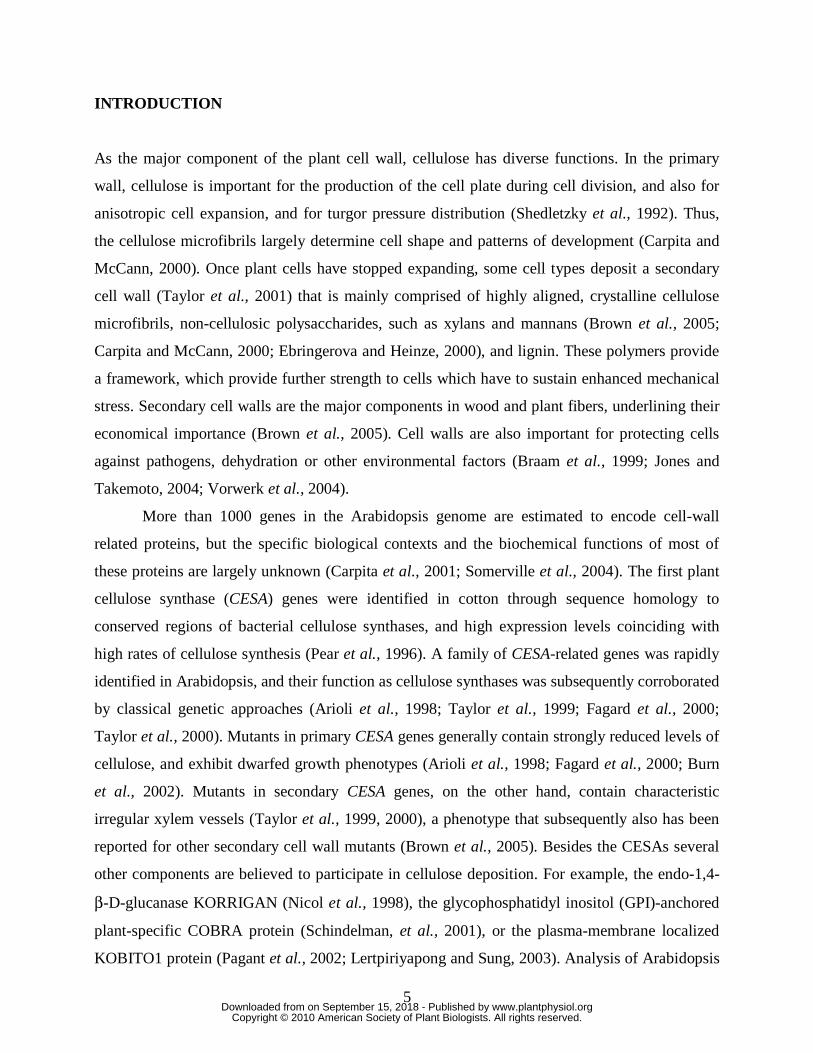
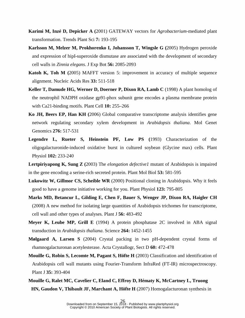
(Potikha and Delmer, 1995). The highly-ordered cellulose found in the cell walls of Arabidopsis
trichomes displays strong birefringence under polarized light, whereas the Arabidopsis tbr mutant
displays no such birefringence (cf. Fig. 1), and the cellulose content in tbr mutant trichomes is
strongly reduced (Potikha and Delmer, 1995). In this work we report the identification of the
gene responsible for the TBR trait, and a further characterization of the Arabidopsis tbr mutant.
www.plantphysiol.orgon September 15, 2018 - Published by Downloaded from Copyright © 2010 American Society of Plant Biologists. All rights reserved.
7
We show that TBR belongs to a plant-specific, poorly described gene family (TBR-like, TBL)
with 46 members in Arabidopsis. TBR and other gene family members are strongly co-expressed
with primary- and secondary CESA genes, respectively. We further provide evidence that TBR
and an additional member of the family influences secondary wall cellulose deposition. Our
results also suggest involvement of the latter genes in pectin modification, more specifically
methylesterification that may affect the deposition of secondary walls.
RESULTS
Novel phenotypes of the tbr mutant
The tbr mutant of Arabidopsis was previously characterized as lacking leaf and stem trichome
birefringence (Potikha and Delmer, 1995; Fig. 1) characteristic of plant cells which contain
highly ordered cellulose in their secondary walls. Consistently, cellulose levels were decreased in
tbr trichomes by >80%. In addition, the cellulose content in the leaf vasculature was decreased
with ~30%, whereas cellulose contents in stems, roots or callus tissue were unaffected (Potikha
and Delmer, 1995). Other tbr phenotypes reported by Potikha and Delmer (1995) included
reduced trichome density, altered trichome shape and surface appearance, lack of trichome
papillae and basal cells, altered stomata shape, and altered patterns of callose deposition.
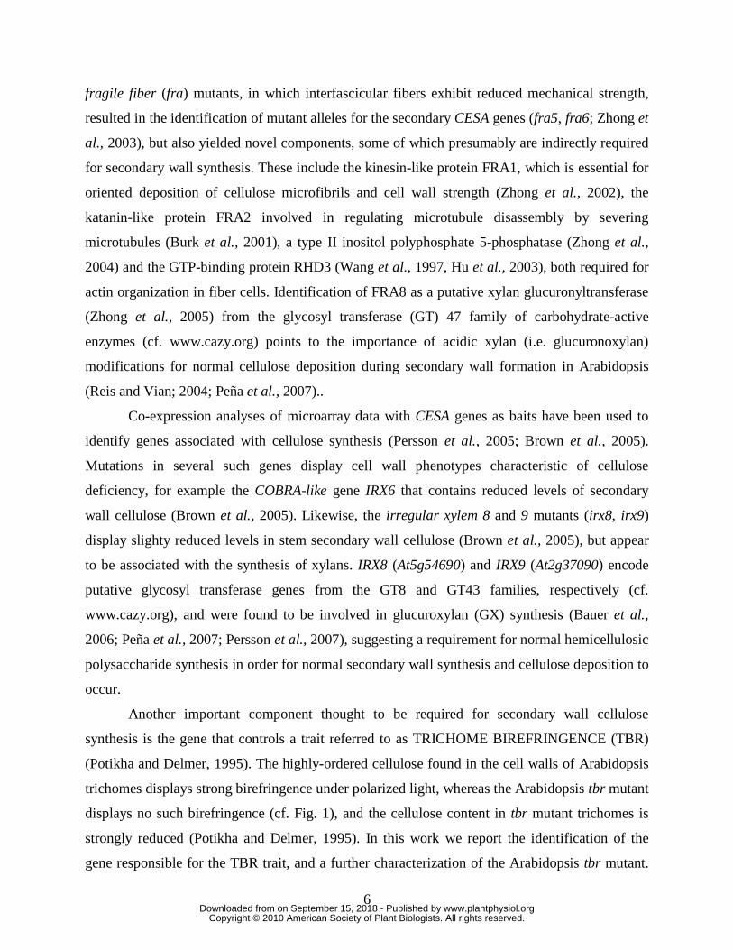
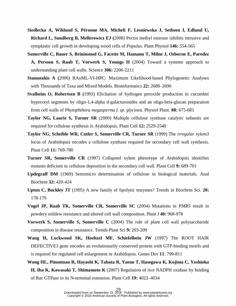
An additional cell wall-related phenotype was detected when we grew tbr mutant
seedlings in the dark. Mutant seedlings frequently displayed a marked reduction in hypocotyl
length (Fig. 2A), reminiscent of etiolated primary cesa mutant seedlings, e.g. prc1-8, ixr1-2 or
rsw1-10 (Fagard et al., 2000; Mouille et al., 2003), and dark-grown wild-type seedlings treated
with cellulose synthesis inhibitors, like thaxtomin A (Scheible et al., 2003; Bischoff et al., 2009)
or DCB (Robert et al., 2004). In addition, young etiolated tbr mutant seedlings occasionally
showed slight isotropic cell expansion symptoms in the upper part of the hypocotyl (Fig. 2B, C),
a phenotype that was reported previously for thaxtomin A-treated seedlings (Scheible et al.,
2003). Such symptom was never observed in wild-type seedlings (Fig. 2D). These results point
towards a function of TBR in primary cell wall synthesis in etiolated seedlings. To assess
whether this is due to reduced levels of primary wall cellulose levels we measured the amount of
crystalline cellulose in tbr and wild-type hypocotyls (Fig. 2E). We did not detect any differences
in cellulose levels between the tbr and the wild-type control, indicating that alterations in other
cell wall polymers may be the cause of the reduced hypocotyl elongation.
www.plantphysiol.orgon September 15, 2018 - Published by Downloaded from Copyright © 2010 American Society of Plant Biologists. All rights reserved.
8
Greenhouse-grown tbr mutants (i.e. progeny with five backcrosses to Col-0 wild type)
also repeatedly displayed a marked growth variation that is unrelated to variation in germination
time. The size of tbr mutants ranged between wild type-like and considerably dwarfed, with the
frequency of the latter being less than 10% (supplemental Fig. S1). Additional visible phenotypes
of tbr mutants included a ~40% reduced stem thickness at the base (0.82±0.12 mm in tbr vs.
1.47±0.21 mm in wild type), and reduced leaf size (supplemental Fig. S1).
Cell wall composition of tbr trichomes
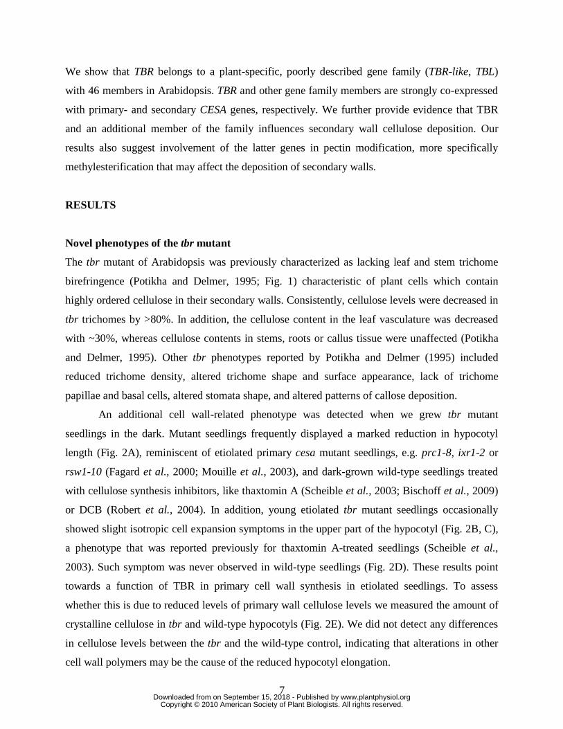
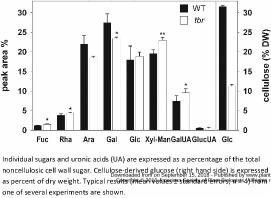
To investigate the effects on cell wall composition of the TBR gene product we isolated mature
trichomes from tbr and wild-type rosette leaves. Cellulose measurements in the trichome
preparations revealed a ~70% decrease in tbr relative to wild type (Fig. 3) thus confirming
previous results (Potikha and Delmer, 1995). We also analyzed the non-cellulosic sugar
composition and uronic acid content in trichomes by HPAEC (Fig. 3). Compared to wild type, tbr
trichomes displayed a 25% increase of the pectic component galacturonic acid. Similarly, another
prominent pectic component, rhamnose was increased by ~15%. In contrast, the relative
galactose- and arabinose-contents were reduced by ~15%, while glucuronic acid, a minor but for
biomineralization processes important compound found in plant cell walls, was unaltered.
Neutral sugars, i.e. xylose and fucose, apparent in the side chains of hemicelluloses such as
xyloglucans, were increased by around 20 % (Fig. 3). A clear separation of xylose and mannose
could not be achieved by the HPAEC method used, but additional data obtained from analysis of
alditol acetates by GC-MS (not shown), suggested that the xylose content was increased in tbr
mutant trichomes, whereas the mannose content was unchanged.
Identification of TBR
The morphological changes and the reduction in cellulose in tbr mutant trichomes are robust and
transmitted to the progeny in a pattern typical for recessive mutants (Potikha and Delmer, 1995).
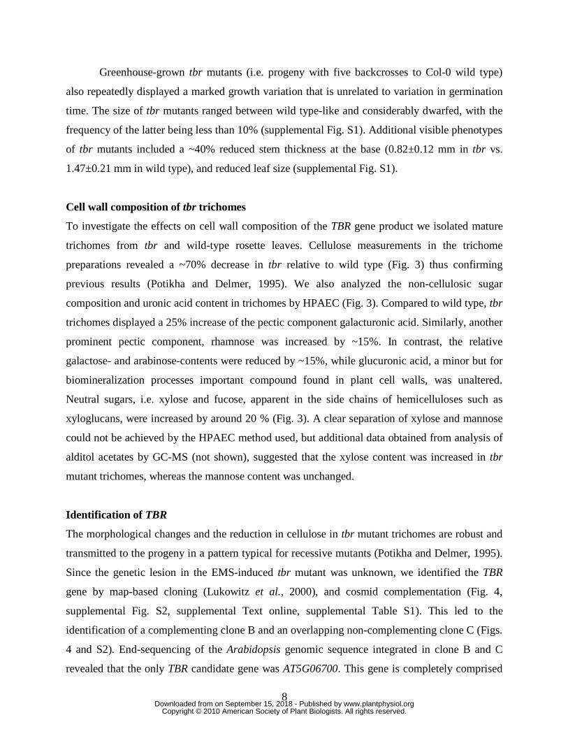
Since the genetic lesion in the EMS-induced tbr mutant was unknown, we identified the TBR
gene by map-based cloning (Lukowitz et al., 2000), and cosmid complementation (Fig. 4,
supplemental Fig. S2, supplemental Text online, supplemental Table S1). This led to the
identification of a complementing clone B and an overlapping non-complementing clone C (Figs.
4 and S2). End-sequencing of the Arabidopsis genomic sequence integrated in clone B and C
revealed that the only TBR candidate gene was AT5G06700. This gene is completely comprised
www.plantphysiol.orgon September 15, 2018 - Published by Downloaded from Copyright © 2010 American Society of Plant Biologists. All rights reserved.
9
in B whereas the first two thirds of the gene are absent in C. Sequencing of AT5G06700 from tbr
mutant DNA subsequently revealed a G�A transition in the third exon of the annotated coding
region, resulting in replacement of Gly427 by Glu in the predicted protein (Fig. 4A). A CAPS
marker was developed based on the single base change found in AT5G06700. Co-segregation of
strong trichome birefringence and the heterozygous CAPS genotype was found in the progeny of
tbr mutants transformed with clone B, as expected (supplemental Fig. S2D).
To provide further and independent confirmation of the identity of TBR, we investigated
additional reduction/loss of function alleles for AT5G06700. To this end, we first tested several
T-DNA insertion lines (SALK_134006, SALK_134014, SALK_058509, SAIL_707_D07) but
were unable to (i) identify homozygous mutants by PCR and (ii) detect plants with reduced or
lacking trichome birefringence. Therefore we next produced an RNA interference (RNAi)
construct to AT5G06700, and introduced it into wild type. Many of the resulting RNAi lines
displayed considerable or complete loss of trichome birefringence (Fig. 4B). In addition,
complementation of the tbr mutant was achieved by expression of the annotated AT5G06700
coding sequence under control of the CaMV 35S promoter (supplemental Fig. S3).
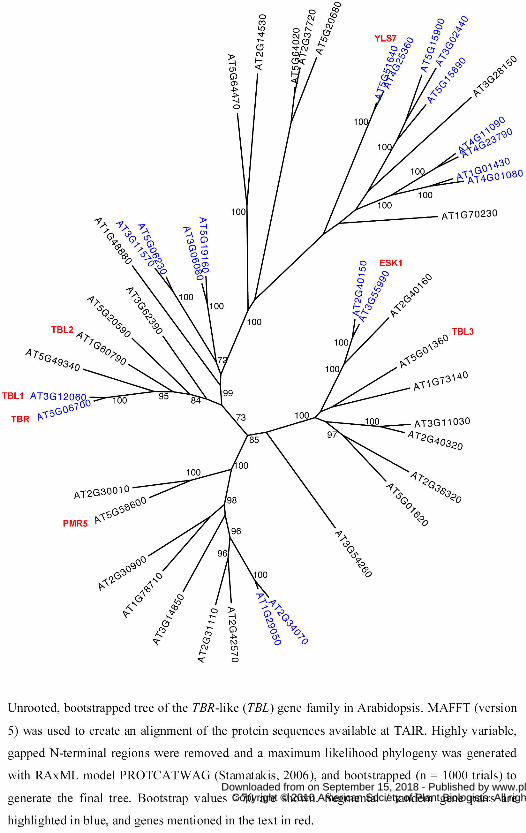
TBR and its homologs are plant-specific DUF231 proteins
According to the annotation provided by TAIR, TBR spans ~2.173 kb (from start to stop codon,
Fig. 4A), contains five exons and encodes a 2.207 kb transcript that yields a predicted 608 amino
acid 67.9 kD protein with an isoelectric point of 9.12. BLASTP and TBLASTN searches with the
TBR protein sequence revealed that TBR is plant-specific, and has 45 homologs (e-values <10-29)
in the Arabidopsis genome. We aligned the protein sequences (Table S2), and created an
unrooted phylogenetic tree (Fig. 5). The proteins cluster into three major branches consistent with
previous analyses (Xin et al., 2007), with TBR (AT5G06700) being part of the smallest clade
(Fig. 5). Within the family, the TIGR database lists eight pairs of segmental duplicated genes and
one triplet that arose through segmental and tandem duplication (highlighted in blue in Fig. 5).
TBR, its duplicated twin AT3G12060 (TBL1), and the closely related AT1G60790 (TBL2)
encode the longest annotated proteins in the family (608, 556 and 541 amino acids), while
AT2G31110 and AT3G14850 appear to encode the shortest TBL proteins with 364 and 356 amino
acids, respectively (supplemental Fig. S4A). All of the TBR and TBR-like (TBL) homologs
harbor a putative transmembrane domain at the N-terminus (TMH), and contain a domain of
unknown function DUF231 (PFAM 3005; IPR004253) near their C-termini (supplemental Fig.
www.plantphysiol.orgon September 15, 2018 - Published by Downloaded from Copyright © 2010 American Society of Plant Biologists. All rights reserved.
10
S4A). The predicted amino acid exchange (Gly427 � Glu) in the tbr mutant introduces a
negative charge just in front of the DUF231 domain (see asterisk in supplemental Fig. S4B). This
Gly residue is conserved in TBR homologs from other plant species (Fig. S4B) as well as 26 of
the Arabidopsis TBLs (supplemental Table S2). Three additional Arabidopsis TBLs have Ala and
the residual 16 an aromatic amino acid (Y, W and F) in this position.
Furthermore, the 46 proteins contain another plant-specific region, typically 87-89
residues long, that has not been recognized previously and which we termed TBL domain
(supplemental Fig. S4). Thirteen amino acids in this domain, including five unequally spaced
cysteine residues, two tryptophans and a glycine-aspartate-serine (GDS) signature are fully
conserved among the 46 family members (see highlighted region in supplemental Fig. S3B). In
addition, similar amino acids are found in another 25 positions of the TBL domains (see
highlighted region in supplemental Fig. S4B; supplemental Table S2). The GDS signature is
followed by a Leu in over half of the proteins (24 proteins), or by a similar non-aromatic,
aliphatic amino acid like Ile and Val or Met, which in turn is preceded by several hydrophobic
amino acids. GDS in the same amino acid context (i.e. GDSL) has previously been found to be a
conserved motif in some esterases/lipases (Upton and Buckley, 1995; Akoh et al., 2004).
Within the 150-180 residues long DUF231 domain we detected another 13 residues with
absolute identity (including another 3 cysteines, 3 histidines and 2 tryptophans) in all 46 proteins,
and 27 residues with conserved similarity (Supplemental Fig. S4B; Supplemental Table 2 online).
There is a remarkable stretch of highly conserved amino acids towards the end of the DUF231
domain including a DxxH motif (Supplemental Fig. S4B; Supplemental Table 2 online), which is
also conserved in the same class of GDSL motif-containing esterases/lipases. Interestingly, the
Asp and His residues in the DxxH motif together with the Ser residue in the distant GDS motif
were determined to form the catalytic triad in the 2.5Å crystal structure of a fungal
rhamnogalacturonan acetylesterase (Mølgaard and Larsen, 2004).
TBR/TBLs have highly similar DUF231 protein homologs in other plant species. Within
the C-terminal part starting with the TBL domain TBR, for example, shares 77% identity (90%
similarity) with grapevine protein CAO23412, 69% identity (82% similarity) with rice lustrin A-
like protein BAD35858 (Os06g0207500) or 61% identity (80% similarity) with Medicago
truncatula protein ABE91344 (Supplemental Fig. S4B).
Expression pattern of TBR
www.plantphysiol.orgon September 15, 2018 - Published by Downloaded from Copyright © 2010 American Society of Plant Biologists. All rights reserved.
11
To investigate where the TBR gene is active we created promoter GUS gene reporter lines. When
expressed under the control of 1.6 kb sequence upstream of the annotated start codon (which
includes 140 bp annotated 5’-UTR), GUS activity in young plantlets was prominent in leaf and
stem trichomes (Figs. 6A to C). However, similar GUS constructs for the close TBR homologs
At3g12060 (TBL1) and At1g60790 (TBL2) showed that these genes were not active in trichomes
(Figs. 6D, and E). Interestingly, the TBL1 coding sequence expressed under control of the TBR
promoter complemented the trichome birefringence phenotype of tbr mutants (data not shown).
These results suggest that the TBL1 is functionally equivalent to TBR, but may work in different
tissues or cell-types compared to TBR.
In 3-weeks-old TBR:GUS gene reporter plants GUS activity was mainly associated with
the leaf vasculature, and was also present in younger expanding/maturating rosette leaves (Fig.
6F), as well as rapidly growing parts of the root, e.g. lateral root tips (data not shown). In 4-
weeks-old plants the signal persisted in the vasculature and trichomes, and was also strong in
rapidly expanding, fortifying inflorescence stems (Fig. 6G), where primary and secondary wall
celluloses are deposited. With increasing age and organ maturation GUS activity continuously
decreased (Fig. 6H) until it was hardly detectable in 6-weeks old plants (Fig. 6I). This expression
pattern is in agreement with the developmental AtGenExpress ATH1 genechip data (Schmid et
al., 2005; for visualization see http://www.bar.utoronto.ca/efp), and is also consistent with the
one expected for a component required during cellulose deposition.
Co-expression of TBR and TBLs with CESA and other cell wall genes
The strong reduction in crystalline cellulose in tbr trichomes (Potikha and Delmer, 1995) led us
to investigate whether the gene is co-expressed with cellulose synthase (CESA) genes. Analyses
using the GeneCAT co-expression tool (http://genecat.mpg.de; Mutwil et al., 2008) revealed that
CESA5, CESA6, CESA3 rank at positions 2, 6 and 12 on the list of genes co-expressed with TBR
(supplemental Table S3). We also found the endochitinase ELP/POM/CTL1 (At1g05850) and the
GPI-anchored protein encoding COBRA gene (At5g60920) on this list. These genes are well-
known components for primary wall cellulose production (Hauser et al., 1995; Schindelman et
al., 2001; Zhong et al., 2002). Similar results were also obtained using other co-expression tools,
including ATTED-II (Obayashi et al., 2009) and AraGenNet (Mutwil et al., 2010). Vice versa,
TBR also appears in the list of co-expressed genes when primary CESA genes are used as baits
(results not shown; Persson et al., (2005).
www.plantphysiol.orgon September 15, 2018 - Published by Downloaded from Copyright © 2010 American Society of Plant Biologists. All rights reserved.
12
To see if also other members of the family may be transcriptionally coordinated with
cellulose related processes we extended the analysis including the additional 35 DUF231 family
members represented on ATH1 genechips. Interestingly, the TBL gene At5g01360 was tightly co-
expressed with the secondary CESA genes, and conversely the analysis of genes co-expressed
with At5g01360 in GeneCAT revealed many of the known or suspected genes important for
secondary wall cellulose synthesis (Brown et al., 2005), including all three secondary CESA
genes, IRX8, IRX9, IRX12 and COBL4 (supplemental Table S3).
Finally, several other DUF231 family members, including At1g78710, At2g37720,
At2g40160, At3g28150, At3g62390, At5g01620, At5g15890 and At5g58600, displayed
remarkable co-expression with other cell wall-related genes, like cellulose synthase-like (Csl),
endo-1,4-beta glucanase, xyloglucan:xyloglucosyl transferase, laccase, polygalacturonase or
pectinesterase genes (cf. http://genecat.mpg.de). Taken together, the co-expression analyses
suggest that, besides TBR, at least At5g01360 and possibly several other members of the plant-
specific TBL/DUF231 domain gene family might have a function in cell wall biology.
Isolation and phenotypic characterization of tbl3 mutants
To investigate the potential involvement of At5g01360/TBL3 in cellulose / cell wall synthesis, we
obtained seeds from a corresponding T-DNA insertion line (SALK_065959) available through
the Nottingham European Arabidopsis Stock Center. Homozygous mutant plants were identified
by PCR screening of genomic DNA samples, and were further characterized for expression of
At5g01360 (subsequently named TBL3) at the cDNA level. No PCR amplification was possible
from mutant cDNA with primers spanning the T-DNA insertion site (supplemental Figs. S5A and
B), and quantitative real-time PCR (qRT-PCR) revealed that the cDNA template corresponding
to the last exon of At5g01360 was more than 1000-fold less abundant in the mutant compared to
the wild-type cDNA pool (supplemental Figs. S5A and C). These results suggest that the isolated
homozygous mutant is a null.
Growth analysis revealed that the homozygous mutant progeny displayed a variety of
growth phenotypes, ranging from mild to severe reductions in inflorescence stem elongation
(supplemental Fig. S6), whereas rosette leaf or root growth was only mildly affected
(supplemental Fig. S6; data not shown). The variation in growth and stem size is similar to what
was observed for the tbr mutants (supplemental Fig. S1). However, TBR gene expression was not
changed in the tbl3 mutants (data not shown). The tbl3 mutants also frequently displayed
www.plantphysiol.orgon September 15, 2018 - Published by Downloaded from Copyright © 2010 American Society of Plant Biologists. All rights reserved.
13
significantly (10-20%) reduced stem diameter (data not shown) as found for tbr mutants and for
other secondary wall mutants (Turner & Somerville, 1997; Taylor et al., 2001). To corroborate
the mutant phenotype we also produced gene-specific RNAi lines for At5g01360, and found the
tbl3 growth phenotypes recapitulated in these (data not shown). These results suggest that TBL3
is required for normal stem development in Arabidopsis.
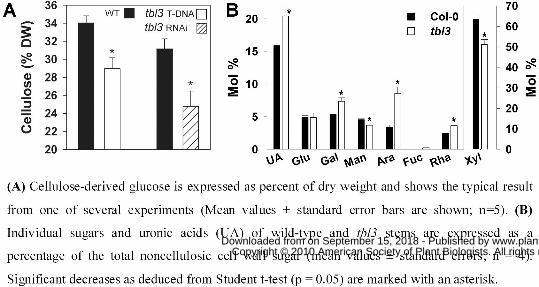
Biochemical analysis of tbl3 T-DNA mutant stems revealed a 15-20% reduction in
cellulose content independent of stem age (Fig. 7A). These data were reproduced using the tbl3
RNAi lines (Fig. 7A). We also detected changes in the non-cellulosic carbohydrate composition
in stem material sampled 20 days after bolting (Fig. 7B). In agreement with previous studies,
wild-type stems showed a high xylose content characteristic of secondary cell walls (Turner and
Somerville, 1997; Brown et al., 2005). Cell wall material from tbl3 mutant stems displayed a
20% relative reduction in xylose content, and a strong relative increase in arabinose, galactose,
rhamnose and fucose, as well as uronic acids (Fig. 7B). These changes resemble the sugar
compositions measured in stems of irx7 or irx8 mutants, and of mutants in other genes co-
expressed with secondary CESAs (Brown et al., 2005; Persson et al., 2007). Finally, stem
sections were cut from the base of the mature inflorescence stem to investigate xylem
morphology of tbl3 mutant and wild type. The xylem vessels of tbl3 mutants were
indistinguishable from wild-type vessels displaying open xylem elements with relatively round
shape (data not shown). These data suggest that while the constituents for several important
secondary wall polymers were decreased in tbl3 the integrity of the wall still is sufficient for
normal xylem morphology.
Additional hypocotyl phenotypes of tbr and tbl3
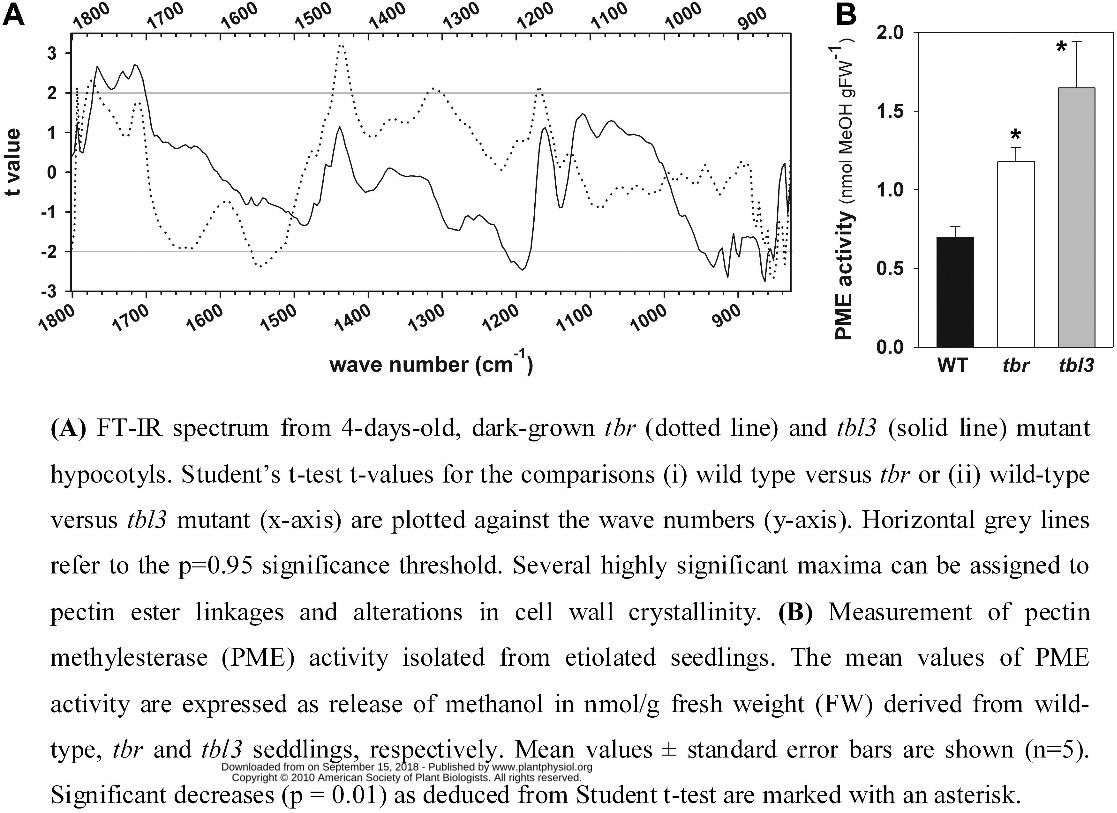
To determine possible structural changes in the cell walls of tbr hypocotyls, we performed
Fourier Transform Infrared (FTIR) microspectroscopy (Fig. 8A). This analysis revealed,
according to Student t-test, significant changes at wave number 1168 cm-1 and also at wave
number 1774 cm-1, which most likely correspond to decrease of ester linkages presumably of
methyl-esterified pectins (Mouille et al., 2003; Sene et al., 1994). The peak at 1546 cm-1 may
represent changes in not further specified cell wall proteins. These results indicate that the tbr
hypocotyls are not holding lower levels of cellulose (Fig. 2E), but there may be structural
changes in the cell wall framework involving esterified pectins. In agreement with this
www.plantphysiol.orgon September 15, 2018 - Published by Downloaded from Copyright © 2010 American Society of Plant Biologists. All rights reserved.
14
observation, a significantly elevated pectin methylesterase (PME) activity was detected in tbr
protein extracts derived from etiolated seedlings (Fig. 8B).
In contrast to tbr (Fig. 2), etiolated tbl3 seedlings did not show a significant reduction in
hypocotyl length (data not shown). The level of crystalline cellulose in these hypocotyls tended
to be slightly increased on a dry weight basis, and FTIR analysis revealed similar changes in cell
wall structures of etiolated tbl3 hypocotyls as seen for tbr (Fig. 8A). We found significant
changes at wave numbers 852, 917, 933, 1191 cm-1, and at wave numbers 1762, 1731 and 1712
cm-1 (Fig. 8A), indicating structural changes of cellulose and esterified pectins in tbl3 compared
to wild-type seedlings. In addition and similar to tbr, PME activity was significantly increased in
tbl3 (Fig. 8B). These data suggest that the tbr and tbl3 seedling hypocotyls hold lower levels of
methylesterified pectins due to enhanced pectin methylesterase activities.
DISCUSSION
In this study we applied forward and reverse genetic approaches to identify two members of the
undescribed DUF231 gene family, i.e. TBR (At5g06700) and TBL3 (At5g01360), as novel
components contributing to secondary wall cellulose synthesis in higher plants. The tbr mutant
was previously reported as a mutant that lacks crystalline cellulose in trichomes, and to lesser
extent also in the vasculature (Potikha and Delmer, 1995). The involvement of TBR in cellulose
synthesis is underpinned by the expression pattern of the gene. TBR displays extraordinary co-
expression with primary CESA genes, such as CESA3, CESA5 or CES6, while co-expression with
secondary CESA genes is inconspicuous. Several genes co-expressed with the primary and
secondary CESA genes were previously shown to be associated with primary and secondary wall
cellulose production, respectively (see introduction and results). The expression patterns obtained
from the microarray datasets were mirrored in the TBR promoter::GUS staining patterns,
corroborating that TBR is similarly expressed as the primary wall CESA genes. Considering the
substantial reduction in trichome associated crystalline cellulose levels in tbr, it is perhaps
surprising that no decrease in primary wall cellulose levels were observed. However, it is
possible that other TBR-related gene products may functionally compensate for the loss of TBR
in these tissues. In support of such scenario, TBL1 could rescue the trichome birefringence
phenotype when expressed under the TBR promoter.
www.plantphysiol.orgon September 15, 2018 - Published by Downloaded from Copyright © 2010 American Society of Plant Biologists. All rights reserved.
15
We showed that the reduction in cellulose content in tbr trichomes was accompanied by
an increased amount of pectic compounds, i.e. galacturonic acids, whereas the relative galactose
and arabinose contents were reduced by ~15%. These results imply increased amounts of HG and
decreased abundance of the galactose- and arabinose-containing side chains of the pectin
rhamnogalacturonan I in the mutant. The amount of glucuronic acid remained stable in
trichomes, in turn. This result confirms that the changes in uronic acids are associated with pectic
structures, and rules out the idea that loss of trichome birefringence is a result of reduced uronic-
acid-dependent bio-mineralization/crystallization. The amount of xylose-mannose was increased
indicating elevated amounts of xyloglucans. Since the effects on pectin/xyloglucan content,
and/or modification, was significant we propose that tbr directly interferes with cellulose
biosynthesis in trichomes, and directly or indirectly influences pectin composition in this organ.
In support of this, several mutants deficient in cellulose production display increased levels of
pectins, most likely homogalacturonans (His et al., 2001).
tbr hypocotyls contained wild-type levels of primary wall cellulose, less methylesterified
pectins, and hold modified cell wall structures as assessed by FTIR analysis. In addition, results
of the PME assay confirm that the lower degree of pectin methylesterification comes along with a
higher methylesterase activity in tbr compared to wild-type seedlings. Many primary CW
mutants e.g. prc1-1, rsw1-10, kor1-1, kob1-1 have less crystalline cellulose and short hypocotyls
(Desnos et al., 1996, Arioli et al., 1998, Nicol et al., 1998, Fagard et al., 2000, Pagant et al.,
2002). However, dwarfed hypocotyls are not necessarily linked to the level of crystalline
cellulose, but can also result from altered levels of methylesterified pectins (Derbyshire et al.,
2007). In addition, it is interesting to note that the tbr hypocotyl phenotypes described partially
resemble the mutant quasimodo2 (qua2), affecting a putative pectin methyltransferase. qua2 has
higher levels of crystalline cellulose, a shorter hypocotyl as well as altered pectin composition
(Mouille et al., 2003, 2007). Our results suggest that TBR, besides its role in secondary wall
cellulose deposition in trichomes, is involved in pectin modifications in etiolated hypocotyls, a
tissue mainly containing primary wall cellulose. Alternatively, the changes described could
reflect a feedback loop from secondary to primary cell wall biosynthesis as has been described
for mur10 (Bosca et al., 2006). However, TBR has a largely non-redundant/critical biological
function in trichomes, as suggested by the missing trichome expression of its close homologs
(Figs. 6D and E). In this context, it is interesting to note (i) that Arabidopsis trichomes do not
appear to express secondary CESA genes (CESA4, CESA7, CESA8) while primary CESA genes
www.plantphysiol.orgon September 15, 2018 - Published by Downloaded from Copyright © 2010 American Society of Plant Biologists. All rights reserved.
16
(CESA1, CESA3, CESA5, CESA6) and KORRIGAN are expressed (Jakoby et al., 2008; Marks et
al., 2008), and (ii) that the Arabidopsis trichome cell wall contains polysaccharides that are more
like those of typical primary walls (Marks et al., 2008). This may suggest that the “secondary
wall” crystalline cellulose deposited in trichomes is a product of the primary CESA complex.
Alternatively, the secondary CESA complex may be recruited from other cell types, such as
neighboring trichome companion or basal cells. However, both interpretations seem to be
rejected by the observation that irx3 (cesa7) mutants as well as primary cesa mutants (cesa3,
cesa2, cesa5, cesa6, cesa2/cesa5 and cesa2/cesa6) display wild-type like trichome birefringence
(V. Bischoff, S. Nita and W. Scheible, unpublished data). Another possibility would be that
cellulose synthase-like (CSL) proteins synthesize cellulose in trichomes, similar to the situation
described for pollen tubes of Nicotiana alata (Doblin et al., 2001).
The second TBL/DUF231 domain gene family member that we functionally implicated in
secondary cell wall and cellulose synthesis is TBL3 (AT5G01360). This gene was strongly co-
expressed with CESA4, CESA7, CESA8, and analysis of a T-DNA insertion null allele and RNA
interference lines confirmed its function in secondary cell wall cellulose deposition. The T-DNA
mutants have shorter and weaker stems at high frequency, contained significantly less cellulose in
the inflorescence stems and displayed a cell wall monosaccharide pattern that highly resembles
those of several irx mutants (Brown et al., 2005). tbl3 stems appear to be enriched in pectin as
judged from increased levels of uronic acids, arabinose, galactose and rhamnose. While xylose
levels decreased by ~20%, this neutral sugar pattern is reminiscent of those found in irx7, irx8 or
irx9 mutants (Brown et al., 2005). The irregular xylem structure is a known criteria for mutants
involved in secondary wall cellulose biosynthesis (Turner and Somerville, 1997). However, the
reduction of cellulose and/or the structural changes in the secondary cell wall of tbl3 were not
sufficient to yield an irregular xylem phenotype with collapsed xylem vessels (data not shown).
This is in agreement with several other mutants notably involved in secondary wall cellulose
biosynthesis. Some of the latter showed normal or modestly disturbed xylem vessels (Brown et
al., 2005; Persson et al., 2005; Brown et al., 2009), others developed the irregular xylem
phenotype only in a double irx background (Brown et al., 2009). Additional analyses showed that
etiolated tbl3 hypocotyls displayed wild type-like phenotypes but, as seen for tbr hypocotyls, had
slightly elevated crystalline cellulose levels and significantly reduced amounts of
methylesterified pectins. Along with the PME assay showing higher PME activity in tbl3, our
results suggest that TBL3, similar to TBR, is involved in secondary wall cellulose biosynthesis in
www.plantphysiol.orgon September 15, 2018 - Published by Downloaded from Copyright © 2010 American Society of Plant Biologists. All rights reserved.
17
specific tissues and also has an influence on the pectic structures in tissue containing mainly
primary cell wall. A role of AT5G01360 in secondary cell wall biosynthesis was previously
suggested as it turned up as member of a core xylem-specific gene set resulting from global
comparative transcriptome analysis (Ko et al., 2006).
What might be the biochemical functions of the plant-specific DUF231 domain genes?
One report (Yoshida et al., 2001) describes YLS7 (AT5G51640) as highly expressed in senescent
leaves, but beyond this the gene remained anonymous. Another member of the gene family
(ESK1; AT3G55990) was found to act as a negative regulator of cold acclimation, as mutations in
the ESK1 gene provide strong freezing tolerance (Xin et al., 2007). In a third report, it was noted
that a null mutation in the PMR5 gene (i.e. AT5G58600) rendered Arabidopsis resistant to
powdery mildew species, and based on cell wall and FTIR analyses pmr5 cell walls were
enriched in pectin and displayed a reduced degree of pectin methylesterifiation relative to wild-
type cell walls, suggesting that the gene affects pectin composition (Vogel et al., 2004). This
observation is in line with our results, as we also noted changes in pectin composition in various
tissues of tbr and tbl3, respectively. Although the alterations in pectin composition in tbr
trichomes and tbl3 stems seem to be due to compensatory effects following the reduction in
cellulose levels, our results from etiolated tbr and tbl3 hypocotyls suggest a more direct impact of
these gene products on pectin modification. FTIR analysis revealed less esterified pectins and this
could be confirmed by an elevated PME activity. These results indicate that at least some
DUF231 gene family members, including PMR5, TBR and TBL3, are required to maintain
higher pectin methylesterification states, by e.g inhibiting PME activity, rather than being PMEs
by themselves, as might be deduced from the conserved GDSL and DxxH motifs known from
some esterases (cf. results).
Whatever the biochemical function of the proteins, loss of TBR or TBL3 appears to
increase PME activity, reduce pectin esterification and to decrease cellulose deposition in
trichomes and stems, respectively. There is existing evidence indicating that correct
modifications of non-cellulosic cell wall polysaccharides are important for growth of cells
undergoing secondary wall thickening, the synthesis of secondary wall cellulose and secondary
wall integrity. For example, the dwarfed Arabidopsis irx8/gaut12 mutant is deficient in GX and
HG (Persson et al., 2007), has alterations of the glycosyl sequence at the GX reducing end (Peña
et al., 2007) and has a significant reduction in secondary cell wall thickness and cellulose content
(Persson et al., 2007). The degree of pectin methylesterification can limit cell growth and
www.plantphysiol.orgon September 15, 2018 - Published by Downloaded from Copyright © 2010 American Society of Plant Biologists. All rights reserved.
18
hypocotyl elongation in Arabidopsis, as shown by ectopic expression of a fungal pectin
methylesterase (Derbyshire et al., 2007). Similarly, pectin methylesterase prevents correct growth
of developing wood cells/fibres in poplar (Siedlecka et al., 2008), suggesting that proper pectin
esterification is likely to be essential for xylem development and lignification (Pelloux et al.,
2007). Furthermore, there is evidence for potent binding of pectins, which are enriched in neutral
side chains, to cellulose (Zykwinska et al., 2005), thus leading to the assumption that pectin-
cellulose interactions are significant for cell wall assembly and normal cellulose deposition
during primary and secondary cell wall formation. In tobacco leaf explants oligogalacturonides
derived from esterified HG were shown to stimulate cellulose deposition and cell wall thickening
(Altamura et al., 1998), and such oligogalacturonides were also shown to elicit production of
hydrogen peroxide (H2O2) (Legendre et al., 1993; Svalheim and Robertson, 1993), probably via
induction of small Rac-type GTPases which activate H2O2-producing plasma-membrane NADPH
oxidases (Keller et al., 1998; Potikha et al., 1999; Wong et al., 2007). In this respect, it should be
noted that the plasma-membrane-localized small GTPase AtRAC2/ROP7 (At5g45970) is
specifically induced during later stages of xylem differentiation in Arabidopsis (Brembu et al.,
2005), and strong co-expression with TBL3 was detected in our study (cf. Supplemental Table
S3). Constant production of low levels of H2O2 stimulates the onset of secondary wall cellulose
synthesis and secondary wall differentiation as shown in cotton fiber cells (Potikha et al., 1999;
Karlsson et al., 2005; Hovav et al., 2008) which from the botanical point of view represent seed
trichomes. It is therefore tempting to speculate that (i) TBR and/or TBL3 function in maintaining
HG esterification, as suggested by our results, and that (ii) this is required for triggering
secondary wall cellulose synthesis in a manner similar to the suite of events outlined above. The
esterification state of pectic polymers might therefore influence the synthesis and deposition of
cellulose in plants.
MATERIALS AND METHODS
Plant materials and growth conditions.
Plants were grown in environmental chambers (120 μE,16h light/8h dark, 60% relative humidity,
20°C) or in the greenhouses of MPI-MPP (150 μE, 12-14h light, ~70% relative humidity, 20-
22°C) on a commercial Arabidopsis substrate (Stender AG; Schermbeck, Germany). Etiolated
seedlings were grown on vertical agar plates (0.7% w/v agar, half-strength Murashige & Skoog
www.plantphysiol.orgon September 15, 2018 - Published by Downloaded from Copyright © 2010 American Society of Plant Biologists. All rights reserved.
19
medium supplemented with 0.5 % (w/v) sucrose) wrapped in aluminium foil. Arabidopsis wild-
type (Col-0) and tbr mutant seeds were obtained from an in-house collection. The tbl3 T-DNA
insertion line SALK_065959 was obtained from The Nottingham Arabidopsis Stock Centre
(NASC; Nottingham University, UK).
Positional cloning of the TBR locus
See Protocol S1
Cell wall analyses and pectin methylesterase activity assay
See Protocols S2 and S3
Analysis of trichome birefringence.
Trichome birefringence of old rosette leaves was analyzed by incubation in methanol for 20 min,
followed by discoloration for 1 h in boiling 85% lactic acid, and rinsing with water (3 times). The
leaves were then observed under a stereomicroscope (Leica MZ 12.5, Germany) equipped with a
polarizer.
Stem cross sections.
50µm cross sections were cut with a vibratome (Leica, Bensheim, Germany). Sections were
stained with 0.1% toluidine blue (analysis of irregular xylem structures) and observed under a
stereo microscope (Olympus, BX41, Hamburg, GERMANY; Leica, MZ 125, Germany).
Fourier-transform infrared microspectrometry.
Four-day-old seedlings were squashed between two barium fluoride windows and rinsed
abundantly with distilled water for 2 min, before drying at 37 °C for 20 min. For each mutant,
twenty spectra were collected from individual hypocotyls of seedlings from four independent
cultures (five seedlings from each culture), as described by Mouille et al. (2003). Normalization
of the data and statistical analyses were performed as described by Mouille et al. (2003).
Normalization of the data set and statistical analyses were performed using the statistical
language R V2.6. (R Development Core Team, 2006). To normalize the spectra, the baseline,
estimated using a linear regression involving 10 points at each end of the spectrum, was
subtracted from each absorbance value and the area was set to one by dividing each absorbance
www.plantphysiol.orgon September 15, 2018 - Published by Downloaded from Copyright © 2010 American Society of Plant Biologists. All rights reserved.
20
value by the sum of all absorbance values. To determine the difference of the composition and
the structure between mutants and wild-type, Student’s t-test was performed.
RNA interference constructs for TBR and TBL3.
A 381 bp TBR-specific PCR fragment was amplified using primers 5’ –
CACCAACCACTCGTCACGCGCC- 3’ and 5’ –GGTTTGGTTGAAGTGACATTGGG- 3’,
subcloned into pENTR/SD/D-TOPO (Invitrogen) and recombined into pK7GWIWG2(II) (Karimi
et al., 2002). A 177 bp TBL3 specific PCR fragment was amplified with primers 5’ –
CACCAATTACCAGATTTGCCACTATGAGC- 3’ and 5’-
TTGAAGAAAGATGAAGACGAAGAGG- 3’ and shuttled into pK7GWIWG2(II) with the
same strategy. The constructs were transferred into Col-0 plants by Agrobacterium mediated
transformation (Clough and Bent, 1998), and T1 transformants were selected on kanamycin and
scored for trichome birefringence as described above.
Promoter–β-glucuronidase constructs and GUS staining.
To obtain a TBR promoter–-glucuronidase (GUS) gene fusion construct, a PCR product was
amplified from genomic DNA of Arabidopsis wild-type Col-0 using PfuTurbo polymerase
(Stratagene, La Jolla, CA) and primers 5'-CAATGTCGACGGAAGATGAGTTGGACAATGC-3'
and 5'-CAATGGATCCGCATATACTTAACGGCGTCTG-3'. The 1.64 kb BamHI/SalI promoter
fragment was subsequently fused to the GUS gene in vector pBI101.1 (Clontech); and after
sequence verification the recombinant vector was introduced into Arabidopsis wild-type Col-0
via A. tumefaciens (GV3101) according to Clough and Bent (1998). Detection of GUS activity
was performed according to Jefferson et al. (1987) by incubation in staining buffer at 37°C
overnight. Promoter–β-glucuronidase lines for the close TBR homologs At3g12060 (TBL1) and
At1g60790 (TBL2) were produced likewise using primers 5'-
CAATGTCGACTTTCGGAGTTTCTAGTCTGGA-3' and 5'-
CAATGGATCCTTCAGGCAATGCGTAATCTAT-3' and primers 5'-
CAATGTCGACCCATTTGTATCAATCTCCCCC-3' and 5'-
CAATCCCGGGAATGAAGCACACGGAAAGTGA-3', respectively.
Molecular analysis of tbl3 mutants.
www.plantphysiol.orgon September 15, 2018 - Published by Downloaded from Copyright © 2010 American Society of Plant Biologists. All rights reserved.
21
A T-DNA insertion line (SALK_065959) for TBL3 (At5g01360) was obtained from the
Nottingham European Arabidopsis Stock Center (Nottingham University, UK). Plant DNA was
extracted as previously described (Lukowitz et al., 2000). Mutant lines were confirmed for T-
DNA insertion using spanning primers LP (5’ –GTATGAAAAACGGACAGCCAGAAACT- 3’)
and RP (5’ -TCCGAAAGAACCCCACAGAGC- 3’) (designed on
http://signal.salk.edu/tdnaprimers.html), and a primer from the left T-DNA border, LBc1 (5’ –
CCGCAATGTGTTATTAAGTTG- 3’). Primer combinations LBC1/LP and LP/RP were used.
PCR conditions were 96 °C for 5 min; 96 °C for 30 s, 58 °C for 40 s, and 72 °C for 1 min; 4 °C
hold. RNA was isolated from tbl3 mutant stems using the Plant RNeasy Mini Kit (Qiagen). RNA
quality checks, DNase I treatment and reverse transcription were performed as described
previously (Czechowski et al., 2005). PCR with two primers 5’ –
GTATGAAAAACGGACAGCCAGAAACT- 3’ and 5’ –TCCGAAAGAACCCCACAGAGC- 3’
spanning the insertion site (cf. Fig. 7A) was performed on wild-type and tbl3 cDNA to confirm
the absence of a PCR product when using the mutant template (cf. Fig. 7B). Quantitative real-
time PCR was used to analyze the expression level of At5g01360, using a Applied Biosystems
HT7900 sequence detection system, SYBR green reagent (Applied Biosystems) and primers 5’ –
ATCGCATCGACGCTCACAC- 3’ and 5’ –TCAGCGGTTAGGATCTTGCC- 3’. For further
details and subsequent data normalization procedures see Czechowski et al. (2005).
Sequence alignment and phylogenetic analysis.
MAFFT version 5 (Katoh et al., 2005) was used to create an alignment of the Arabidopsis
DUF231 protein sequences available at TAIR and of other plant TBR homologs available at
NCBI. The alignment was processed with Boxshade 3.21 (http://www.ch.embnet.org) and
arranged manually into the final layout (cf. supplemental Fig. S4B, Table S2). Prior to computing
a phylogeny, the highly variable and gapped N-terminal region was removed manually, and all
columns with 50% or more gaps were removed with the software REAP (Hartmann and Vision,
2008). RAxML v7 was then used to compute an unrooted maximum likelihood phylogeny from
the masked alignment using the PROTCATWAG model (Stamatakis, 2006). The phylogenetic
tree was subsequently bootstrapped (n = 1000 trials) to create the final tree in FigTree version
1.1.2 (http://tree.bio.ed.ac.uk/software/figtree/) as shown in Fig. 5.
Co-expression analysis.
www.plantphysiol.orgon September 15, 2018 - Published by Downloaded from Copyright © 2010 American Society of Plant Biologists. All rights reserved.
22
Co-expression analyses were performed using the web-based tool GeneCAT (Mutwil et al.,
2008), ATTED-II (Obayashi et al., 2009).
Accession numbers.
Sequence data from this article can be found in the Arabidopsis Genome Initiative under the
following locus identifiers: AT5G06700 (TBR), AT3G12060 (TBL1), AT1G60790 (TBL2) and
AT5G01360 (TBL3). The mutant tbr sequence is available from GenBank (ID XXX).
Supplemental Material
The following materials are available in the online version of this article.
Supplemental Figure S1. Growth Phenotype of tbr mutants
Supplemental Figure S2. Identification of TBR by recombinational mapping and cosmid
complementation.
Supplemental Figure S3. Complementation of tbr mutant by 35S::TBR.
Supplemental Figure S4. Structure and sequence alignment of DUF231 domain proteins
Supplemental Figure S5. Molecular characterization of tbl3 T-DNA mutants
Supplemental Figure S6. Growth phenotype of tbl3 mutants
Supplemental Table S1. Specifications and primer sequences of mapping markers and the tbr
CAPS marker.
Supplemental Table S2. Partial alignment of the 46 Arabidopsis DUF231 proteins.
Supplemental Table S3. Genes co-expressed with TBR or TBL3.
Protocol S1. Positional cloning of the tbr locus
Protocol S2. Biochemical analyses of tbr and tbl3 cell walls
Protocol S3: Pectin methylesterase (PME) extraction and activity assay
ACKNOWLEDGMENTS
We are grateful to Kian Hematy, Gregory Mouille (INRA, Versailles) and Eugenia Maximova
(MPI-MPP) for excellent advice and help with respect to microscopy, and Joachim Selbig and
Stefanie Hartmann (University of Potsdam) for help with phylogenetic analysis. We also
acknowledge Erwin Grill (TU München, Germany) for providing the Col-0 cosmid library,
www.plantphysiol.orgon September 15, 2018 - Published by Downloaded from Copyright © 2010 American Society of Plant Biologists. All rights reserved.
23
CEREON Genomics for supplying information on Col/Landsberg erecta polymorphisms, and the
Nottingham European Arabidopsis Stock Center (NASC) for seeds of the T-DNA insertion line
SALK_065959. Volker Bischoff was supported by the European Union Framework Program 6
(FP6) “CASPIC” NEST-CT-2004-028974 and the German Science Foundation (DFG, SCHE
548/2).
LITERATURE CITED
Akoh CC, Lee GC, Liaw YC, Huang TH, Shaw JF (2004) GDSL family of serine
esterases/lipases. Prog Lipid Res 43: 534-52
Altamura MM, Zaghi D, Salvi G, De Lorenzo G, Bellincampi D (1998) Oligogalacturonides
stimulate pericycle cell wall thickening and cell divisions leading to stoma formation in tobacco
leaf explants. Planta 204: 429-436
Arioli T, Peng L, Betzner AS, Burn J, Wittke W, Herth W, Camilleri C, Höfte H, Plazinski
J, Birch R, Cork A, Glover J, Redmond J, Williamson RE (1998) Molecular analysis of
cellulose biosynthesis in Arabidopsis. Science 279: 717-720
Bauer S, Vasu P, Persson S, Mort AJ, Somerville CR (2006) Development and application of
a suite of polysaccharide-degrading enzymes for analyzing plant cell walls. Proc Natl Acad Sci
USA 103: 11417-11422
Bevan M (1984) Binary Agrobacterium vectors for plant transformation. Nucleic Acids Res 12:
8711-8721
Bischoff V, Cookson SJ, Wu S, Scheible WR (2009) Thaxtomin A affects CESA-complex
density, expression of cell wall genes, cell wall composition, and causes ectopic lignification in
Arabidopsis thaliana seedlings. J Exp Bot 60: 955-965
Bosca S, Barton CJ, Taylor NG, Ryden P, Neumetzler L, Pauly M, Roberts K, Seifert GJ
(2006) Interactions between MUR10/CesA7-dependent secondary cellulose biosynthesis and
primary cell wall structure. Plant Physiol 142: 1353-63
Braam J (1999) If walls could talk. Curr Opin Plant Biol. 2: 521-524
Brembu T, Winge P, Bones AM (2005) The small GTPase AtRAC2/ROP7 is specifically
expressed during late stages of xylem differentiation in Arabidopsis. J Exp Bot 56: 2465-2476
www.plantphysiol.orgon September 15, 2018 - Published by Downloaded from Copyright © 2010 American Society of Plant Biologists. All rights reserved.
24
Brown DM, Zeef LA, Ellis J, Goodacre R, Turner SR (2005) Identification of novel genes in
Arabidopsis involved in secondary cell wall formation using expression profiling and reverse
genetics. Plant Cell 17: 2281-2295
Brown DM, Zhang Z, Stephens E, Dupree P, Turner SR (2009) Characterization of IRX10
and IRX10-like reveals an essential role in glucuronoxylan biosynthesis in Arabidopsis. Plant J
57: 732-46
Burk DH, Liu B, Zhong R, Morrison WH, YE ZH (2001) A katanin-like protein regulates
normal cell wall biosynthesis and cell elongation. Plant Cell 13: 807-827
Burn JE, Hocart CH, Birch RJ, Cork AC, Williamson RE (2002) Functional analysis of the
cellulose synthase genes CesA1, CesA2, and CesA3 in Arabidopsis. Plant Physiol 129: 797-807
Carpita N, McCann M (2000) The cell wall. In B Buchanan, W Gruissem, R Jones, eds,
Biochemistry and Molecular Biology of Plants, American Society of Plant Physiologists, pp
52-108
Carpita NC, Tierney M, Campbell M (2001) Molecular biology of the plant cell wall:
searching for the genes that define structure, architecture, and dynamics. Plant Mol Biol 47: 1–
5
Clough SJ, Bent AF (1998) Floral dip: a simplified method for Agrobacterium-mediated
transformation of Arabidopsis thaliana. Plant J 16: 735-743
Curtis MD, Grossniklaus U (2003) A gateway cloning vector set for high-throughput functional
analysis of genes in planta. Plant Physiol 133: 462-469
Czechowski T, Stitt M, Altmann T, Udvardi MK, Scheible WR (2005) Genome-wide
identification and testing of superior reference genes for transcript normalization in
Arabidopsis. Plant Physiol 139: 5-17
Desnos T, Orbović V, Bellini C, Kronenberger J, Caboche M, Traas J, Höfte H (1996)
Procuste1 mutants identify two distinct genetic pathways controlling hypocotyl cell elongation,
respectively in dark- and light-grown Arabidopsis seedlings. Development 122: 683-93
Doblin MS, De Melis L, Newbigin E, Bacic A, Read SM (2001) Pollen tubes of Nicotiana alata
express two genes from different beta-glucan synthase families. Plant Physiol 125: 2040-2052
Derbyshire P, McCann MC, Roberts K (2007) Restricted cell elongation in Arabidopsis
hypocotyls is associated with a reduced average pectin esterification level. BMC Plant Biol 7:
31
www.plantphysiol.orgon September 15, 2018 - Published by Downloaded from Copyright © 2010 American Society of Plant Biologists. All rights reserved.
25
Ebert B, Zoeller D, Erban A, Fehrle I, Hartmann J, Niehl A, Kopka J and Fisahn J (2010)
Metabolic profiling of Arabidopsis thaliana epidermal cells. Journal of Experimental Botany
(accepted)
Ebringerova A, Heinze T (2000) Naturally occurring xylans: Structures, isolation procedures
and properties. Macromol Rapid Commun 21: 542–556
Fagard M, Desnos T, Desprez T, Goubet F, Refregier G, Mouille G, McCann M, Rayon C,
Vernhettes S, Höfte H (2000) PROCUSTE1 encodes a cellulose synthase required for normal
cell elongation specifically in roots and dark-grown hypocotyls of Arabidopsis. Plant Cell 12:
2409-2424
Filisetti-Cozzi TM, Carpita NC (1991) Measurement of uronic acids without interference from
neutral sugars. Anal Biochem 197: 157-162
Hartmann S, Vision TJ (2008) Using ESTs for phylogenomics: can one accurately infer a
phylogenetic tree from a gappy alignment? BMC Evo. Biol 8: 95
Hauser MT, Morikami A, Benfey PN (1995) Conditional root expansion mutants of
Arabidopsis. Development 121: 1237–1252
His I, Driouich A, Nicol F, Jauneau A, Höfte H (2001) Altered pectin composition in primary
cell walls of korrigan, a dwarf mutant of Arabidopsis deficient in a membrane-bound endo-1,4-
β-glucanase. Planta. 212: 348-58.
Hovav R, Udall JA, Chaudhary B, Hovav E, Flagel L, Hu G, Wendel JF (2008) The
evolution of spinnable cotton fiber entailed prolonged development and a novel metabolism.
PLoS Genetics 4: e25
Hu Y, Zhong R, Morrison WH, Ye ZH (2003) The Arabidopsis RHD3 gene is required for cell
wall biosynthesis and actin organization. Planta 217: 912-21
Jakoby MJ, Falkenhan D, Mader MT, Brininstool G, Wischnitzki E, Platz N, Hudson A,
Hülskamp M, Larkin J, Schnittger A (2008) Transcriptional profiling of mature Arabidopsis
trichomes reveals that NOECK encodes the MIXTA-like transcriptional regulator MYB106.
Plant Physiol 148: 1583-1602
Jefferson RA, Kavanagh TA, Bevan MW (1987) GUS fusions: beta-glucuronidase as a
sensitive and versatile gene fusion marker in higher plants. Embo J 6: 3901-7
Jones DA, Takemoto D (2004) Plant innate immunity - direct and indirect recognition of general
and specific pathogen-associated molecules. Curr Opin Immunol 16: 48-62
www.plantphysiol.orgon September 15, 2018 - Published by Downloaded from Copyright © 2010 American Society of Plant Biologists. All rights reserved.
26
Karimi M, Inzé D, Depicker A (2001) GATEWAY vectors for Agrobacterium-mediated plant
transformation. Trends Plant Sci 7: 193-195
Karlsson M, Melzer M, Prokhorenko I, Johansson T, Wingsle G (2005) Hydrogen peroxide
and expression of hipI-superoxide dismutase are associated with the development of secondary
cell walls in Zinnia elegans. J Exp Bot 56: 2085-2093
Katoh K, Toh M (2005) MAFFT version 5: improvement in accuracy of multiple sequence
alignment. Nucleic Acids Res 33: 511-518
Keller T, Damude HG, Werner D, Doerner P, Dixon RA, Lamb C (1998) A plant homolog of
the neutrophil NADPH oxidase gp91-phox subunit gene encodes a plasma membrane protein
with Ca21-binding motifs. Plant Cell 10: 255–266
Ko JH, Beers EP, Han KH (2006) Global comparative transcriptome analysis identifies gene
network regulating secondary xylem development in Arabidopsis thaliana. Mol Genet
Genomics 276: 517-531
Legendre L, Rueter S, Heinstein PF, Low PS (1993) Characterization of the
oligogalacturonide-induced oxidative burst in cultured soybean (Glycine max) cells. Plant
Physiol 102: 233-240
Lertpiriyapong K, Sung Z (2003) The elongation defective1 mutant of Arabidopsis is impaired
in the gene encoding a serine-rich secreted protein. Plant Mol Biol 53: 581-595
Lukowitz W, Gillmor CS, Scheible WR (2000) Positional cloning in Arabidopsis. Why it feels
good to have a genome initiative working for you. Plant Physiol 123: 795-805
Marks MD, Betancur L, Gilding E, Chen F, Bauer S, Wenger JP, Dixon RA, Haigler CH
(2008) A new method for isolating large quantities of Arabidopsis trichomes for transcriptome,
cell wall and other types of analyses. Plant J 56: 483-492
Meyer K, Leube MP, Grill E (1994) A protein phosphatase 2C involved in ABA signal
transduction in Arabidopsis thaliana. Science 264: 1452-1455
Mølgaard A, Larsen S (2004) Crystal packing in two pH-dependent crystal forms of
rhamnogalacturonan acetylesterase. Acta Crystallogr, Sect D 60: 472-478
Mouille G, Robin S, Lecomte M, Pagant S, Höfte H (2003) Classification and identification of
Arabidopsis cell wall mutants using Fourier-Transform InfraRed (FT-IR) microspectroscopy.
Plant J 35: 393-404
Mouille G, Ralet MC, Cavelier C, Eland C, Effroy D, Hématy K, McCartney L, Truong
HN, Gaudon V, Thibault JF, Marchant A, Höfte H (2007) Homogalacturonan synthesis in
www.plantphysiol.orgon September 15, 2018 - Published by Downloaded from Copyright © 2010 American Society of Plant Biologists. All rights reserved.
27
Arabidopsis thaliana requires a Golgi-localized protein with a putative methyltransferase
domain. Plant J 2007 50: 605-14.
Mutwil M, Obro J, Willats WG, Persson S (2008) GeneCAT--novel webtools that combine
BLAST and co-expression analyses. Nucleic Acids Res 36: W320-326
Mutwil M, Usadel B, Schütte M, Loraine A, Ebenhöh O, Persson S (2010) Assembly of an
interactive correlation network for the Arabidopsis genome using a novel heuristic clustering
algorithm. Plant Physiol 152: 29-43
Neff MM, Turk E, Kalishman M (2002) Web-based primer design for single nucleotide
polymorphism analysis. Trends Genet 18: 613-615
Nicol F, His I, Jauneau A, Vernhettes S, Canut H, Höfte H (1998) A plasma membrane-bound
putative endo-1,4-beta-D-glucanase is required for normal wall assembly and cell elongation in
Arabidopsis. EMBO J 17: 5563-5576
Obayashi T, Hayashi S, Saeki M, Ohta H, Kinoshita K. (2009) ATTED-II provides
coexpressed gene networks for Arabidopsis. Nucleic Acids Res. 37: D987-991
Pagant S, Bichet A, Sugimoto K, Lerouxel O, Desprez T, McCann M, Lerouge P,
Vernhettes S, Höfte H (2002) KOBITO1 encodes a novel plasma membrane protein necessary
for normal synthesis of cellulose during cell expansion in Arabidopsis. Plant Cell 14: 2001-
2013
Pear JR, Kawagoe Y, Schreckengost WE, Delmer DP, Stalker DM (1996) Higher plants
contain homologs of the bacterial celA genes encoding the catalytic subunit of cellulose
synthase. Proc Natl Acad Sci USA 93: 12637-12642
Pelloux J, Rustérucci C, Mellerowicz EJ (2007) New insights into pectin methylesterase
structure and function. Trends Plant Sci 12: 267-77
Peña MJ, Zhong R, Zhou GK, Richardson EA, O'Neill MA, Darvill AG, York WS, Ye ZH
(2007) Arabidopsis irregular xylem8 and irregular xylem9: implications for the complexity of
glucuronoxylan biosynthesis. Plant Cell 19: 549-563
Persson S, Wei H, Milne J, Page GP, Somerville CR (2005) Identification of genes required
for cellulose synthesis by regression analysis of public microarray data sets. Proc Natl Acad Sci
USA 102: 8633-8638
Persson S, Caffall KH, Freshour G, Hilley MT, Bauer S, Poindexter P, Hahn MG, Mohnen
D, Somerville C (2007) The Arabidopsis irregular xylem8 mutant is deficient in
www.plantphysiol.orgon September 15, 2018 - Published by Downloaded from Copyright © 2010 American Society of Plant Biologists. All rights reserved.
28
glucuronoxylan and homogalacturonan, which are essential for secondary cell wall integrity.
Plant Cell 19: 237-255
Potikha T, Delmer DP (1995) A Mutant of Arabidopsis thaliana displaying altered patterns of
cellulose deposition. Plant J 7: 453-460
Potikha TS, Collins CC, Johnson DI, Delmer DP, Levine A (1999) The involvement of
hydrogen peroxide in the differentiation of secondary walls in cotton fibers. Plant Physiol 119:
849-858
R Development Core Team (2006) R: A language and environment for statistical computing.
URL: http://www.R-project.org
Reis D, Vian B. (2004) Helicoidal pattern in secondary cell walls and possible role of xylans in
their construction. C R Biol 327: 785-790
Reiter WD, Chapple CC, Somerville CR (1993) Altered growth and cell walls in a fucose-
deficient mutant of Arabidopsis. Science 261: 1032-1035
Robert S, Mouille G, Höfte H (2004) The mechanism and regulation of cellulose synthesis in
primary walls: lessons from cellulose-deficient Arabidopsis mutants. Cellulose 11: 351–364
Scheible WR, Fry B, Kochevenko A, Schindelasch D, Zimmerli L, Somerville S, Loria R,
Somerville CR (2003) An Arabidopsis mutant resistant to thaxtomin A, a cellulose synthesis
inhibitor from Streptomyces species. Plant Cell 15: 1781-94
Schindelman G, Morikami A, Jung J, Baskin TI, Carpita NC, Derbyshire P, McCann MC,
Benfey PN (2001) COBRA encodes a putative GPI-anchored protein, which is polarly localized
and necessary for oriented cell expansion in Arabidopsis. Genes Dev 15: 1115-1127
Schmid M, Davison TS, Henz SR, Pape UJ, Demar M, Vingron M, Scholkopf B, Weigel D,
Lohmann JU (2005) A gene expression map of Arabidopsis thaliana development. Nat Genet
37: 501-506
Scott, TA, Melvin EH (1953) Determination of dextran with anthrone. Anal Chemistry 25:
1656–1661
Sene C, McCann MC, Wilson RH, Grinter R (1994) Fourier-Transform Raman and Fourier-
Transform Infrared Spectroscopy (An Investigation of Five Higher Plant Cell Walls and Their
Components). Plant Physiol 106: 1623-1631
Shedletzky E, Shmuel M, Trainin T, Kalman S, Delmer D (1992) Cell wall structure in cells
adapted to growth on the cellulose-synthesis inhibitor 2,6-Dichlorobenzonitrile: A comparison
between two dicotyledonous plants and a graminaceous monocot. Plant Physiol 100: 120-130
www.plantphysiol.orgon September 15, 2018 - Published by Downloaded from Copyright © 2010 American Society of Plant Biologists. All rights reserved.
29
Siedlecka A, Wiklund S, Péronne MA, Micheli F, Lesniewska J, Sethson I, Edlund U,
Richard L, Sundberg B, Mellerowicz EJ (2008) Pectin methyl esterase inhibits intrusive and
symplastic cell growth in developing wood cells of Populus. Plant Physiol 146: 554-565
Somerville C, Bauer S, Brininstool G, Facette M, Hamann T, Milne J, Osborne E, Paredez
A, Persson S, Raab T, Vorwerk S, Youngs H (2004) Toward a systems approach to
understanding plant cell walls. Science 306: 2206-2211
Stamatakis A (2006) RAxML-VI-HPC: Maximum Likelihood-based Phylogenetic Analyses
with Thousands of Taxa and Mixed Models. Bioinformatics 22: 2688–2690
Svalheim O, Robertsen B (1993) Elicitation of hydrogen peroxide production in cucumber
hypocotyl segments by oligo-1,4-alpha d-galacturonides and an oligo-beta-glucan preparation
from cell walls of Phytophthora megasperma f. sp. glycinea. Physiol Plant. 88: 675-681
Taylor NG, Laurie S, Turner SR (2000) Multiple cellulose synthase catalytic subunits are
required for cellulose synthesis in Arabidopsis. Plant Cell 12: 2529-2540
Taylor NG, Scheible WR, Cutler S, Somerville CR, Turner SR (1999) The irregular xylem3
locus of Arabidopsis encodes a cellulose synthase required for secondary cell wall synthesis.
Plant Cell 11: 769-780
Turner SR, Somerville CR (1997) Collapsed xylem phenotype of Arabidopsis identifies
mutants deficient in cellulose deposition in the secondary cell wall. Plant Cell 9: 689-701
Updegraff DM (1969) Semimicro determination of cellulose in biological materials. Anal
Biochem 32: 420-424
Upton C, Buckley JT (1995) A new family of lipolytic enzymes? Trends in Biochem Sci. 20:
178-179
Vogel JP, Raab TK, Somerville CR, Somerville SC (2004) Mutations in PMR5 result in
powdery mildew resistance and altered cell wall composition. Plant J 40: 968-978
Vorwerk S, Somerville S, Somerville C (2004) The role of plant cell wall polysaccharide
composition in disease resistance. Trends Plant Sci 9: 203-209
Wang H, Lockwood SK, Hoeltzel MF, Schiefelbein JW (1997) The ROOT HAIR
DEFECTIVE3 gene encodes an evolutionarily conserved protein with GTP-binding motifs and
is required for regulated cell enlargement in Arabidopsis. Genes Dev 11: 799-811
Wong HL, Pinontoan R, Hayashi K, Tabata R, Yaeno T, Hasegawa K, Kojima C, Yoshioka
H, Iba K, Kawasaki T, Shimamoto K (2007) Regulation of rice NADPH oxidase by binding
of Rac GTPase to its N-terminal extension. Plant Cell 19: 4022–4034
www.plantphysiol.orgon September 15, 2018 - Published by Downloaded from Copyright © 2010 American Society of Plant Biologists. All rights reserved.
30
Xin Z, Mandaokar A, Chen J, Last RL, Browse J (2007) Arabidopsis ESK1 encodes a novel
regulator of freezing tolerance. Plant J 49:786-799
Yoshida S, Ito M, Nishida I, Watanabe A (2001) Isolation and RNA gel blot analysis of genes
that could serve as potential molecular markers for leaf senescence in Arabidopsis thaliana.
Plant Cell Physiol 42: 170-178
Zhang X, Oppenheimer DG (2004) A simple and efficient method for isolating trichomes for
downstream analyses. Plant Cell Physiol 45: 221-224
Zhong R, Kays SJ, Schroeder BP, Ye ZH (2002) Mutation of a chitinase-like gene causes
ectopic deposition of lignin, aberrant cell shapes, and overproduction of ethylene. Plant Cell 14:
165-179
Zhong RQ, Morrison WH, Freshour GD, Hahn MG, Ye ZH (2003) Expression of a mutant
form of cellulose synthase AtCesA7 causes dominant negative effect on cellulose biosynthesis.
Plant Physiol 132: 786-795
Zhong R, Ye ZH (2004) Molecular and biochemical characterization of three WD-repeat-
domain-containing inositol polyphosphate 5-phosphatases in Arabidopsis thaliana. Plant Cell
Physiol 45: 1720-1728
Zhong RQ, Pena MJ, Zhou GK, Nairn CJ, Wood-Jones A, Richardson EA, Morrison WH,
Darvill AG, York WS, Ye ZH (2005) Arabidopsis fragile fiber8, which encodes a putative
glucuronyltransferase, is essential for normal secondary wall synthesis. Plant Cell 17: 3390-
3408
Zykwinska AW, Ralet MCJ, Garnier CD, Thibault JFJ (2005) Evidence for in vitro binding
of pectin side chains to cellulose. Plant Physiol 139: 397-407
www.plantphysiol.orgon September 15, 2018 - Published by Downloaded from Copyright © 2010 American Society of Plant Biologists. All rights reserved.
31
FIGURE LEGENDS
Figure 1. Lack of trichome birefringence in tbr mutants.
(A) Birefringence phenotype of a wild-type Col-0 and (B) of a tbr mutant leaf. Appearance of
individual wild-type and tbr mutant trichomes under (C, D) polarized and (E, F) non-polarized
white light.
Figure 2. Hypocotyl phenotype of etiolated tbr mutants.
(A) Aspects of 4-days-old etiolated wild-type and tbr mutant seedlings. The length distribution
among tbr seedlings is indicated by percent numbers. Numbers at the bottom (13.98 ± 1.37, 7.78
± 2.48, n=42) indicate the mean value (in millimeters) ± standard deviation of hypocotyl length
in wild-type and tbr populations (B, C) Cell swelling phenotype occasionally observed at the top
of tbr mutant hypocotyls, and in comparison (D) the invariant, regular wild-type phenotype. (E)
Crystalline cellulose determination as measured by the Updegraff protocol. Mean values ±
standard deviations (n=3) are given.
Figure 3. Altered cellulose and non-cellulosic sugar composition in tbr mutant trichomes.
Individual sugars and uronic acids (UA) are expressed as a percentage of the total noncellulosic
cell wall sugar. Cellulose-derived glucose (right hand side) is expressed as percent of dry weight.
Typical results (mean values ± standard errors; n = 4) from one of several experiments are
shown.
Figure 4. Identification of TBR.
(A) View of the ~5kb region responsible for complementation of the tbr phenotype. The structure
of the one annotated gene (At5g06700) in that region is given. Exons are depicted as white boxes
and untranslated regions of At5g06700 i.e. TBR as grey boxes. The arrow indicates the direction
of transcription. The point mutation in the tbr-1 allele (G�A) leads to a predicted glycine to
glutamate exchange at position 427 of the encoded protein. (B) Knock-down of TBR by RNA
interference (cf. the three photos to the right showing leaves of different RNAi lines) leads to
strongly reduced trichome birefringence as compared to wild type (photo to the left).
Figure 5. Unrooted tree of the TBL gene family.
www.plantphysiol.orgon September 15, 2018 - Published by Downloaded from Copyright © 2010 American Society of Plant Biologists. All rights reserved.
32
Unrooted, bootstrapped tree of the TBR-like (TBL) gene family in Arabidopsis. MAFFT (version
5) was used to create an alignment of the protein sequences available at TAIR. Highly variable,
gapped N-terminal regions were removed and a maximum likelihood phylogeny was generated
with RAxML model PROTCATWAG (Stamatakis, 2006), and bootstrapped (n = 1000 trials) to
generate the final tree. Bootstrap values >70 are shown. Segmental / tandem gene pairs are
highlighted in blue, and genes mentioned in the text in red.
Figure 6. GUS expression of TBR.
(A) Staining of a nine-days-old seedling transformed with GUS driven by the TBR promoter. (B)
Magnified view of stained leaf trichomes of a nine-days-old seedling. (C-E) Staining of stem
trichomes of seedlings transformed with (C) TBR:GUS, (D) TBL1:GUS or (E) TBL2:GUS. (F-I)
Typical staining of (F) a three-weeks-old, (G) a four-weeks-old, (H) a five-weeks-old, and (I) a
six-weeks-old TBR:GUS transgenic plant.
Figure 7. Biochemical cell wall phenotypes of tbl3 T-DNA mutants.
(A) Cellulose-derived glucose is expressed as percent of dry weight and shows the typical result
from one of several experiments (Mean values ± standard error bars are shown; n=5). (B)
Individual sugars and uronic acids (UA) of wild-type and tbl3 stems are expressed as a
percentage of the total noncellulosic cell wall sugar (mean values ± standard errors; n = 4).
Significant decreases as deduced from Student t-test (p ≤ 0.05) are marked with an asterisk.
Figure 8. FT-IR spectra and PME activity in tbr and tbl3 mutants.
(A) FT-IR spectrum from 4-days-old, dark-grown tbr (dotted line) and tbl3 (solid line) mutant
hypocotyls. Student’s t-test t-values for the comparison between wild type and tbr or tbl3 mutant
(x-axis) are plotted against the wave numbers (y-axis). Horizontal grey lines refer to the p=0.95
significance threshold. Several highly significant maxima can be assigned to pectin ester linkages
and alterations in cell wall crystallinity. (B) Measurement of pectin methylesterase (PME)
activity isolated from etiolated seedlings. The mean values of PME activity are expressed as
release of methanol in nmol/g fresh weight (FW) derived from wild-type, tbr and tbl3 seddlings,
respectively. Mean values ± standard error bars are shown (n=5). Significant decreases (p ≤ 0.01)
as deduced from Student t-test are marked with an asterisk.
www.plantphysiol.orgon September 15, 2018 - Published by Downloaded from Copyright © 2010 American Society of Plant Biologists. All rights reserved.
www.plantphysiol.orgon September 15, 2018 - Published by Downloaded from Copyright © 2010 American Society of Plant Biologists. All rights reserved.
www.plantphysiol.orgon September 15, 2018 - Published by Downloaded from Copyright © 2010 American Society of Plant Biologists. All rights reserved.
www.plantphysiol.orgon September 15, 2018 - Published by Downloaded from Copyright © 2010 American Society of Plant Biologists. All rights reserved.
www.plantphysiol.orgon September 15, 2018 - Published by Downloaded from Copyright © 2010 American Society of Plant Biologists. All rights reserved.
www.plantphysiol.orgon September 15, 2018 - Published by Downloaded from Copyright © 2010 American Society of Plant Biologists. All rights reserved.
www.plantphysiol.orgon September 15, 2018 - Published by Downloaded from Copyright © 2010 American Society of Plant Biologists. All rights reserved.
www.plantphysiol.orgon September 15, 2018 - Published by Downloaded from Copyright © 2010 American Society of Plant Biologists. All rights reserved.
www.plantphysiol.orgon September 15, 2018 - Published by Downloaded from Copyright © 2010 American Society of Plant Biologists. All rights reserved.