Safety and feasibility of fasting incombination with platinum-basedchemotherapyTanya B. Dorff1, Susan Groshen2, Agustin Garcia1, Manali Shah1, Denice Tsao-Wei2, Huyen Pham3, Chia-Wei Cheng4,Sebastian Brandhorst4, Pinchas Cohen4, Min Wei4, Valter Longo4* and David I. Quinn1*
Abstract
Background: Short-term starvation prior to chemotherapy administration protects mice against toxicity. Weundertook dose-escalation of fasting prior to platinum-based chemotherapy to determine safety and feasibility incancer patients.
Methods: 3 cohorts fasted before chemotherapy for 24, 48 and 72 h (divided as 48 pre-chemo and 24 post-chemo)and recorded all calories consumed. Feasibility was defined as ≥ 3/6 subjects in each cohort consuming ≤ 200 kcalper 24 h during the fast period without excess toxicity. Oxidative stress was evaluated in leukocytes using theCOMET assay. Insulin, glucose, ketones, insulin-like growth factor-1 (IGF-1) and IGF binding proteins (IGFBPs) weremeasured as biomarkers of the fasting state.
Results: The median age of our 20 subjects was 61, and 85 % were women. Feasibility criteria were met.Fasting-related toxicities were limited to≤ grade 2, most commonly fatigue, headache, and dizziness. The COMET assayindicated reduced DNA damage in leukocytes from subjects who fasted for ≥48 h (p = 0.08). There was anon-significant trend toward less grade 3 or 4 neutropenia in the 48 and 72 h cohorts compared to 24 h cohort (p = 0.17). IGF-1 levels decreased by 30, 33 and 8 % in the 24, 48 and 72 h fasting cohorts respectively after the first fastingperiod.
Conclusion: Fasting for 72 h around chemotherapy administration is safe and feasible for cancer patients. Biomarkerssuch as IGF-1 may facilitate assessment of differences in chemotherapy toxicity in subgroups achieving the physiologicfasting state. An onging randomized trial is studying the effect of 72 h of fasting.
Trial registration: NCT00936364, registered propectively on July 9, 2009.
BackgroundPlatinum chemotherapy is a mainstay of combinationsystemic therapy for many solid tumors, with the abilityto reduce the risk of cancer recurrence after curativesurgery in some situations, or to extend survival in
advanced disease. However toxicity frequently limits theamount of chemotherapy that can be administered. Boththe efficacy and toxicity of chemotherapy agents, includ-ing platinum drugs, are related to oxidative cellulardamage. Preclinical studies have shown that the heart,liver, and renal tissue may be protected from toxicity bythe concurrent administration of antioxidants [1–3]. Thelimitation of this approach has been concern over a pos-sible attenuation of efficacy against malignant cells, al-though this has not been substantiated in the availablerandomized trial data [4]. A more appealing approachwould be to differentially induce protection in normalhost cells without reducing, or potentially even increasing,susceptibility of cancer cells to chemotherapy. Cell culture
* Correspondence: [email protected]; [email protected] Longo and David I. Quinn are co-senior authors, having supervisingresponsibility for the laboratory and the clinical aspects of the data,respectively.4Longevity Institute, University of Southern California Davis School ofGerontology, Department of Biological Sciences, 3715 McClintock Avenue,Los Angeles 90089, CA, United States1USC Keck School of Medicine, Norris Comprehensive Cancer Center, 1441Eastlake Ave. #3440, Los Angeles, CA 90033, USAFull list of author information is available at the end of the article
experiments have identified that chemotherapy toxicity tonormal primary cells was reduced when cultured in condi-tions mimicking fasting, while neoplastic cells did not ex-perience the same protection, and in some cases besensitized to the chemotherapeutic cytotoxicity in thelow-glucose and low growth factor environment [5, 6].Further experiments with xenografts in mice revealed thatshort-term starvation (STS) for 48 h prior to chemother-apy treatment significantly reduced side effects and deathfrom high-dose chemotherapy when compared to micefed with standard diets prior to receiving chemotherapy,leading to a hypothesis that fasting induces oxidativestress resistance [5]. The mice subjected to STS regainedmost of the weight lost during the 4 days after chemother-apy, whereas the control mice lost a significant proportionof their weight in the same post-chemotherapy period, po-tentially reflecting their experience of chemotherapy toxic-ities of anorexia and nausea. The overall response of themice exposed to STS was encouraging for the safety oftranslating this concept into human cancer patients.Powerful and wide-ranging metabolic and gene expres-
sion changes are induced by calorie restriction in normalcells, including upregulation of antioxidants and DNArepair pathways, in part mediated by dampening thenutrient-sensing and pro-proliferative pathways such asIGF-1/Akt and mTOR [6]. Oncogene expression, affect-ing the same pro-growth signaling cascades amongothers, prohibit a fasting-like response in cancer cellswhich continue to proliferate, and cancer cells may actu-ally be sensitized to toxins in the setting of nutrientdeprivation [7]. Studies in healthy volunteers have re-vealed that within 22 and 48 h of fasting, blood glucoseand insulin levels decrease significantly, and blood ke-tones increase [8, 9]. STS has been shown to induce a40 % reduction in circulating insulin-like growth factor-1 (IGF-1) as well as changes in IGF-1 binding protein(IGFBP) levels in mice [10, 11]. These changes representa potential set of biomarkers for identifying when a pro-tective state may occur, although they only represent asubset of the changes induced by fasting.As a first step in exploring the ability of fasting to
induce differential stress resistance in humans, weperformed a clinical trial to determine the safety andfeasibility of fasting prior to chemotherapy adminis-tration in human cancer patients. We sought to iden-tify a recommended fasting duration to be studied ina subsequent randomized trial, embedding correlativestudies to generate preliminary data regarding bio-markers of the fasting state and to evaluate oxidativestress in host leukocytes as proof of principle. Toassess safety and compliance, we designed a dose-escalation protocol in a “real-world” setting of pa-tients with advanced cancer receiving platinum-basedcombination chemotherapy.
MethodsEligible patients had cancer for which platinum-basedcombination chemotherapy without concurrent radiationwas being recommended with curative (peri-operative)or palliative intent. Because fasting was timed aroundthe administration of platinum, regimens in which plat-inum was administered consecutively for more than2 days (ex: Bleomycin, Etoposide, Cisplatin for germ celltumors) were not eligible. Patients may have begun re-ceiving platinum chemotherapy (1–2 cycles of thechemotherapy could have already been administered),provided at least 2 more cycles were planned duringwhich fasting could occur. Chemotherapy was adminis-tered at the treating physician’s discretion; standard anti-emetics were administered, including dexamethasoneand 5HT3 inhibitors. Subjects were excluded if they haddiabetes, low body mass index (<20.5), or had lost morethan 10 % of their weight in the preceding year.Escalation of fasting began at a “dose” level of 24 h,
and each cohort consisted of 6 subjects; the design issummarized in Fig. 1. Subjects were instructed to con-sume zero calories, but ample water and non-caloricbeverages. However subjects were advised that if theyhad symptoms related to fasting (such as feeling faint,weak, dizzy, etc.) that they should consume a smallamount of juice or food, aiming to stay under 200 kcalin a 24 h period. All food consumed was recorded in afood diary, including quantity, so that calorie intakecould be estimated. There were no specific requirementsfor what to eat on all other days of the chemotherapycycle, although a transition diet (ex: starting with smallquantities of soft cooked foods, then advancing to regu-lar diet) was recommended at the completion of longerfasting durations.A patient was classified as “compliant” if he or she
consumed fewer than 200 kcal/24 h during the fastingperiod for 2 consecutive courses of chemotherapy. Feasi-bility for the fasting regimen was defined as 3 or morecomplaint subjects who did not experience unacceptablefasting-related toxicity. Unacceptable fasting-related tox-icity was defined as: patients being hospitalized duringthe fasting period (for reasons that are not attributed todisease, chemotherapy or post-operative complications)OR patients experiencing any Grade 3+ adverse eventsnot attributed to disease, chemotherapy or post-operative complications during the fasting period. Toxic-ities were recorded at the start of each chemotherapycycle, and were graded according to CTCAE v4.0. Thesafety benchmark was set at 0 subjects in the 6 personcohort experiencing unacceptable toxicity related tofasting.If the 24 h fasting cohort did not meet the criteria for
safety AND feasibility, the protocol would be terminated.If safety and feasibility were met, escalation would
Dorff et al. BMC Cancer (2016) 16:360 Page 2 of 9
proceed to 48 h, and then if the 48 h cohort met the cri-teria for safety and feasibility, the plan was to escalate to72 h fasting (48 h before and 24 h after completion ofplatinum chemotherapy, the split timing based on pre-clinical observations). If feasibility or safety were notmet at 48 h, a 48 h fasting cohort would be opened witha specific low-calorie diet plan. If the 72 h cohort wasopened but was found to be not safe or feasible, expansionof the 48 h cohort would occur (see Fig. 1).After enrollment, baseline levels of glucose, insulin,
and IGF-1 pathway markers were measured. Subjectswere instructed to begin the fast 24 (or 48) hours beforethe expected completion of their platinum infusion, thusrequiring coordination with the infusion centers andcareful estimation of pre-medication and hydration infu-sion times. Fasting was undertaken prior to chemother-apy during 2 chemotherapy treatment cycles. Subjectswere allowed to choose whether to continue fasting orto consume a regular diet prior to subsequent chemo-therapy treatments.Blood samples were collected after fasting but before
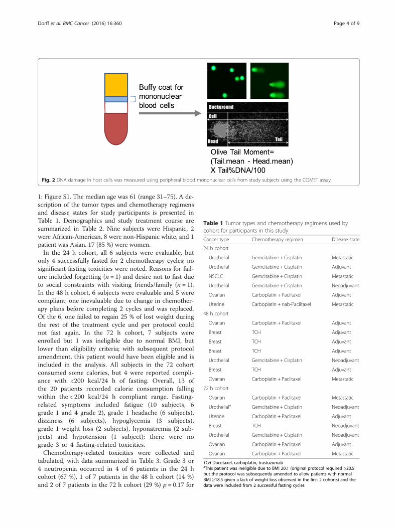
administration of premedications and chemotherapy,and then again 24 h after completion of chemotherapy.Serum samples were analyzed for IGF-1 and IGFBP1 byan in-house ELISA developed in the Cohen laboratory[12, 13]. Single cell gel electrophoresis assay (cometassays [14]) for the detection of DNA damage was
performed with Trevigen Comet Assay kits according tothe manufacturer’s protocols Fig. 2. In brief, cells iso-lated from buffy coats were embedded in Comet LMAgarose and then lysed followed by electrophoresis inTBE buffer. Cells were stained with SYBR Green and im-aged. Analysis was performed using CometScore™, wherevalues of Olive moment were generated [15]. Biomarkerdata, except the Comet assay, were logarithm-transformed prior to analysis; to compare differencesamong the 3 fasting cohorts, only “compliant” patientswere included. Geometric means and associated 95 %confidence intervals were calculated; p-values for thebiomarkers analyses were based on the F-test from re-gression analyses with patients classified as a random ef-fect. Categorical data were summarized with numbersand percentages; Fisher’s exact test was used to test as-sociation and Mantel-Haenszel test for trend was usedfor testing for trend. Intent-to-treat analysis was usedfor clinical endpoints.
ResultsAfter Institutional Review Board approval, 20 subjectsprovided written informed consent and were enrolledfrom October 2009 to November 2012 at the Universityof Southern California (USC) Norris ComprehensiveCancer Center and the Los Angeles County/USC MedicalCenter. Trial design schema is provided in Additional file
Fig. 1 Schema of the trial design; cohorts and strategy for escalating fasting duration. R24r signifies 24 hours of fasting without planned food,only rescue. R48r is 48 hours of fasting prior to chemotherapy, without planned food, only rescue. If safety or feasibility failed in the cohort of 48hours fasting, we planned a “de-escalation” to a 48 hour fasting period with planned rescue food: R48p. However, if 48 hours fasting was safe andfeasible, we would escalate to 72 hours fasting, broken down as 48 hours before chemotherapy and 24 hours after, R48/24r
Dorff et al. BMC Cancer (2016) 16:360 Page 3 of 9
1: Figure S1. The median age was 61 (range 31–75). A de-scription of the tumor types and chemotherapy regimensand disease states for study participants is presented inTable 1. Demographics and study treatment course aresummarized in Table 2. Nine subjects were Hispanic, 2were African-American, 8 were non-Hispanic white, and 1patient was Asian. 17 (85 %) were women.In the 24 h cohort, all 6 subjects were evaluable, but
only 4 successfully fasted for 2 chemotherapy cycles; nosignificant fasting toxicities were noted. Reasons for fail-ure included forgetting (n = 1) and desire not to fast dueto social constraints with visiting friends/family (n = 1).In the 48 h cohort, 6 subjects were evaluable and 5 werecompliant; one inevaluable due to change in chemother-apy plans before completing 2 cycles and was replaced.Of the 6, one failed to regain 25 % of lost weight duringthe rest of the treatment cycle and per protocol couldnot fast again. In the 72 h cohort, 7 subjects wereenrolled but 1 was ineligible due to normal BMI, butlower than eligibility criteria; with subsequent protocolamendment, this patient would have been eligible and isincluded in the analysis. All subjects in the 72 cohortconsumed some calories, but 4 were reported compli-ance with <200 kcal/24 h of fasting. Overall, 13 ofthe 20 patients recorded calorie consumption fallingwithin the < 200 kcal/24 h compliant range. Fasting-related symptoms included fatigue (10 subjects, 6grade 1 and 4 grade 2), grade 1 headache (6 subjects),dizziness (6 subjects), hypoglycemia (3 subjects),grade 1 weight loss (2 subjects), hyponatremia (2 sub-jects) and hypotension (1 subject); there were nograde 3 or 4 fasting-related toxicities.Chemotherapy-related toxicities were collected and
tabulated, with data summarized in Table 3. Grade 3 or4 neutropenia occurred in 4 of 6 patients in the 24 hcohort (67 %), 1 of 7 patients in the 48 h cohort (14 %)and 2 of 7 patients in the 72 h cohort (29 %) p = 0.17 for
Fig. 2 DNA damage in host cells was measured using peripheral blood mononuclear cells from study subjects using the COMET assay
Table 1 Tumor types and chemotherapy regimens used bycohort for participants in this study
Cancer type Chemotherapy regimen Disease state
24 h cohort
Urothelial Gemcitabine + Cisplatin Metastatic
Urothelial Gemcitabine + Cisplatin Adjuvant
NSCLC Gemcitabine + Cisplatin Metastatic
Urothelial Gemcitabine + Cisplatin Neoadjuvant
Ovarian Carboplatin + Paclitaxel Adjuvant
Uterine Carboplatin + nab-Paclitaxel Metastatic
48 h cohort
Ovarian Carboplatin + Paclitaxel Adjuvant
Breast TCH Adjuvant
Breast TCH Adjuvant
Breast TCH Adjuvant
Urothelial Gemcitabine + Cisplatin Neoadjuvant
Breast TCH Adjuvant
Ovarian Carboplatin + Paclitaxel Metastatic
72 h cohort
Ovarian Carboplatin + Paclitaxel Metastatic
Urotheliala Gemcitabine + Cisplatin Neoadjuvant
Uterine Carboplatin + Paclitaxel Adjuvant
Breast TCH Neoadjuvant
Urothelial Gemcitabine + Cisplatin Neoadjuvant
Ovarian Carboplatin + Paclitaxel Adjuvant
Ovarian Carboplatin + Paclitaxel Metastatic
TCH Docetaxel, carboplatin, trastuzumabaThis patient was ineligible due to BMI 20.1 (original protocol required ≥20.5but the protocol was subsequently amended to allow patients with normalBMI ≥18.5 given a lack of weight loss observed in the first 2 cohorts) and thedata were included from 2 successful fasting cycles
Dorff et al. BMC Cancer (2016) 16:360 Page 4 of 9
24 h compared to the 48 + 72 h cohort patients. Routinegranulocyte colony stimulating factor (GCSF) supportwas not prohibited, but was not used in our study popu-lation except in one patient after neutropenia had oc-curred. All 6 subjects in the 24 h fasting cohortexperienced grade 1 or 2 nausea, compared to 6 of 7(87 %) in the 48 h cohort and 3 of 7 (43 %) in the 72 hcohort (p = 0.019, test for trend); 5/6 (83 %) subjects inthe 24 h fasting cohort experienced grade 1 or 2 vomit-ing, compared to 3/7 (43 %) in the 48 h cohort and none(0 of 7) in the 72 h cohort (p = 0.003, test for trend).Pathologic complete responses in the setting of radio-
graphic complete responses were observed in 2 patients(Table 2). Partial radiographic responses were seen in 6of the 20 patients, of which 1 patient subsequently wasfound to have pathologic complete response. Three pa-tients had stable disease as their best radiographic re-sponse, of which 2 underwent surgery and hadpathologic complete response. Four of the pathologiccomplete responses were seen in the 72 h cohort; theother was in the 48 h cohort. One patient had progres-sive disease during treatment (48 h cohort), and 6 werenot evaluable because they were treated in the adjuvantsetting.Changes in insulin, glucose, IGF-1, IGFBP, and β-
hydroxybutyrate levels are summarized in Table 4,with graphical summary of IGF-1 levels in Additional
file 1: Figure S1. Among the compliant patients,blood glucose did not change significantly or consist-ently (p = 0.35). In the 24 h cohort, 4 of 6 subjects re-ported compliance with fasting and recorded <200kCal consumed; in these, 4 patients, insulin levels de-creased by a mean of −56 % just after chemotherapyfollowing the first fast. In the 48 h cohort, in 6 com-pliant patients, the insulin level decreased by 27 %and in the 7 compliant patients in the 72 h cohort,insulin levels decreased by 42 % at 48 h after fasting.Although patterns were suggestive, given the patient-to-patients variability, the 3 cohorts did not differ sig-nificantly in terms of changes over time (p = 0.35).Just after chemotherapy following the first fast, IGF-1levels decreased by a mean of −30 % (−44 %, −12 %)in the 24 h cohort, −33 % in the 48 h cohort, and−8 % in the 72 h cohort (p = 0.32 comparing all 3groups overall the times). After chemotherapy follow-ing fasting, the serum beta-hydroxybutyrate levels
Table 3 Chemotherapy related toxicities. Rates of selectedchemotherapy-related toxicities experienced by patients in thefasting cohorts, with CTC adverse event v4.0 grading. Only thegrades for which events occurred are shown
were elevated in the 48- and 72-h cohorts; in contrastthe levels in the 24-h cohort were decreased (seeTable 4). Overall the changes in the 48- and 72-hcohorts differed from the changes in the 24-h cohort(p = 0.037), depicted in Additional file 1: Figure S2.Serum pre-albumin levels did not change consistentlywith fasting based on the 10 compliant patients whohad data available.DNA damage in normal cells was measured using per-
ipheral blood mononuclear cells from study subjectsusing the COMET assay (Fig. 2). DNA damage increasedin all cohorts after chemotherapy, however after fasting48 or 72 h there was a decrease in Olive tail momentwhereas the 24 h cohort continued to have evidence ofincreased DNA damage. At baseline the Olive tail mo-ments, based on the COMET assay, were similar for the3 fasting cohorts (see Fig. 3) and were essentially un-changed just after chemotherapy following the first fast(p = 0.61 based on least significant difference method ofpairwise comparisons following overall p-value forchanges over time p < 0.001). 24 h later the peripheralblood mononuclear cell DNA damage had increasedslightly for all three groups (p = 0.042). In contrast, bythe second course, the DNA damage decreased for the
48-h and the 72-h cohorts (range 0.9 to 20.7 – all valuesless than baseline), but not for the 24-h cohort (p = 0.08).
DiscussionCalorie restriction has been associated with an increasein longevity in animals [16], and with lower rates of de-veloping aging-related diseases such as spontaneous can-cer formation [16, 17]. However, calorie restrictionrequires long periods to be effective and can cause anumber of side effects which includes impaired immunefunction and wound healing. In contrast, brief cycles ofSTS lead to profound changes in gene expression andcellular metabolism that render normal cells more resist-ant to oxidative stress [6, 18]. There has been interest instudying the effect of calorie intake and nutritional com-position of diet on the risk of disease progression or re-currence in patients with cancer diagnoses [19, 20].Fasting has been studied as an adjunct to traditionaltherapy in rheumatoid arthritis [21] and as a means ofpreventing chronic disease [22, 23]. However, the use ofSTS to reduce chemotherapy side effects in cancerpatients has, to our best knowledge, never been studiedin a clinical trial. We present the first dose-escalationstudy of fasting in human cancer patients receiving
Table 4 Baseline levels and median changes in insulin, glucose, IGF1/IGFBP1
Biomarker cohort Baseline Median % change after 1st fast Median % change after 2nd fast P value
These are presented only for self-reported compliant patients, and only for those with pre- and post-fasting samples available, separated by fasting durationcohort. The post-fasting blood draws were taken after 24 h of fasting in the 24 h cohort, and after 48 h of fasting in both the 48 and 72 h cohorts, and were doneprior to any premedications or chemotherapy*p-value comparing changes in levels from baseline, between the 3 fasting cohorts**p-value comparing changes from baseline between the 24 h cohort compared to the 48 + 72 h cohorts combined
Dorff et al. BMC Cancer (2016) 16:360 Page 6 of 9
chemotherapy, and found that up to 72 h of calorieintake <200 kcal per 24 h was safe and feasible in apopulation of men and women aged 31 to 75. Therewere no grade 3 toxicities attributed to fasting, and onlyone subject failed to regain any of the fasting associatedweight prior to the next chemotherapy cycle. We didnot find any evidence of malnutrition, although we wereunable to obtain pre-albumin data in all subjects. Im-portantly, the safety of fasting prior to chemotherapycan only be extrapolated to a selected population of on-cology patients, as we excluded those with >10 % recentweight loss, body mass index <20.5, or diabetes mellitus.Although the study was not designed to compare
the toxicity experience between cohorts, we did planto look at changes in fasting biomarkers as part ofthe determination of which duration of fasting wouldbe optimal to pursue in the randomized portion ofthe trial. IGF-1 has been shown to be one of themajor growth factors that promote cell proliferationand growth. In model organism, inhibition of theIGF-1 signaling is associated with enhanced cellularprotection against various stresses including toxins.By contrast, most tumor cells harbor oncogenic muta-tions in the IGF-1 signaling pathway. Constitutive ac-tivation of IGF-1 downstream mediators renders thetumor cell irresponsive to fasting-induced cellularprotection. IGF-1 is significantly reduced by fasting[24], while its binding proteins exhibit divergent pat-terns of change in response to fasting. IGFBP-1 in-creases rapidly even with overnight fasting and isquickly suppressed by calorie intake [25]. The IGF-1
axis has been implicated in conferring differential stress re-sistance [5, 18]. Chemotherapy toxicity was found to bedramatically reduced using the liver IGF-1 deficient (LID)mouse model, which has a tissue-specific knockdown ofIGF-1 resulting in approximately 70 % lower circulatingIGF-1 levels than in normal mice [18]. In our subjects,there was some significant reduction of IGF-1 after fasting,which continued at the 24 h post-chemotherapy point, des-pite resuming normal diet. Although we would have ex-pected greater reductions in IGF-1 in the cohorts withlonger durations of fasting, the degree of change was simi-lar between the 24 h fasting and longer duration fastinggroups. The variability in insulin and IGF-1 changes likelyreflects some non-compliance, perhaps a greater degree inthe cohorts with longer fasting. Furthermore, values duringthe second fasting cycle may have been affected by dexa-methasone administration with the previous cycle ofchemotherapy. Since many patients joined the study afterhaving completed one cycle of chemotherapy, even the firstfasting cycle results could have been impacted.Because there was no control group eating a regular
diet, we cannot yet address the hypothesis of whetherpatients experience fewer or less severe chemotherapyside effects after fasting. However, we found preliminaryevidence of reduced DNA-damage evident in host leuko-cytes after chemotherapy exposure for subjects whofasted for 72 h compared to 24 h. This is one mechan-ism by which we hypothesize fasting may not only re-duce toxicity to normal tissues, but improve cancertreatment efficacy: by reducing damage to hematopoieticprecursors and promoting hematopoietic stem cell-
Fig. 3 Olive moments, indicating DNA damage in peripheral blood mononuclear cells, are shown here by cohort, comparing the chemotherapy-freebaseline (BL) to a sample taken after fasting, but before chemotherapy (C1D1 = cycle 1, day 1) and 24 hours after chemotherapy (C1D2 = cycle 1, day2) and again after fasting but before chemotherapy (C2D1 = cycle 2, day 1). The difference comparing the 48 + 72 hour cohorts to the 24 hour cohortis p=0.08 by F test with ANOVA
Dorff et al. BMC Cancer (2016) 16:360 Page 7 of 9
dependent regeneration [26, 27], blood counts might bemaintained at higher levels, which would allow patientsto receive their chemotherapy at full dose, on time. Thereduced DNA damage in peripheral blood mononuclearcells seen based on the COMET assay results (Fig. 3)may translate into clinical benefit if host tissues, such ashematopoietic precursor cells, are protected and regen-erated. Our findings in the study population mirror whatwe have observed in mice. We previously reported pre-liminary evidence of hematopoietic protection from thispatient population, with less depletion of lymphocytecounts noted after repeated cycles of chemotherapy inpatients who fasted for 72 h compared to those whofasted 24 h [26, 27]. Now we present additional evi-dence for protection against myelosuppression (Table 3),with fewer patients experiencing grade 3 or 4 neutropeniain the 48 and 72-h fasting cohorts, as well as lower ratesof grade 1 and 2 thrombocytopenia. This, of course, isconfounded by the varying chemotherapy regimens, andspecifically by the fact that there was more gemcitabine/cisplatin in the 24 h cohort (4/6) compared to the 48- and72-h cohorts (1/7 and 2/7, respectively). Furthermore, pa-tients did not always enroll in the clinical trial during theirfirst cycle of chemotherapy, so that the number of cyclesan individual had experienced at the time point blood wasdrawn was not uniform. Nevertheless, the trends are fa-vorable, and the lower rate of neuropathy in the 48 and72 h fasting cohorts is particularly intriguing, given thegreater number of taxane-containing regimens in thesecohorts. Additional data from the ongoing randomizedphase II portion of this trial will provide direct compara-tive data to evaluate whether STS indeed protects cancerpatients against chemotherapy toxicity.One of the reasons anti-oxidants and similar com-
pounds have not been met with enthusiasm by the on-cology community as potential methods via which toreduce chemotherapy toxicity is the concern for redu-cing chemotherapy efficacy. Thus, while evaluating treat-ment response was not feasible as an endpoint for thisstudy due to the heterogeneity of cancer types andchemotherapy regimens as well as the inclusion of pa-tients receiving treatment in the adjuvant setting, we feltit was important to at least evaluate whether we saw alack of response to chemotherapy. Our observation ofpathologic complete responses and radiographic re-sponses provides reassuring preliminary evidence thatthe anti-neoplastic effect of chemotherapy was not nega-tively affected by fasting, in agreement with results pub-lished in animal studies. Our ongoing phase II trial willfocus on patients being treated in the metastatic or neo-adjuvant setting, and will have larger numbers of pa-tients receiving the same chemotherapy regimens so thatwe can better speak to this concern, and evaluatewhether enhancement of chemotherapy effect occurs.
Limitations of our study include the possibility of in-complete compliance and the variability of the compos-ition of the “rescue” food consumed. Despite ourencouragement to study subjects to honestly disclose allfood and beverage consumed during the fasting period,the lack of consistent changes in glucose or ketone gen-eration may indicate that more than 200 kcal/24 hperiod were consumed even in subjects whose food diar-ies reported <200 kcal/24 h. Furthermore, the proteincontent and derivation of rescue foods could theoretic-ally influence the perception by host tissues of lack ofnutrients in the environment, which may have contrib-uted to the variable results. In the randomized trialwhich is currently underway, we will perform intention-to-treat analysis, but will also analyze a subgroup of“biochemically compliant” subjects to determine whetherachievement of the protective fasting state, not just theattempt to achieve a fasting state, is associated with re-duced chemotherapy toxicity. In addition, a separatestudy is underway which employs a specific fasting-mimic diet, in which subjects are provided all the foodthey should consume during the “fasting” period aroundchemotherapy (NCT01802346). This approach will elim-inate the variability in the composition of the rescuefood consumption and may be more acceptable to pa-tients than aiming for zero calorie intake.
ConclusionsIn conclusion, fasting for up to 72 h, divided as 48 h be-fore and 24 h after chemotherapy infusion, is safe andfeasible in human cancer patients receiving platinumcombination chemotherapy. Preliminary evidence fromcorrelative studies supports the hypothesis that fastingmay confer some protection to host tissues againstchemotherapy damage to normal tissues.
Additional files
Additional file 1: Figure S1. Changes in IGF-1 represented as absolutechanges (left side) and percentage change from baseline (right side) forthe 24, 48, and 72 hour cohorts. Figure S2. Changes in beta-hydroxybutyrate(ketone) levels in fasting subjects, shown as absolute levels (left column) bycohort, and % changes (right column). (DOCX 182 KB)
AbbreviationsCOMET, gel electrophoresis method for measuring DNA strand breaks incells; CTCAE, common terminology criteria for adverse events; DNA,deoxyribonucleic acid; ELISA, Enzyme-linked immunosorbent assay; Etc., et-cetera; Ex, example; GCSF, granulocyte colony stimulating factor; IGF-1,insulin-like growth factor-1; IGFBP, insulin-like growth factor binding protein;kCal, kilocalorie; mTOR, mammalian target of rapamycin; STS, short term star-vation; USC, University of Southern California.
AcknowledgementsThe authors would also like to thank Kristi Massopust and Charlean Ketchensfor their care of study subjects.
FundingThis study was funded by a grant from the V Foundation and was supportedin part by award number P30CA014089 from the National Cancer Institute.
Availability of data and materialsAll relevant materials are provided in the manuscript or Additional files.
Authors’ contributionsTBD participated in study design, served as principal investigator for theclinical trial, and oversaw the data analysis. SG served as the leadbiostatistician for study design and conduct, and supervised the analysis. AGcontributed to protocol design and conduct. MS coordinated biospecimenhandling and analysis of correlative data. DT-W performed the clinical dataanalysis. HP contributed to protocol conduct/amendments. CWC performedthe ELISA assays. SB performed the COMET assays. PC contributed to designof correlative studies and conduct of ELISA assays. MW analyzed the correlativedata. VDL participated in study design and supervised all aspects of correlativescience. DIQ participated in study design and contributed to study conduct andanalysis. Conception and design summary: TBD, SG, MW, VL, DIQ. Acquisition ofdata: TBD, AG, MS, HP, CWC, SB, PC, MW, VL, DIQ. Analysis and interpretation ofdata: TBD, SG, AG, MS, DTW, HP, CWC, MW, VL, DIQ. Drafting of manuscript: TBD,SG, AG, DTW, VL, DIQ. All authors read and approved the final manuscript.
Competing interestsThe authors declare no conflicts of interest related to the manuscript withthe exception of V.D.L. who has equity interest in L-Nutra, a company thatdevelops medical food.
Consent for publicationIndividual consent provisions are: Not Applicable.
Ethics approval and consent to participateThis study was reviewed and approved by the University of SouthernCalifornia Health Sciences Institutional Review Board (HS:09/00010). Allsubjects provided written informed consent prior to participation.
DisclaimerThe content is solely the responsibility of the authors and does notnecessarily represent the official views of the National Cancer Institute or theNational Institutes of Health.
Author details1USC Keck School of Medicine, Norris Comprehensive Cancer Center, 1441Eastlake Ave. #3440, Los Angeles, CA 90033, USA. 2Department of PreventiveMedicine, USC Keck School of Medicine, 1441 Eastlake Ave, #4427, LosAngeles 90033, CA, United States. 3USC Keck School of Medicine,Department of Obstetrics and Gynecology, 1441 Eastlake Ave, #3440, LosAngeles 90033, CA, United States. 4Longevity Institute, University of SouthernCalifornia Davis School of Gerontology, Department of Biological Sciences,3715 McClintock Avenue, Los Angeles 90089, CA, United States.
Received: 16 October 2015 Accepted: 19 May 2016
References1. Appenroth D, Frob S, Kersten L, et al. Protective effects of vitamin E and C
on cisplatin nephrotoxicity in developing rats. Arch Toxicol. 1997;71:677–83.2. DeMartinis BS, Bianchi MD. Effect of vitamin C supplementation against
cisplatin-induced toxicity and oxidative DNA damage in rats. Pharmacol Res.2001;44:317–20.
3. Yuce A, Atessahin A, Ceribasi AO, Aksakal M. Ellagic acid prevents cisplatininduced oxidative stress in liver and heart tissue of rats. Basic Clin PharmToxicol. 2007;101:345–9.
4. Block KI, Koch AC, Mead MN, Tothy PK, Newman RA, Gyllenhaal C. Impact ofantioxidant supplementation on chemotherapeutic toxicity: a systematicreview of the evidence from randomized controlled trials. Int J Cancer.2008;123:1227–39.
5. Raffaghello L, Lee C, Safdie FM, et al. Starvation-dependent differential stressresistance protects normal but not cancer cells against high-dosechemotherapy. Proc Natl Acad Sci. 2008;105:8215-20.
6. Csizsar A, Labinskyy N, Jimenez R, et al. Anti-oxidative and anti-inflammatoryvasoprotective effects of caloric restriction in aging: role of circulatingfactors and SIRT-1. Mech Age Develop. 2009;130:518–27.
7. Lee C, Raffaghello L, Brandhorst S, et al. Fasting cycles retard growth oftumors and sensitize a range of cancer cell types to chemotherapy. SciTransl Med. 2012;4:124–7.
8. Mansell PI, Macdonald IA. The effect of starvation on insulin-inducedglucose disposal and thermogenesis in humans. Metabolism.1990;39:502–10.
9. Romijin JA, Godfried MH, Hommes MJT, et al. Decreased glucose oxidationduring short-term starvation. Metabolism. 1990;39:525–30.
10. NgoTH BRJ, Tymchuk CN, et al. Effect of diet and exercise on serum insulin,IGF-1, and IGFBP-1 levels and growth of LNCaP Cells in Vitro. Cancer CausesControl. 2002;13:929–35.
11. O’Sullivan U, Gluckman PD, Breier BH, et al. Insulin-like growth factor-1 (IGF-1)in mice reduces weight loss during starvation. Endocrinology. 1989;125:2793–4.
12. Muzumdar RH, Ma X, Fishman S, Yang X, Atzmon G, et al. Central andopposing effects of IGF-1 and IGF-binding protein-3 on systemic insulinaction. Diabetes. 2006;55:2788–96.
13. Milman S, Atzmon G, Huffman DM, Wan J, Crandall JP, et al. Low insulin-likegrowth factor-1 level predicts survival in humans with exceptionallongevity. Aging Cell. 2014;13:769–71.
14. Ostling O, Johanson KJ. Biochem Biophys Res Commun. 1984;123:291–8.15. Olive PL, Wlodek D, Durand RE, Banath JP. Factors influencing DNA
migration from individual cells subjected to gel electrophoresis. Exp CellRes. 1992;198:259–67.
16. Everitt AV, LeCouteur DG. Life extension by calorie restriction in humans.Ann NY Acad Sci. 2007;1114:428–33.
17. Dunn SE, Ehrlich M, Sharp NJH, et al. A dominant negative mutant of theinsulin-like growth factor-1 receptor inhibits the adhesion, invasion andmetastasis of breast cancer. Cancer Res. 1998;58:3353–61.
18. Kritchevsky D. Diet and cancer: what’s next? J Nutr. 2003;133:3827S–9.19. Lee C, Safdie FM, Raffaghello L, et al. Reduced levels of IGF-1 mediate
differential protection of normal and cancer cells in response to fasting andimprove chemotherapeutic index. Cancer Res. 2010;70:1564–72.
20. Rock CL, Demark-Wahnefried W. Nutrition and survival after the diagnosis ofbreast cancer: a review of the evidence. J Clin Oncol. 2002;20:3302–16.
21. Chan JM, Gann PH, Giovannucci GL. Role of diet in prostate cancerdevelopment and progression. J Clin Oncol. 2005;23:8152–60.
22. Kjeldsen-Kragh J, Borchgrevink CF, Laerum E, et al. Controlled trial offasting and one-year vegetarian diet in rheumatoid arthritis. Lancet.1991;338:899–902.
23. Varady KA, Hellerstein MK. Alternate day fasting and chronic diseaseprevention: a review of human and animal trials. Am J Clin Nutr.2007;86:7–13.
24. Weindruch R, Sohal RS. Caloric intake and aging. New Engl J Med.1997;337:986–94.
25. Ngo TH, Barnard RJ, Anton T, Tran C, Elashoff D, Heber D, et al. Effect ofisocaloric low-fat diet on prostate cancer xenograft progression toandrogen independence. Cancer Res. 2004;64:1252.
26. Smith WJ, Underwood LE, Clemmons DR. Effects of caloric or proteinrestriction on insulin-like growth factor-1 (IGF-1) and IGF-binding protein inchildren and adults. J Clin Endocrinol Metab. 1995;80:443–9.
27. Cheng C-W, Adams GB, Perin L, et al. Prolonged fasting reduces IGF-1/PKAto promote hematopoietic stem cell-based regeneration and reverseimmunosuppression. Cell Stem Cell. 2014;14:810–23.
• We accept pre-submission inquiries
• Our selector tool helps you to find the most relevant journal
• We provide round the clock customer support
• Convenient online submission
• Thorough peer review
• Inclusion in PubMed and all major indexing services
• Maximum visibility for your research
Submit your manuscript atwww.biomedcentral.com/submit
Submit your next manuscript to BioMed Central and we will help you at every step:


![Fasting and Power [Draft]imranhosein.org/inhmedia/books/fasting&power-new.pdf · Fasting in Islam – its basic objective 21 Fasting and internal spiritual power 26 Religion and the](https://static.documents.pub/doc/80x56/5ed9610cf59b0f56f45f61bd/fasting-and-power-draft-amppower-newpdf-fasting-in-islam-a-its-basic-objective.jpg)