NATIONAL ACADEMY OF SCIENCES SANFORD LOUIS PALAY 1918–2002 A Biographical Memoir by ALAN PETERS, JACK ROSENBLUTH, GEORGE PAPPAS, LAWRENCE KRUGER, AND ENRICO MUGNAINI Biographical Memoirs , VOLUME 84 PUBLISHED 2004 BY THE NATIONAL ACADEMIES PRESS WASHINGTON , D . C . Any opinions expressed in this memoir are those of the authors and do not necessarily reflect the views of the National Academy of Sciences.
Transcript
N A T I O N A L A C A D E M Y O F S C I E N C E S
S A N F O R D L O U I S P A L A Y1 9 1 8 – 2 0 0 2
A Biographical Memoir by
A L A N P E T E R S , J A C K R O S E N B L U T H ,
G E O R G E P A P P A S , L A W R E N C E K R U G E R , A N D
E N R I C O M U G N A I N I
Biographical Memoirs, VOLUME 84
PUBLISHED 2004 BY
THE NATIONAL ACADEMIES PRESS
WASHINGTON, D.C.
Any opinions expressed in this memoir are those of the authorsand do not necessarily reflect the views of the
National Academy of Sciences.
Har
vard
Med
ical
Sch
oo
l ( P
ub
lic
Aff
airs
)
271
SANFORD LOUIS PALAY
September 23, 1918–August 5, 2002
B Y A L A N P E T E R S , J A C K R O S E N B L U T H ,
G E O R G E P A P P A S , L A W R E N C E K R U G E R , A N D
E N R I C O M U G N A I N I
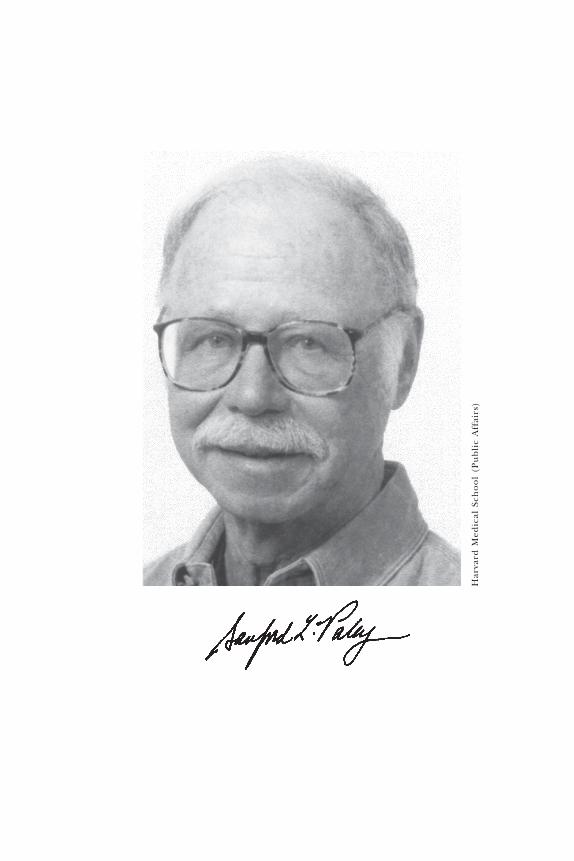
SANFORD LOUIS PALAY, a member of the National Academyof Sciences since 1977, died on August 5, 2002, at the
age of 83. He was buried in the Sleepy Hollow cemetery inConcord, Massachusetts. With his death modern neuro-cytology lost one of its founders. From the beginning offine structural studies of the nervous system the high qual-ity of electron micrographs produced by Sanford Palay setstandards that others would strive to emulate, and he con-tributed much toward the interpretation of electron micro-graphs of the nervous system and the advancement of knowl-edge on the principles of organization of the nervous system.It must be remembered that prior to fine structural studies,stains had been developed that could be used selectively toshow specific components of the nervous system in lightmicroscopic preparations. In electron micrographs all ofthe diverse neuronal and glial components are seen, butmuch of the continuity of cell bodies and processes is lostin the extremely narrow plane of the ultrathin section. There-fore, one of the earliest challenges toward which Sanford
Based on a memorium published in the Journal of Neurocytology, vol. 31, 2002. Withpermission of the publishers, Kluwer Academic Publishers.
272 B I O G R A P H I C A L M E M O I R S
Palay made many contributions was to determine which pro-files belonged to which parts of cells and which criteriacould be used to selectively identify the myriad profiles en-countered in electron micrographs.
Sandy was born in Cleveland, Ohio, of Russian Jewishimmigrant parents. In 1940 he received his bachelor’s de-gree in English from Oberlin College, a place for which hehad such fond memories that he donated his collection ofneuroscience journals and histological slides to the college.In 1940 he entered the School of Medicine at Western Re-serve University (now Case Western Reserve), with the in-tention of becoming a bacteriologist. In the spring of hisfirst year at medical school he applied for a fellowship thatwould allow him to do research in the summer break. Hechose to work in the laboratory of Ernst and Berta Scharrer,where he was given the project of trying to stain droplet-laden cells in the meninges of the toad. In 1944 he pub-lished the results of this investigation. The Scharrers taughtSandy a great deal about scientific investigation, about neu-roanatomy, and about cytology, and eventually Sandy wenton to work on neurosecretion (1945,1), which was ErnstScharrer’s prime interest (1945,2). Sandy continued to workwith the Scharrers throughout his time in medical school,and he developed a close relationship with Ernst Scharrer,who was to have a great influence in guiding Sandy’s scien-tific career.
After completing his M.D. degree in 1943 Sandy spent ayear as an intern at New Haven Hospital, where in the eve-nings he continued his research in the Department ofAnatomy at Yale University. He worked on tracing the neu-rosecretory pathway from the preoptic nucleus to the neu-rohypophysis in catfish, using material that he had broughtfrom Cleveland. At the end of the internship Sandy re-turned to Western Reserve University as a resident in medi-
273S A N F O R D L O U I S P A L A Y
cine, with appointments as a teaching fellow in medicineand a research fellow in anatomy. This allowed him to con-tinue his association with the Scharrers, and he took part ina study on chemical sense and taste in gourami and searobins (1947). It was Ernst Scharrer who suggested thatSandy ought to meet and work with Albert Claude, who wasat the Rockefeller Institute, pursuing his pioneering studieson the biochemistry of cellular components. Sandy appliedand was awarded a postdoctoral fellowship to work withClaude, but when his residency in Cleveland came to anend in 1946, he was called up to serve with the Army Medi-cal Corps as a member of the forces in occupied Japan. Asa result of that service Sandy began his lifelong interest inJapanese art and culture, leading him to collect Japaneseart and to cultivate many bonsai specimens. He returned toJapan in 1978 for an extended period as a visiting professorat the University of Osaka.
After leaving the Army in 1948 Sandy joined Albert Claudeas a fellow at the Rockefeller Institute and spent the yearexamining the chromosomes of the salivary gland by elec-tron microscopy. Formvar replicas were used (1949), be-cause at that time there were no suitable techniques avail-able for preparing thin sections. It was at the RockefellerInstitute that Sandy met George Palade, who had recentlybecome a refugee from Romania, which had been occu-pied by the Russians and fallen under a Communist re-gime.
After a year, in 1949, he returned to Yale, where he wasfirst appointed instructor and then assistant professor ofanatomy. He continued his work on neurosecretion, and itwas under his direction that Milton Brightman and StevenWissig completed their doctoral theses on the relationshipbetween neurosecretion and lactation in rats and on thy-roid secretion also in rat.
274 B I O G R A P H I C A L M E M O I R S
By 1952 important technical advances had been madein preparing tissue for electron microscopy. As Sandy pointedout in one of his later publications (1992,1), “Palade hadintroduced his Veronal buffered osmium tetroxide for opti-mal fixation of tissues. Borysko, Swerdlow and colleagueshad introduced a satisfactory method for embedding tissuein butyl methacrylate, and Harrison Latta had invented away to break plate glass into useful knives for thin section-ing.” So when George Palade invited Sandy to return to theRockefeller Institute to work with him for six months andto learn the new techniques, Sandy enthusiastically acceptedthe offer. It was at this time that he began his definitivestudies on the fine structure of the nervous system, and hisfirst success in achieving good fixation of neurons was ob-tained by injecting osmic acid into the fourth ventricle ofthe rat, since here the motor cells of the abducens nucleusare close to the surface, as are the cells of the overlyingcerebellum. The outcome was that in 1955 he and GeorgePalade were able to publish a pioneering article in the firstvolume of the Journal of Biophysical and Biochemical Cy-tology (now the Journal of Cell Biology) on “The Fine Struc-ture of Neurons.” This article described the Nissl substanceand the mitochondria of nerve cells, as well as long fila-ments that were subsequently recognized as neurofilaments.This study was soon followed by the one of which Sandy wasmost proud, the first description of the fine structure ofsynapses in the mammalian nervous system (1956). Sandyrecounted (1992) how one Saturday early in August of 1953,when he was alone in the laboratory, he was using the elec-tron microscope to examine thin sections of the abducensnucleus, and on the surfaces of dendrites and cell bodieshe encountered clublike profiles that were filled with mito-chondria and contained vesicles that were aggregated againstthe presynaptic membrane. Earlier that summer George
275S A N F O R D L O U I S P A L A Y
Pappas had shown Sandy electron micrographs showing thefine structure of a contractile vacuole in Amoeba proteussurrounded by clusters of small vesicles (30-40 nm diam-eter). Sandy remembered the close similarity between vesiclessurrounding the Amoeba contractile vacuole and those inthe presynaptic terminals on the soma of the motor neu-rons. Also the pre- and postsynaptic membranes were thick-ened and appeared denser, indicating a zone of intimateadherence, and most importantly these membranes wereseparated by a thin intercellular space, thus directly con-firming Cajal’s inference about the synaptic junctions be-tween nerve cells. He later reminisced: “I became very ex-cited, and having no one with whom to share this greatnews directly, I telephoned George Pappas at home to con-vey my exhilaration. Fortunately, he was there, or I wouldhave burst. I was extraordinarily privileged to be the firstone to see the synaptic junction, and I recognized my goodfortune” (1992). Recollections of this period are availablein a videotaped interview of Sandy Palay by Lawrence Krugerin the summer of 2001. The tape is deposited in the historyarchives of the Society for Neuroscience. The discovery ofcharacteristic vesicles at the mammalian synapses was re-ported in a joint paper with Palade at a meeting of theAmerican Association of Anatomists (1954). In the sameyear E. D. P. DeRobertis and H. S. Bennett also reportedthe finding of vesicles in nerve terminals of frog sympa-thetic ganglia and earthworm nerve cord at the meeting ofthe Federation of Societies for Experimental Biology meet-ing (Submicroscopic vesicular components of the synapse.Fed. Proc. 13[1954]:13:35).
Sandy returned to Yale, and in 1955 he was promoted toassociate professor. While continuing to work on the brainfine structure, he began a study on intestinal villi and thepathway of fat absorption with his graduate student L. J.
276 B I O G R A P H I C A L M E M O I R S
Karlin, and that study was brought to completion a fewyears later (1959,1,2). Sandy stayed in New Haven only oneyear because his prominence as a neurocytologist gainedhim the position of chief of the Section on Neurocytologyat the National Institutes of Health in Bethesda, Maryland.He was given an electron microscopy laboratory in the base-ment of Building 9. His new instrument turned out to haveserious problems, but it was finally replaced with a latermodel that did function properly. His associates there in-cluded Jack Rosenbluth, Mary Grillo, Milton Brightman,David Wolf, Spencer Gordon, Jr., Sam McGee Russell, andthe research assistant Catherine Crigler. Three years later,in 1960, he was promoted to chief of the Laboratory onNeuroanatomical Sciences at NIH. Sandy became interestedin the tendency of astrocytes to swell after immersion fixa-tion. Dispute with Sarah Luse and Ed Dempsy over the mean-ing of this artifact provided part of the impetus to developperfusion fixation. Sandy had a strong intuition regardingwhat was true structure and what was artifact (myelin splits,swollen astrocytes, large extracellular spaces in the centralnervous system), and this led him to develop and perfectthe buffered osmic acid perfusion fixation method for rats,while continuing to work on synapses (1958,1), neuroglia(1958,2), and neurosecretion (1958,3). A major improve-ment occurred when the laboratory switched from meth-acrylate to Araldite embedding, which helped improve theappearance of compact myelin and resulted in a paper onthe nerve cell bodies and their myelin sheaths in the eighthnerve ganglion of the goldfish (1961). Sandy’s interest inmyelin had also been spurred by a visit from Harry Webster,who brought spectacular electron micrographs of myelinatedperipheral nerve fibers. There were engaging lunchtimemeetings with Eric Kandel and W. Alden Spencer, who bothhad two-year appointments in the NIH neurophysiology labo-
277S A N F O R D L O U I S P A L A Y
ratory. At this time in Bethesda Sandy bought a Mercedesconvertible that he drove for many years.
In 1961 Sandy relinquished his position at NIH whenDon Fawcett invited him to become the Bullard Professorof Neuroanatomy at Harvard Medical School. Sandy was adominant figure in neurocytology, producing high-qualityelectron micrographs and lucid descriptions of structuresthat set standards for others to try to emulate. Soon afterhis arrival at Harvard he and his colleagues published animportant article on the perfusion fixation with osmic acidthey had developed at NIH (1962). Until then fixation ofnervous tissues for electron microscopy was mostly achievedby immersing pieces of central nervous system in bufferedosmic acid solutions. The resulting preservation was neververy good. Using the perfusion technique with buffered os-mium tetroxide, which produced black and often brittlebrains, Sandy and his collaborators obtained superior pres-ervation. This enabled them to begin to better analyze themorphological features of various components of the cen-tral and peripheral nervous systems, and accumulate a largeportfolio of electron micrographs illustrating the fine struc-ture and principles of organization of the central nervoussystem, especially the cerebellar cortex. Sandy was deeplyimpressed by how images from Golgi-impregnated materialcould be used to analyze electron micrographs and becamevery fond of the Golgi method, of which there were fewpractitioners in the United States. Visitors to Sandy’s labo-ratory were regularly treated with a show of his impressiveillustrations from the cerebellar cortex and emerged greatlyinspired, and many returned for extended periods of col-laboration.
Sandy was insistent on the value of artificial respirationof the animal prior to perfusion fixation with osmium tetrox-ide, as it had been shown that this improved the preserva-
278 B I O G R A P H I C A L M E M O I R S
tion of ultrastructure, especially that of the glial cells. Sub-sequently, as glutaraldehyde was introduced as a fixativeand as aldehydes began to be purified, he and his traineesadopted the perfusion fixation of the nervous system bybuffered aldehydes. Putting these techniques to good use,he and his wife, Victoria (“Vickie”) Chan-Palay, completeddetailed studies of the cerebellum. The results were pub-lished in a series of articles and culminated in the publica-tion of the definitive description of the components of thecerebellum in their book Cerebellar Cortex: Cytology andOrganization (1974). This book, which led to a better un-derstanding of the neuronal circuits in the cerebellum, wascited among the Fifty Best Books of 1974 at the Interna-tional Book Fair in Frankfurt.
As well as cooperating in science, Sandy and Vickie raisedtwo caring daughters, Victoria (from Vickie’s previous mar-riage) and Rebecca. They were a great comfort to Sandy inhis waning years, and visits by his grandchildren, his daugh-ter Victoria’s two children, were always a particular joy tohim.
By the late 1960s a wide range of structures in the cen-tral and peripheral nervous systems had been examined byelectron microscopy, and it was becoming possible to makegeneralized descriptions of the various neuronal and neu-roglial components of the nervous system. Sandy and AlanPeters talked about producing a book describing and illus-trating the fine structural features of components of thenervous system when Alan Peters worked with Sandy on thelateral geniculate nucleus of the cat in 1963-64, but theyconcluded that the time was not yet ripe to undertake thatproject. The basic reason was that one important compo-nent of the nerve cell, the axon hillock and the axon initialsegment that were the most plausible candidates for thespike initiation site, had not been recognized in electron
279S A N F O R D L O U I S P A L A Y
micrographs. However, over the course of the next two yearsit became evident that the initial axon segment is consis-tently characterized by bundles of microtubules linked bycross-bridges and electron dense undercoating of the plasmamembrane, and this led to another landmark publicationin the Journal of Cell Biology (1968). Consequently in 1968it was decided to go ahead and write the book, and Sandyand Alan Peters, who was then at Boston University, askedHarry Webster, who was at the Massachusetts General Hos-pital, to join them and to undertake the description of theperipheral nervous system. The hope was that such a bookwould serve as a guide to help others in the analysis ofelectron micrographs and promote the understanding ofthe principles of organization of the entire nervous system.The first edition of The Fine Structure of the Nervous Sys-tem was published in 1970 as a rather thin book of some200 pages, but by the third edition in 1991 it had grown toabout 500 pages, with over 130 plates, many of which con-tinue to be reproduced in various text books.
Sandy’s great strength and charm was that he alwayshad time for others and he was never prepared to acceptsecond best. No doubt this latter aspect of his make-up ledhim to accept the position of editor in chief of the Journalof Comparative Neurology, when Max Cowan stepped downfrom that position in 1980. Sandy’s attention to detail andhis goal to strive for high standards of both text and illus-trations resulted in the journal’s receipt of increasing num-bers of first-rate articles and ultimately becoming a weeklypublication. Sandy examined each article submitted to thejournal, and after reading the reviews he always wrote acarefully constructed letter to the corresponding authoroutlining which changes might be necessary to improve thearticle before publication. Even after retiring from Harvardin 1989 Sandy continued as editor in chief, running the
280 B I O G R A P H I C A L M E M O I R S
journal from his home in Concord; after he stepped downas editor in chief in 1993 he continued to play a role aseditor in chief emeritus. In 1991 he wrote an in-depth ar-ticle on the founding of the journal (1991,2).
Sandy’s ability to evaluate scientific work in the neuro-sciences put him in great demand as a member of editorialboards, and so in addition to the Journal of ComparativeNeurology Sandy served on several other editorial boards,including the board of the Journal of Neurocytology. A num-ber of his friends and colleagues contributed an issue ofthe Journal of Neurocytology in honor of his retirementfrom Harvard (October 1990). Sandy was also a member ofthe editorial boards of a number of other journals, includ-ing the Journal of Cell Biology, Brain Research, Experi-mental Neurology, Anatomy and Embryology, Neuroscience,and Experimental Brain Research.
Sandy’s achievements in neuroanatomy were recognizedby several important awards and honors, and among thesewe can cite the Lashley Award by the American Philosophi-cal Society; his election to the American Academy of Artsand Sciences in 1963; to the National Academy of Sciencesin 1977; and to the American Philosophical Society in 1997;his election as president of the American Association ofAnatomists, an association that awarded him its Henry GrayAward for his contributions to anatomy; the Ralph GerardAward for Contributions to Neuroscience by the Society forNeuroscience; and numerous invited lectureships. His re-tirement as Bullard Professor of Neuroanatomy from Harvardin 1989 by no means diminished Sandy’s interest in sci-ence. As stated above, he continued for several years as theeditor in chief of the Journal of Comparative Neurology,and in 1994 Boston College gave him an appointment asDistinguished Scholar in Residence in the Department ofBiology. He enjoyed this appointment and often went to
281S A N F O R D L O U I S P A L A Y
Boston College, where he offered a graduate course in thehistory of neuroscience. Even when his health started todecline Sandy continued to teach this course, with the stu-dents coming to his home in Concord.
When one went to visit him at home, or later in thehospital, Sandy always had scientific journals beside his chairand was invariably ready to engage in a discussion of recentfindings that he had read about and to put those findingsin the context of earlier knowledge. Sandy had a deep andcomprehensive understanding of the history of neuroscience,and he loved to peruse older publications. One regret isthat he did not write more about this subject, for there arefew with his depth of knowledge, insight, and love of thehistory of neuroscience (see 1987) and the close links be-tween brain science and philosophy.
An aspect of Sandy’s multifaceted personality that en-deared him to many of his friends was his keen intellectualinterest in the physical world, human nature, literature,music, and the fine arts. Sandy was also an accomplishedpianist, and it was a source of great regret to him thattoward the end of his life he was no longer able to play theclassical pieces that he loved. Although Sandy was endowedwith a reserved temperament, when visiting with him onefelt in contact with a person of culture in the broadestsense of the word. In all probability this is the image thathe would like us to remember. He will never be forgottenby those who knew him.
282 B I O G R A P H I C A L M E M O I R S
S E L E C T E D B I B L I O G R A P H Y
1944
The histology of the meninges of the toad (Bufo). Anat. Rec. 88:257-70.
1945
Neurosecretion. VII. The preopticohypophysial pathway in fishes. J.Comp. Neurol. 82:129-43.
With E. Scharrer and R. G. Nigles. Neurosecretion. VIII. The Nisslsubstance in secreting nerve cells. Anat. Rec. 92:23-29.
1947
With E. Scharrer and S. W. Smith. Chemical sense and taste in thefishes, Prionotus and Trichogaster. J. Comp. Neurol. 86:183-98.
1949
With A. Claude. An electron microscopic study of salivary glandchromosomes by the replica method. J. Exp. Med. 89:431-38.
1954
With G. E. Palade. Electron microscopic observations of interneu-ronal and neuromuscular synapses. Anat. Rec. 118:335.
1955
With G. E. Palade. The fine structure of neurons. J. Biophys. Biochem.Cytol. 1:69-88.
1956
Synapses in the central nervous system. J. Biophys. Biochem. Cytol.2(Suppl.):193-202.
1958
The morphology of synapses in the central nervous system. Exp. CellRes. 5(Suppl.):275-93.
An electron microscopic study of neuroglia. In Biology of Neuroglia,ed. W. F. Windle, pp. 24-38. Springfield: Thomas.
283S A N F O R D L O U I S P A L A Y
The morphology of secretion In Frontiers in Cytology, ed. S. L. Palay,pp. 305-42. New Haven: Yale University Press.
1959
With L. J. Karlin. An electron microscopic study of the intestinalvillus. I. The fasting animal. J. Biophys. Biochem. Cytol. 5:363-72.
With L. J. Karlin. An electron microscopic study of the intestinalvillus. II. The pathway of fat absorption. J. Biophys. Biochem. Cytol.5:373-84.
1962
With S. M. McGee-Russell, S. Gordon, and M. A. Grillo. Fixation ofneural tissues for electron microscopy by perfusion with solu-tions of osmium tetroxide. J. Cell Biol. 12:385-410.
1966
With A. Peters. The morphology of laminae A and A1 of the dorsalnucleus of the lateral geniculate body of the cat. J. Anat.(Lond.)100:451-86.
1968
With C. Sotelo, A. Peters, and P. Orkand. The axon hillock and theinitial segment. J. Cell Biol. 38:193-201.
1970
With A. Peters and H. D. Webster. The Fine Structure of the NervousSystem: The Cells and Their Processes. New York: Hoeber-Harper.
1974
With V. Chan-Palay. Cerebellar Cortex: Cytology and Organization. NewYork: Springer-Verlag.
1987
A century of neuroanatomy in America. In The American Associationof Anatomists, 1888–1987, ed. J. E. Pauly, pp. 134-47. Baltimore:Williams and Wilkins.
284 B I O G R A P H I C A L M E M O I R S
1991
With A. Peters and H. D. Webster. The Fine Structure of the NervousSystem: The Neurons and Their Supporting Cells. 3rd ed. New York:Oxford University Press.
The founding of the Journal of Comparative Neurology. J. Comp.Neurol. 314:1-8.
1992
A concatenation of accidents. In The Neurosciences: Pathways of Dis-covery II, eds. F. E. Samson and G. Adelman, pp. 191-212. Boston:Birkhäuser.
With V. Chan-Palay, E. Mugnaini, and E. Voogd. Cerebellum. InElsevier’s Encyclopedia of Neuroscience, eds. G. Adelman and B. J.Smith, pp. 321-28. Amsterdam: Elsevier.
1996
With A. Peters. The morphology of synapses. J. Neurocytol. 25:701-16.