29
SARCOMERE, SARCOTUBULAR SYSTEM DR.NILESH KATE. M.D. ASSOCIATE PROFESSOR, DEPARTMENT OF PHYSIOLOGY, ESIC MEDICAL COLLEGE & HOSPITAL, GULBARGA.
| Date post: | 19-Jul-2015 |
| Category: |
Health & Medicine |
| Upload: | nileshkate79 |
| View: | 239 times |
| Download: | 3 times |

SARCOMERE, SARCOTUBULAR SYSTEM
DR.NILESH KATE. M.D.
ASSOCIATE PROFESSOR,DEPARTMENT OF PHYSIOLOGY,
ESIC MEDICAL COLLEGE & HOSPITAL,
GULBARGA.

OBJECTIVES. Introduction Classification of Muscles Structure of Skeletal Muscle Myofibrils Sarcomere Molecular level of Myosin and Actin Action of Actin, Myosin and Calcium in contraction Walk – Along –Theory Energy source of Muscle contraction Iso metric and Iso Tonic contraction

Introduction We are born with about 300 to 350 bones but
some of the bones fuse together as we grow and by the time we reach young adulthood we have on an average about 206 bones.
There are about 650 muscles in the body, and they are divided into three different types, skeletal, visceral and cardiac.

Introduction About 40% of your body weight is accounted for by
muscles. largest --The gluteus maximus muscle, located in the
buttocks. Longest– Sartorius. Smallest – stapedius. Strongest – Masseter in the jaw. Shortest– styloglossus. Tensor tympani (skeletal) Contrary to what people assume, muscles do not
push, but can only pull.
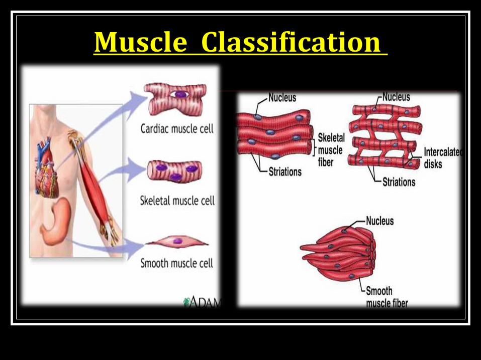
Muscle Classification

Functional anatomy & organization.Structural organization•Endomysiym.
•Fasciculi – Perimysium.
•Muscle belly – Epimysium.
•Tendons.

Structural organization

Structure of muscle fibre.Long cylindrical.
Sarcolemma.
Sarcoplasm.Cell organelle.Sarcoplasmic reticulum.Myofibrils.Sarcotubular system.
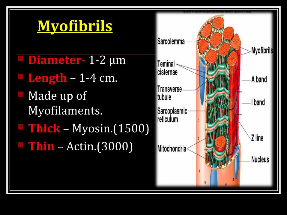
Myofibrils
Diameter- 1-2 μm Length – 1-4 cm. Made up of
Myofilaments. Thick – Myosin.(1500) Thin – Actin.(3000)

Myofilaments
Arrangement of Myofilaments under light microscope. Alternate light & dark
bands.

Striations of muscle fibres.
Due to difference in the refractive index. ‘A’ Band – Dark band
Anisometropic to polarized light. Length --- 1.5 μm ‘H’ zone – centre of A band
H means Henson or “Hell” – light.

Striations of muscle fibres.
“M” Line – centre of H zone. Pronounced during muscle contraction.
I Band - Light band. Isotropic to polarized light. “Z” Line – Dark
Z Wischenscheibe – German- Between disc.

Sarcomere A portion of myofibril
that lies between two successive Z discs is called sarcomere.
Sarcomere = ½ I band + A band +
½ I band. The structural &
functional unit of muscle fiber.

Myofilaments. Contractile apparatus.
Contractile proteins. Myosin Actin
Regulatory proteins. Troponin Tropomyosin.
Anchoring proteins. α Actinin Titin. Nebulin Dystrophin

Thick filaments. Twice the diameter. Each thick filament
surrounded by 6 thin flaments.
Myosin . Mol.wt – 4,80,000. 6 polypeptide chains 4 – light 2 – heavy.

Myosin .
Globular head. Myosin II The tails of myosin molecules
bundles to form the body of myosin filament with heads hangs to the side.
The part of head hangs from body form the arm
Thus the protruding arms and heads -- cross- bridges
Participate is actual muscle contraction.

Thin filaments. Head end – extend into
A band Tail end – anchored to
the Z line. Contractile protein
Actin Regulatory proteins
Tropomyosin Troponin.
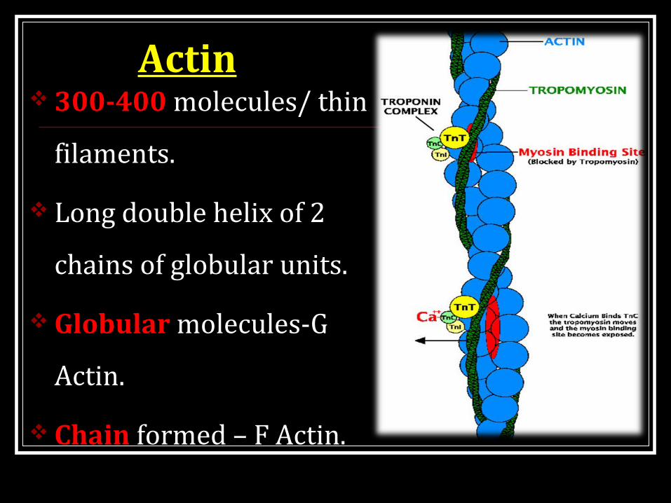
Actin 300-400 molecules/ thin
filaments.
Long double helix of 2
chains of globular units.
Globular molecules-G
Actin.
Chain formed – F Actin.

Tropomyosin 40-60 molecules/ thin
filaments. Long filaments. Lie in groove between
2 actin filaments. Covers binding sites
on actin. Thus regulatory
proteins.

Troponin Small globular units.
3 subunits.
Troponin ‘I’ – Affinity for
Actin.
Troponin ‘T’ – Affinity
for Tropomyosin.
Troponin ‘C’- Affinity for
Calcium

Anchoring proteins. α Actinin – cross link the Actin
filaments in the area of Z line.
Titin. – Interconnect Z line.
Nebulin.– connect α actinin with
troponin- tropomyosin complex.
Dystrophin-glycoprotein
complex.– provide structural
support & strength.

Sarcotubular system. Sarcotubular system –
with sarcoplasmic reticulum
Imp role in internal conduction of depolarization within the muscle fibre.
Formed by Transverse tubular
system (T) Longitudinal
sarcoplasmic reticulum.

Transverse tubular system (T) Invagination of
sarcollema into muscle fibre at junction of A & I bands.
Rapid transmission. Its membranes contains
voltage gated Ca channels Dihydropyridine receptors.
Activate longitudinal SR.

Longitudinal sarcoplasmic reticulum.
Sarcoplasmic tubules of sarcoplasmic reticulum.
Run in long axis of muscle fibres.
Do not open to exterior. Terminal cisterns. Triad- T tubule + 2
terminal cisterns. Ryanodine receptors.

Energy source for Muscle Contraction The energy source -- ATP
Large amount of ATP are cleaved during contraction process. When greater the work is done more ATP is cleaved. This is called Fenn effect.
ATP – 1 to 2 seconds
Phosphocreatine – 5 to 8 seconds
Glycogen – 1 minute
Oxidative metabolism – for many hours.

Isotonic Contraction Isotonic contraction:
In this muscle contract with change in length of muscle fibre but no change in tension.
Eg., Walking, Running,
Lifting a load

Isometric contraction:In this type the muscle contract
with change in tension but no change in length of the muscle fibre.
Eg., Muscle which helps in
maintaining posture against gravity.
Contraction of the arm muscles when trying to push a wall.

Summary Introduction Classification of Muscles Structure of Skeletal Muscle Myofibrils Sarcomere Molecular level of Myosin and Actin Action of Actin, Myosin and Calcium in contraction Energy source of Muscle contraction Iso metric and Iso Tonic contraction

Thank you.


