18
Science of Veterinary Medicine Urinary System Unit Handouts

Science of Veterinary
Medicine
Urinary System
Unit Handouts


Urinary System
Functions of the Urinary System
• Elimination of waste products
– __________________________
– __________________________
– __________________________
• Regulate aspects of homeostasis
– __________________________
– __________________________
– __________________________
– __________________________
– __________________________
– __________________________
Organs of the Urinary system
• __________________________
• __________________________
• __________________________
• __________________________
The Kidney

Nephrons
• _______________________________________________________
• _______________________________________________________
Urine Formation Processes
• __________________________
• __________________________
• __________________________
Filtration
• _______________________________________________________________
• _______________________________________________________________
• _______________________________________________________________
• _______________________________________________________________
Reabsorption
• The peritubular capillaries reabsorb several materials
– __________________________
– __________________________
– __________________________
– __________________________

• Some reabsorption is ____________________, most is _________________ (____________________)
• Most reabsorption occurs in the _________________________________________
• Materials NOT reabsorbed
– ___________________________________________ (Urea, Uric acid, Creatinine)
– __________________________
Secretion – Reabsorption in Reverse
• Some materials move from the peritubular capillaries into the renal tubules
– __________________________
– __________________________
– __________________________
• Materials left in the renal tubule move toward the ________________ and become _________________
Formation of Urine
Ureters
• Slender tubes attaching the kidney to the bladder
– ______________________________________________
– ______________________________________________
– ______________________________________________
• Peristalsis (_____________________________________) aids gravity in urine transport

Urinary Bladder
• ______________________________________________
• ______________________________________________
• __________________ – three openings
– __________________________________
– __________________________________
Urinary Bladder Wall
- _______________________________________________________________________
- _______________________________________________________________________
- _______________________________________________________________________
Urethra
• Thin-walled tube that carries urine from the bladder to the outside of the body by __________________
• Release of urine is controlled by two sphincters
– ___________________________________________________
– ___________________________________________________
• Function
– Females – ______________________________________________________
– Males – ________________________________________________________
Student Name: _____________________________________
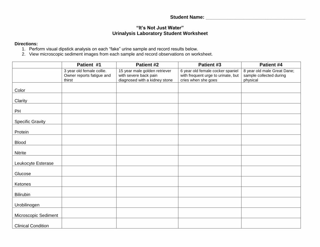
“It’s Not Just Water” Urinalysis Laboratory Student Worksheet
Directions:
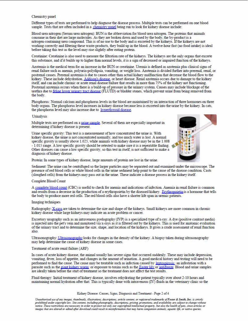
1. Perform visual dipstick analysis on each “fake” urine sample and record results below. 2. View microscopic sediment images from each sample and record observations on worksheet.
Patient #1 Patient #2 Patient #3 Patient #4
3 year old female collie. Owner reports fatigue and thirst
15 year male golden retriever with severe back pain diagnosed with a kidney stone
6 year old female cocker spaniel with frequent urge to urinate, but cries when she goes
8 year old male Great Dane; sample collected during physical
Color
Clarity
PH
Specific Gravity
Protein
Blood
Nitrite
Leukocyte Esterase
Glucose
Ketones
Bilirubin
Urobilinogen
Microscopic Sediment
Clinical Condition

Directions: 1. Perform visual dipstick analysis on each real urine sample and record results below. 2. View microscopic sediment images from each sample and record observations on worksheet.
Patient #1 Patient #2
Color
Clarity
PH
Specific Gravity
Protein
Blood
Nitrite
Leukocyte Esterase
Glucose
Ketones
Bilirubin
Urobilinogen
Microscopic Sediment
Clinical Condition
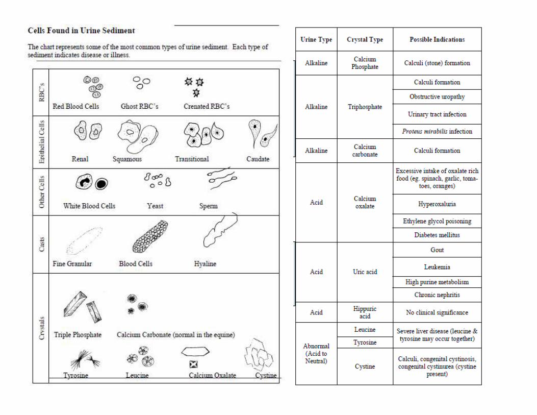
Look at each sample under the microscope.
Sketch the image and try to identify cells and sediment seen.
Patient #1
Patient #2


Name: ________________________________________ Date: ____________________

Questions
1. What is the function of the glomerulus and the bowman's capsule?
2. What is the function of the loop of henle?
3. Compare the processes of the distal tubule to the proximal tubule.
4. Describe the path the blood takes as it flows through the nephron.
5. Describe the path the filtrate takes as it flows through the nephron (beginning at the glomerulus).
6. Diffusion is a process where molecules move from areas of high concentration to low concentration. Toxins
diffuse from the blood and into the tubules of the nephron. How might this process be altered if there were
fewer blood vessels intertwined with the tubules?
7. Dialysis is a process where a person with
nonfunctioning kidneys can have their blood
filtered by a machine. The image to the right
outlines this process. Relabel the image to
indicate which parts would match the anatomy of
the kidney:
renal artery
renal vein
ureter
nephron
bladder

The kidney is a bean shaped organ that has an outer area called the cortex. The inner area, the renal medullais
composed of seven cone shaped renal pyramids (only 3 of them are shown in the image) with the tubes visible
from them making up a collection of nephrons. The renal pyramids merge to form the renal pelvis at the center
of the kidney, urine collects here before draining into the ureter and travelling to the bladder for storage.
Color the medulla area light green. Color the cortex pink, and the renal pelvis and ureter yellow. The nephrons
pictured on the kidney should be colored orange .
Note the two vessels attached to the kidney, color the artery red and the vein blue .
If you view a nephron close up, as shown in the second picture, you can see that it is a complex structure
composed of many tubes, and each kidney has about 1 million nephrons. The nephron's primary function is to
filter waste from the blood. The nephron has three major parts: the glomerulus, the Bowman's Capsule, and the
tubules, which consist of the promimal and distal tubule and the Loop of Henle.
Blood enters the kidney from the renal artery and moves into the glomerulus, where filtration occurs.
Filtration is the process by which water and dissolved particles are pulled out of the blood. The resulting liquid,
called filtrate contains many of the toxic substances that might have accumulated in the blood (like ammonia).
The glomerulus is enclosed by the Bowman's capsule, small molecules and water can pass through this area,
but larger molecules do not. The filtrate is then collected in the Bowman's capsule for transport through the
nephron.
Color the renal artery red on both images. On the second image the artery enters the glomerulus and then
exits to twist around the larger tubules. Color the renal vein blue , it is also twisted around the tubules. These
two vessels, the artery and the veins meet near the loop of henle, color this area purple .
Color the Bowman's capsule brown , leave the glomerulus white, you should have already colored the
arteries inside it red.
The nephron itself will restore vital nutrients and water back into the blood, while retaining the waste products
the body needs to eliminate. Two processes accomplish this task: tubular reabsorption and tubular secretion.
During tubular reabsorption, cells in the proximal tubule remove water and nutrients from the filtrate and pass
them back into the blood, wastes such as urea are retained in the tubule. During tubular secretion, wastes that
were not initially filtered out in the bowman's capsule are removed from the blood in the distal tubule.
Ammonia and many drugs are removed from the blood during tubular secretion.
Color the proximal tubule dark green , until it reaches the loop of henle. The loop of henle should be colored
pink , and then when it changes into the distal tubule, color the distal tubule light green .
Notice the capillaries that wrap around the tubules (you colored them red). At the points of contact with the
tubule and the capillaries, water and nutrients are reabsorbed into the blood. In addition, wastes remaining in the
blood after filtration are passed to the tubule. The filtrate flows from the proximal tubule and into the Loop of
Henle. The loop of henle concentrates the filtrate, by removing more water from it, and passes it to the distal
tubule. From the distal tubule it travels to the collecting duct - now called urine. The collecting duct prepares
the urine for transport out of the body, it is collected in the renal pelvis where it eventually enters the ureter.
From there it goes to the bladder.
Color both the collecting duct and the ureter yellow .

URINALYSIS CRISS-CROSS
1
2
3
4
5
6
7
8
9
10
11
12
13
14
15
16
17
18
19
20
ACROSS
3. The functional unit of the kidney that filters blood and produces urine
6. The word used to describe the dark orange color of concentrated urine
8. To determine the proper concentration, it is important to read the colors on the strip at the specified __
9. Description for the sky when you can't see the sun or a urine sample you can't see through
10. Specialized microscopic structures containing nuclei and organelles, sloughed off along the urinary
tract, appearing in urine sediment
12. A polymer of amino acids that can be detected in urine.
13. Microorganisms that cause infection and can be seen in the sediment of urine under the microscope
14. The type of sugar that is detected on a urine strip
16. When salts solidify they form _______ that can be seen in the sediment of urine under the microscope
18. A urine sample with a pH above 7 is _________
20. Disease in which sugar may be excreted in the urine
DOWN
1. It is best to test urine when it is _______ or the best condition to eat produce
2. The property of a urine sample assessed by specific gravity testing
4. When the kidney stops working, the patient is in renal ________
5. The color of normal urine
7. The organ responsible for the production of urine
11. Professionals who perform urinalysis testing are called clinical laboratory _________
15. When urine contains something detectable on the chemical strip it produces a color ________
17. The chemicals used for urinalysis are in filter paper on plastic ________
19. An adjective that refers to the kidney