13
Sedimentation and the ESR Test A Simple and Useful Separation Technique
Sedimentation and the ESR Test
A Simple and Useful Separation Technique

Why do we need to shake well before serving or using?We are always told to shake juice bottles before serving.
This is printed as label instructions.
Shake it up!“Natural juice products settle.
Shake well before serving.“
“Shake it up!” instructions for soymilk, inhalers and
oral suspensions.
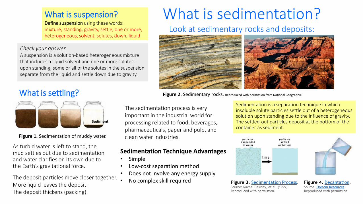
Check your answerA suspension is a solution-based heterogeneous mixture that includes a liquid solvent and one or more solutes; upon standing, some or all of the solutes in the suspension separate from the liquid and settle down due to gravity.
What is suspension?Define suspension using these words: mixture, standing, gravity, settle, one or more, heterogeneous, solvent, solutes, down, liquid
What is settling?
What is sedimentation? Look at sedimentary rocks and deposits:
As turbid water is left to stand, the mud settles out due to sedimentation and water clarifies on its own due to the Earth’s gravitational force.
The deposit particles move closer together.More liquid leaves the deposit.The deposit thickens (packing).
The sedimentation process is very important in the industrial world for processing related to food, beverages, pharmaceuticals, paper and pulp, and clean water industries.
Sedimentation is a separation technique in which insoluble solute particles settle out of a heterogeneous solution upon standing due to the influence of gravity. The settled-out particles deposit at the bottom of the container as sediment.
Sedimentation Technique Advantages• Simple• Low-cost separation method• Does not involve any energy supply• No complex skill required Figure 3. Sedimentation Process.
Source: Rachel Casiday, et al. (1999) Reproduced with permission.
Figure 4. Decantation.Source: Oresom Resources.Reproduced with permission.
Sediment
Figure 2. Sedimentary rocks. Reproduced with permission from National Geographic.
Figure 1. Sedimentation of muddy water.
From macro-sedimentation to micro-sedimentation
To understand blood sedimentation better, watch https://www.youtube.com/watch?v=gwsNcC4ZFHw
What difference do you notice between the sedimentation in the two glitter bottles?The sedimentation rate can be different for different solutions.
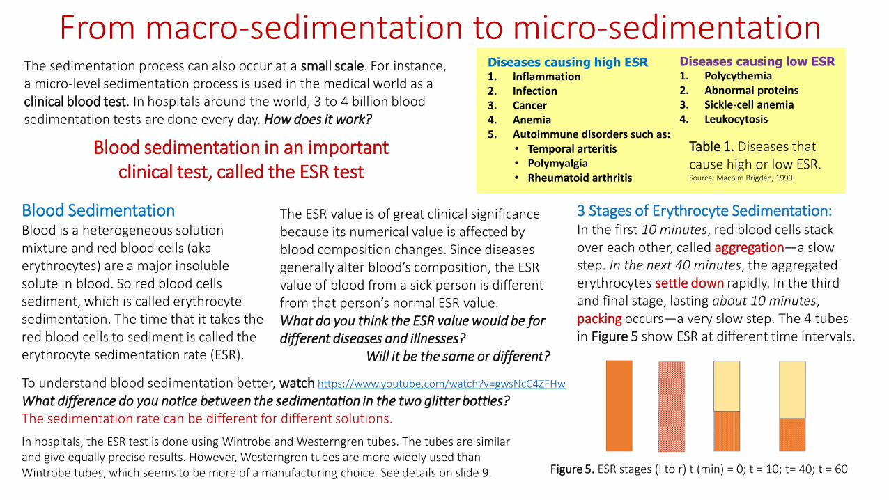
The ESR value is of great clinical significance because its numerical value is affected by blood composition changes. Since diseases generally alter blood’s composition, the ESR value of blood from a sick person is different from that person’s normal ESR value. What do you think the ESR value would be for different diseases and illnesses?
Will it be the same or different?
Blood SedimentationBlood is a heterogeneous solution mixture and red blood cells (aka erythrocytes) are a major insoluble solute in blood. So red blood cells sediment, which is called erythrocyte sedimentation. The time that it takes the red blood cells to sediment is called the erythrocyte sedimentation rate (ESR).
Figure 5. ESR stages (l to r) t (min) = 0; t = 10; t= 40; t = 60
In hospitals, the ESR test is done using Wintrobe and Westerngren tubes. The tubes are similar and give equally precise results. However, Westerngren tubes are more widely used than Wintrobe tubes, which seems to be more of a manufacturing choice. See details on slide 9.
The sedimentation process can also occur at a small scale. For instance, a micro-level sedimentation process is used in the medical world as a clinical blood test. In hospitals around the world, 3 to 4 billion blood sedimentation tests are done every day. How does it work?
Blood sedimentation in an important clinical test, called the ESR test
3 Stages of Erythrocyte Sedimentation: In the first 10 minutes, red blood cells stack over each other, called aggregation—a slow step. In the next 40 minutes, the aggregated erythrocytes settle down rapidly. In the third and final stage, lasting about 10 minutes, packing occurs—a very slow step. The 4 tubes in Figure 5 show ESR at different time intervals.
Diseases causing high ESR 1. Inflammation2. Infection3. Cancer4. Anemia5. Autoimmune disorders such as:
• Temporal arteritis• Polymyalgia• Rheumatoid arthritis
Diseases causing low ESR 1. Polycythemia2. Abnormal proteins3. Sickle-cell anemia4. Leukocytosis
Table 1. Diseases that cause high or low ESR. Source: Macolm Brigden, 1999.
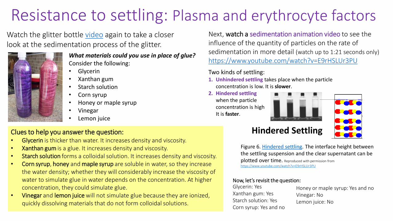
What materials could you use in place of glue? Consider the following:• Glycerin• Xanthan gum• Starch solution• Corn syrup• Honey or maple syrup• Vinegar• Lemon juice
Watch the glitter bottle video again to take a closer look at the sedimentation process of the glitter.
Clues to help you answer the question:• Glycerin is thicker than water. It increases density and viscosity.• Xanthan gum is a glue. It increases density and viscosity.• Starch solution forms a colloidal solution. It increases density and viscosity.• Corn syrup, honey and maple syrup are soluble in water, so they increase
the water density; whether they will considerably increase the viscosity of water to simulate glue in water depends on the concentration. At higher concentration, they could simulate glue.
• Vinegar and lemon juice will not simulate glue because they are ionized, quickly dissolving materials that do not form colloidal solutions.
Now, let’s revisit the question:Glycerin: YesXanthan gum: YesStarch solution: YesCorn syrup: Yes and no
Next, watch a sedimentation animation video to see the influence of the quantity of particles on the rate of sedimentation in more detail (watch up to 1:21 seconds only)
https://www.youtube.com/watch?v=E9rHSLUr3PU
Two kinds of settling: 1. Unhindered settling takes place when the particle
concentration is low. It is slower.2. Hindered settling
when the particle concentration is high.It is faster.
Resistance to settling: Plasma and erythrocyte factors
Figure 6. Hindered settling. The interface height between the settling suspension and the clear supernatant can be plotted over time. Reproduced with permission from
https://www.youtube.com/watch?v=E9rHSLUr3PU
Hindered Settling
Honey or maple syrup: Yes and noVinegar: NoLemon juice: No
The influence of particle size and shape
• Spherical or near-spherical particles • Heavy particles • Dilute slurries• Particles whose diameter does not rival
that of the container• Flocculation or "clumping" of particles
into spherical shapes• Auto coagulation due to chemical traits
inherent in the particle
Question: In the glitter bottles, what could you have done differently with the glitter (not the solutions) to speed up the fall? Consider the following scenarios:
1. Increase the glitter particle size.2. Decrease the glitter particle size3. Use a mix of bigger and smaller glitter particles4. Use sequins of moon shape mixed with the glitter.5. Add some starch powder to the solution
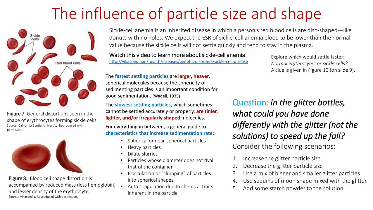
Figure 7. General distortions seen in the shape of erythrocytes forming sickle cells. Source: California Baptist University. Reproduced with permission.
Figure 8. Blood cell shape distortion is accompanied by reduced mass (less hemoglobin) and lesser density of the erythrocyte.Source: Vikaspedia. Reproduced with permission.
Sickle-cell anemia is an inherited disease in which a person’s red blood cells are disc-shaped—like donuts with no holes. We expect the ESR of sickle-cell anemia blood to be lower than the normal value because the sickle cells will not settle quickly and tend to stay in the plasma.
Watch this video to learn more about sickle-cell anemia: http://vikaspedia.in/health/diseases/genetic-disorders/sickle-cell-disease
The fastest settling particles are larger, heaver, spherical molecules because the sphericity of sedimenting particles is an important condition for good sedimentation. (Wadell, 1935)
The slowest settling particles, which sometimes cannot be settled accurately or properly, are tinier, lighter, and/or irregularly shaped molecules.
For everything in between, a general guide to characteristics that increase sedimentation rate:
Explore which would settle faster:Normal erythrocytes or sickle cells? A clue is given in Figure 10 (on slide 9).

Exploring cause and effects
1. Increase the size of the glitter particles
2. Decrease the size of the glitter particles
3. Have a mix of bigger and smaller glitter particles
4. Have sequins of moon shape mixed with the glitter
5. Add some starch powder to the solution
1. Yes. I could have done this! Hindered settling is promoted.
2. No! This would not work! Smaller particles do not settle out.
3. This would work, but the sedimentation rate would not be high! Although hindered settling is promoted.
4. No! This would not work! Lack of sphericity.
5. No! This would not work! Starch would mix with water, increasing the solution’s density and viscosity; however, if too much starch is added, all glitter will settle because the starch would pull them all down.Another reason for particles not settling is the charge on
particles: Generally, in water medium, particles are negatively charged. Because of the repulsion of charge on the particles, the particles stay dispersed.
Let’s explore these circumstances:1. You constantly vibrate the desk on which you
placed the bottle for sedimentation: Will the sedimentation be effective or not?
2. You keep the tube/bottle in a slanted position: Will the sedimentation be effective or not?
3. You keep the bottom of the tube immersed in warm water: Will the sediment harden or not?
Outcomes of the circumstances:1. Sedimentation is not effective if vibrated, so avoid vibrations.2. Sedimentation is not effective in a slanted position, so keep
vessels upright at 90 degrees.3. Sediment will get hardened soon and will alter the rate of
sedimentation, so avoid high temperatures
Summary: Three factors that affect sedimentation:1) fluid factors, 2) particle factors, and 3) mechanical factorsTo investigate sedimentation, we avoid interference of mechanical factors and carry out the sedimentation in vibration-free upright containers and do not increase the temperature.
In the glitter experiment, what could you have done with the glitter (not the solutions) to speed up the fall? Consider the following scenarios:

The laboratory investigation: ESR test in the classroom
Working in groups of five, prepare five blood sample models: 1 represents normal blood2 have low ESR disease condition2 have high ESR disease conditions
Your lab station tray includes the following materials used to represent different blood components :• Fibrous tomato drink/V8• Olive oil• Butter• Petroleum jelly• Beet extract• Starch solution• Beet shaving
Plus: 5 graduated test tubes with screw caps, droppers, tweezer and test tube stand
During the 60 minutes when the ESR sedimentation is taking place, work on the following assessment activities:
Post-Lab QuizAnswer the post-lab inquiry questions. Feel free to use the slide notes printout and web searches.
HomeworkAnswer the two free-response questions about clinical engineering careers. You are strongly encouraged to do Internet research to answer the questions.
In the ESR test:Fluid factors = plasma factorsParticle factors = erythrocyte factors
Use the handout as a guide for doing the lab. It includes instructions on how to prepare the blood models that corresponding to normal blood and four diseased blood models for rheumatoid arthritis, anemia, leukocytosis and sickle-cell anemia.
All the solutions and apparatus needed to do the lab are at the lab bench at each lab station.
Prepare the model blood samples by mixing the different specific ingredients.
As soon as you have prepared a blood model that corresponds to a disease, conduct its ESR test by leaving it undisturbed for 60 minutes in a test tube stand.
At the 60th minute, be ready to measure the ESR!
Simulation Protocol:• Erythrocytes = fibrous tomato (V8 drink)• Plasma = olive oil• Globulins = butter • Fibrinogen = petroleum jelly• Reduced protein condition = beet extract• White blood cells = starch solution• Sickle cell = beet shavings

Real-world, relevant workIn a typical ESR test, a small sample of blood is placed in a very thin and long test tube, called an ESR tube. Before placing the blood in the tube, the tube is internally coated with an anticoagulant that prevents blood clotting. This is done because blood clotting interferes with sedimentation. In your lab, it is not necessary for you to add any anti-coagulant because we’re not using real blood.
Then the tube with the blood is left to stand in a rack/stand on a vibration-free flat surface. After 60 minutes, the plasma height, which has clarified over the erythrocyte sediment, is measured in millimeters (mm).
Two kinds of tubes are used for ESR tests (see Figure 9). The tubes are the same except for the dimensions. The Wintrobe tube is smaller in diameter than the Westergren tube. Both tubes can be used with or without stoppers. As shown in Figure 10, the specific gravity of erythrocytes causes them to settle out of solution (fall down) due to gravity.
An ESR tube is 250–350 mm long with an internal diameter of 2.5–3.5 mm. The tube is graduated with marks from zero to 200 mm (or 20 cm) so researchers can measure the height of the plasma that clarifies from the sediment. The small diameter and length require only a very small amount of blood. The tube dimensions also help the sedimentation to be completed within one hour.
The ESR test is so convenient and easy to perform and needs only small amounts of blood that doctors use the test as a baseline and follow-up monitoring tool to determine the success of medications and other treatments.
Figure 9. Westerngren (left) and Wintrobe (right) ESR tubes. Source: 2016 Giri Dhurba. Reproduced with permission.
Figure 10. Percent composition and specific gravity of separated layers of blood. The average specific gravity of normal human blood is 1.060. Source: Theresa Stec. Reproduced with permission.
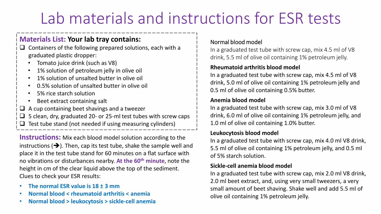
Lab materials and instructions for ESR tests
Instructions: Mix each blood model solution according to the
instructions (). Then, cap its test tube, shake the sample well and place it in the test tube stand for 60 minutes on a flat surface with no vibrations or disturbances nearby. At the 60th minute, note the height in cm of the clear liquid above the top of the sediment. Clues to check your ESR results:
• The normal ESR value is 18 ± 3 mm• Normal blood < rheumatoid arthritis < anemia• Normal blood > leukocytosis > sickle-cell anemia
Normal blood modelIn a graduated test tube with screw cap, mix 4.5 ml of V8 drink, 5.5 ml of olive oil containing 1% petroleum jelly.
Rheumatoid arthritis blood modelIn a graduated test tube with screw cap, mix 4.5 ml of V8 drink, 5.0 ml of olive oil containing 1% petroleum jelly and 0.5 ml of olive oil containing 0.5% butter.
Anemia blood modelIn a graduated test tube with screw cap, mix 3.0 ml of V8 drink, 6.0 ml of olive oil containing 1% petroleum jelly, and 1.0 ml of olive oil containing 1.0% butter.
Leukocytosis blood modelIn a graduated test tube with screw cap, mix 4.0 ml V8 drink, 5.5 ml of olive oil containing 1% petroleum jelly, and 0.5 ml of 5% starch solution.
Sickle-cell anemia blood modelIn a graduated test tube with screw cap, mix 2.0 ml V8 drink, 2.0 ml beet extract, and, using very small tweezers, a very small amount of beet shaving. Shake well and add 5.5 ml of olive oil containing 1% petroleum jelly.
Materials List: Your lab tray contains: Containers of the following prepared solutions, each with a
graduated plastic dropper:• Tomato juice drink (such as V8)• 1% solution of petroleum jelly in olive oil• 1% solution of unsalted butter in olive oil• 0.5% solution of unsalted butter in olive oil• 5% rice starch solution• Beet extract containing salt
A cup containing beet shavings and a tweezer 5 clean, dry, graduated 20- or 25-ml test tubes with screw caps Test tube stand (not needed if using measuring cylinders)
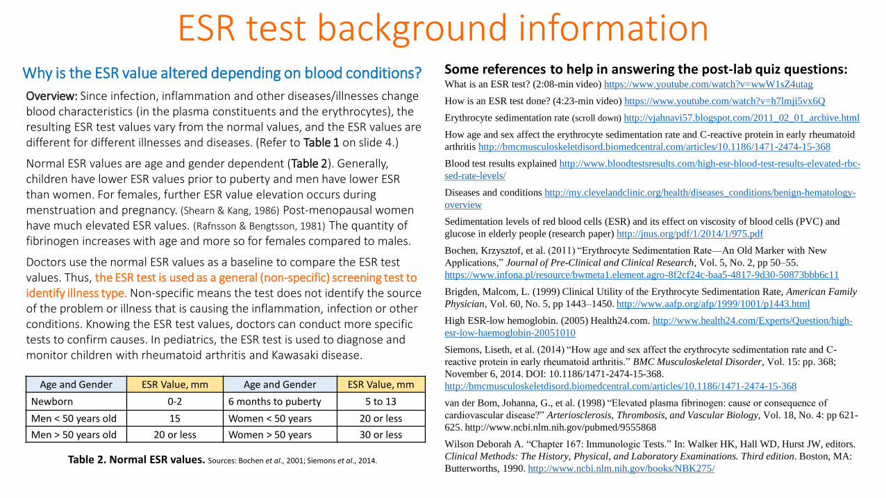
Why is the ESR value altered depending on blood conditions?
Overview: Since infection, inflammation and other diseases/illnesses change blood characteristics (in the plasma constituents and the erythrocytes), the resulting ESR test values vary from the normal values, and the ESR values are different for different illnesses and diseases. (Refer to Table 1 on slide 4.)
Normal ESR values are age and gender dependent (Table 2). Generally, children have lower ESR values prior to puberty and men have lower ESR than women. For females, further ESR value elevation occurs during menstruation and pregnancy. (Shearn & Kang, 1986) Post-menopausal women have much elevated ESR values. (Rafnsson & Bengtsson, 1981) The quantity of fibrinogen increases with age and more so for females compared to males.
Doctors use the normal ESR values as a baseline to compare the ESR test values. Thus, the ESR test is used as a general (non-specific) screening test to identify illness type. Non-specific means the test does not identify the source of the problem or illness that is causing the inflammation, infection or other conditions. Knowing the ESR test values, doctors can conduct more specific tests to confirm causes. In pediatrics, the ESR test is used to diagnose and monitor children with rheumatoid arthritis and Kawasaki disease.
Age and Gender ESR Value, mm Age and Gender ESR Value, mm
Newborn 0-2 6 months to puberty 5 to 13
Men < 50 years old 15 Women < 50 years 20 or less
Men > 50 years old 20 or less Women > 50 years 30 or less
Table 2. Normal ESR values. Sources: Bochen et al., 2001; Siemons et al., 2014.
Some references to help in answering the post-lab quiz questions:What is an ESR test? (2:08-min video) https://www.youtube.com/watch?v=wwW1sZ4utag
How is an ESR test done? (4:23-min video) https://www.youtube.com/watch?v=h7lmji5vx6Q
Erythrocyte sedimentation rate (scroll down) http://vjahnavi57.blogspot.com/2011_02_01_archive.html
How age and sex affect the erythrocyte sedimentation rate and C-reactive protein in early rheumatoid
arthritis http://bmcmusculoskeletdisord.biomedcentral.com/articles/10.1186/1471-2474-15-368
Blood test results explained http://www.bloodtestsresults.com/high-esr-blood-test-results-elevated-rbc-
sed-rate-levels/
Diseases and conditions http://my.clevelandclinic.org/health/diseases_conditions/benign-hematology-
overview
Sedimentation levels of red blood cells (ESR) and its effect on viscosity of blood cells (PVC) and
glucose in elderly people (research paper) http://jnus.org/pdf/1/2014/1/975.pdf
Bochen, Krzysztof, et al. (2011) “Erythrocyte Sedimentation Rate—An Old Marker with New
Applications,” Journal of Pre-Clinical and Clinical Research, Vol. 5, No. 2, pp 50–55.
https://www.infona.pl/resource/bwmeta1.element.agro-8f2cf24c-baa5-4817-9d30-50873bbb6c11
Brigden, Malcom, L. (1999) Clinical Utility of the Erythrocyte Sedimentation Rate, American Family
Physician, Vol. 60, No. 5, pp 1443–1450. http://www.aafp.org/afp/1999/1001/p1443.html
High ESR-low hemoglobin. (2005) Health24.com. http://www.health24.com/Experts/Question/high-
esr-low-haemoglobin-20051010
Siemons, Liseth, et al. (2014) “How age and sex affect the erythrocyte sedimentation rate and C-
reactive protein in early rheumatoid arthritis.” BMC Musculoskeletal Disorder, Vol. 15: pp. 368;
November 6, 2014. DOI: 10.1186/1471-2474-15-368.
http://bmcmusculoskeletdisord.biomedcentral.com/articles/10.1186/1471-2474-15-368
van der Bom, Johanna, G., et al. (1998) “Elevated plasma fibrinogen: cause or consequence of
cardiovascular disease?” Arteriosclerosis, Thrombosis, and Vascular Biology, Vol. 18, No. 4: pp 621-
625. http://www.ncbi.nlm.nih.gov/pubmed/9555868
Wilson Deborah A. “Chapter 167: Immunologic Tests.” In: Walker HK, Hall WD, Hurst JW, editors.
Clinical Methods: The History, Physical, and Laboratory Examinations. Third edition. Boston, MA:
Butterworths, 1990. http://www.ncbi.nlm.nih.gov/books/NBK275/
ESR test background information
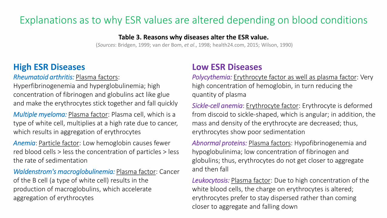
Explanations as to why ESR values are altered depending on blood conditions
Table 3. Reasons why diseases alter the ESR value.(Sources: Bridgen, 1999; van der Bom, et al., 1998; health24.com, 2015; Wilson, 1990)
High ESR DiseasesRheumatoid arthritis: Plasma factors: Hyperfibrinogenemia and hyperglobulinemia; high concentration of fibrinogen and globulins act like glue and make the erythrocytes stick together and fall quickly
Multiple myeloma: Plasma factor: Plasma cell, which is a type of white cell, multiplies at a high rate due to cancer, which results in aggregation of erythrocytes
Anemia: Particle factor: Low hemoglobin causes fewer red blood cells > less the concentration of particles > less the rate of sedimentation
Waldenstrom's macroglobulinemia: Plasma factor: Cancer of the B cell (a type of white cell) results in the production of macroglobulins, which accelerate aggregation of erythrocytes
Low ESR DiseasesPolycythemia: Erythrocyte factor as well as plasma factor: Very high concentration of hemoglobin, in turn reducing the quantity of plasma
Sickle-cell anemia: Erythrocyte factor: Erythrocyte is deformed from discoid to sickle-shaped, which is angular; in addition, the mass and density of the erythrocyte are decreased; thus, erythrocytes show poor sedimentation
Abnormal proteins: Plasma factors: Hypofibrinogenemia and hypoglobulinima; low concentration of fibrinogen and globulins; thus, erythrocytes do not get closer to aggregate and then fall
Leukocytosis: Plasma factor: Due to high concentration of the white blood cells, the charge on erythrocytes is altered; erythrocytes prefer to stay dispersed rather than coming closer to aggregate and falling down
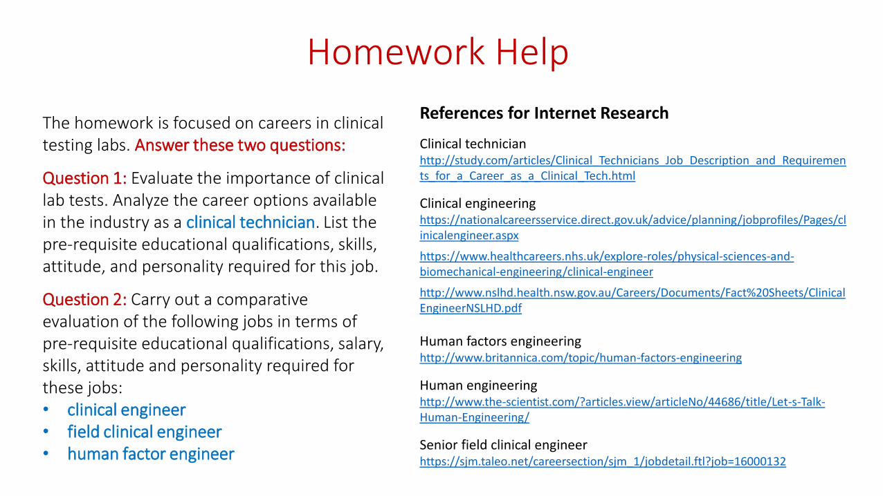
Homework Help
References for Internet Research
Clinical technicianhttp://study.com/articles/Clinical_Technicians_Job_Description_and_Requirements_for_a_Career_as_a_Clinical_Tech.html
Clinical engineering https://nationalcareersservice.direct.gov.uk/advice/planning/jobprofiles/Pages/clinicalengineer.aspx
https://www.healthcareers.nhs.uk/explore-roles/physical-sciences-and-biomechanical-engineering/clinical-engineer
http://www.nslhd.health.nsw.gov.au/Careers/Documents/Fact%20Sheets/ClinicalEngineerNSLHD.pdf
Human factors engineeringhttp://www.britannica.com/topic/human-factors-engineering
Human engineeringhttp://www.the-scientist.com/?articles.view/articleNo/44686/title/Let-s-Talk-Human-Engineering/
Senior field clinical engineerhttps://sjm.taleo.net/careersection/sjm_1/jobdetail.ftl?job=16000132
The homework is focused on careers in clinical testing labs. Answer these two questions:
Question 1: Evaluate the importance of clinical lab tests. Analyze the career options available in the industry as a clinical technician. List the pre-requisite educational qualifications, skills, attitude, and personality required for this job.
Question 2: Carry out a comparative evaluation of the following jobs in terms of pre-requisite educational qualifications, salary, skills, attitude and personality required for these jobs: • clinical engineer• field clinical engineer• human factor engineer


![anaemia and disease activity in patients · proves anaemia and reduces disease activity in patients with rheumatoid ... activity, and erythrocyte sedimentation rate (ESR)] improve.](https://static.documents.pub/doc/80x56/5e831e92b884ce6106762d7e/anaemia-and-disease-activity-in-patients-proves-anaemia-and-reduces-disease-activity.jpg)


![Sedimentatie: Alles kan beter CAT. Waar gaat het over? Sedimentatie ESR Erythrocyte sedimentation rate Bezinkingssnelheid RBC (volbloed) [MESH] “Blood.](https://static.documents.pub/doc/80x56/5551a0ef4979591f3c8b945e/sedimentatie-alles-kan-beter-cat-waar-gaat-het-over-sedimentatie-esr-erythrocyte-sedimentation-rate-bezinkingssnelheid-rbc-volbloed-mesh-blood.jpg)


