Self-assembly of ultra-small micelles fromamphiphilic lipopeptoids†
King Hang Aaron Lau,*a Valeria Castelletto,b Thomas Kendall,c Jan Sefcik,d
Ian W. Hamley,*b Mehedi Rezae and Janne Ruokolainene
Poly(N-substituted glycine) ‘‘peptoids’’ constitute a promising class
of peptide-mimetic materials. We introduce the self-assembly of
lipopeptoids into spherical micelles ca. 5 nm in diameter as well as
larger assemblies by varying the peptoid sequence design. Our results
point to design rules for the self-assembly of peptoid nanostructures,
enabling the creation of stable, ultra-small peptidomimetic
nanospheres.
Peptidic systems that self-assemble into nanostructures havecaptured great attention due to their resemblance to proteinsand other biological structures. In particular, a large literaturedescribes peptide amphiphiles, specifically lipo-peptides, thatshow significant potential in biomedicine.1 Nanofibrils or worm-like micelles are the most commonly reported structural motif.1c,2
They are typically observed to arise from the self-assembly ofpeptide headgroups with (extended) beta-sheet structures.Spherical micelles, formed if hydrogen bonding between peptidechains is de-emphasized,3 have been less commonly reported.2b
Naturally occurring lipopeptides that form micelles highlight thebiomedical potential of such nanostructures.4
Peptoids are structural isomers of peptides with sidechainattachment shifted to the amide nitrogens (Fig. 1A).5 As a classof novel peptide-mimetic polymers, they are emerging as aversatile platform for materials applications and they possessseveral attractive features for biomedical applications due to thesidechain shift.5,6 First, proteolysis is essentially inhibited,5,7
which enables applications requiring resistance against bio-degradation (e.g. antimicrobial peptidomimics,8 and long-termbiointerfaces and biomaterials).6a,9 Second, sequence-specificpeptoids may be conveniently synthesized by a ‘‘submonomer’’solid phase protocol10 and bioactive sequences may be discoveredfrom combinatorial library searches.5,11 Moreover, peptoids areconformationally flexible since they can adopt both cis- and trans-backbone conformations and they lack intra- and inter-backbonehydrogen bonding.5 A rich family of secondary structures – helices,ribbons and sheets – may be induced by focusing on sidechainselection and design,5 and these have been exploited for protein-mimetic structures,12 cell membrane penetrating helices,8a,13
and scaffolds for displaying biorecognition sequences.14
However, peptoid self-assembly into nanostructures has receivedrelatively less attention, despite the fact that polypeptoids obtained
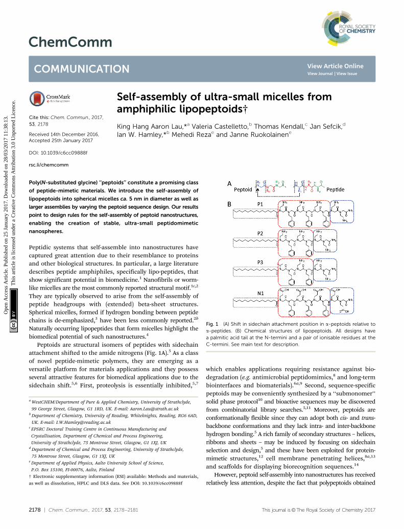
Fig. 1 (A) Shift in sidechain attachment position in a-peptoids relative toa-peptides. (B) Chemical structures of lipopeptoids. All designs havea palmitic acid tail at the N-termini and a pair of ionisable residues at theC-termini. See main text for description.
a WestCHEM/Department of Pure & Applied Chemistry, University of Strathclyde,
99 George Street, Glasgow, G1 1RD, UK. E-mail: [email protected] Department of Chemistry, University of Reading, Whiteknights, Reading, RG6 6AD,
UK. E-mail: [email protected] EPSRC Doctoral Training Centre in Continuous Manufacturing and
Crystallisation, Department of Chemical and Process Engineering,
University of Strathclyde, 75 Montrose Street, Glasgow, G1 1XJ, UKd Department of Chemical and Process Engineering, University of Strathclyde,
75 Montrose Street, Glasgow, G1 1XJ, UKe Department of Applied Physics, Aalto University School of Science,
P.O. Box 15100, FI-00076, Aalto, Finland
† Electronic supplementary information (ESI) available: Methods and materials,as well as dissolution, HPLC and DLS data. See DOI: 10.1039/c6cc09888f
Received 14th December 2016,Accepted 25th January 2017
from living polymerization could self-assemble similar to con-ventional block-copolymers (BCP),15 and sequence-specific peptoidshave been used as models for studying conventional BCP self-assembly.16 Among the limited peptoid-specific structuresreported, nanosheets appear to be a prominent morphology.An alternating hydrophilic–hydrophobic motif has been shownto form nanosheets extending tens of microns laterally.14
Hybrid oligo(peptoid–peptide) diblock amphiphiles have beenshown to form hydrogels composed of nanosheets or nano-fibers depending on the appended peptide sequence,17 andstrips of nanosheets may curl into nano- or micro-tubules.18
Interestingly, sequence-specific control of a basic structuralform such as self-assembled micelles has not been reported.
Inspired by peptide self-assembly, we coupled palmitoyllipid ‘‘tails’’ to amphiphilic peptoid sequences to direct theself-assembly of the resulting lipopeptoids (Fig. 1B). The narrowcross-section of the palmitoyl chain ensures that the peptoidsform a relatively large head-group. A pair of ionizable residues wasalso introduced at the C-terminus to enhance head-group solubility.The combined geometric and solubility head-to-tail asymmetrieswere expected to promote the self-assembly of small well-definedmicelles with a high surface curvature.19
The main peptoid sequence has an alternating XY amphi-philic motif, where X is an ionizable residue and Y is thearomatic N-phenylethyl glycine (Npe). Npe residues have beenreported to contribute to the assembly of peptoid nanosheetsthrough hydrophobic and p–p interactions,14 and we speculatedthat these interactions would also stabilize any self-assembledlipopeptoid structure. P1/P2/P3 have positively (P) charged Nae(N-aminoethyl glycine) residues. The number of XY motifs isdecreased from 3 in P1 to 2 in P2, to probe whether shorteningthe peptoid relative to the hydrophobic palmitoyl tail influencesthe self-assembly. P3 is the same length as P2 but the last Naeresidue is replaced by Nval (the analogue of valine) to further probethe effect of increased hydrophobicity. N1 has glutamic acidanalogues (Nglu) in place of Nae to create a negatively (N) chargedcounterpart to P1 for verifying the self-assembly of the underlyingamphiphilic design.
The peptoids were synthesized using standard submonomersolid phase synthesis. In contrast to lipopeptides (a.k.a. peptideamphiphiles) with alternating amphiphilic motifs that oftenlead to nanofibers composed of extended beta-sheet structures,1c,20
we found that the lipopeptoids assembled into well-defined andvery stable micelles.
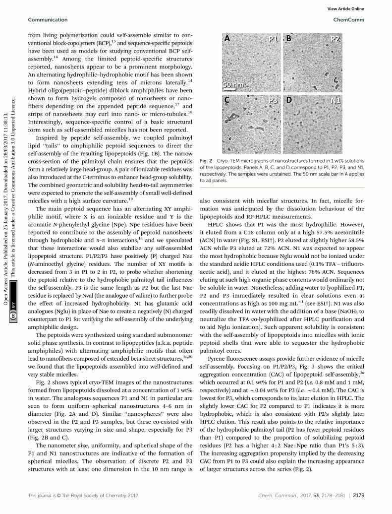
Fig. 2 shows typical cryo-TEM images of the nanostructuresformed from lipopeptoids dissolved at a concentration of 1 wt%in water. The analogous sequences P1 and N1 in particular areseen to form uniform spherical nanostructures 4–6 nm indiameter (Fig. 2A and D). Similar ‘‘nanospheres’’ were alsoobserved in the P2 and P3 samples, but these co-existed withlarger structures varying in size and shape, especially for P3(Fig. 2B and C).
The nanometer size, uniformity, and spherical shape of theP1 and N1 nanostructures are indicative of the formation ofspherical micelles. The observation of discrete P2 and P3structures with at least one dimension in the 10 nm range is
also consistent with micellar structures. In fact, micelle for-mation was anticipated by the dissolution behaviour of thelipopeptoids and RP-HPLC measurements.
HPLC shows that P1 was the most hydrophilic. However,it eluted from a C18 column only at a high 57.5% acetonitrile(ACN) in water (Fig. S1, ESI†). P2 eluted at slightly higher 58.5%ACN while P3 eluted at 72% ACN. N1 was expected to appearthe most hydrophobic because Nglu would not be ionized underthe standard acidic HPLC conditions used (0.1% TFA – trifluoro-acetic acid), and it eluted at the highest 76% ACN. Sequenceseluting at such high organic phase contents would ordinarily notbe soluble in water. Nonetheless, adding water to lyophilized P1,P2 and P3 immediately resulted in clear solutions even atconcentrations as high as 100 mg mL�1 (see ESI†). N1 was alsoreadily dissolved in water with the addition of a base (NaOH; toneutralize the TFA co-lyophilized after HPLC purification andto aid Nglu ionization). Such apparent solubility is consistentwith the self-assembly of lipopeptoids into micelles with ionicpeptoid shells that were able to sequester the hydrophobicpalmitoyl cores.
Pyrene fluorescence assays provide further evidence of micelleself-assembly. Focusing on P1/P2/P3, Fig. 3 shows the criticalaggregation concentration (CAC) of lipopeptoid self-assembly,3a
which occurred at 0.1 wt% for P1 and P2 (i.e. 0.8 mM and 1 mM,respectively) and at B0.04 wt% for P3 (i.e. B0.4 mM). The CAC islowest for P3, which corresponds to its later elution in HPLC. Theslightly lower CAC for P2 compared to P1 indicates it is morehydrophobic, which is also consistent with P2’s slightly laterHPLC elution. This result also points to the relative importanceof the hydrophobic palmitoyl tail (P2 has fewer peptoid residuesthan P1) compared to the proportion of solubilizing peptoidresidues (P2 has a higher 4 : 2 Nae : Npe ratio than P1’s 5 : 3).The increasing aggregation propensity implied by the decreasingCAC from P1 to P3 could also explain the increasing appearanceof larger structures across the series (Fig. 2).
Fig. 2 Cryo-TEM micrographs of nanostructures formed in 1 wt% solutionsof the lipopeptoids. Panels A, B, C, and D correspond to P1, P2, P3, and N1,respectively. The samples were unstained. The 50 nm scale bar in A appliesto all panels.
Small angle X-ray scattering (SAXS) further characterized thephysical dimensions of the lipopeptoid assemblies. Samplesabove the CACs at 1, 5 and 10 wt% were measured, and allexhibited similar results. The 1 wt% dataset, corresponding tothe cryo-TEM samples, is shown in Fig. 4. A simple sphericalcore and shell model (with shell thickness R1 and core radiusR0) was used to analyze the SAXS data. Excellent fits wereobtained and the structural and scattering parameters obtainedare shown in Table 1.
The average micelle diameter Dtotal was 5.4 nm and 4.2 nmfor the P1 and N1 micelles, respectively. These dimensions arein excellent agreement with the TEM results, given the resolutionof the unstained cryo-TEM images and the uncertainty in theSAXS fitting. Dtotal of P2 micelles (i.e. Dtotal(P2)) is 5.2 nm andmatches the fraction of spherical structures seen in cryo-TEM(Fig. 2B). The fact that the Dtotal(P2) is not higher, as might beexpected from the larger structures seen in TEM, may be due toreduced contrast for this sample. Similar to P2, SAXS measuredonly a slightly larger Dtotal(P3) = 7.0 nm, corresponding to thefraction of smaller micelles seen in TEM (Fig. 2C). Nonetheless,the polydispersities obtained increased from 0.44 (P1) to 0.57 (P2)and then to 0.71 (P3), which is consistent with the increasedmorphological inhomogeneity seen in TEM images.
Dynamic light scattering (DLS) measurements focused onP1 and P2 to better understand the SAXS and cryo-TEM data.
Although DLS is significantly more sensitive for larger particles,we have been successful in using the technique to characterizemolecular clusters with hydrodynamic radii (Rh) as small as1 nm.21 Fig. 5 shows the measured normalised intensity auto-correlation functions (ACF) together with the theoretical ACFscalculated for monodisperse populations of particles withvarious Rh. The P1 data was best fitted using Rh = 2.8 nm(i.e. Dtotal = 5.6 nm), which is in excellent agreement with thediameters measured by both SAXS and cryo-TEM. For P2, theinitial ACF decay at short to 0.02 ms overlaps with the P1 data,indicating the presence of micelles with diameter B5 nm.However, a broad shoulder extends to 2 ms, corresponding toa relatively small polydisperse population of particles with Rh
ranging from 10 to 60 nm. The presence of a small populationof such larger micelles (detected due to their more intensescattering) may stand out prominently in TEM images (Fig. 2C),but may not be detected by SAXS (Table 1).
The consistent micelle diameters measured by cryo-TEM, SAXSand DLS, using samples prepared separately and at multipleconcentrations above the CAC, give confidence to the measureddimensions and indicate reproducible self-assembly. Indeed, P1and P2 samples re-measured by DLS after storage for 11 weeks at4 1C gave very similar results (average Rh = 3 nm; see Fig. S5 andTable S3, ESI†), which demonstrated that the lipopeptoid designsgave highly stable micelles.
Interestingly, the observed nanostructures are smaller thanexpected from the dimensions of the lipopeptoids (e.g. P1 and
Fig. 3 Pyrene fluorescence intensity plotted against log concentrationof P1, P2 and P3. The intensities were normalized to the highestvalues recorded at 0.5 wt% lipopeptoid. The pyrene concentration was1.3 � 10�5 wt% and fluorescence was measured at 373–375 nm. Thebreaks in the plots identify the CAC. See Fig. S3 (ESI†) for representative fullspectra of the fluorimetry measurements.
Fig. 4 SAXS form factor data of the lipopeptoids P1, P2, P3 and N1dissolved at 1 wt% in water (symbols) and fitting by SASfit according toa spherical core–shell model (red traces). The fitted parameters andstructural dimensions are described in Table 1.
Table 1 Fitted structural dimensions according to a spherical core–shellmodel. The SAXS data are shown in Fig. 4. The scattering contrast(proportional to electron difference contrast with respect to solvent) isin arbitrary units corresponding to the intensity scale, which is not inabsolute units
Fig. 5 Normalized intensity autocorrelation functions (ACF) of P1 and P2dissolved at 0.3 wt% in water. DLS measurements were taken at 901 usinglaser wavelength of 632.8 nm. Calculated ACFs based on single exponentialdecays for Rh = 2.8 nm (best fit for P1), and 10 and 60 nm are also displayed(to illustrate the polydispersity for P2).
N1 have contour lengths = 4.8 nm).‡ A simplistic picture ofspherical micelles with fully extended sequences would resultin a diameter twice the contour length, i.e. around 10 nm for P1.Also, SAXS indicated the same B1 nm peptoid shell thickness R1
for all designs (Table 1) despite their different sequence lengths.Since it has been shown that peptoid backbones are inherentlyflexible, with persistence lengths ranging from 0.5 to 1 nm,22 wespeculate that the peptoid sequences in the micelles are ‘‘bent’’over to form relatively thin shell layers. The shell thickness wouldthen be dictated by the same peptoid chain cross-section for allsequences (Fig. 1). Indeed, such a configuration could facilitate theordering of the hydrophilic Nae/Nglu residues towards the waterinterface and the hydrophobic Npe residues towards the palmitoylcore. Thus lipopeptoid micelles with ‘‘ultra-small’’ diameters lessthan indicated by the peptoid sequence lengths could be formed.
The formation of micellar structures from all the lipopeptoids,as intended, suggests that the designed geometric asymmetry andhead-to-tail hydrophobicity contrast are useful for controllingpeptoid self-assembly. In comparison, previously reportedlipopeptoids with lower degrees of asymmetry (using ‘‘fatter’’double-tailed phospholipids and more hydrophobic residuemotifs) formed spherical aggregates 80–600 nm in diameter.23
In addition, the fact that backbone hydrogen bonding issuppressed in peptoids might also enhance the importance(and conceptual simplicity) of basic geometric and hydro-phobicity considerations in peptoid self-assembly. Indeed, asa counterpoint, computer simulation models of peptide self-assembly suggest that weakened hydrogen bonding favors theformation of spherical peptide micelles over beta-sheets andcylindrical morphologies.3b
In conclusion, we designed lipopeptoids with high geometricand hydrophobicity head-to-tail asymmetries by coupling palmitoyltails to amphiphilic peptoid sequences, in order to direct peptoidself-assembly into micelles. Unprecedented ultra-small, uni-form and stable spherical micelles with ca. 5 nm diametersand 0.1 wt% CAC were obtained by appropriate peptoidsequence design. The micelles resemble many globular proteinsin size and could be explored as protein mimics. The small sizecould be due to the inherent conformational flexibility of thepeptoid backbone, and could facilitate the transport of thesemicelles across tissues and blood vessels for drug delivery andbiosensing applications. Increases in the sequence hydrophobi-city were observed to lead generally to increased aggregation andpolydispersity. Comparison with previous reports suggests thatthe basic considerations of the overall molecular shape and thedistribution of hydrophobicity content could play prominent rolesin peptoid self-assembly.
KHAL thanks Mikyoung Goo for her outstanding assistancewith peptoid synthesis, and the Strathclyde Academic InvestmentScheme and Tenovus Scotland (S15/29) for financial support. IWHthanks EPSRC for the award of a Platform grant (EP/L L020599/1).We are grateful to the ESRF for the award of beamtime (ref.MX-1769) and to Gabriele Gachin for support during the beamtime.
TK thanks EPSRC and the Doctoral Training Centre in ContinuousManufacturing and Crystallisation (EP/K503289/1) for funding.Research data available at: http://dx.doi.org/10.15129/15d4068d-b268-45fd-bd6a-22b0cbae2ede.
References‡ Calculated from 0.25 nm for every pair of methylenes and 0.35 nm perpeptoid residue.14
1 (a) M. J. Webber, E. A. Appel, E. W. Meijer and R. Langer, Nat.Mater., 2016, 15, 13–26; (b) J. Boekhoven and S. I. Stupp, Adv. Mater.,2014, 26, 1642–1659; (c) F. Versluis, H. R. Marsden and A. Kros,Chem. Soc. Rev., 2010, 39, 3434–3444.
2 (a) T. Aida, E. W. Meijer and S. I. Stupp, Science, 2012, 335, 813–817;(b) J. F. Miravet, B. Escuder, M. D. Segarra-Maset, M. Tena-Solsona,I. W. Hamley, A. Dehsorkhi and V. Castelletto, Soft Matter, 2013, 9,3558–3564.
3 (a) A. Dehsorkhi, V. Castelletto and I. W. Hamley, J. Pept. Sci., 2014,20, 453–467; (b) Y. S. Velichko, S. I. Stupp and M. O. de la Cruz,J. Phys. Chem. B, 2008, 112, 2326–2334.
4 I. W. Hamley, Chem. Commun., 2015, 51, 8574–8583.5 A. S. Knight, E. Y. Zhou, M. B. Francis and R. N. Zuckermann,
Adv. Mater., 2015, 27, 5665–5691.6 (a) K. H. A. Lau, Biomater. Sci., 2014, 2, 627–633; (b) C. Secker,
S. M. Brosnan, R. Luxenhofer and H. Schlaad, Macromol. Biosci.,2015, 15, 881–891.
7 S. M. Miller, R. J. Simon, S. Ng, R. N. Zuckermann, J. M. Kerr andW. H. Moos, Drug Dev. Res., 1995, 35, 20–32.
8 (a) A. M. Czyzewski, H. Jenssen, C. D. Fjell, M. Waldbrook, N. P.Chongsiriwatana, E. Yuen, R. E. Hancock and A. E. Barron, PLoSOne, 2016, 11, e0135961; (b) M. L. Huang, S. B. Y. Shin, M. A.Benson, V. J. Torres and K. Kirshenbaum, ChemMedChem, 2012, 7,114–122.
9 K. H. A. Lau, T. S. Sileika, S. H. Park, A. M. L. Sousa, P. Burch,I. Szleifer and P. B. Messersmith, Adv. Mater. Interfaces, 2015,2, 1400225.
10 R. N. Zuckermann, J. M. Kerr, S. B. H. Kent and W. H. Moos,J. Am. Chem. Soc., 1992, 114, 10646–10647.
11 Y. Gao and T. Kodadek, Chem. Biol., 2013, 20, 360–369.12 B. C. Lee, T. K. Chu, K. A. Dill and R. N. Zuckermann, J. Am. Chem.
Soc., 2008, 130, 8847–8855.13 A. M. Czyzewski and A. E. Barron, AIChE J., 2008, 54, 2–8.14 E. J. Robertson, A. Battigelli, C. Proulx, R. V. Mannige, T. K. Haxton,
L. Yun, S. Whitelam and R. N. Zuckermann, Acc. Chem. Res., 2016,49, 379–389.
15 C. U. Lee, T. P. Smart, L. Guo, T. H. Epps and D. H. Zhang,Macromolecules, 2011, 44, 9574–9585.
16 A. M. Rosales, R. A. Segalman and R. N. Zuckermann, Soft Matter,2013, 9, 8400–8414.
17 Z. D. Wu, M. Tan, X. M. Chen, Z. M. Yang and L. Wang, Nanoscale,2012, 4, 3644–3646.
18 (a) R. C. Elgersma, G. E. Mulder, J. A. W. Kruijtzer, G. Posthuma,D. T. S. Rijkers and R. M. J. Liskamp, Bioorg. Med. Chem. Lett., 2007,17, 1837–1842; (b) H. K. Murnen, A. M. Rosales, J. N. Jaworski,R. A. Segalman and R. N. Zuckermann, J. Am. Chem. Soc., 2010, 132,16112–16119.
19 J. N. Israelachvili, Intermolecular and Surface Forces, Academic Press,San Diego, 3rd edn, 2011, pp. 535–576.
20 E. De Santis and M. G. Ryadnov, Chem. Soc. Rev., 2015, 44, 8288–8300.21 A. Jawor-Baczynska, J. Sefcik and B. D. Moore, Cryst. Growth Des.,
2013, 13, 470–478.22 A. M. Rosales, H. K. Murnen, S. R. Kline, R. N. Zuckermann and
R. A. Segalman, Soft Matter, 2012, 8, 3673–3680.23 (a) C.-Y. Huang, T. Uno, J. E. Murphy, S. Lee, J. D. Hamer, J. A.
Escobedo, F. E. Cohen, R. Radhakrishnan, V. Dwarki andR. N. Zuckermann, Chem. Biol., 1998, 5, 345–354; (b) Y. U. Konca,K. Kirshenbaum and R. N. Zuckermann, Int. J. Nanomed., 2014, 9,2271–2285.


![Open Access Nanoscale Drug Delivery and Hyperthermia: The ...€¦ · based liposomes [11, 12]. Other self-assembling systems— polymeric micelles formed from amphiphilic block co-polymers](https://static.documents.pub/doc/80x56/5fa6ffd11f655536fd2de424/open-access-nanoscale-drug-delivery-and-hyperthermia-the-based-liposomes-11.jpg)
