ORIGINAL ARTICLE Serum Antibody Against NY-ESO-1 and XAGE1 Antigens Potentially Predicts Clinical Responses to Anti–Programmed Cell Death-1 Therapy in NSCLC Yoshihiro Ohue, MD, PhD, a Koji Kurose, MD, PhD, a Takahiro Karasaki, MD, PhD, b Midori Isobe, PhD, a Takaaki Yamaoka, BSc, a Junichiro Futami, PhD, c Isao Irei, MD, PhD, d Takeshi Masuda, MD, PhD, e Masaaki Fukuda, MD, PhD, f Akitoshi Kinoshita, MD, PhD, g Hirokazu Matsushita, MD, PhD, h,i Katsuhiko Shimizu, MD, PhD, j Masao Nakata, MD, PhD, j Noboru Hattori, MD, PhD, e Hiroyuki Yamaguchi, MD, PhD, k Minoru Fukuda, MD, PhD, k,l Ryohei Nozawa, PhD, m Kazuhiro Kakimi, MD, PhD, h,i Mikio Oka, MD, PhD n, * a Department of Respiratory Medicine, Kawasaki Medical School, Okayama, Japan b Department of Thoracic Surgery, The University of Tokyo, Tokyo, Japan c Department of Medical Bioengineering, Graduate School of Natural Science and Technology, Okayama University, Okayama, Japan d Department of Pathology, Kawasaki Medical School, Okayama, Japan e Department of Respiratory Internal Medicine, Hiroshima University Hospital, Hiroshima, Japan f Department of Respiratory Medicine, The Japanese Red Cross Nagasaki Genbaku Hospital, Nagasaki, Japan g Department of Respiratory Medicine, Nagasaki Prefecture Shimabara Hospital, Nagasaki, Japan h Department of Immunotherapeutics, Graduate School of Medicine, The University of Tokyo, Tokyo, Japan i Cancer Immunology Data Multi-level Integration Unit, Medical Science Innovation Hub Program, RIKEN, Tokyo, Japan j Department of General Thoracic Surgery, Kawasaki Medical School, Okayama, Japan k Department of Respiratory Medicine, Nagasaki University Graduate School of Biomedical Sciences, Nagasaki, Japan l Clinical Oncology Center, Nagasaki University Hospital, Nagasaki, Japan m Faculty of Health and Welfare Services Administration, Kawasaki University of Medical Welfare, Okayama, Japan n Department of Immuno-Oncology, Kawasaki Medical School, Okayama, Japan Received 16 January 2019; revised 5 August 2019; accepted 13 August 2019 Available online - 23 August 2019 ABSTRACT Introduction: Programmed cell death-1 (PD-1) inhibitors effectively treat NSCLC and prolong survival. Robust biomarkers for predicting clinical benefits of good response and long survival with anti–PD-1 therapy have yet to be identified; therefore, predictive biomarkers are needed to select patients with benefits. *Corresponding author. Drs. Ohue and Kurose contributed equally to this work. Disclosure: Dr. Ohue has received grants from JSPS Kakenhi, Kawasaki Medical School, Medical Research Encouragement Prize of the Japan Medical Association, and Okayama Health Foundation; and has a patent for a method for examining therapeutic effect on cancer and composition for inducing immune response. Dr. Kurose has received grants from JSPS Kakenhi, Takeda Science Foundation, and Kawasaki Medical School and has a patent for a method for examining thera- peutic effect on cancer and composition for inducing immune response. Dr. Hattori has received grants from Ono Pharmaceutical Co., Ltd.; and has received personal fees from Boehringer Ingelheim Japan, Inc., Ono Pharmaceutical Co., Ltd., MSD K.K., a subsidiary of Merck & Co., Inc., and AstraZeneca K.K. Dr. Yamaguchi has received grants from Novartis Pharma K.K., Ely Lilly Japan K.K., and Boeh- ringer Ingelheim Japan, Inc.; and has received personal fees from AstraZeneca K.K., Boehringer Ingelheim Japan, Inc., Taiho Pharma- ceutical Co., Ltd., Eli Lilly Japan K.K., and Bristol-Myers Squibb Company. Dr. Minoru Fukuda has received grants from MSD; and has received personal fees from Ono Pharma and MSD. Dr. Oka has received grants from JSPS Kakenhi and Kawasaki Medical School; has received personal fees from The University of Tokyo, Thyas Co., Ltd., and Pole Star Co., Ltd.; and has a patent for a method for examining therapeutic effect on cancer and composition for inducing immune response. The remaining authors declare no conflict of interest. Address for correspondence: Mikio Oka, MD, PhD, Department of Immuno-Oncology, Kawasaki Medical School, 577 Matsushima, Kura- shiki, Okayama 701-0192, Japan. E-mail: [email protected]. jp ª 2019 International Association for the Study of Lung Cancer. Published by Elsevier Inc. This is an open access article under the CC BY-NC-ND license (http://creativecommons.org/licenses/by-nc-nd/ 4.0/). ISSN: 1556-0864 https://doi.org/10.1016/j.jtho.2019.08.008 Journal of Thoracic Oncology Vol. 14 No. 12: 2071-2083
Transcript
ORIGINAL ARTICLE
Serum Antibody Against NY-ESO-1 and XAGE1Antigens Potentially Predicts Clinical Responses toAnti–Programmed Cell Death-1 Therapy in NSCLC
aDepartment of Respiratory Medicine, Kawasaki Medical School, Okayama, JapanbDepartment of Thoracic Surgery, The University of Tokyo, Tokyo, JapancDepartment of Medical Bioengineering, Graduate School of Natural Science and Technology, Okayama University,Okayama, JapandDepartment of Pathology, Kawasaki Medical School, Okayama, JapaneDepartment of Respiratory Internal Medicine, Hiroshima University Hospital, Hiroshima, JapanfDepartment of Respiratory Medicine, The Japanese Red Cross Nagasaki Genbaku Hospital, Nagasaki, JapangDepartment of Respiratory Medicine, Nagasaki Prefecture Shimabara Hospital, Nagasaki, JapanhDepartment of Immunotherapeutics, Graduate School of Medicine, The University of Tokyo, Tokyo, JapaniCancer Immunology Data Multi-level Integration Unit, Medical Science Innovation Hub Program, RIKEN, Tokyo, JapanjDepartment of General Thoracic Surgery, Kawasaki Medical School, Okayama, JapankDepartment of Respiratory Medicine, Nagasaki University Graduate School of Biomedical Sciences, Nagasaki, JapanlClinical Oncology Center, Nagasaki University Hospital, Nagasaki, JapanmFaculty of Health and Welfare Services Administration, Kawasaki University of Medical Welfare, Okayama, JapannDepartment of Immuno-Oncology, Kawasaki Medical School, Okayama, Japan
Received 16 January 2019; revised 5 August 2019; accepted 13 August 2019Available online - 23 August 2019
Drs. Ohue and Kurose contributed equally to this work.
Disclosure: Dr. Ohue has received grants from JSPS Kakenhi, KawasakiMedical School, Medical Research Encouragement Prize of the JapanMedical Association, and Okayama Health Foundation; and has apatent for a method for examining therapeutic effect on cancer andcomposition for inducing immune response. Dr. Kurose has receivedgrants from JSPS Kakenhi, Takeda Science Foundation, and KawasakiMedical School and has a patent for a method for examining thera-peutic effect on cancer and composition for inducing immuneresponse. Dr. Hattori has received grants from Ono PharmaceuticalCo., Ltd.; and has received personal fees from Boehringer IngelheimJapan, Inc., Ono Pharmaceutical Co., Ltd., MSD K.K., a subsidiary ofMerck & Co., Inc., and AstraZeneca K.K. Dr. Yamaguchi has receivedgrants from Novartis Pharma K.K., Ely Lilly Japan K.K., and Boeh-ringer Ingelheim Japan, Inc.; and has received personal fees fromAstraZeneca K.K., Boehringer Ingelheim Japan, Inc., Taiho Pharma-ceutical Co., Ltd., Eli Lilly Japan K.K., and Bristol-Myers SquibbCompany. Dr. Minoru Fukuda has received grants from MSD; and has
biomarkers for predicting clinical benefits of good responseand long survival with anti–PD-1 therapy have yet to beidentified; therefore, predictive biomarkers are needed toselect patients with benefits.
received personal fees from Ono Pharma and MSD. Dr. Oka hasreceived grants from JSPS Kakenhi and Kawasaki Medical School; hasreceived personal fees from The University of Tokyo, Thyas Co.,Ltd., and Pole Star Co., Ltd.; and has a patent for a method forexamining therapeutic effect on cancer and composition for inducingimmune response. The remaining authors declare no conflict ofinterest.
Address for correspondence: Mikio Oka, MD, PhD, Department ofImmuno-Oncology, Kawasaki Medical School, 577 Matsushima, Kura-shiki, Okayama 701-0192, Japan. E-mail: [email protected]
ª 2019 International Association for the Study of Lung Cancer.Published by Elsevier Inc. This is an open access article under theCC BY-NC-ND license (http://creativecommons.org/licenses/by-nc-nd/4.0/).
ISSN: 1556-0864
https://doi.org/10.1016/j.jtho.2019.08.008
Journal of Thoracic Oncology Vol. 14 No. 12: 2071-2083
2072 Ohue et al Journal of Thoracic Oncology Vol. 14 No. 12
Methods: We conducted a prospective study to explorewhether serum antibody against NY-ESO-1 and/or XAGE1cancer-testis antigens predicted primarily good clinicalresponse and secondarily long survival with anti–PD-1therapy for NSCLC. The serum antibody was detected byenzyme-linked immunosorbent assay, and tumor immunemicroenvironment and mutation burden were analyzed byimmunohistochemistry and next-generation sequencing.
Results: In the discovery cohort (n ¼ 13), six antibody-positive NSCLC cases responded to anti–PD-1 therapy(two complete and four partial responses), whereas sevenantibody-negative NSCLC cases did not. Antibody positivitywas associated with good response and survival, regardlessof tumor programmed death ligand 1 (PD-L1) expression,mutation burden, and CD8þ T-cell infiltration. In the vali-dation cohort (n ¼ 75), 17 antibody-positive NSCLC casesresponded well to anti–PD-1 therapy as compared with 58negative NSCLC cases (objective response rate 65% versus19%, p ¼ 0.0006) and showed significantly prolongedprogression-free survival and overall survival. Antibody ti-ters highly correlated with tumor reduction rates. In themultivariate analysis, response biomarkers were tumorprogrammed death ligand 1 expression and antibody posi-tivity, and only antibody positivity was a significantly betterpredictive biomarker of progression-free survival (hazardratio ¼ 0.4, p ¼ 0.01) and overall survival (hazard ratio ¼0.2, p ¼ 0.004).
Conclusions: Our results suggest that NY-ESO-1 and/orXAGE1 serum antibodies are useful biomarkers for pre-dicting clinical benefits in anti–PD-1 therapy for NSCLC andprobably for other cancers.
� 2019 International Association for the Study of LungCancer. Published by Elsevier Inc. This is an open accessarticle under the CC BY-NC-ND license (http://creativecommons.org/licenses/by-nc-nd/4.0/).
Keywords: Biomarker; Anti–programmed death 1 therapy;NSCLC; Cancer-testis antigen; Serum antibody
IntroductionLung cancer is the leading cause of death from
cancer worldwide.1 Most lung cancers, especiallyNSCLC, are diagnosed in the advanced stages and areresistant to conventional chemotherapy, resulting inpoor prognosis. Recently, immunotherapy usingimmune-checkpoint inhibitors has prolonged NSCLCpatient survival.2 Programmed cell death-1 (PD-1) ofan immune-checkpoint molecule is expressed on acti-vated CD8þ T cells and binds to programmed deathligand 1 (PD-L1) on tumor cells, resulting in T-cellexhaustion.3 Therapeutic antibodies (Abs) for PD-1,nivolumab and pembrolizumab, inhibit this bindingand reactivate CD8þ T-cell with cytotoxic function
(cytotoxic T lymphocytes [CTLs]).3,4 A number ofclinical trials have since shown that anti–PD-1 therapyis effective in the treatment of various solid and he-matologic malignancies including NSCLC. The responserate of NSCLC to anti–PD-1 monotherapy is approxi-mately 20%; therefore, response biomarkers have beenextensively investigated.4-8 Although biomarkers suchas tumor PD-L1 expression (tumor proportion score[TPS]), tumor mutation burden (TMB), microsatelliteinstability, T cell infiltration, and circulating PD-1þCD8þ T cell are described, these markers are not souseful and convenient due to the reliability, cost, andtime demands.4,5,7-9
Melanoma antigen-1 (MAGE-1, renamed MAGE-A1) ofcancer-testis (CT) antigen was discovered as the firsthuman tumor antigen, and hundreds of CT antigens havesince been identified.10,11 MAGE-A family members andNew York esophageal squamous cell carcinoma-1 (NY-ESO-1) are broadly expressed in various human malig-nancies, and MAGE-A1, MAGE-A3, NY-ESO-1, SSX, andXAGE1 among CT antigens elicit spontaneous T cell andhumoral immune responses in cancer patients. NY-ESO-1has been extensively investigated as a target of cancervaccines and T-cell therapy because it exhibits thehighest immunogenicity among CT antigens.10-18 XAGE1is expressed in approximately 40% to 60% of lung ad-enocarcinomas, and the XAGE1 serum Ab is a goodprognostic marker in advanced lung adenocarcinomapatients, as we reported previously.14,19,20
In cancer patients with serum Ab against NY-ESO-1and XAGE1, the antigen-specific CD4þ and CD8þ T cellare frequently detected in peripheral blood.12,14,15 Thesefindings suggest that NY-ESO-1 and XAGE1 are majorimmunodominant antigens and play important roles inthe immune surveillance of NSCLC. The serum Abpositivity probably reflects the presence of activatedantigen-specific T cell and PD-1/PD-L1–mediated im-mune suppression, suggesting good responses to anti–PD-1 therapy (Supplementary Fig. 1). Accordingly, wehypothesized that NY-ESO-1 and XAGE1 serum Abs havepotential as response biomarkers in anti–PD-1 therapyfor NSCLC, and conducted a prospective multicenterstudy to verify this hypothesis.
Materials and MethodsStudy Design and Patients
This biomarker study was prospectively designedand performed to explore whether NY-ESO-1 andXAGE1 serum Abs predicted clinical responses andfurther patient survival with anti–PD-1 therapy, ac-cording to the Reporting Recommendations forTumour Marker Prognostic Studies criteria as listed inthe guideline.21 This study was approved by the
December 2019 Predictive Biomarkers for Anti-PD-1 Therapy of NSCLC 2073
Institutional Ethics Committee of Kawasaki MedicalSchool (number 2071-7; clinical study registry, UMIN-CTR 000016678) and four medical centers in Japan,and was performed in accordance with the Declarationof Helsinki. For reference, we have investigatedexpression and immune responses and monitoringabout CT antigens in human cancers including NSCLCfor a long time.15,17,19,20,22-24
Patients who have advanced NSCLC with metastatic/recurrent and unresectable stages and postoperativerecurrence were consecutively enrolled into this studyfrom the above five medical centers between March2016 and December 2018. Patient inclusion criteriawere good performance status 0-2, good organ function,measurable lesions, and informed consent to this study.Exclusion criteria were active multiple primary malig-nancies and infections, autoimmune diseases, receivingintensive immunosuppressive agents, and pregnancy.Anti–PD-1 monotherapy with nivolumab or pem-brolizumab was administered as a standard therapy forthese NSCLC patients in a first-line (TPS � 50% atdiagnosis) or later setting according to governmentapproval in Japan. Before anti–PD-1 therapy, NY-ESO-1and XAGE1 serum Abs were measured using enzyme-linked immunosorbent assay (ELISA) by laboratoryscientists.
Patients in the discovery cohort were stratified bytheir NY-ESO-1/XAGE1 serum Ab status for enrollmentin this observational study and were analyzed. Then,when objective response rate (ORR) of anti–PD-1monotherapy was 20% overall and at least 50% in theAb-positive patients, and when the Ab-positive pro-portion was minimum 20% and maximum 25% inadvanced NSCLC patients, the required sample size was75 and 56 in the independent validation cohort,respectively. Finally, 75 patients with NSCLC wereenrolled in the validation study. It was calculated in apriori power analysis for Fisher’s exact test with thepower level 0.8 and the significance level 0.05 byG*Power calculator.25 The reasons for estimated ORR20% and 50% were based on the results of priorclinical trials and the ORR 50% in NSCLC of TPS greaterthan or equal to 50% treated with pembrolizumab,respectively, and the Ab-positive proportion 20% atminimum and 25% at maximum were referred toSupplementary Table 1.2,5 To assess the status of theAb response, sera from patients were collected within2 months before anti–PD-1 therapy, and Ab titers weremeasured. Clinical responses to anti–PD-1 therapy andthe Ab status were double-blinded with each other byclinicians and laboratory scientists.
The primary and secondary endpoints in this studywere the ORR to anti–PD-1 therapy, and progression-freesurvival (PFS) and overall survival (OS) after anti–PD-1
therapy, respectively, according to the NY-ESO-1/XAGE1serum Ab status.
Clinical Samples, Clinical Efficacy, and SurvivalAnalysis
Patients had consented to Institutional ReviewBoard–approved protocols permitting blood and tissuesample collection and sequencing. Tumor tissues andsera were obtained from all patients before anti–PD-1therapy. Tumor tissues for whole-exome sequencingwere freshly frozen material from only the discoverycohort patients before anti–PD-1 therapy. The EGFRmutation (EGFRmt) or EML4-ALK-fusion was identifiedby peptide nucleic acid–locked nucleic acid polymerasechain reaction clamp and fluorescence in situ hybridi-zation, respectively. As a negative control, sera fromnonmalignant donors were independently obtainedfrom previous study between 2015 and 2016.22
Target lesions at baseline were assessed according tothe Response Evaluation Criteria in Solid Tumors version1.1 (RECIST v1.1), and the baseline sum of the longestdiameters for target lesions was recorded and used todetermine objective responses.26 Initial responses toanti–PD-1 therapy were assessed by investigator-assessed RECIST v1.1 criteria, and a complete response(CR) and partial response (PR) were confirmed by arepeat imaging occurring at least 4 weeks after the initialidentification of response.26 Unconfirmed responseswere considered stable disease (SD) or progressive dis-ease (PD) dependent on the results of the second chest x-ray or computed tomography scan. PD showed morethan 20% increase in the sum of diameters of targetlesions from baseline sum or the appearance of new le-sions.26 All images were investigator-assessed by blin-ded reviewers.
PFS rates were assessed according to RECIST v1.1and PFS and OS were analyzed by the Kaplan-Meiermethod. Differences in PFS and OS between patientsubgroups were analyzed using the Log-rank test, andp values less than 0.05 were considered to be signifi-cant. PFS and OS for living patients were censored atthe date of last known contact. The dates of PFS and OSafter anti–PD-1 therapy were updated as December31, 2018.
Antibody Responses to NY-ESO-1 and XAGE1Ab responses to the NY-ESO-1 and XAGE1 pro-
teins were examined by ELISA, as we reported pre-viously.20,27 The recombinant NY-ESO-1 and syntheticXAGE1 protein (GL Biochemistry, Shanghai, China) (1mg/mL) in a coating buffer was adsorbed onto 96-well ELISA plates (Nunc, Roskilde, Denmark) andincubated at 4�C overnight. Plates were washed with
2074 Ohue et al Journal of Thoracic Oncology Vol. 14 No. 12
phosphate-buffered saline and blocked with 5% fetalcalf serum/phosphate-buffered saline (200 mL/well)at 37�C for 1 hour. After washing, 100 mL of seriallydiluted serum was added to each well and incubatedat 4�C for 2 hours. After washing, alkaline phospha-tase affinipure goat anti-human immunoglobulin G,Fcg fragment-specific (1:5000) (Jackson ImmunoResearch, West Grove, Pennsylvania) was added tothe wells and the plates were incubated at 37�C for 1hour. After washing and development (AttoPhos APfluorescent substrate system, Promega, Madison,Wisconsin), absorbance was read at an excitation of440/30 and emission of 560/40 with a gain of 50.
The cutoff value was based on the reactivity ofnegative control sera from non-malignant donors (n ¼60) and was defined as follows: the 95% confidenceinterval (CI) upper limit optical density (OD) value ofthe negative control serum pool. The extrapolatedtiter of patient serum samples was defined as theminimal dilution factor for which an O.D. greater thanthe cutoff was examinable. The Ab response wasdefined as positive for serum with extrapolated titersexceeding or equal to 100 (�100). The cutoff value ofgreater than or equal to 100 for positive was deter-mined by previous findings that NY-ESO-1/XAGE1–specific T cells were frequently detected from cancerpatients with Ab titer greater than or equal to100.15,28
Immunohistochemistry for ImmuneMicroenvironment and CT Antigens
The tumor PD-L1 and major histocompatibility(MHC)–class I expression, and CD8þ T cell, CD20þ Bcell, and CD163þ M2-macrophage infiltration in tumortissues before anti–PD-1 therapy were analyzed byimmunohistochemistry (IHC).22 Details of analysismethods are fully provided in the SupplementaryMaterials. Tumor NY-ESO-1 and XAGE1 antigenexpression before anti–PD-1 therapy were alsoanalyzed by IHC. Four-micrometer–thick sections weredeparaffinized with xylene and ethanol. Antigenretrieval was performed by microwave heating in an-tigen retrieval buffer (10 mM citrate buffer, pH 6.0)with a pressure cooker for 10 minutes. After theinactivation of endogenous peroxidase with 0.3% H2O2
for 15 and 5 minutes, respectively, specimens werepre-incubated with serum-free blocking solution(Nacalai Tesque and DakoCytomation, Kyoto, Japan) forNY-ESO-1 and XAGE1, respectively. After washing, ananti–NY-ESO-1 mouse monoclonal Ab (clone E978,1:100, Invitrogen, Carlsbad, California) and USO 9-13monoclonal Ab (2 mg/mL) were added and incubated at4�C and room temperature overnight for NY-ESO-1 and
XAGE1, respectively. After washing, sample slides werestained by the streptavidin-biotin complex (SimpleS-tain MAX-PO kit; Nichirei, Tokyo, Japan), followed by areaction with 3, 30-diaminobenzidine in H2O2 andcounterstained with hematoxylin solution.
Whole-Exome Sequencing and Detection ofTumor Somatic Mutations
DNA and RNA samples were prepared using AllPrepDNA/RNA/miRNA Universal Kits (Qiagen, Hilden, Ger-many) according to the manufacturer’s instructions.Genomic DNA was converted to DNA libraries for DNAsequencing using the SureSelect Target EnrichmentSystem Capture Process (Agilent Technologies, SantaClara, California). Details of analysis methods are fullyprovided in the Supplementary Materials.
RNA Sequencing and Immunogenomic Analysis ofthe Tumor Microenvironment
The characterization of the tumor immune microen-vironment before anti–PD-1 therapy was furtheranalyzed by next-generation sequencing. The expressionof immune-related genes was extracted.29 The enrich-ment of tumor-infiltrating lymphocytes was estimatedby a single sample Gene Set Enrichment Analysis(ssGSEA) using gene sets provided by Charoentonget al.30-32 Details of analysis methods are fully providedin the Supplementary Materials.
Statistical AnalysesStatistical analyses were performed with the two-
sided Mann–Whitney U test for two groups usingGraphPad Prism v.6 (Graphpad Prism Software, SanDiego, California) and IBM SPSS Statistics 23 forWindows (IBM, New York, New York). In analyses ofthe relationship between each parameter, Spearman’scorrelation analysis was performed. PFS and OS wereanalyzed by the Kaplan-Meier method. Differences inPFS and OS between patient subgroups wereanalyzed using the Log-rank tests. To assess therelationship between a factor and PFS and OS, uni-variate and multivariate analyses were performedusing Cox’s proportional hazards regression model.We previously reported in lung adenocarcinoma thatXAGE1 serum Ab was a good prognostic markerregardless of EGFR status and that XAGE1 andGalectin-9 were poor prognostic markers and tumorPD-L1 expression and T-cell infiltration were likelyto be good prognostic ones.20,22 These factors rele-vant to survival and the present study were inves-tigated by a multivariate analysis. A multivariateanalysis was performed by all factors using Cox’sregression analysis (backwards stepwise model).
December 2019 Predictive Biomarkers for Anti-PD-1 Therapy of NSCLC 2075
Results are expressed as the mean or 95% CI, andthe threshold for significance was p less than 0.05.
ResultsRelationship Between Antibody Responses to NY-ESO-1 and XAGE1 and Clinical Benefits of GoodResponse and Long Survival With Anti–PD-1Therapy for NSCLC
In the discovery cohort, 13 patients with NSCLC whowere treated with nivolumab were enrolled (Table 1),and the median follow-up time from the registration was3.6 months (range, 0.5 to 24.0 months). These patientshad previously been heavily treated with systemicchemotherapy. The CT antibody, serum Ab against theNY-ESO-1 and/or XAGE1 antigens, was positive in re-sponders (CR and PR) and negative in nonresponders(SD and PD) (Fig. 1A). Two patients with CR had oneeach of NY-ESO-1 and XAGE1 Ab, whereas two of fourpatients with PR had NY-ESO-1 Ab and the remainingtwo had XAGE1 Ab. The CT antibody was not detected in
Values shown are n (%) unless otherwise stated.CT, cancer-testis antibody, serum antibody against NY-ESO-1 and/or XAGE1antigen.
patients who were nonresponders, and there was a sig-nificant difference in the CT antibody titer between re-sponders and nonresponders (Fig. 1A) (p < 0.01). Thebest changes from the baseline of target lesions showedmarked differences between CT antibody-positive and-negative patients (Fig. 1B). However, objective re-sponses appeared to be associated with PD-L1 expres-sion levels but not with the infiltration of variousimmune cells, cytokines, chemokines, or tumor MHC-class I expression levels analyzed by IHC and next-generation sequencing (Fig. 1B, Supplementary Fig. 2).CT antibody-positive patients obtained prolonged sur-vival with anti–PD-1 therapy. Hazard ratios (HRs) of PFSand OS between CT antibody-positive and -negative pa-tients were 0.17 (95% CI: 0.04–0.66) (Fig. 2A) and 0.15(95% CI: 0.04–0.60) (Fig. 2B), respectively.
In the independent validation cohort, 75 patientswith NSCLC were consecutively enrolled and receivednivolumab or pembrolizumab (Table 1), and the medianfollow-up time from the registration was 7.3 months(range, 0.5 to 24.0 months). The CT antibody was posi-tive in 17 of 75 (23%) patients (Table 1, Fig. 1A),comprising 10 XAGE1-positive Ab, 6 NY-ESO-1 Ab, and 1with both Abs being positive (Fig. 1A). First-line anti–PD-1 therapy was given in 2 (12%) and 7 (12%) of 17 CTantibody-positive and 58 negative patients, respectively,and 66 of 75 (88%) patients received anti–PD-1 therapyafter the second-line therapy, and 30 (40%) did afterthird-line therapy (Fig. 2C). Eleven of 17 (65%; 5 CRsand 6 PRs) CT antibody-positive patients responded toanti–PD-1 therapy, in contrast to 11 of 58 (19%; 1 CRand 10 PRs) CT antibody-negative patients (ORR 65% vs.19%, p ¼ 0.0006), resulting in 29% (22 of 75) ORR inthe validation cohort (Fig. 1A, Table 2). The sensitivityand specificity of CT antibody for response were 0.65(95% CI: 0.38–0.86) and 0.81 (95% CI: 0.69–0.90),respectively. A significant difference was observed in theCT antibody titer between responders and non-responders (Fig. 1A, p < 0.001), and the greatest changesfrom the baseline of target lesions showed marked dif-ferences between CT antibody-positive and -negativepatients (Fig. 1B). However, objective responses werenot associated with CD8þ T-cell infiltration or tumor PD-L1 and MHC-class I expression levels in appropriateavailable tissues before anti–PD-1 therapy (Fig. 1B). CTantibody-positive patients obtained prolonged survivalwith anti–PD-1 therapy; HRs of PFS and OS between CTantibody-positive and -negative patients were 0.42 (95%CI: 0.24–0.75) (Fig. 2A) and 0.21 (95% CI: 0.10–0.44)(Fig. 2B), respectively, even after the second line or thethird line (Fig. 2C). In the multivariate analysis, responsemarkers were smoking (odd ratio ¼ 11, 95% CI: 1.0–113), high PD-L1 expression (odd ratio ¼ 5.0, 95%: CI1.0–26) and CT antibody positivity (odd ratio ¼ 9.6, 95%
A S A A A A A A A A A S A A A A S A S A S A A A A S S A A A A A A A A S A A S S A A A A N A N
-100-80-60-40-20
020406080
100200300
CR(5/6)
PR(6/16)
SD(3/17)
PD(3/36)
CR(2/2)
PR(4/4)
SD(0/1)
PD(0/6)
Figure 1. Relationship between antibody responses to NY-ESO-1 and XAGE1 cancer-testis (CT) antigens and clinical responseswith anti–programmed death 1 (PD-1) therapy. A, Extrapolated CT antibody (serum antibody against NY-ESO-1 and/or XAGE1CT antigen) titers in the discovery (n ¼ 13) and validation (n ¼ 75) cohorts according to clinical responses with anti–PD-1therapy. Patient sera were collected within 2 months before anti–PD-1 therapy, and CT antibody titers were measured byenzyme-linked immunosorbent assay (ELISA) and defined as positive for serum with an extrapolated titer exceeding or equalto 100 (�100). Clinical responses were assessed according to the Response Evaluation Criteria in Solid Tumors version 1.1(RECIST v1.1) guidelines. The numbers in parentheses indicate the number of CT antibody-positive patients/the number ofpatients with complete response (CR), partial response (PR), stable disease (SD), or progressive disease (PD). (*p < 0.01; ***p< 0.001 by two-sided Mann-Whitney U test.) B, Target lesions and their size at baseline were assessed according to RECIST v1.1. Best change from baseline in the sum of the longest diameters of target lesions in patients who had greater than or equalto one evaluable post-baseline tumor assessment. Horizontal dotted-line represents the 20% threshold for declaring pro-gression of target lesions and the -30% threshold for declaring response per RECIST v1.1. In the discovery and validationcohorts, all patients with NSCLC were treated with nivolumab or pembrolizumab, and 6 of 13 NSCLC and 21 of 75 NSCLCcases, respectively, in these cohorts were squamous cell carcinoma (S). Six patients in the discovery cohort had an EML4-ALKfusion or Kras-mutation tumor, and 7 in the validation cohort had an EGFR-mutation tumor. Programmed death ligand 1 (PD-L1) expression on tumor cells, CD8þ T-cell, B-cell, and M2-macrophage infiltration, and major histocompatibility (MHC) class Iand CTantigen expression levels in tumor tissues were evaluated by immunohistochemistry. A, adenocarcinoma; S, squamouscell carcinoma; L, large cell carcinoma.
2076 Ohue et al Journal of Thoracic Oncology Vol. 14 No. 12
CI: 1.6–57), and only CT antibody positivity was asignificantly better predictive biomarker of PFS (HR ¼0.4, 95% CI: 0.2–0.9) and OS (HR ¼ 0.2, 95% CI: 0.1–0.8)after anti–PD-1 therapy (Table 2). Consequently, CTantibody-positive patients in the validation cohort alsoachieved clinical benefits of good response and longsurvival with anti–PD-1 therapy, even though they wereheavily treated patients.
Relationships Among CT Antigen Expression, CTAntibody, and Clinical Responses to Anti–PD-1therapy
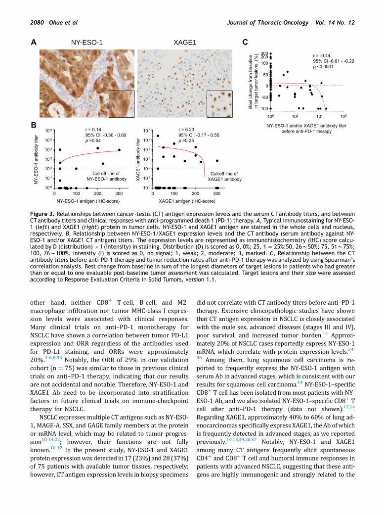
We investigated the relationship between NY-ESO-1/XAGE1 expression levels and CT antibody titers in thediscovery and validation cohorts. Figure 3A showsrepresentative staining of tumor NY-ESO-1 and XAGE1
protein. No apparent correlations between NY-ESO-1/XAGE1 expression levels and CT antibody titers wereobserved (Fig. 3B). Then, we investigated the relation-ship between CT antibody titers before anti–PD-1 ther-apy and tumor reduction rates after anti–PD-1 therapy,resulting in a strong correlation between them (Fig. 3C)(p < 0.0001). This indicates that CT antibody is usefulbiomarker predicting responses to anti–PD-1 therapy.
Relationships Among the CT Antibody, TumorMissense-Mutation Burden (TMB), PD-L1Expression, CD8 T-Cell Infiltration, and ClinicalBenefits With Anti–PD-1 Therapy for NSCLC
We evaluated TMB, tumor PD-L1 expression, CD8þ T-cell, B-cell, and M2-macrophage infiltration, and tumorMHC-class I expression levels in the discovery cohort
0
20
40
60
80
100
Pro
gres
sion
free
surv
ival
(%)
A
CT antibody-positiveCT antibody-negative
0 3 6 9 12 15 18 21 24
HR, 0.17 (95% CI, 0.04-0.66)Log-rank ap =0.001
0
20
40
60
80
100
0 3 6 9 12 15 18 21 24
Discovery cohort (n=13) Validation cohort (n=75)C
Ove
rall
surv
ival
(%)
Time after anti-PD-1 (months)
0
20
40
60
80
100B HR, 0.15 (95% CI, 0.04-0.60)Log-rank p =0.005
0 3 6 9 12 15 18 21 24
No. at risk
CT Ab posCT Ab neg
6 6 5 4 4 4 4 4 27 2 0 0 0 0 0 0 0
CT Ab posCT Ab neg
17 12 10 8 6 4 3 2 158 27 12 7 5 3 2 2 1
No. at risk
CT Ab posCT Ab neg
6 6 5 5 5 5 5 5 27 3 1 0 0 0 0 0 0
CT Ab posCT Ab neg
17 17 12 10 8 7 7 5 458 44 32 20 14 8 5 3 2
0
20
40
60
80
100
0 3 6 9 12 15 18 21 24
HR, 0.21 (95% CI, 0.10-0.44)Log-rank p =0.004
Validation cohort
0 3 6 9 12 15 18 21 240
20
40
60
80
100
0
20
40
60
80
100
Ove
rall
surv
ival
(%)
Pro
gres
sion
free
surv
ival
(%) After 2nd line (n=66)
Time after anti-PD-1 (months)
HR, 0.37 (95% CI, 0.20-0.67)Log-rank p =0.006
HR, 0.22 (95% CI, 0.10-0.45) Log-rank p =0.005
HR, 0.42 (95% CI, 0.24-0.75)Log-rank p =0.011
0 3 6 9 12 15 18 21 24
HR, 0.11 (95% CI, 0.04-0.32)Log-rank p =0.008
0
20
40
60
80
100
0 3 6 9 12 15 18 21 24
After 3rd line (n=30)
HR, 0.30 (95% CI, 0.13-0.67)Log-rank p =0.004
0
20
40
60
80
100
0 3 6 9 12 15 18 21 24
No. at risk
CT Ab posCT Ab neg
15 11 9 7 5 3 3 2 151 23 9 6 4 3 2 2 1
CT Ab posCT Ab neg
8 6 6 6 4 3 3 2 122 10 4 0 0 0 0 0 0
No. at risk
CT Ab posCT Ab neg
15 15 11 9 7 9 6 5 451 39 27 19 13 8 5 3 2
CT Ab posCT Ab neg
8 8 7 7 6 6 6 5 422 17 12 8 5 3 2 2 2
Figure 2. Kaplan-Meier curves for the progression-free survival (PFS) and overall survival (OS) of patients according tocancer-testis (CT) antibody status in the discovery and validation cohorts after anti–programmed death 1 (PD-1) therapy.A, Kaplan-Meier curves for the PFS of patients according to the CT antibody (serum antibody against NY-ESO-1 and/or XAGE1CT antigen) status in the discovery and validation cohorts after anti–PD-1 therapy. PFS was calculated from the date of anti–PD-1 therapy to the date of disease progression or death from any cause. Hazard ratio (HR) for PFS of CT antibody-positivepatients, as compared to that of -negative patients, was calculated with Cox regression analysis, and 95% confidence interval(CI) was shown. B, Kaplan-Meier curves for the OS of patients according to the CT antibody status in the discovery andvalidation cohorts after anti–PD-1 therapy. OS was calculated from the date of anti–PD-1 therapy to the date of death fromany cause. HR for OS of CT antibody-positive patients, as compared to that of negative patients, was calculated with Coxregression analysis, and 95% CI was shown. C, Kaplan-Meier curves for the PFS and OS of patients according to the CTantibodystatus in the validation cohorts after the second line (n ¼ 66) and the third line (n ¼ 30) anti–PD-1 therapy. PFS and OS werecalculated as described above. HR for PFS and OS of CT antibody-positive patients, as compared to those of -negative pa-tients, was calculated with Cox regression analysis, and 95% CI was shown. Ab, antibody; pos, positive; neg, negative.
December 2019 Predictive Biomarkers for Anti-PD-1 Therapy of NSCLC 2077
before anti–PD-1 therapy (Fig. 1B, Fig. 4A), and repre-sentative staining was shown in Supplementary Fig. 3.ORR with anti–PD-1 therapy appeared to be associatedwith a high CT antibody titer (100%), TMB (75%), andPD-L1 expression (83%) (Fig. 4B), but not with CD8þ T-cell, B-cell, and M2-macrophage infiltration or tumorMHC-class I expression levels (Fig. 1B). PFS, OS, and ORRaccording to TMB and the PD-L1 expression status, andthe combination of these two were analyzed, showingbetter PFS and ORR with high TMB and PD-L1 expres-sion than with low TMB and PD-L1 expression(Supplementary Fig. 4).
We investigated whether the combination of the CTantibody and tumor PD-L1 expression or CD8þ T-cellinfiltration has potential as a biomarker for clinical re-sponses with anti–PD-1 therapy in 72 available tumortissues (Supplementary Fig. 5). Regardless of the PD-L1
expression status, CT antibody-positive patients hadhigher ORR and longer PFS and OS than CT antibody-negative patients. Furthermore, regardless of CD8þ T-cell infiltration, the former had higher ORR than thelatter. For example, a patient who had lung adenocar-cinoma with XAGE1 Ab and EML-4/ALK-fusion achievedCR with anti–PD-1 therapy, and the marked intratumorinfiltration of lymphocytes was observed after anti–PD-1therapy (Supplementary Fig. 6). These results indicatethat the CT antibody, even alone, is an independentlyuseful biomarker for predicting clinical benefits of goodresponse and long survival with anti–PD-1 therapy.
DiscussionThe present study showed that NSCLC patients with
NY-ESO-1 and/or XAGE1 Ab obtained the significant
Table 2. Objective Response Rate, Progression-Free Survival, and Overall Survival With Anti–Programmed Death 1 Therapy, According to Patient Characteristics andImmunological Status
Characteristics
No. ofPatientsWith CRor PR/Total
CR or PR Rate(95% CI) %
Odds Ratio forCR or PR(95% CI)
MedianPFS
pValue
UnivariateAnalysis forPFSHR (95% CI)
MultivariateAnalysis forPFSHR (95% CI)
MedianOS
pValue
UnivariateAnalysis forOSHR (95% CI)
MultivariateAnalysis forOSHR (95% CI)
SexMale 16/55 29 (18 to 43) — 2.8 0.58 1.0 (0.6 to 1.9) — NR 0.56 2.1 (0.9 to 4.9) —
Female 6/20 30 (12 to 54) 4.3 18.4Age, yr<65 7/24 29 (13 to 51) — 1.9 0.32 1.3 (0.7 to 2.3) — 12.5 0.25 1.5 (0.8 to 3.0) —
� 65 15/51 29 (17 to 44) 3.6 18.4Smoking statusCurrent or former
smoker17/53 32 (20 to 46) 11 (1.0 to 113) 3.3 0.37 0.7 (0.4 to 1.3) 0.4 (0.2 to 0.8) 18.4 0.72 1.1 (0.6 to 2.4) 0.8 (0.3 to 2.1)
Never-smoker 5/22 23 (8 to 45) 2.8 14.1Tumor typeSquamous
cell carcinoma6/21 29 (11 to 52) — 2.8 0.97 1.3 (0.7 to 2.4) — 8.3 0.13 1.8 (0.8 to 3.7) —
Non-squamous cellcarcinoma
16/54 30 (18 to 44) 3.2 16.0
Disease stageMetastatic/
recurrentor unresectable
18/59 31 (19 to 44) 4.1 (0.5 to 34) 2.8 0.72 1.2 (0.6 to 2.2) 0.8 (0.4 to 1.9) 14.0 0.84 1.1 (0.5 to 2.5) 0.5 (0.2 to 1.5)
Postoperativerecurrence
4/16 25 (7 to 52) 3.3 14.1
Brain metastasisPresent 6/20 30 (12 to 54) 0.3 (0.04 to 1.6) 2.4 0.34 1.3 (0.7 to 2.3) 2.2 (1.0 to 4.6) 5.2 0.09 1.8 (0.9 to 3.7) 5.2 (2.0 to 13)None 16/55 29 (18 to 43) 3.3 14.1
Regimen lineFirst or second line 15/45 33 (20 to 49) — 3.3 0.35 0.8 (0.5 to 1.4) — 14.0 0.91 1.0 (0.5 to 1.9) —
Later line 7/30 23 (10 to 42) 2.7 14.1Driver mutation
status (EGFR)Positive 2/7 29 (4 to 71) 6.3 (0.2 to 198) 4.6 0.50 1.3 (0.6 to 2.9) 0.7 (0.2 to 2.6) NR 0.31 0.5 (0.2 to 1.8) 0.5 (0.1 to 3.1)Negative 18/59 31 (19 to 44) 3.2 14
CD8þ T-cellinfiltration
High 9/28 32 (16 to 52) — 2.3 0.67 1.1 (0.6 to 2.0) — 14.0 0.72 0.9 (0.4 to 1.9) —
Low 10/31 32 (17 to 51) 3.2 14.1
(continued)
2078
Ohue
etal
Journalof
Thora
cicOncology
Vol.
14No.
12
Table2.C
ontinu
ed
Cha
racteristics
No.
ofPa
tien
tsWithCR
orPR
/Total
CRor
PRRa
te(95%
CI)%
Odd
sRa
tiofor
CRor
PR(95%
CI)
Med
ian
PFS
p Value
Univa
riate
Ana
lysisfor
PFS
HR(95%
CI)
Multiva
riate
Ana
lysisfor
PFS
HR(95%
CI)
Med
ian
OS
p Value
Univa
riate
Ana
lysisfor
OS
HR(95%
CI)
Multiva
riate
Ana
lysisfor
OS
HR(95%
CI)
PD-L1ex
pression
score
�213
/32
41(24to
59)
5.0(1.0
to26
)3.2
0.27
0.7(0.4
to1.3)
0.7(0.3
to1.4)
16.0
0.65
0.8(0.4
to1.8)
0.6(0.2
to1.4)
<2
5/27
19(6
to38
)2.8
7.9
CTan
tige
nex
pression
Positive
15/3
050
(31to
69)
—5.3
0.02
0.5(0.3
to0.9)
—20
.40.17
0.6(0.3
to1.3)
—
Neg
ative
5/32
16(5
to33
)2.1
9.2
CTan
tibo
dyPo
sitive
11/1
765
(38to
86)
9.6(1.6
to57
)13
.20.01
0.4(0.2
to0.9)
0.4(0.2
to0.9)
NR
0.00
40.2(0.1
to0.7)
0.2(0.1
to0.8)
Neg
ative
11/5
819
(10to
31)
2.8
10.2
Multiva
riatean
alyses
werepe
rformed
usingLo
gistic
regression
mod
elto
pred
ictco
ntribu
tion
sof
each
variab
lesto
CRor
PRprob
ability.
Univa
riatean
dmultiva
riatean
alyses
werepe
rformed
usingCox
’sprop
ortion
alha
zardsregression
mod
elto
assess
therelation
ship
ofallfactorswithPF
San
dOS.
p<
.05was
considered
tobe
sign
ifica
nt.CR,
completerespon
se;PR
,pa
rtialrespon
se;CI,co
nfide
nceinterval;NR,
notreac
hed;
PFS,
prog
ression-free
survival;OS,
overallsurvival;HR,
hazard
ratio;
PD-L1,
pro-
gram
med
deathlig
and1;
CT,
canc
ertestis
antige
n.
December 2019 Predictive Biomarkers for Anti-PD-1 Therapy of NSCLC 2079
clinical benefits of high ORR and long PFS and OS withanti–PD-1 therapy, as compared with the Ab-negativepatients. Additionally, Ab titers highly correlated withtumor reduction rates in anti–PD-1 therapy. Thus, ourresults suggest that strong immune surveillance waspresent in CT antibody-positive NSCLC patients whoresponded well to anti–PD-1 therapy, reversing immunesuppression or tolerance. Therefore, NY-ESO-1 andXAGE1 Ab are considered to be convenient and usefulbiomarkers predicting clinical benefits of good responseand long survival in anti–PD-1 therapy for NSCLC. Ournovel findings in NSCLC would be extended to biomarkerstudy in immunotherapies of other cancers because NY-ESO-1 is broadly expressed in many types of malignancy.
Here, we for the first time reported a strong cor-relation between NY-ESO-1 and XAGE1 Ab titers andtumor reduction rates with anti–PD-1 therapy forNSCLC. Similarly, Yuan et al.28 reported a good corre-lation between NY-ESO-1 Ab titers and clinical re-sponses and a close association between Ab-positivitywith CD8þ T-cell response and long survival, withanti-CTL antigen 4 (CTLA-4) therapy in melanoma. Inaddition, they addressed a close association betweenNY-ESO-1–specific Ab and CD8þ T cell in patients whowere responders. We also detected NY-ESO-1–specificCD8þ T cell from peripheral blood of patient with CRafter anti–PD-1 therapy (data not shown). Thus, it isspeculated that CT antibody titers reflect cytotoxicactivity levels of CT antigen-specific CD8þ T cell.
In this study, many patients had been heavily treatedwith conventional chemotherapy; however, it is unlikelythat CT antibody is associated with sensitivity tochemotherapy. Rather, CT antibody is considered to be aspecific response biomarker for anti–PD-1 and –CTLA-4therapy. There are few clinical reports focusing on CTantibody itself or the relationship between the antibodyand cancer therapy, although Yuan et al.28 showed goodcorrelations between NY-ESO-1 Ab and clinical benefitswith anti–CTLA-4 therapy in melanoma as describedabove. These results indicate the presence of activatedCT antigen-specific CD8þ T cell and checkpointmolecule–mediated strong immunosuppression in CTAb-positive patients, reflecting good responses tocheckpoint inhibitors even in patients heavily treatedwith chemotherapy. In the near future, these issueswould be investigated in detail.
NY-ESO-1 and XAGE1 Ab were strongly associatedwith high response rates in anti–PD-1 therapy forNSCLC; however, the well-known biomarkers of tumorPD-L1 expression and TMB were also associated withbetter responses in the present study, as reportedpreviously.4-8 However, no relationship was noted be-tween these Ab and the two biomarkers, indicating thatthese Ab are independent biomarkers in NSCLC. On the
Figure 3. Relationships between cancer-testis (CT) antigen expression levels and the serum CTantibody titers, and betweenCTantibody titers and clinical responses with anti–programmed death 1 (PD-1) therapy. A, Typical immunostaining for NY-ESO-1 (left) and XAGE1 (right) protein in tumor cells. NY-ESO-1 and XAGE1 antigen are stained in the whole cells and nucleus,respectively. B, Relationship between NY-ESO-1/XAGE1 expression levels and the CT antibody (serum antibody against NY-ESO-1 and/or XAGE1 CT antigen) titers. The expression levels are represented as immunohistochemistry (IHC) score calcu-lated by D (distribution) � I (intensity) in staining. Distribution (D) is scored as 0, 0%; 25, 1 ¼ 25%:50, 26w50%; 75, 51w75%;100, 76w100%. Intensity (I) is scored as 0, no signal; 1, weak; 2, moderate; 3, marked. C, Relationship between the CTantibody titers before anti–PD-1 therapy and tumor reduction rates after anti–PD-1 therapy was analyzed by using Spearman’scorrelation analysis. Best change from baseline in sum of the longest diameters of target lesions in patients who had greaterthan or equal to one evaluable post-baseline tumor assessment was calculated. Target lesions and their size were assessedaccording to Response Evaluation Criteria in Solid Tumors, version 1.1.
2080 Ohue et al Journal of Thoracic Oncology Vol. 14 No. 12
other hand, neither CD8þ T-cell, B-cell, and M2-macrophage infiltration nor tumor MHC-class I expres-sion levels were associated with clinical responses.Many clinical trials on anti–PD-1 monotherapy forNSCLC have shown a correlation between tumor PD-L1expression and ORR regardless of the antibodies usedfor PD-L1 staining, and ORRs were approximately20%.4-6,8,33 Notably, the ORR of 29% in our validationcohort (n ¼ 75) was similar to those in previous clinicaltrials on anti–PD-1 therapy, indicating that our resultsare not accidental and notable. Therefore, NY-ESO-1 andXAGE1 Ab need to be incorporated into stratificationfactors in future clinical trials on immune-checkpointtherapy for NSCLC.
NSCLC expresses multiple CT antigens such as NY-ESO-1, MAGE-A, SSX, and GAGE family members at the proteinor mRNA level, which may be related to tumor progres-sion10-14,22; however, their functions are not fullyknown.10-12 In the present study, NY-ESO-1 and XAGE1protein expressionwas detected in17 (23%) and28 (37%)of 75 patients with available tumor tissues, respectively;however, CT antigen expression levels in biopsy specimens
did not correlate with CT antibody titers before anti–PD-1therapy. Extensive clinicopathologic studies have shownthat CT antigen expression in NSCLC is closely associatedwith the male sex, advanced diseases (stages III and IV),poor survival, and increased tumor burden.14 Approxi-mately 20% of NSCLC cases reportedly express NY-ESO-1mRNA, which correlate with protein expression levels.34-36 Among them, lung squamous cell carcinoma is re-ported to frequently express the NY-ESO-1 antigen withserum Ab in advanced stages, which is consistent with ourresults for squamous cell carcinoma.14 NY-ESO-1–specificCD8þ T cell has been isolated from most patients with NY-ESO-1 Ab, and we also isolated NY-ESO-1–specific CD8þ Tcell after anti–PD-1 therapy (data not shown).12,14
Regarding XAGE1, approximately 40% to 60% of lung ad-enocarcinomas specifically express XAGE1, the Ab ofwhichis frequently detected in advanced stages, as we reportedpreviously.14,15,19,20,37 Notably, NY-ESO-1 and XAGE1among many CT antigens frequently elicit spontaneousCD4þ and CD8þ T cell and humoral immune responses inpatients with advanced NSCLC, suggesting that these anti-gens are highly immunogenic and strongly related to the
LC04
1LC
068
LC07
5LC
114
LC07
2LC
056
LC09
2LC
065
LC08
3LC
113
LC13
7LC
168
LC05
2
0
200
400
600
0
10
20
30
Mis
sens
e-m
utat
ion
CR/PR
CT
antib
ody
titer
A
Mutation
High Low
PD-L1
CD8+
75 29
83 14
CT antibodyB
100 00
20406080
100
020406080
100
020406080
100
020406080
100
0
1
2
3
ND
ND
SD/PD
PD
-L1
scor
e%
CD
8+
Imm
unoh
isto
chem
istry
Clinical response
43 50
Obj
ectiv
ere
spon
sera
te (%
)
104
102
100
106
Figure 4. Relationship between four predictive biomarkersand clinical responses with anti–programmed death 1 (PD-1)therapy. A, Relationships among the CT antibody (serumantibody against NY-ESO-1 and/or XAGE1 CT antigen), tumormissense-mutation burden, programmed death ligand 1 (PD-L1) expression, CD8þ T-cell infiltration, and clinical re-sponses with anti–PD-1 therapy in the discovery cohort (n ¼13). Clinical responses were assessed according to ResponseEvaluation Criteria in Solid Tumors, version 1.1 guidelines.B, Objective response rates were shown according to highand low levels of the above four factors. High levels of eachfactor were defined for titer greater than or equal to 100 forthe CT antibody, greater than or equal to 178 (median value)nonsynonymous single nucleotide variants for mutations,greater than or equal to score 2 (mean value) for PD-L1expression, and greater than or equal to 10% (mean value)for %CD8þ T cell, and a low level was less than the highlevel values. CT antigen-specific CD4þ and CD8þ T cellswere frequently detected in patients with CT antibodytiter greater than or equal to 100. CR, completeresponse; PR, partial response; SD, stable disease; PD, pro-gressive disease.
December 2019 Predictive Biomarkers for Anti-PD-1 Therapy of NSCLC 2081
immune surveillance of NSCLC.14,15,23,37 Thus, NY-ESO-1and XAGE1 immunity in NSCLC are promising targets forimmune-checkpoint therapy reversing immunesuppression.
Our NSCLC patients with NY-ESO-1 and XAGE1 Abresponded particularly well to anti–PD-1 therapy. One
explanation for this result is the high immunogenicity ofthe NY-ESO-1 and XAGE1 antigens in NSCLC, as describedabove. Another reason is that some NSCLC patients’ T cellssimultaneously recognize multiple tumor antigensincluding CT antigens, as previously reported, and thatanti–PD-1 and –CTLA-4 therapy elicited neoantigen-specific CD8þ T-cell responses in NSCLC and melanoma,respectively.13,14,38-40 Consequently, many antigens areconsidered to elicit antigen-specific CD8þ T-cell responsesdestroying tumor cells. However, all tumor antigens do notalways elicit CD8þ T-cell responses; a previous study re-ported that mutant p53 peptides elicited CD4þ T cell andhumoral, but not CD8þ T-cell responses.41 The immuno-genicity of each tumor antigen expressed inNSCLCmust beinvestigated in more detail for future immunotherapies.
Although this study only included 88 NSCLC patients,two of seven patients with EGFRmt and XAGE1 Ab ach-ieved CR and PR with anti–PD-1 therapy, and only onelung adenocarcinoma with EML4-ALK fusion and XAGEAb had CR (Supplementary Fig. 5). These results indicatethat XAGE1 Ab-positive NSCLC with EGFRmt or EML4-ALK-fusion responds to anti–PD-1 therapy. Several clin-ical trials on anti–PD-1 therapy have shown that NSCLCwith EGFRmt usually does not have the clinical benefitsof a better response and survival with anti–PD-1 ther-apy.7,42 This may be because these NSCLC patients werenon- or light smokers with low TMB.39,43 Accordingly,EGFRmt may be a poor-response biomarker in anti–PD-1therapy for NSCLC, whereas NY-ESO-1 and XAGE1 Abmay be good-response markers because NY-ESO-1 andXAGE1 are highly immunogenic in NSCLC, as wedescribed above. NY-ESO-1 and XAGE1, as well as drivermutations of EGFRmt and EML4-ALK fusion, are tumorprogression factors; however, CT antigens are highlyimmunogenic, whereas driver mutations may not bein NSCLC patients. Thus, NSCLC with EGFRmt maybe divided into two types: CT antibody-positive and-negative, and CT antibody-positive NSCLC with EGFRmtmay obtain clinical benefits with anti–PD-1 therapy, asshown here. This issue must be clarified in clinical trialson NSCLC with EGFRmt as soon as possible.
The limitations of the present study were the smallnumber of patients included, the short follow-up timeafter anti–PD-1 therapy, and many analyses of small bi-opsy specimens. Larger clinical studies are needed toconfirm the usefulness of the CT antibody as a predictivebiomarker of the clinical benefits of good response andlong survival associated with anti–PD-1 therapy. Now weare planning a large-scale study of patients with NSCLC.
In conclusion, patients who have NSCLC withNY-ESO-1 and XAGE1 Abs obtained significant clinicalbenefits of good response and long survival with anti–PD-1 therapy, and Ab titers correlated well with tumorreduction rates after anti–PD-1 therapy. NY-ESO-1 and
2082 Ohue et al Journal of Thoracic Oncology Vol. 14 No. 12
XAGE1 Ab are predictive biomarkers of clinical benefitswith anti–PD-1 therapy for NSCLC, and must be incor-porated into future clinical trials on immune checkpointinhibitors as stratification factors.
AcknowledgmentsThis work was supported by grants from JSPS KAKENHI(15K09235, 16K18463, 16K21533, 18K15226, and18K07306) to Drs. Oka, Ohue, and Kurose; by grantsfrom the Takeda Science Foundation to Drs. Ohue andKurose; by a grant from the Medical Research Encour-agement Prize of The Japan Medical Association to Dr.Ohue; by a grant from the Okayama Health Foundationto Dr. Ohue; and by grants from Kawasaki MedicalSchool to Drs. Oka, Ohue, and Kurose. Dr. Oka receivedcollaborative research funds from the University ofTokyo and Thyas Co., Ltd. The Department of Immuno-Oncology is endowed by Pole Star Co., Ltd.
The authors thank Professor Eiichi Nakayama at Ka-wasaki Medical School for his distinguished teaching andhis long-term commitment to cancer immunologyresearch as our leader. Prof. Nakayama died suddenly onJuly 20, 2017. We are all very pleased to have completedthis work for Prof. Nakayama.
Supplementary DataNote: To access the supplementary material accompa-nying this article, visit the online version of the Journal ofThoracic Oncology at www.jto.org and at https://doi.org/10.1016/j.jtho.2019.08.008.
References1. World Health Organization (WHO). Cancer, Fact sheet,
February 2017. http://www.who.int/mediacentre/factsheets/fs297/en/. Accessed January 10, 2017.
2. Reck M, Rabe KF. Precision diagnosis and treatment foradvanced non–small-cell lung cancer. N Engl J Med.2017;377:849–861.
3. Okazaki T, Chikuma S, Iwai Y, et al. A rheostat for im-mune responses: the unique properties of PD-1 and theiradvantages for clinical application. Nat Immunol.2013;14:1212–1218.
4. Balar AV, Weber JS. PD-1 and PD-L1 antibodies in cancer:current status and future directions. Cancer ImmunolImmunother. 2017;66:551–564.
5. Nishino M, Ramaiya NH, Hatabu H, et al. Monitoringimmune-checkpoint blockade: response evaluation andbiomarker development. Nat Rev Clin Oncol.2017;14:655–668.
6. Lopes G, Wu Y-L, Kudaba I, et al. Pembrolizumab(pembro) versus platinum-based chemotherapy(chemo) as first-line therapy for advanced/metastaticNSCLC with a PD-L1 tumor proportion score (TPS)� 1%: open-label, phase 3 KEYNOTE-042 study. J ClinOncol; 2018. https://ascopubs.org/doi/10.1200/JCO.2018.36.18_suppl.LBA4?expanded¼undefined. AccessedJanuary 10, 2018.
7. Topalian SL, Taube JM, Anders RA, et al. Mechanism-driven biomarkers to guide immune checkpoint blockadein cancer therapy. Nat Rev Cancer. 2016;16:275–287.
8. Tray N, Weber JS, Adams S. Predictive biomarkers forcheckpoint immunotherapy: current status and chal-lenges for clinical application. Cancer Immunol Res.2018;6:1122–1128.
9. Kamphorst AO, Pillai RN, Yang S, et al. Proliferation ofPD-1þ CD8 T cells in peripheral blood after PD-1-targeted therapy in lung cancer patients. Proc NatlAcad Sci U S A. 2017;114:4993–4998.
10. Simpson AJ, Caballero OL, Jungbluth A, et al. Cancer/testis antigens, gametogenesis and cancer. Nat RevCancer. 2005;5:615–625.
11. Whitehurst AW. Cause and consequence of cancer/testisantigen activation in cancer. Annu Rev Pharmacol Tox-icol. 2014;54:251–272.
12. Caballero OL, Chen YT. Cancer/testis (CT) antigens:potential targets for immunotherapy. Cancer Sci.2009;100:2014–2021.
13. Hofmann O, Caballero OL, Stevenson BJ, et al. Genome-wide analysis of cancer/testis gene expression. Proc NatlAcad Sci U S A. 2008;105:20422–20427.
14. Chiriva-Internati M, Pandey A, Saba R, et al. Cancertestis antigens: a novel target in lung cancer. Int RevImmunol. 2012;31:321–343.
15. Ohue Y, Eikawa S, Okazaki N, et al. Spontaneous anti-body, and CD4 and CD8 T-cell responses against XAGE-1b(GAGED2a) in non–small cell lung cancer patients. Int JCancer. 2012;131:E649–E658.
16. Rapoport AP, Stadtmauer EA, Binder-Scholl GK, et al. NY-ESO-1-specific TCR-engineered T cells mediate sustainedantigen-specific antitumor effects in myeloma. Nat Med.2015;21:914–921.
17. Takeoka T, Nagase H, Kurose K, et al. NY-ESO-1 proteincancer vaccine with poly-ICLC and OK-432: rapid andstrong induction of NY-ESO-1-specific immune responsesby poly-ICLC. J Immunother. 2017;40:140–147.
18. Cheever MA, Allison JP, Ferris AS, et al. The prioritiza-tion of cancer antigens: a national cancer institute pilotproject for the acceleration of translational research.Clin Cancer Res. 2009;15:5323–5337.
19. Nakagawa K, Noguchi Y, Uenaka A, et al. XAGE-1 expres-sion in non–small cell lung cancer and antibody response inpatients. Clin Cancer Res. 2005;11:5496–5503.
20. Ohue Y, Kurose K, Mizote Y, et al. Prolongation of overallsurvival in advanced lung adenocarcinoma patients withthe XAGE1 (GAGED2a) antibody. Clin Cancer Res.2014;20:5052–5063.
21. McShane LM, Altman DG, Sauerbrei W, et al. Reportingrecommendations for tumor marker prognostic studies(REMARK). J Natl Cancer Inst. 2005;97:1180–1184.
22. Ohue Y, Kurose K, Nozawa R, et al. Survival of lungadenocarcinoma patients predicted from expression ofPD-L1, galectin-9, and XAGE1 (GAGED2a) on tumor cellsand tumor-infiltrating T cells. Cancer Immunol Res.2016;4:1049–1060.
23. Saito K, Nakayama E, Valmori D. Immune responses tothe cancer testis antigen XAGE-1b in non small cell lungcancer Caucasian patients. PLoS One. 2016;11:e0150623.
December 2019 Predictive Biomarkers for Anti-PD-1 Therapy of NSCLC 2083
24. Isobe M, Eikawa S, Uenaka A, et al. Correlation of highand decreased NY-ESO-1 immunity to spontaneousregression and subsequent recurrence in a lung cancerpatient. Cancer Immun. 2009;9:8.
25. G*Power: Statistical Power Analyses. http://www.gpower.hhu.de/. Accessed January 20, 2019.
27. Eikawa S, Kakimi K, Isobe M, et al. Induction of CD8 T-cell responses restricted to multiple HLA class I alleles ina cancer patient by immunization with a 20-mer NY-ESO-1f (NY-ESO-1 91-110) peptide. Int J Cancer.2013;132:345–354.
28. Yuan J, Adamow M, Ginsberg BA, et al. Integrated NY-ESO-1 antibody and CD8þ T-cell responses correlatewith clinical benefit in advanced melanoma patientstreated with ipilimumab. Proc Natl Acad Sci U S A.2011;108:16723–16728.
29. Karasaki T, Nagayama K, Kuwano H, et al. An immuno-gram for the cancer-immunity cycle: towards personal-ized immunotherapy of lung cancer. J Thorac Oncol.2017;12:791–803.
30. Barbie DA, Tamayo P, Boehm JS, et al. Systematic RNAinterference reveals that oncogenic KRAS-driven cancersrequire TBK1. Nature. 2009;462:108–112.
31. Godec J, Tan Y, Liberzon A, et al. Compendium of im-mune signatures identifies conserved and species-specific biology in response to inflammation. Immunity.2016;44:194–206.
32. Charoentong P, Finotello F, Angelova M, et al. Pan-cancerimmunogenomic analyses reveal genotype-immunophe-notype relationships and predictors of response tocheckpoint blockade. Cell Rep. 2017;18:248–262.
33. Hanna N, Johnson D, Temin S, et al. Systemic therapy forstage IV non–small-cell lung cancer: American Society of
Clinical Oncology clinical practice guideline update.J Clin Oncol. 2017;35:3484–3515.
34. Gure AO, Chua R, Williamson B, et al. Cancer-testisgenes are coordinately expressed and are markers ofpoor outcome in non–small cell lung cancer. Clin CancerRes. 2005;11:8055–8062.
35. Cho HJ, Caballero OL, Gnjatic S, et al. Physical inter-action of two cancer-testis antigens, MAGE-C1 (CT7) andNY-ESO-1 (CT6). Cancer Immun. 2006;6:12.
36. Chueh AC, Liew MS, Russell PA, et al. Promoter hypo-methylation of NY-ESO-1, association with clinicopatho-logical features and PD-L1 expression in non–small celllung cancer. Oncotarget. 2017;8:74036–74048.
37. Talebian Yazdi M, Loof NM, Franken KL, et al. Local andsystemic XAGE-1b-specific immunity in patients withlung adenocarcinoma. Cancer Immunol Immunother.2015;64:1109–1121.
38. Germain C, Gnjatic S, Tamzalit F, et al. Presence of Bcells in tertiary lymphoid structures is associated with aprotective immunity in patients with lung cancer. Am JRespir Crit Care Med. 2014;189:832–844.
39. Rizvi NA, Hellmann MD, Snyder A, et al. Cancer immu-nology. Mutational landscape determines sensitivity toPD-1 blockade in non–small cell lung cancer. Science.2015;348:124–128.
40. van Rooij N, van Buuren MM, Philips D, et al. Tumorexome analysis reveals neoantigen-specific T-cell reac-tivity in an ipilimumab-responsive melanoma. J ClinOncol. 2013;31:e439–e442.
41. Tsuji T, Gnjatic S. Split T-cell tolerance as a guide for thedevelopment of tumor antigen-specific immunotherapy.Oncoimmunology. 2012;1:405–407.
42. Lee CK, Man J, Lord S, et al. Checkpoint inhibitors inmetastatic EGFR-mutated non–small cell lung cancer-ameta-analysis. J Thorac Oncol. 2017;12:403–407.
43. Vogelstein B, Papadopoulos N, Velculescu VE, et al. Can-cer genome landscapes. Science. 2013;339:1546–1558.