+ All Categories
Home > Documents > seRUM GLUTAMATE nEHYDROGENASE DETERMINAT fON INdigitool.library.mcgill.ca/thesisfile63391.pdf ·...

seRUM GLUTAMATE nEHYDROGENASE DETERMINAT fON INdigitool.library.mcgill.ca/thesisfile63391.pdf ·...

Date post: 31-Mar-2018
Category:
Upload: nguyenkiet
View: 218 times
Download: 1 times
Share this document with a friend
94
'" 1 seRUM GLUTAMATE nEHYDROGENASE DETERMINAT_fON IN CHRONIC ALCOHOLICS by Richard Jan pivon " A th&sis submi tted ta the Facu1ty of Graduate Studies lt a né!." Research in partial fu1fillment of the requirernents for the degree of Master of Science Departrnent of Experimental Surgery McGi1l university Montreal, Canada August 1985. ® pivon 1985 . .,
Transcript

'"

1

seRUM GLUTAMATE nEHYDROGENASE DETERMINAT_fON

IN

CHRONIC ALCOHOLICS

by

Richard Jan pivon

"

A th&sis submi tted ta the Facu1ty of Graduate Studies lt

a né!." Research in partial fu1fillment of the

requirernents for the degree of

Master of Science

Departrnent of Experimental Surgery

McGi1l university

Montreal, Canada August 1985. ® pivon 1985 .

.,

CHAPTER l

CHA PT ER II

CHAP'PER III

CHAPTER IV

- l -

TABLE OF CONTENTS - ./,

PREFACE

INTRODUCTION

HEPATOTOXICITY

SERUM GLUTAMATE DEHYDROGENASE AS REFLECTING LlVER FUNCTION

METHODOLOGY

1

A - Methodology· pt! Serum GDH Determination 0 L

B Studies in Chronic Alcoholics l - Studies in :Norma l Sub jects 2 - Patient Population of Chronic Alcoholics

..

a) Studies a~ the Montreal General Hospital b) Studies on Patients admitted to Half-Way

Center 3 - Dipstick Methodology of Serum Alcohol

Determina tIon

CHAPTER V RESULTS ~

A - Serum GDH and Other Li ver Enz'yme Determinations in Control Subjects

B - ~erum GDH and Other Li ver Enzyme Determinations in Chronic Alcoholics

1 ...: Pilot Studies 2 - Studies of serum enzyme levels in Chronic

Alcoholics admi tted to l:he Half~Way Center a) Relationship between the different rang~s

of serum a>lcohol level and the serum GDa concentration ranges

b) Relationship between the different ranges of serum alcohol levels and the GGTP, Alk. Phos., SGOT and SGPT levels on admission

t)

CHAPTER VI DISCUSSION

A - Methodology of GDH Determina tion B -" Assays of GDH and other serum enzymes in

Chronic Alcoholics C - Perspectives of GDH assay in liver diseases

CHAPTER VII CONCLUSIONS

REFERENCES

PAGE 1

6

10

17

33

56

69

81

83

}

FIGURES

Fig.l

Fig.2

Fig.3

Fig.4

Fig.5

Fig.6

Fig.7

Fig.8

Fig.9

TABLES

Table l

- II -

LIST'· OF tLLUSTRATIONS

structural changes in serum GDH during transamination

The reversible reaction catalyzed by GDH

Glutamate Dehydrogenase Reactions with Different Transaminases

niagram illustrating the two ports of entry into the mitochondrial respiratory chain

The Dependence of the speed of GDH reaction on co-enzyme~ substrate and EDTA concentration, as weIl as pH level

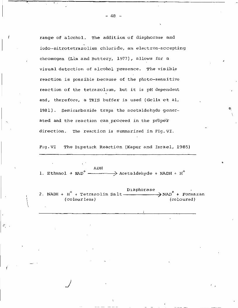
The Dipstick Reaction

Blood Alcohol Level vs Serum GDH

Blood Alcohol Level vs ç;GTP

Comparison' of GDH and GGTP

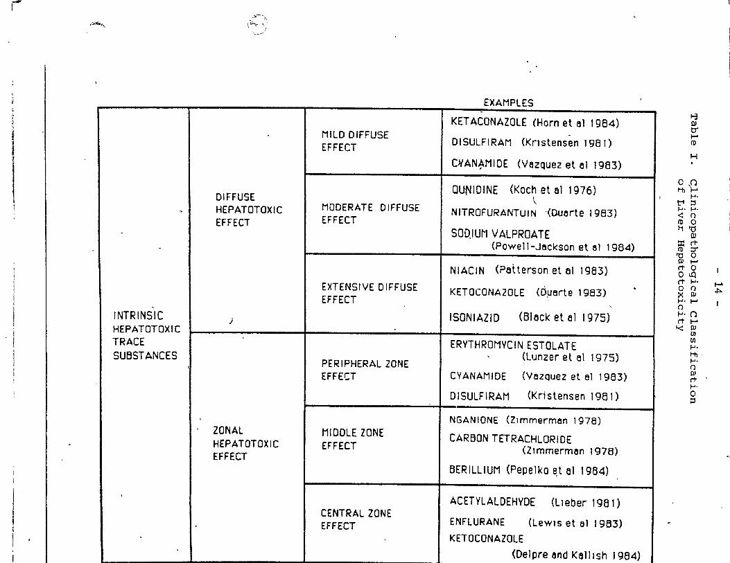
'Clinicopa thological Classification of Liver Hepatotoxicity

Table II GDH content in h\,lman tissue

Table III GDH content of tumour tissue in b hurnan sub jects

Table IV verification' of Dipstick Methodology using a PMO 2-Zeiss chromatogram Spectrophotorneter

PAGE

p.24

p.25

p·.28

p.31

p.34

p.48

p.62

p.64

p.65

.

p.14

p.20

B~21

p.51

Table V

Table VI

Table VII

- III -

\

Classification of Alcohol Dipst~ck Results - Scale for Low Serum Ethanol Content

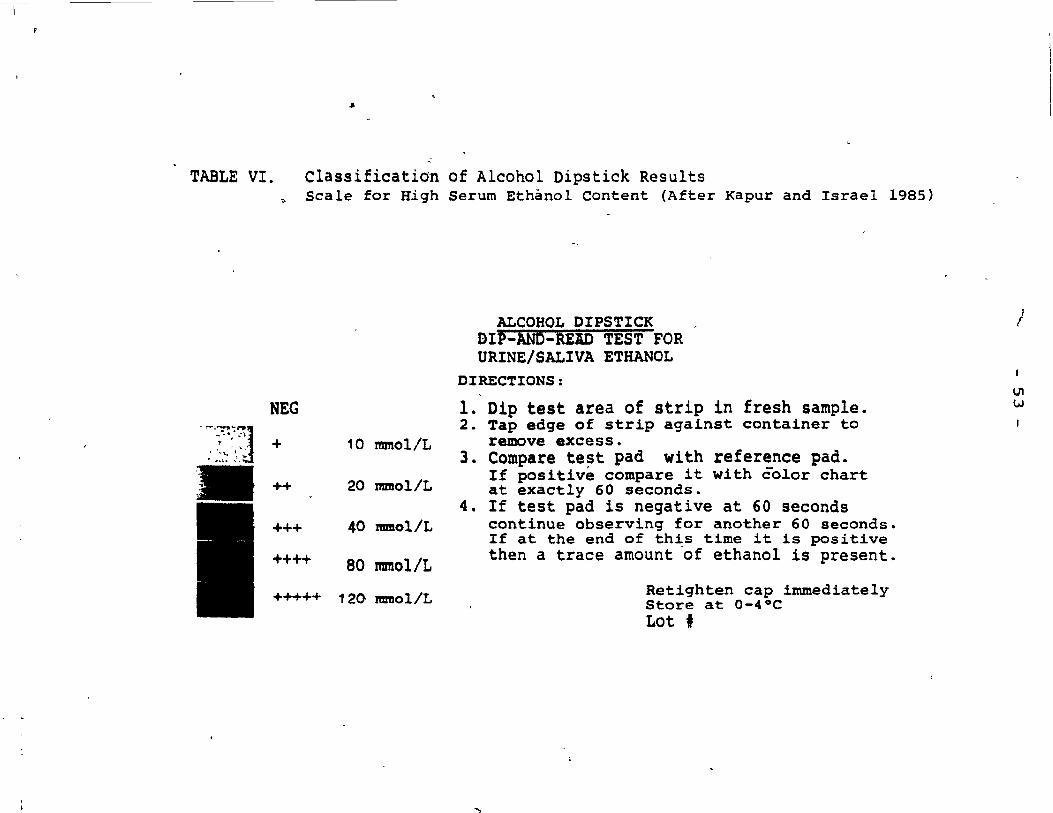
Classification of Alcohol Dipstick R~sults - S~ale for High Serum Ethanol Content

Compar~son of urine ethanol determin­ation by Alcohol Dipstick Methodology with'U.V. methodology

Table VIII Comparative study of alcohol levels in serum using Dipstick Methodology and Gas Chromatography

Table IX

Table X

• Table XI

Table XII

1

Normal Values of serum enzyme levels as reflecting hepatic function

Serum ~DH determinations inf12 Chronic Alcoholics ~

Serum GGTP and Alkaline Phosphatase in 12 Chronic Alcoholics

Relationship between serum GDH level and serum Alcohol level on admission (60 cases)

p.52

p.53

p.54

p.55

p.56

p.S8

p.59

p.63

Table XIII Nurnber of cases with NorJ;llal (N=) and p.68 significantly elevated (H=) enzymes in the four groups of serum ethanol conc'entrations on admission (60 cases)

Table XIV Serum GOH and other liver enzyme .. values in a patient with obst~uctive

jaundice l fOliowing choledocho-.' jejunostorny

p.79

o

, - 'IV

Résumé

DETERMINATION DE LA DESHYDROGENASE DU GLUTAMATE (GOlO SERIQUE CHEZ LES .,ALCOOLIQUES CHRONIQUES

Les facteurs sériques GDB, GGTP, Alk. Phos .. SGOT et SGPT furent ,

étudiés chez 72 patients souffrant d'alcoolisme. chronique. pAu cours de

projets pilotes impliquant 12 patients, une modification fut apportée à la

méthode de détermination de la GDH et les au tres enzymes furent analysés. Cette

méthode modifiée de détermination du facteur GDH (Koch et Pivon) s'est avérée

plus précise et plus simple que la méthode utiÙsée jusqu'alors. Chez

60 alcooliques chroniques admis dans un centre de réhabilitation, les niveaux

d'enzymes furent: déterminés aux 1er et 21 ième jours de leur s,éjour et mis 'en

corrélation avec les diffé~ents niveaux d'éthanol sérique. Ceci fut f!tabli

d'après le procédé de jaugeage (Kapur et Israel - 1985). Lors de crises

intenses d'alcoolisme, la.hausse du taux de la GDH sérique était directement

rekiée au niveau d'éthanoi dans le sang (Sensibilité:. 81.25%; Sp~cificité: 90.91%;

Valeur de prédiction positive: 76.47%; Valeur de prédiction négative: 93.02%).

Au 2lième jour, tous les taux de la GDH se situaient dans les limites normales. /

Même au-delà de 48 heures après le retour à la normale des taux de la GDH, le

facteur GGTP s~ révéla un bon indicateur de la condition hépat:i.~ue. Cette

méthode modifiée .... de détermination de la GDH sérique chez les alcooliques

chroniques semble apporter un nouveau paramètre pour - évaluer l'état de ceux

qui, lors d'épisodes aigus d r alcoolisme t présentent des lésions aux

mitochondries des cellules du foie et possiblement pour dépister d'autres

maladies hépatiques.

"

. . IV ..:.

Abstract ,

SERUM GLUTAMATE DEIIYDROGENASE DETERMINATION IN CHRONIC ALCOHOLICS

Serum GDH, GGTP, Alk. Phos., SGOT and SGPT were s tudied

alcohol~c patients. In pl lot studies on 12 in 72 chr~c

patients, the modification of GDII determination was devel-

oped, and the other enzynles tested. The modiflcation of ~

GDH determinatlon (Koch and Pivon) was found to be more

accurate and simple than previous methodology. In 60

chronic aicoholics admi tted te a half-way center 1 the , )

enzyme levels were determined at days land 21 and correla'ted

to serum ethanoi levels, determined by dipstick methodology

(Kapur and Israel 1985). During acute alcoholic episodes 1

serum GDH elevation was directly roelated to ethanol b100d

levei (Sensitivity: 81.25%; Specificity: 90.91%; positive

Predictive Value: 76.47%: Negative Predictive value: 93~02%).

At day 21, aIl GDH valueti. were within normal range .. GGTP

was found to be a godd indicator of li ver status beyond •

48 hours 1 when GDH values retu'rn to normal. Serum GDH

o

determination in chronic alcoholics appears to add a new

parameter te the assessment of chronic alcoho1ics, r-eflecting

mitochondrial injur.y in the acute episode and possibly in

other liver diseases.

_ t

-- -----,

l.>

t

- l -

PREFACE

Since ~y graduation in medicine, the subject of

liver function tas fasclnated me. I remember several

professors stressing how Ilttle is known about the

"metabollc factory" of the body. l have a 150 wondered

how We can assess liver status prior ta surgery, without

performlng laparatomy or biopsy. It is important for

the surgeon ta know as much as possible about the patlent

before placing him on the operating table, 50 that

decisions, taken d~ring the operation,;tave a more valid·

biochemical background.

Whil'e working at the Biology Department of McGill

University, my "research lnstincts" were encauraged by

.". "-

the Ch~ef of the Department, Dr. G. Maçlachlan, who used

to say: "rf you don't know, you have ta "find out for

yourself and the best way ta do it is ta carry out a

controlled experlment" This saying stuck in my mind,

50 that after graduation, r decided to 'pursue a research

career. After working one year as a volunteer in the

Divlsion of Clinical Investigatlon of the Department of

p

Surgery, Dr. Skoryna, Dlrector of the Unit and the Gastro-

intestinal Research Laboratory, suggested to me that I

" - 2 -

take formai training at McGill. When l asked him

whether there 'were any pro jects dealing wi th the liver 1

he said that this i~ one of t;pe current concerns of

many surgeons, particular ly wi th reference to the

assessment of liver function. The question then came

up as to what subJects should be s'tudied. l suggested

that there was a need for a group of patients wi th . ".

demonstrable liver in jury who could be ·studied for a

prolonged period of time. At this point, l had also

met Dr. Paul' Koch of the Montreal General Hospital, . . where l previously worked' as a' student. wi th his help,

l was able tq" design the pro'ject, after contacting ,

Dr. Jose Negrete, Chief of the Alcohol Research Unf.t of

\

the Hospital, whl)se patients were studied initially.

Further studies were carried out on patients from Maison t. Il

Jean Lapointe, whose pro gram director, Mr. Jacques Perras"

was most cooperative, not only in obtaining the samp-les

but also in analyzing psychometrie data on these patients.

In addition to, these individuals who guided my

studies, l must thank à number of persons who he~ped me

to complete the pro ject. l have enjoyed attending the v'

research seminars ,and surgical rounds at St. Mary's

'1

- 3 -

Hospital and the Montréal "General Hospital. l' became

, , acquainted with the wide scope and divers~-cy of '/

research projects in the Department of Surgery, which

added signi~icantly to my pool of general knowledge.

l wish to thank particular ly Dr. D.B. Tonks, Chief of

the Dept. of Làboratories, and Dr.DDavid ~. Mulder,

Chief of the Dept. of Surgery of the Montreal General

~

Hospital, for their encouragement and coooperation in . establishing the techniques of serum enzyme determin-

ation at the Special Clinical Testing ~aboratory of the

Hospital. Miss Pauline Reed, Chief Technician, instructed'

the author in methodology and was extremely helpful in

the initial stages of the investigation. Mrs. Marta/

Fuska, Chief Technician of the Clinical'Investigation

Service of st. Mary's Hospital, continuously helped in " .

furthér enzyme analysis. Miss Huguette Bacon and her i

associates at La Maison Jean Lapointe processed serum 1

. /

samples from chronic alcoholics at required intervals

in order to fulfil the resear~ protocol requJ-rements.

Dr. Jose Rodriguez, surgeon-in-Chief, ëtnd Dr. John

Keyserlingk, in charge of Surgical Teaching, at st.

Mary's Hospital, were most cooperëjltive in the course

)

l' i

, "

... 4

( -u

of these studies. Dr. Julius Gordon, Director of

Surgical Resj!arch of the Dept. of Surgery, and Miss

Brenda Bewick, Administrative Asslstant, were very

cooperative ln completing aIl the necessary document-

ation for, registration and submission of the thesis.

l should also mention my colleagues 1 Dr. Gilles

Hedder.l..ch at St. Mary 1 s, and Dr. George Chuang at the

Montreal General Hospital, who were very helpful in

the course of my studies. Mr. Jan T.Z. Nolan, Computer

System. Integrator, helped the author tremendously in , ~

the statistical and mathematical analysis of the

computerized data. Mrs. Jean Cornellier, Secretary of

the Gastrointestinal Researeh Laboratory, is thanked

sineerely for t"yping the manuscrlpt. Mr. Carmen Cristo-

faro, Chief of the Audiovis ual DJpartment, and his

associates provid~d a Il the photographie material.

Finally," l must thank my wife, Joanna, for her patience

when l was virtually absent from home in the course of b •

my studies, and my mother, Alexandra,' for her support.

Withput aIl this help, the work could llot be done.

Albert Einstein once said thc;:tt curiosity has i ts

own reason for existence: Il One 'cannat he lp but be in

- 5

awe when he contempla tes the mystery of the marvelous

structure of life and reali ty" . Perhaps "life" should

read "liver". l sincerely hope that, by establishing

a new p&edure for serum GDH determinations in chronic

alcoholics, this work will contribute in a P?sitive way

ta the development of research of liver diseases.

, August 17 1 1985.

Richard J. Pivon, M.D. Research Fellow ,( Divisidn ai Surgical Research Department of Surgery McGill University.

----

\

- 6 -

'INTRODUCTION

In the search of knowledge for tests reflecting

liver function, the serum Glutamate Dehydrogenase

activity was neglected for a relatively long period

of time. The first test used clinically was probably

urine bilirubin deterrnination, based on the observation

of Paul Ehrlich in 1883 (Henry et al 1974l, who had

noticed that uriQe of patients suffering from liver

disease contained biiirubin. Alkaline phosphatase

activity in blood was demonstrated by Kay in 1930. The

heterogenous nature of alkaline phosphatase that can \ Q

be derived not only from the cells lining the biliary

tr~ct of the liver but also from bone osteoblasts, the

mucosal cells o~ the small intestine and the placenta,

renders the valùe of this assay irnprecise, unless other

enzymes are determined sirnultaneously. The determination

of serum Glutarnic O~aloacetic transaminase (SGOT), ~

Glutamic pyruvic transaminase (SGPT) and" Glutarnyl trans-

peptidase was developed around the early fifties. The

transaminases are used as indicators of diffuse hepato-

cellular in jury; however, not only are they not organ ~

specific, but they are found in large quantities in

the myocardiurn, skeletal muscles and kidneys. The

.p

- 7 -

gammag1utamy1 transpeptidase, a1though not elevated

in bone disease, is a sensit1ve indicator of hepatie

disease, but is not spee1fie sinee it 1S also elevated

in panereatie disease and eareinoma as weIl as inf1am-

matory disease. It was only in the ear1y fifties

that Hogeboom and Schneider (1953), in the eleventh ,

ehapter of their monumental work on intraee1lular

distribution of enzymes, deseribed the glutamate dehy-

drogenase aetivity 1n the 1iver.

Sinee ehronie aleohol abuse ranks high in the

hea1th effeets problem due to high morbidity and

t associated socialogiea1 and eeonomie impa7t {Meyer et

al 1981, Vierling 1983}, it is not surprising that

the 1ite!ature pn serum enzyme determinations associated

with ethano1 toxieity beeame voluminous. Lieber and

his associates (1977) proposed to use the Glutamate

Dehydrogenase determination as an indieator of 1iver

damage during the acute phase of a1eoho1 abuse.

-Obvious1y, sinee GDH is of pure1y mitoehondria1 origin,

it appears to the author that a new parameter for the

assessment of 1iver funetion has been found, namely the

ref1ection of the extent of mitochondrial damage.

--- ----~----" _.- ,

-----.,.----------------------------- ------------------ ----~-- ----

1

'­--,

- 8 -

The significance of changes in serum Glutamate

Dehydrogenase activity may play an important role

in other forms of liver in jury due to obstructive

/

jaundice, various forms of hepatitis and the effects

of hepatotoxic substances such as certain medications " .

and anaesthetic agents. The mitochondria have a

remarkable capa~1ty to recov~r after ln jury, including

that caused by the most potent physiological agent -

the high concentration of intracellular calcium. It

was rather fortunate that, under the supervision of

Dr. Paul ~~ch, we were able to develop a n~ and

accurate modif1cation for serum' Glutamate Dehydrogenase

determination. This method has the potential to

.. eventually beco~e useful in the assessment of various

surgical and non-surgical conditions affecting the liver;

however, a considerable amount of studies remain to be

carried out o~ Glutamate Dehydrogenase activity, taking

into account the variability of factors affecting the

.; clinical course of these processes. In this respect,

o

the studyOof Chronic Alcoholics offered a rather unique

opportunity for obtaining data from a relatively homo-

1

genous group of patients, in which the degree of damage

- ------------------- ---,---- -------------

__ 1

- 9 -

to the "powerhouse" of the cell, as reflected by

serum GDH levels, could be-related to the level

of the toxic substance in the serum.

o

. .

10 -

CHAPTER II HEPATOTOXICITY

....... Trace sub~tances that may adverse1y affect 1iver

function in man inc1ude such diverse groups as trace

e1ements, certain drugs, chemica1s and ethano1. ln a

vast m~jority of cases, hepatotoxicity is due to trace

substances, since aLJLthe compounds listed be10w are

present in the b100d in trace amounts.' A1coho1 affects

, the 1iver only when taken in excessive ~mounts; however,

f •

the resu1ting damage to the 1iver is due to acetylaldehyde,

a trace product of ethanoi metabolism. Small amounts of

ethanol faund in nàr~aL human subjects\is be1ieved ta

. originate from bacter~a1 fermentation in the gastrointes-

tinal tract (Geokas, 1981). The purpose of this present-

ation 'is to present sorne of the prob1ems re1ated to the

investigation of hepatotoxicity as weIl as to discuss

the perspectives in further studies of diagnostic pro-

cedures. Repeated liver biopsies, although necessary,

are impractical a~d difficult fr~m the c1inical viewpoint, ~/-\

particular1y in chronic a1coholics. A battery of 1iver

function tests is used today: BSP, which ref1ects the

degree'of cho1estasis; Indocyanine Green (IeG), to

determine improved conjugation of Bromsu1faphtalein~

..

- --------

- 11 -

PTT and PT to assess coagulation status. These are

" just a few examples. ,

The main area of developments occurred in the

determination of serum liver enzymes, as an non-

invasive method for assessment of liver function.

This refers to both the use of serum enzyme ratios as

well as enzyme levels per se and to sorne relatively

new tests such as glutamate dehydrogenese determin-

ations (GDH) , introduced by Ellis and Golqberg (1972)

and Lieber and his associates (1977) which reflects

~hanges in mitochondrial metabolism. Needless to say,

advancements in this field are of significance not only

in the assessment of hepatotoxicity or trace substances

but also encompass a much wider field. This refers to 11'

c1inical evaluation of liver status (French a'ad Burbige,

1979 and Sherloc~, 1982) in medical·and surgical con-

ditions particularly since liver transplants and hepatic

resections are being carried out with increased 'frequency.

Befor~ we dis~uss the assessment of hepatotoxicity, 1

it is USQful to review the classification of hepatotoxi­

r city. The basis of currently used classification of

hepatotoxicity is that proposed by Zimmerman (1978,1983)

..

- 12 -

who has divided the hepatotoxic substances into those

that have idiosyncratic effects and those that are \

intrinsic hepatotoxic substances. The compounds exer-

ting idiosyncratic action concern a relatively small

group oThthe patient popula~ion; their effects are

bases on hypersensitivity reactions and, therefore,

the incidence is relatively low. Idiosyncratic subst-

ances can produce both diffuse and zonal changes in the

liver lobule. According to Zimmerman (1978) diffuse

necrosis can be produced by such substances as C-methyl-

dopa, Doxorubicin, Salicylates; massive necrosis may

result in sorne cases from prolonged use of Isoniazid,

.1

Phenytoin, Ketoconazolé and Selenium compounds (Zimmerman,

1978). tn connection with the idiosyncratic hepatotoxi-

'"' city, it shouldjJe emphasized that the level of dosage

, is of significance; certain medications, such as salicy-

1

lates (Zimmerman, '1978), produce the idiosyncratic effect

in a small group of patients, if the dosage is relatively

high. Another point that should be mentioned with refer-

ence to idiosyncratic agents is that when small doses

are used, the effect is usually diffuse ~nd relatively ,

mild; with higher doses, massive necrosis may result

._------

(

\

13

in individuals who are immunologically less competent

(Zimmerman, 1983). However, in a srnall group of

patients, even therapeutic doses of Isoniazid, Pheny­

toin, Selenium and Ketoconazole can produce massive .

necrosis (Zimmerman, 1978).

The largest group of compounds, which exert hepato­

toxic action, are ~hose which have an intrinsic effect

on the liver (Table I). 0 These cornpounds can be classified

clinicopathologically into those that produce either

diffuse changes or exert an effect on the peripheral,

rniddle or the central zone of the Qepatic lobule. Substances

which produce diffuse changes can be subdivided into those

inducing only mild changes (Klatskin, 1974, Kristensen,

'1981 and Vazquez et al, 1983) usually of inflammatory

nature (such as those resulting frorn use of Ketoconazole

(Klatskin, 1974»: moderate changes (Koch et al, 1976,

Powell-Jackson et al, 1984 and Sippel and Agger, 1981)

are related to subsequent steatosis and proliferation

-of fibrocytes: extensive changes (Black et al, 1975,

Diaz-Rivera et al, 1950, Duarte et al, 1983, Patterson

et al, 1983 and witzleben, 1972) result in necrosis

with consecutive development of cirrhosis. Zonal

,<1,","-,

1

- 1

,

INTRINSIC HEPATOTOXIC TRACE SUBSTANCES

\~; ~~) ,---j

DIFFUSE HEPATOTOXIC EFFECT

)

ZONAL HÈPATOTOXIC EFFECT

MilO DIFFUSE EFFECT

MODERATE DIFFUSE EFFECT

EXTENSIVE DIFFUSE EFFECT

PERIPHERAL ZONE EFFECT

MIDDLE ZONE EFFECT

CENTRAL ZONE EFFECT

EXAMPlES

KETACoNAZoLE (Horn et al t 984)

DISULFIRAM (Knslensen 1 ge 1)

C>t'AN~MIDE (VazQuez et al 1983)

QUNIDINE (Koch et 61 1976)

" NITROFURANTulN -(Quarte 1983)

500.1 UN V ALPROA TE (Powell-Jeckson et al 1984)

NIACIN (Patterson et 61 1983)

KETOCONAZOLE (d,~arte 1983)

ISONIAZIO (Black et 61 1975)

ERVTHROMVCIN ESToLATE (Lunzer et 61 t 975)

CVANAMIDE (VazQuez el al 1983)

DISULFIRAM (Kristensen 1981)

NGANIONE (Zlmmerman 1978)

CARBON TETRACHLORIDE (Zlmmerman 1978)

BERILLIUM (Pepelko e,t 61 1984)

ACET'r'LALDEHVDE (lleber 1981)

ENFLURANE (LeWIS et al 1983)

KETOCONAZOLE

(Delpre ond Kelllsh 1984)

1-3 Dl tl" ..... CD

H

o J) Iil\--

.... -t"' ::::1 .... - .... -<: () CD 0 f1 1:1

Dl ::r: rt CD ::r '0 0 III ..... rt 0 0\.0 rt .... o () >: III .......... ()

... ·0 rt .....

'<: III en CIl ..... Iil­..... () III rt­..... o ::::1

...... ~

"1 1

---------- -----------~------------------------

.J - 15 -

hepatotoxic effects of the peripheral zone of the

lobule we~e reported following exposure to elemental

.. , phosphorus (Diaz-Rivera et al, J950), manganese-

b~lirubin compounds (witzleben, 1972) and erythromycin q

estolate (Lunzer et al, 1975). Ferrous sulfate when

taken in high doses has also been reported ta induce

changes in the peripheral zone of the lobule (Horn et

al, 1984). Beryllium toxicity due to industrial expo-

sure is known ta praduce changes in the middle zone

(Aldridge et al, 1949 and Pepelko et al, 1984): another

example of hepatotoxic'effects in this zone is the action

of carbon tetrachloride and Nganione (Zimmerman, 1978).

o

The central (perineural) zone of ,the hepatic lobule

(Rappaport zone III) has a decreased oxygen tension'

(Rappaport et al, 1954). ~poxia renders this zone

more susceptible to hepatotoxic effects. Enfluorane

has been reported to induce occasional1y toxic,changes

in this zone (Lewis et al, 1983). Effects of ethanol

received a great deal of attention, since Lieber (1981,

and Lieber and Leo, 1982) pointed out that this zone

is initial1y affe~ted by chronic alcohol abuse, even

if the intake of ethanol is not extreme1y high but

\

"-----------------------

- 16

chronic. It should be stressed that sorne of these

, '

substances may affect cortcurrently different areas

of the hepatic lobule or produce qoth a zonal change

and a diffuse ·change. For instance, Ketoconazole

produces initiallx central zO,ne chang,es (De1pre and

Ka1lish, 1984) but ~a'y a1so cause diffuse changes,

ranging from rnild to extensive (Duarte et al, 1983 and

, ,

K1atskin, 1974). Erythrornycin estolate and ethyl-

su'ccinate rnay affect occasionally bath the central

zone as weIl as the peripheral zone (Lunzer et al,

1975).

~-- ----tt"'-.. -- ---~~

- 17

CHAPTER III - SERUM GLUTAMATE DEHYDROGENASÉ AS REFLECTING LIVER FUNCTION

A. Localisation and Development of Glu~amate Dehydrogenase Determination

Interest in Glutamate Dehydrogenase localisation }

" in hepatocytes was first developed fo11owing the

" studies, of Hogeboom and Schneider and pallade (1948).

These investigators developed the methodology of dif-

ferential centri:fugation, which was subsequently improved

by Schneider and Hogeboom (1950) apd Hogeboo,m et al (1952-'-3). \

DifferentiaI centrifugation enabled them to estab1ish the

location of GDH in the mitochondria of the hepatocyte - ,

using spectrophotometry and a reaction mixture qlose to o

, 1 that later us~d by Ellis and Go~dberg (1972) for serum

enzyme determinations. During this study, it was es.tab-

D

'lished that GDH is confined to mitochondria and that

initially minimal or no transfer into other intracellu1ar

compartments occurs without damage to the mitochoncl'rial

membrane. Studies by DeDuve's group (1955) further

advanced liver cell fractionation. DeDuve et al (1955)

had separated the mitochondrial layer ~nto two fractions:

a, heavy one consisting of a den~e bottom layer, and a

light mitochondrial fraction was first isolated together

,

0.,

-- 18

/ wi th the supernatant fluid and recovered with the

microsomal fraction. DeDuve' s work has concluded Il' ,

that the DPNH and TPNH cytochrome c reductase aC,tiv-

ities reflect the presence of two different systems

associated with mitochondria and the microsomes "

respectively. St4Hdies by christie and Judah (1955) ,

using the differential centrifugation method of

--...:.=> ___ .,S-~="f~ied the intr,a-

s pectro photometr ic

methods. Mitochondria were found to contain 90% of

the homogenate activity. christie and Judah (1955)

also confirmed the dependence of mitochondrial locali-

~ation of GDH on the intactness of the mitochondrial

membrane. Water treatment "rendered" the co-factor

or a substrate more accessiblè to the GDH system,

suggesting that the "barrier" (mi tochondrial membrane)

must be disrupted before GDH can be released from the

mi tochondr ia.

A Montreal group, consisting ~of A11ard, DeLamirande

and cantero (1957), separated the mitochondrial fraction'

of the hepatocyte and found that during fasting, GDH

activity per mi tochondria is increased. ' They reported

( GDH activity as a possible indicator of the rate of

- --~!

(

- 19 -

gluconeogenesis from protein. In 1959, deDuve's

group (Beaufay, Benda11, Baudhuim and deDuve, 1959)

fina11y est~blished the location of enzymes in the

mitochondr ial and lysosymal fracti.on 1 an issue that

was contested prior to their publication. The GDH

was f'Ound to be 10calised in the heavy (mitochondria1)

fraction.

It appears that Schmidt and schmidt (1962) were

the first to correlate qe"rum GDH levels with liver

biopsies. The data from Schmidt and Schmidt (1962)

l in Tab-l-e II show the amount of GDH present in various

tissues (measured in mu/g of fresh tissue). In the

liver tissue obtained from 30 subjects, the GDH content

averaged 38610 mU/g, while the adrenal cortex occupied ;'

the second position wi th 6675 mU/g i brain cortex, wi th " --

4115 mU/g, was in the third posit'ion. It is of interest

o l

that the intestinal mucosa contains amounts ranging

from 1481 mU/g in' the stomach to 3658 mU/g in' the sigmoid

colon. According to Schmidt and Schmidt (1962') 1 eryth-

rocytes do not contain any GDH (~ mU/g). The content of

GDH in different types of tumours, listed in Table lII,

ranged from 1170 mU/g in testicular tumours to 6602 mU/g

in colonie tumours.

"

- 20 -

Table II. GDH content in human tissues (after Schmidt and Schmidt 1962)

No. mu/g qf , mU/g of fresh extracted

Organ sarnples JJ - tissue pro te in--.

Liver 30 3~610 398 Adrena1 cortex 5 6675 120 Brain cortex 3 4115 250 S igmoid colon mucosa l 3658 164 Lymph nodes 5 2743 51 Thyt-oid 2 2469 203 Lungs 4 2469 43 Brain rnedul1a 3 2304 190

, Adrena 1 medu lIa 6 2286 45

"'-.. R~~1 mucosa l 1737 42 Cere e11um 2 1554 93

"'- Gastric mucosa 7 1481 v 27 Fat tissue 6 1280 95 Uterine musc le 3 1115 9 Heart muscle 6 1097 23 Gastric muscle 3 676 14 Skeleta1 musc le 5 548 5 Pancreas 4 53'0 14

Cl

~ Breast 2 438 13 Bronchial mucosa l 365 33 Testicle l 311 20 Erythrocyte 4 fO fO Skin 2 fO fO

\ -

/

Table III. GDH content of tumour tissiles in human subjects (after Schmidt and Schmidt 1962)

No. mU/g of mU/g of 'fresh extracted

, , Organ samples tissue protein , i

, 1

! 1

'(

Colonie cancer 3 6602 86

Gas.,tric cancer 4 3840 73 1 i l' N ' 1

Lymphosarcoma 1 2578 42 ; 1 ~

,1 1 1 Thyroid cancer 1 2396 38 ,

, i 1 1

Prostatic adenorna 2 2140 60 ' 1

',1 --1 Metastasis (Hypernephrorna) 3 1920 27

u J.I~'

/~

; 1 l' 1 Seminoma 2 1170 42

.)

'.

- 22 -

Since the liver is the main source of GOH , the

estimation of serum GOH has been used as an indicator

of liver function for many years (Schmidt and Schmidt,

1962J; however,. the methodology of GOH determinations

has been modified several times and it is only reeently

that the factors affecting the GOH level in serum are

more c1early understood. Because of certain amounts

of GDH found in cardiac and skeletal muséles, We have

found in our studies that simultaneous determination

of Creatinine Phosphokinase (CPK) and Creatinine are

valuable adjuncts in the differentiation of hepatotoxic "

effects of various drugs and ethanol, from those due to

cardiomyopathies and skeletal muscle sources (Table II).

B. Properties of Glutamate Dehydrogenase

According to the official nomenclature adopted by

Enzyme commission, the following classification was

approved: EC 1.4.1.2. (Armstrong, 1983); (1) or Class l

oxireduetase: (4) or the group oxidized, in this case NH4+

(1) refers to the oxid~zing agent or coenzyme NAD+i (2) re-

'fers to the specifie reaction or glutamate dehydrogenase,

respectively. It was presumed that GOH was a Cl?ss II

enzyme, but the use of inhibi tor techniques has shown "'-, .;;....

1.

- 23 -

that GDH is a Class l enzyme, whose mechanism for

binding is totally random and not based on the amount

of ammonia osatura tion (purich 1983). rr:he structural ~

changes during transamination are shown in Fig.l.

The Km values of GDH, according to Fersht (1983) are:

2 X 10⁻³ for OC-ketogl'l?-tarate, 5.7 X 10⁻² for NH4 +

and 1.8 X 10⁻⁵ for NADH. It appears from these values

+< that GDH has the largest affinity fo~ NH4 production.

GDH has a molecular ueight of 2 X 10⁶

(Ellis and

Goldberg 1972). GDH is an al10steric enzyme and one

of the most active amongst the dehydrogenases, whose

activity is controlled by activators (ADP ,AMP ,GDP)

and inhibitors (ATP,GTP,NAD(P)H) respective1y (Smith

and Thiers 1981, I,'owers and Meister 1982). It appears

that a drop in NAD(P)H wou1d enhance the oxidative

process of GDH, while a rise in NAD(P)H levels could

inhibit GDR activity. GDH catalyzes the oxidative

deamination of L-Glutamate to dc-Ketoglutarate, a

reversible reaction illustrated in the diagrarn (Fig.2).

'-

~

q-

Q

Fig.1 Structural changes in serum GDH during transamination (after Rawn 1983)

œ ~2N

. C02e

o

e œ , 9-02 H3N~nz

c(- Ketoglutarate

o e Il

O-C ~ N$Enz

~

N

"'" .'

25

Fig.2 The reversible reaction catalyzed by GDH. The reaction involves a single step trans-

{" amination of the oc: -carbon of œ-Ketoglurarate.

NH+ coo9 4 Cooe

1 1 H,3N C-H c=o

1 1 H - C-H H - C-H

l 1 t' H - C-H H - C-H

1 NAD+ NADH 100Q cooG NADP-+---- NAD PH

L-Gluti:unate OC -Ketog lutara te

According to Smith and Thier (1981), ehe reaction

involves a single step transamination during which an

amino group is trans f~rred to the œ -carbon on œ -keto-

glutarate to replace the OC -keto group to form L-Glutamate:

in reverse: NH4 + is given off and œ-ketoglutarate is formed.

Ca tabolism of proteins .. leads to a higher concentra tion of l '1.

glutamate than that when anunonia and glutamate under{;Jo , .

oxidative deamination by GDH, even though this reaction is

not directed towards an equilibrium (McGilvery, 1983; Powers

and Meis ter, 1983). After extraction of NH4 + by. GDH from

o ,-

L-Glutama te, the œ -ketoglutarate formed can enter the

1

Krebs Cycle to form urea. Armstrong (1983) pointed ou·t that

the synthesis, of glutamate by L-glutamate dehydrogenase

{

- 26 -

is one 'of the pathways used for conversion of inorganic

nitroge'n into carbon-baund amino groups; in this way, o

the nitrogen enters the metabalic pathway. Since the

reaction is reversifile, the carbon atoms in excess of

L-glutamate become available as OC-ketoglutarate for

. \ catabolism in the K~ebs Cycle. Armstrong (1983) also

pOinte~ out that aftrr transam~natian of gl,utamate,

Œ-ketoglutarate is r~generated and once more an inorganic 1

NH3 is incorporated by GDH.

Mitochondria disposes of excess glutamate by two ,

processes: nitrogen transfer ta anotner keto group or

by GDH oxidation to release NH4 + ~ in both cases the

CX-ketoglutarate is ifree to e,nter the Krebs cycl~ - (McGi Ive::çy,

1983). This reaction, according ta Newsholme and Leech

(1983), is analogous ta a secondary alcohol being oxidized

to a ketone, but with the exception ofaxygen replacement

by nitrogen, which is released in the forro of arnmonia

when the amino acid is hydrolysed. The reâction catalysed

by GDH represents a port of entry for certain amino acids,

such as arginine, histidine, glutamine and praline, which

can take part ~n the, formation of glutamate and enter Krebs

Cycle (Sm~th and Thier, 1981). Since glutamate dehydro-

",-.-------

.'

- 27 ~-

genase can take part in the process of transamination (Fig.3)

of many other amino acids, during which their amino

groups can be transferred to OC-ketoglutarate; according

to Newsholme and,Leech (1983); «-ketoglutarate plays a

more significant 'role in the process of transamination

than previously suggested. ln order for deamination

to continue, «-ketoglutarate must Be maintained in a

free form for transamination, a,condition which Glutamate

Dehydrogenase fulfills by catabolising glutamate to

OC-ketog luta ra te. In the process of amino acid degrad-

ation, the amino acid is usually lost by one of two

processes: either by oxidative re~ction (referred to as

deamination) or by transfer to an oxacid (referred to

as transamination). Newsholme and Leech (1983) concluded

that pyridoxal phosphate forros a transient covalent com-

plex (Schiff's base) with the ami no acid during trans-

amination. A realignment then occurs and "the oxacid is

split off, leaving the amine group attached to the pyri-

doxae phos:ehate or pyridoxine phosphate". Therefore, the

oxacid involved in transmination is available for future

. reaction. Since glutamate dehydrogenase catalyses not

only transamination but also deamination, according to

1-

Fig.3 Glutamate Dehydrogenase Reactions with Different Transaminases after Rawri (1983)

Transaminase

Glutamate-alanine

~

Glutamate-aspartate

Glutamate-cysteine

Glutamate-glycine

Glutamate-leucine

Glutamate-phosphohistidinol

Il

Glutamate-tyrosine

Source

AnimaIs, plants

Animais, plants, bacteria

Animals, (esp~cially liver)

Animais, plants, bacteria

Animals, plants, bacteria

Molds

Animais, bacteria plants

Reaction catalyzed

L-Glutamate + pyruvate ~ œ-ketoglutarate + L-alanine

L-Glutamate + oxalacetate~ œ- ketoglutarate + L-aspartate

L-Glutamate -+ mercaptopyruvate r> œ- ketoglutarate + cysteine

L-Glutamate + glyoxylate~ œ - ketoglutarate + glycine

L-Glutamate + œ- ketoisocaproate~ 0::- ketoglutarate + L-leucine

L-Glutamate + imidazole acetol phosphate;! œ-ketoglutarate + histidinol phosphate

L-Giutamate + p-hydroxyphenyipyruvate~~ 0::- ketoglutarate + L-tyrosine

IV (Xl

-,.

- 29 -

NewshQ1rne and Leech (1983), the reaction is ca11ed '-

transdeanimation~ according to Krebs (Krebs et .al, 1973),

the reaction 1 s direction is determined by the concentra-

tion of substrates involved. These reactions are appar-

ently the reason for sorne of the accumulation of high

energy bonds formed during mitochondrial oxidation of

glutamate .. According to McGilvery (1983), as rnany as

12 (ADP + p) are changed to 12 ATP. The Krebs Cycle is

considered by Armstrong (1983) t"o be amphibo1ic, since

the O:::-ketog1utarate is used not only as a port of entry

int~ the Kreb; Cycle, but also OC-keto'g).utarate, by, the

reverse reaction of GDH, can be used in L-g1ut'amélte

synthesis. The significance of the Krebs Cycle in dis-

posing of arnrnonia is evident in patients with a1coholic

cirrhosis, where arnmonia accumulation g~rs and tremors

Q

may deve10p (Smith and Thier, 1981). It has been sugges-

ted by Smith and Thier (1981) that the arnrnonia is a'ccum-

ulated as a resu1t of glutamate dehyd~ogenase catalysing

, ,

the reverse reaction by which depletion of OC-ketoglutarate

occurs and interferes with the Krebs Cycle in carrying out

itlS function, disposaI of ammonia or urea synt,hesis. It

is obvious that the reactions of transaminases (SGOT, SGPT)

- 30 -

, "

and glutamate dehydrogenase must be in close equili-, , 1

~ \ 1

br1u~. However, as Newsholme and Leech (1983) point

out, certain conditions must be met: oxacids, such as

ketoglutarate, oxaloace'tate, pyruvate, must 'be avail-

able; the process must be reversible i and the amino

acids, such as glutamate, asparate and alanine" must

not only be degraded b~t a1so synthesized.

WeIl ba1anced rneals' provide conditions for trans-

deamination, but as stated by Newsho1rne and Leech (1983),

certain criteria have ta be present: the ainrnonia and

oxacids musLundergo further metaboll.sm; ?nd the NAD(P)H

must be oxidized to NAD(P) by passing through the 'eIectron

transport chain, where it is first cata1yzed by NADH dehy-G

drogenase, according to SmitJ:;l. and Thier (1981), where l ,

ATP is produced and two more ATP are generated along the"

, chain giving a total of 3 A.TP generated for 1 NAD(P)H

oxidized (Fig.4).

One possibi1ity that should' be considered in GDH

s~ructure i~ that two biochemical1y different forms of

G~utamate dehydrogenase may exist in human subjects: ~

one form consisting of NADP-GDH, used fox: synthesis of

glutamate and a second forro consisting of NAD-GDIt which \"J 'j,

Q

- - -- - -

".

.,.,

.1

1 ,

l , ~ 1

,,', .'

0"

~,

't

Fig.4 Diagra~ i~lustratin~~he two ports Df entry into the mitochondria1 respiratory chain.' Port l shows entry of'NADH; Port II shows site,

, "-

of entry of FADH2. The pathway from Port l pro~s 3 'ATP a10ng the respiratory chain, whi1e the pathway from Port II produces only 2 ATP through the tèspiratory chain. (Diagram modified from pub1~cations of wnite et al (1973), Harper (1973), smith and Thier (1981) .

'\.?

~ li'

----

l' port 1 port 1I

"FADH FAD

(Fe-S) 0: >-< (Fe-S)Re

l

1 " 1

NAD(P)H2

NAO(P)

FMN X[Fe-Sl ' , Re

FMNH2 [Fe;Slox

ADP+Pl AlE.

Q

~

. ....

Q X Cyl b[Fe2+]X Cytc 13+

Q'~ Cyt bIFe 3+] '; CYt~-12+_,

ADP+Pl .AI.!:

/ ~

. ,

"

Î

Cylc 2+xcyt t)3+ X' Cyt 113 [Fe2+]

Cyt c 3+ Cyt a2+ Cyt <:I3[Fe2+]

ADP+P'l ATP

"

'-

,0

~

~

1/2 °2

H20

W 1-"

(

- 32

is used in the o-xidative deamination of glutamate

resulting in OC -ketogll.:ltarate formation: This presump-

tion is s trengthened by the finding 6f Henunings (1984),

who found that " yeas t has the capacity to synthesize

two genetically distinct glutamate dehydrogenases". /

if '.

-

IP

\

o

-------------------- -

< ,

, . . 1 "1 .~

} 1 1

----.,.=-,

(

{)

CHAPTER IV

- 33 -}

t ,

MET HO DO LOGY

A. Methodology of Glutamate Dehydrogenas~ Determinations.

The methodology of serum GDH determinations was

developed during the last 20 years. The original

methodology has been modified several times and the

results correlated with morphological findings,

obtained from 11ver biopsy materia1. Schmidt and

Schmidt (1962) used a reaction mixture of DPNH2 ,

TRAD (triethano1amine), EDTA and Ammonium Acetate

with serum and found the Sérum GDH conc~ntrations

tp be 100% (±S) on the first day and gradual1y decl-

ining to 70% (±21) within 12 àays. , when GDH was

injected intrave~ously in dogs, the activity remained

high in the first 2 ·~ours (over 80%) and then declined

./'

to .20% over a period of 54 hoursi a marked, drop. trom

60% occurred at Il hours. Schmidt and scpmidt (1962)

studied 127 normal individuals and found the GDH

activity to be below 1 mU/ml.. They have shown the

dependence of GDH activity on coenzymes, substrate, pH

and EDTA concentration (Flg.5). Ellis and Goldberg

(1~72) assayed GDH activity and established the u1ti-

l ,', mat'e conditions of reaction mlxtures 1 activators and

~

Fig.5 The Dependence of the speed of GDH reaction on co-enzyme substrate and EDTA concentration as weIl as pH lev~. (After Schmidt and Schmidt 1962)

"""'" .... ",., ~ .... " .. ,. lIo"sl,nl' ~ .. ,,,.,, 1'"""'"' ",'O"JH IG '.h"i' H DPNH ,."ftl' H OPNH t,6.,fi"~ DP,* ~(j.I(J' ,.., OPNII 1,0.1#1'114 lIHi. ',0 ,'~, H NH, UddJ H KGS 6,6.,::J1M Y.GS 6,6.'0"1 • .., J(G~ U.,(/J/lfEDrA 1.",(/J /If lDrA 1, •• ,61 H EDlA ',o,rü'H NH/ ,.o.,~, H Nil,· pH U pH U pH 1.5 pH 7,5 2,fuf(JJ,., EDlA l'-

li

2. w

\ .j:>.

Dt 1 \ 1 1 1 \ Il \ 1 lA Q

10

" " ,. Il

1" i , ,

~ , NH~·I l' :1 DPNHI ..

pH ... ·KGS _ EOrA . .. 1 , 1 " , • 10 J " G , ID Il ." tG l " Goa 7.l 7.6 4,0 1.' .1tJC1I _ .,d Il .• ld2 H .,O~

- 35 -

reagents. ~hey,found that ADP was the best activator

and that the pH should be optimally at 7.4 and the ,

temperature at 37 oC. Oxoglutarate was used in these

studies as the initlatùr of the reaction. In previous

studies, serum liver transaminase levels did not

o •

correlate with histological findings obtained in the --course of liver biopsies (Rallai et al, 1964). It was

VanWaes and Lieber (1977) who first carried out GDH

measurements and correlated them with liver biopsy

materia1 obtained from chronic alcoholics. Worner

and Lieber (1980a) carried out a study to determine the

clinicai ap~lication of GDH determinations and use as

a marker in assessing liver damage produced by aicohoi

abuse (Worner and Lieber, 1980b). This study was

carried out on aicoholics who were admitted for detox-

lfication or treatment of complicatlons arising from

severe alcohol abuse. GDH was chosen as an indicator

of aicohoi toxicity to liver mitochondria. A very

high concentration of GOH was observed in liver tissue

extracts of these patients. It was found that not only

does ethanol exert its main effect on the central zone

of the hepatic lobule, but aiso that GDH appears to

- 36 -

play a major role in the derangement of mitochondrial

metabo1ism in chronic alcoholics. The half-1ife of,

GDH is short, therefore,measurements of GDH were

carried out on blood samples obtained within 24 hours

of the intoxication episode. Since cellular lesions

persist longer than serum GDH e1evations, the biopsies

were still positive at day 10 after admission. Other r"

tests carried out by Worner and ~ieber {198üa,b}

included PT, SGOT, SGPT, GGTP and bilirubin. No sig-

nificant or consist~nt relationship wa~ found between

these parameters and liver b~opsies, because of the

large overlaps and perhaps due to differences. in time, when

serum samples were tak~n.

The liver biopsies were carried out using double

blind methodology (VanWaes and Lieber, 1977; Worner and \

Lieber, 1980a,b). The morphological changes observed

included Ma110ry bodies, fibrosis and necrosis, as

weIl as steatosis and inflammatory changes. VanWaes

and Lieber {1977} graded the degree of necrosis as

folrows: O-absence of necrosis an parenchymal inflam-

. 1+ . 1 11 d matlon; occaSlona ce rop-o shown by

the inflammatory reaction, mononuclear in type;

- 37 -

2+ scattered foci of necrotic cells in the paren-

chyma with polymorphonuclear infiltration predom-

inantly in the centrolobular area (classified as

"mild alcoholic hepati tis "); 3+ diffuse parenChyma} -,~

neçrosis with polymorphonucle~r infiltrates (class-

ified as Il frank alcoholic hepatl. tis")(Vanwaes and

Ll.eber, ,1977).0

The serum GDH determinations were classified

into two groups: patients without n~crosis (GDH< 12 lU

or < 2~ times the upper 1imit of normal) and patients

with 2+ or 3-P- necrosis (GDH) 12 lU). This division

showed a good correlation between serum GDH values

and liver biopsies (vanWaes and Lieber, 1977). In

group 1+ necrosis, the GDH determinations varied from

normal to clearly elevated, but not over the preset

limit of 12 rU. The highest elevations of GDH were at 1)

the time of admission; at days '2~3, a marked decrease

was noted'and at days 5-9, the majority of patients

had GDH values which returned to < 12 lU (VanWaes and

Lieber, 1977). The results obtained from serum sam- ,

pIes, taken after 5 days of alcohol abstinence, showed

no correlation between GDH levels and morphological

- 38 -<}'

changes observed during biopsies; while the results

of serum GDH levels determined on'blood ~~mples

" drawn within 48 hours of alcohol abuse correlated

weIl with the liver tissue biopsy results. The , ,

findings ~ere canfirmed in two separate studies by

WanWaes (1977) as weIl as Worner and Lieber (1980 a,b). /"

A group of'32 patients were followed for up tQ 15

months after discharge. The following parameters

wer'e evaluated: hepatomegaly, weight, jaundice, ascites,

edema, anemia and coagulopathies. Blood tests included:

Ht, SGOT, SGPT, PT: bilirubin and GDH determinations.

When our studies of GDH determinations in chronic

alcoholics commenced, it became obvious that GDH deter-,

minations are nat carried out in Quebec hospitals, nor

are they included in,the Governmental laboratory tests .pro •

list. It became necessary to develop a suitable method-

ology which would be bath simple and practical. The \

author has carried out these studies at the Laboratories

of the Montreal General Hospital under the supervision

of Dr. Paul Koch. Previously used methods of Schmidt

and Schmidt (1962), WanWaes and Lieber (1977) and ,

Worner and Lieber (1983) were initially tested in 10

patientsi in each patient, the samples ~re tested five

------,::---------._--------------- ----- -- - -

- 39 -

times. The reaction mixture with serum was used as

a "blank" (Ellis and Goldberg 1972). The purging was

carried out using an automatic analyzer in which the

absorbence is automatica11y set to zero. using this

method. it was found that the variation range between

readings varied from 1.5 to 2.2.

Therefore, the same patients were tested using the f

method proposed by Dr. Koch (Koch and pivon 1985), ,

introducing changes which, according to our resu1ts,

have improved the accuracy of GDH determinations. It

was realized that the variability of resu1ts using

standard methodo1ogy may have been due to the fact that

automatic resetting of absorbence reading to zero does

not take into consideration whether there is any residua1

enzyme 1eft from the previous measurement which may

inf1uenc~ the current reading. Manua1 methodo1ogy

permitted us to bring the absorbence read~ng to zero

prior to each reading: therefore, the number of purgings

required varied but the resu1t was uniformly constant.

Deionized water was used for purging instead of

reaction mixture with serum. Instead of pipetting

the four ingredients (triethano1amine buffer, NADH

------------------------------------- -- -

- 40 -

solution, ADP solution, ammonium solution) separately

into a reaction tube, the ingredients were premixed

and their concentrations adjusted so that the final

reaction mixture volume was 2 ml; 0.5 ml of patient

serum instead of 0.6 ml was th en added. After 15

minutes, the reaction was then triggered by adding

0.1 ml of OC-Ketoglutarate (instead of 0.15 ml of

Oxoglutarate) solution adjusted to account for the \ f

decrease in volume. Six-timed readings at 30-second

intervals were taken on a 300N Gilford spectrophoto-

meter. The multiplication factor was calculated

according to Ellis and Goldberg (1972) taking into

consideration the changed volumes. The modified

methodology was repeated on the same samples from

10 patients ~nd, as stated above, the results were

~

more accurate; in the repeated reading of each [

sample, the variability ranged between 0.00 to 0.1.

1 - -- -----=. .->4= ~.. w l

- 41 -

B - Studies in Chronic Alcoholics

1. Studies in Normal Subjects

The serum of normal subjects was analyzed for

baseline data utilizing the procedure outlined apove.

After blood samples were drawn (test tubes ,wi~hout

heparin or EDTA), approximately 8 ml, they were le ft

to stand for 20 minutes, after which the y were centri-

fuged at 2,500 r.p.m. for 15 minutes. The serum was

immediately pipetted and placed into two new test tubes.

The serum of one tube was analyzed for standard serum

values (SMAl6). The serum in the second tube was used

for determining the GDH level with a Gilford Stasar III

Spectrophotometer and the modification developed by Koch ,

,(Koch and Pivon, 1985). The GDH level was calculated

, with a modified multiplication factor as follows:

Il tJ. E340 X 2 X 836, where E340 = the average difference

between the six readings 9btained at 30-second intervals.

The use of this multiplication factor yields the mean

GDH value, using the mean difference in  E340 ' in lU

per liter. An automated sampler was not employed;

~instead the whole procedure of GDH determinations was

man~ally controlled.

.... ,

- 42 -

The medical history of the normal control subJects

was reviewed in order to rule out alcohol abuse as weIl

as any medical condition or medication which mighb have

an influence on our results. After.a normal SMA 16 was

obtained, the GDH determinations were carried out on

the serum. Since the results of GDH tests were found

to'be close to the previously established normal range

(Worner and Lieber, 1980 a,b), the results were tabul­

ated. The total number of control subjects obtained

was 80. The values of their GDH determinations can be

seen in Table III 1n the chapter on Results.

2. Patient Population of Chronic Alcoholics

a) Studies at the Montreal General Hospital

The initial studies of serum GDH determina­

tions and other serum liver enzymes, such as SGOT, SGPT,

GGTP and Alkaline Phosppatase, were carried out on 12

subjects, who were treated as chronic alco~olics at the

Alcohol Treatment Unit of the Montreal General Hospital.

The age of the population ranged from 44-59 in men and

from 34-55 in womeFl. All"subJects had been followed

from 2 to 9 months (average 6 months) and detailed his­

tories were ta ken at the hospital with reference ta

- 43

chronic alcohol abuse, which was found to last from

10-42 years (a~erage 25 years). The methodology of

GDH determlnations, established prè'V'iously uSlng

normal c~trol subjects ln whom alcohol abuse was

comp~etely excluded, was utilized in chronic alèoholics

to ascertain the relationship between GDH determinations

and other enzyme levels.

Israel (1983) pOlnted out that alcoholism repres-

ents a full range of diseases including physlcal and

pharmacologlcal criteria, suchas tolerance and depen-

dence. A questl0nnalre was used to obtaln information

pertaining to the patient's alcohol abuse and lncluded

the following details: how long has alcohol been 'abused,

the pattern of alcohol abuse, preference of alcoholic

beveragesi are withdrawal symptoms experlenced and are

hepatotoxlC medications being taken? The questionnaire

forms are not incorporated here.

The blood samples were obtained at admission to

the Treatment Unit. Nine patients were seen at the peak

of j.:heir a lcohol abuse.. while 3 cases Were seen wi thin

24 hours of the acute episode. The blood was d'rawn into

a vacutainer, left to stand for 20 minutes, centrifuged

for 15 minutes at 2,500 r.p.m. and the serum pipetted

into two unuRed test tubes for determinations. The

",; ,

SMA 16 was usua11y carried out within an houroof

obtaining the sample. The GDH determinations were

carried out e1ther the same day or with~na few days.

since frozen serum, according to Ellis and GOldberg

(1972), can be stored for several weeks with no loss

of aetivity. During the fol1ow-up visits, GDa de ter-

minations were carried out the same day or within two

days, as weIl as certain other tests suéh as: CPK,

creatinine, LDH, bilirubin, urea nitrogen, urie acid,

Triglicerides, glucose, protein and albumin, and also'

certa~n oligoelements such as strontium and zinc. A

follow-up questionnaire was administered to determine

L

their alcohol abuse"sinee the last visit, as weIl as ~

changes in weight or apetite.

b) Studies., on patients admitted to Half-Way Center

The study carri"ed out at the Montreal General

HOspital pe~itted us to develap the new methodology for

serum GDH determl~ation as weIl as to streamline the

procedure for testing other serum enzyme levels. However,

Ji

..

( \ ,

, .

" , 0

,

45

<1

.. ~ it was realized from the beginning that the levels'

, '

of enzyme activity have to be correlated with ethanol

blood levels. Fortunately, at the time of this studYJ ~ "

Drs. Kapur and Israel offered us samples to conduet i

clinical testing with the new methodology of,alcohQl

serum level determination, ~rior to release for gènerai

us~ge. Th~ method has already.~een validated in the "

'or,.igina 1 laboratory by comparing the resul ts wi th those . . '

obtained by standard gas chromatography and ultra-violet,~ , ,

,

spectroscopy. studies were undertaken on consecutive

cases admitted to La Maison Jean Lap~inte, where patients

abstained from ethanol intake during their 2l-day, stay.

Blood samples w~re obtained bx. the nurs.ing stcfff of

the Center ~nde~ the supervisïon of Mr. Jacques Perras.

, The blood was centrifuged,~the serum was extracted and

irnmediately frozen. In arder to maintain l'the double ....

blind method, the Center labelled the samples under a " ...-;

medicare number without any further information. The

c serum alcohol level was measured by a different tech-

nicia~1 using Al~hOl Dipstick

seale described in section (3)

Methodology and the

(Tables IV and V). , GDF!

'was determined on the Gilford Stasar ·111 ~~cording to "t

'ti,he method described above '(Koch and pi von 1985 In press).

,.

il

(

'1>

46 ~

Serum GGl'P, Alkaline Phosphatase, SGOT and SGPT were

d~termined s imu~taneous ly on the s,ame appa ratus. The,

data was entered in the appropriate file on the IBM

computer and stored in the following manner: each ,.

patient was assigne~ a file number and a let ter accor-

, ding to the time of sample procureme~t: A = acute phase,

D ~ discharge at 21 ,days and J = interval periode Data "

were then subjected to statistical analysis, taking into

consideration serüm enzyme levels <land serum ethanol

level.

The fo llowing groups of patients were studied:

I. Serum GDH, GGTP, Alk. Phos., SGOT, SGPT levels on

admission compareq. to "the ethanol blood level

(acute and subacute phases) .

..II. -Serum GDH, GGTP, Alk.Phos., SGOT, SGi?T at da,Y 2l.

III. Serum GDH, GGTP; Alk. Phos., SGOT, SGPT during the

interval phase .

. AlI patients admitted to ~ Maison Jean Lapointe had

. a SMAC carried out and psychometrie testing to determine

their alcohol abuse pattern, dependence and family history. "

These studies are not the sub ject of this thesis 1 but they

will be reported in a separate paper in a correlative

, fasnion (perras et al, In Preparation). '

/

(

. ,

- 47 -

3. Dipstick Methodol.ogy of Serum Alcohol Determination

,Current methods require not only specifie

, instruments but also trained personnel to determine

~

the alcohol level ln body flulds _ ,The dipstick tech-

nique was deve loped ln order to make a lcoho l deterrn-

ination simple and useful even for technica lly untrained

per~onnel. Although aicohol level ~as usually determined

in the blood, it has been shown that the urlne and saliva

can also be used Wl t'h the dlpstlck methodology_ The

dipstick method 15 a sensitive and semlquantltative

method for assesslng alcohol in body f luids . It is a

s i'mple, , effic.~:~n~t and1ap.ld, ~hC~enique WhlCh could be

used in almost any sltuation and requlres only one minute

to :cfirry out.

The dipstick methodology uses a competlt1.Ve .lnhib-"

1

i tbr of alcohol dehydrogenase (ADfI) 1 pyrazo le (ReynIer,

1969) 1 at the 'site of reaction wl~h ethanol. The lnhlb-

1. tor is used because ADH has a 10\1 Km (Sund and Theorell,

1963) and would become saturated at low concentrations , ,

of alcohol. The enzymatic reactlon can 1 therefore,

proceed in a 9uasi-linear mode wi th respect to substrate

concentration ln v'1.rtua1,1y any desired concentration

jr:'"

;

",

\

.. ,

- 48 -

range of alcohol. The addition of diaphorase and

iodo-nitrotetrazolium chloride, an electron-accepting

chromogen (L1.m and Buttery, 1977), allows for a

visual detection of a lcohol presence. The visibl,e

reaction is possible because of the photo-sensitive

reaction of the tetra zollurn, but i t is pH dependent

and, therefore, a TRIS buffer is used (Ge lIa et al,

1981). Semicarbazide traps the acetaldehyde gener-

ated and the reaction can ,proceed in the proper

direction. The reaction is summprized in Fig. VI.

Flg _ VI The Dlpstlck Reaction (Kapur and Israel, 1985)

ADH 1. Ethanol -f NAD+ -----»~ Acetaldehyde + NADH + H+

Dlaphorase 2 + l' 1 ~ + " NADH + H + Tetrazo lm Sa t -------~7 NAD + Forma zan

(colourless) (coloured)

J

,.

~,

\

- 49 -

The enzyme stabi lizer is a~bumin from bovine serum,

with either dithiothreitol or dithloerythritol. A

predetermined amount of buffered enzyrne-cofactor-

inhibitor is used to impregnate strips of filter

\ . paper, WhlCh are 111:0philised and attached to a plastic

strip at one end. This allowed for determining alcohol

ln the range of 5-160 mg/dl (1-35 mmol/L and 20-120 mmol/L)

/ by comparing to a scaled chart tne color change observed

(from light pink to dark red) on the strlp tested.

f' The dipsticks were first checked on a PMQ 2-Zeiss Chroma-

togram-Spectrophotometer at 530nm, wi th light absorption

relating weIl to the square root of a1cohol ~evel

(Table IV). It tnrned out that the naked eye cou Id al so

discriminate colour changes for the specific ranges

considered, using a six-point colour chart (Tables V and VI).

Patients from various centers were tested. Dipstick ~nalysis

was carrled out on 931 urine samples and 631 serum samples.

For urine, alcohol 1evel ver ifica tIan (Table VII) was

carrled out uSlng a U.V. spectrophotometer and the correlatIon

coeffIcient was fa und to be 0.90 for ranges 0-160 mg/dl and

2-160 mg/dl. The serum alcohol levels were verified by gas

chromatography (Table VIII) with a correlatIon coefficient 7

- 50 -

of 0.90 for the ranges of 0-140 mg/dl and 3-140 mg/dl.

Saliva specimens were also studied on 51 subjects

and the correlation coefficient was greater than 0.90.

The sensitivity was found to be 98.8%, specificity ,

was 97.5% and efficiency 98%. The a1cohol dipstick

can also be converted into a quantltative method by

using reflectance spectrophotometry to measure the

co1our lntenslty. The false positlves were found to

be below 2.2 mM (10 mg/dl). The dipstlck at -15 0 C

is stable for 12 months ~ at 4°C for 7 months and at

room temperature for up to 31 days.

In our studies, the dipstick methodology was, used

to correlate o ethano1 leve1s i~ body f1ulds with serum

1iver enzyme concentrations, both of WhlCh were deter-

mined on the same individual serum sample of each patient.

This permitted correlation of serum liver enzyme leve1s

with actual concentration of alcohol in serum in the

same sample.

- 51 -

TABLE IV. Verification of Dipstick Methodology

150

125

Peak 100 Height (mm) 75

50

25

,

as checked for reflectance densitornetry on a PMQ 2-zeiss Chrornatograrn Spectro­photometer at 530 nm. The square root of the ethano1 level was cornpared to peak heights of deflection for urine, serum and saliva (after Kapur and Israel 1985)

URINE SERUM SALIVA

r = 0.99 r = 0.95 r = 0.99

• 1

.i 1

20 30 40· 10 20 30 40 10 20 30 40

\/ Ethanol Concentration (mg/I) 1

II}

TABLE v.

NEG

~~<'J + _. ,"-- ... ;,,':.,"'~

++

+++

++++ .

~

~

Classification of Alcohol Dipstick Results Sca1e for Low Serum Ethanol Content (After Kapur and Israel 1985)

3 mmo+/L

6 mm61/L

13 mmo1/L

21 mmo1/L

ALCOHOL DIPSTICK OIP-ANb-REiD TEST FOR URINE/SALIVA ETHANOL

DIRECTIONS: 1. 2.

J.

4.

Dip test area of strip in fresh sample. Tap edge of strip against container to remove excess. Compare test pad with reference pad. If positive compare it with ëo1or chart at exactly 60 seconds. If test pad is neqative at 60 seconds continue observinq for another 60 seconds. If at the end of this time it is positive then a trace amount of ethanol ls present.

+++++ 30 mmol/L .. ... Reference Pad 'It+ Test Pad

Retighten cap immediate1y Store at 0-4°C Lot f

jO.

/

'\ llbJ

V1

'"

..

TABLE VI. Classification of Alcohol Dipstick Results

-~'''''''~1'1 ~~~",'" .:'-y •

• ' .. \." ~ fl

\-.;.f • _lo, ~''''''

1

'" Scale for High Serum Ethànol Content (After Kapur and Israel 1985)

NEG

+ 10 mmol/L

++ 20 mmol/L

+-1-+ 40 mmol/L

++++ 80 mmol/L

+++++ 120 mmol/L

ALCOHOL OIPSTICK DIP-AND-REiD TEST FOR URINE/SALIVA ETHANOL

DIRECTIONS:

1. Dip test area of strip in fresh sample. 2. Tap edge of strip against container to

remove excess. 3. Compare test pad with reference pad •

If positive compare it with ëolor chart at exactly 60 seconds.

4. If test pad ls negative at 60 seconds continue observing for another 60 seconds. If at the end of this time 1t 1s positive then a trace amount 'of ethanol ls present.

">

Retighten cap immediately Store at 0-4°C Lot t

)

V1 W

------------------------

- 54 -

T~LE VII. comparison of urine ethanol determination by Alcohol Dipstick Methodology with that obtained by u.v. Methodology using standards with pre-determined optimal ranges (after

~ Kapur and Israel 1985)

uv­ETHANOL

(mg/dl)

>160

120 - 160

80 - 120

40- 80

20 - 40

5 - 20

<5

o

do>

.. .. .. .. .. • •

'-

U~INE • • .. .. f- ETHANOL

• • .... • .. • . .... ..".

, • • • • •• • • • • • ••• .. • •• .. • .. • • • * ••

*** ••• • * • • .. ••• .. • .*. • •• ••• • • • • .. • '* •• ...

• .. • .. ••• •• .. ". ,

** ••• ••••• "680' '* .. ...... • • •••••

o Troce 1 + 2+ 3+ 4+ DIPSTICK ETHANOL

...... ......

...... ..L*

.. 28 .. . ....... *101·

••• w • .... " ,. .......... ••••• ••• • • .. •••

• • • .. ". • • •

-r = 0.90

5+ 6+

- 55 -

TABLE VIII. Comparative study of alcohol levels using Dipstick Methodology and Gas Chromatography carried out on serum standards with pre-determined optimal ranges (after Kapur and Israel 1985)

GC­ETHANOL

(mg/dl)

>140

100 - 140

60 - 100

30-60

5-30

<5

o

f- SERUM ETHANOL

• • • .. ..

••••• • • * * • • • • .. ......

• • • • .. ••• • ••

••••• ••• ......... ...... *307· ••••• * ••• * ••••• ••••• ..*

.. ... ••••• ••••

• ...... • • * ••••

•• •• ** ••• • • ••••• • •••••

• ••• ••••• •••• *

••••• ....... ......... ........ • 66 • '*137 • · ....... ... ..... ••••• ..... .. ...... . ......... • 26 • ........ ••••• ..* ..*

r = 0.90

-

o TRACE 1 + 2+ 3+ 4+ 5+ DIPSTICK ETHANOL

-~~---.

- 56 -

CHAPTER V RESULTS

A. Serum GDH and Other Liver Enzyme Determlnation in control Subjects.

prior to studles in chronic alcoholics, the serum

enzymes were determined ln 80 controt subjects. The

subje€~s studied were all hospltallzed without evidence

or history of liver disease, WhlCh was verlfied by

hospital records.

Table IX. Normal Values of serum enzyme levels as reflecting hepatic function.

BNZYMB VARIATION RANGB MBAN VALUBS

~-~~~~~--~---------------------~~------------------~---~-~-------

GDll

GGTP AI. Ph.

SGOT SGPT

0.33 - 5.68 U/L

7 - 55 30 -110

U/L U/L

8 3

40 U/L 45 UI L

MEAN: 2.24

GGTP 1 Al. Ph. MEAN: 0.2.9 •

SGOT/SGPT MEAN: 0.8

- 57 -

B. Serum GDH and other Liver Enzyme~Determination in chronlc Alcoholics.

1. pilot Studies

The results of the lnitial longitudinal studies on

twelve chronic alcoholic patients at the Montreal General

Hospital and St. Mary's Hospltal are listed in Table X.

These results demonstrate clearly that GDH values are

markedly elevated during the acute phase, the mean being

approximately 10 tlmes hlgher than in control subjects.

Th~ samples were obta~ned on a monthly basis for a

perio~ of 6 months or more. When alcohol abuse was

continued, the values remalned élevated, variability

dependlng on the amount of alcohol lntake. It is signi-

flc~nt that none of ,these patients had GDH value wlthin

the normal range.

A~ far as serum levels of GGTP and Alkaline Phospha-

tase are concerned (Table XI), both were significantly

elevated. SGOT and SGTP levels varied, overlapplng with

normal subjects and no conclùslons could be reached .

. Unfortunately, during this period, the alcohol dipstick

methodology was stlll ~n the process of development and

the alcohol serum levels were not determ~ned.

\

TABLE X. Serum GDH determ1nat10ns in 12 chronic a1coho1ics 4

PHASE VARIATION RANGE MEAN

NORMAL CONTROL 0.33 5.68 2.24

ACUTE PHASE 6.69 66.88 . 22.0 U1 CD ,.

INTER VAL PHASE 2.34 6.02 3.86 ~ .

CONTINUOUS ABUSE 12.70 37.79 22.73

"

~

" • tI

1 ~

\ " ~

1

o ;

. ,

o/~

TABLE XI.

BNZYME

GOTP Al. PH. RATIO

ENZYME

GG'PP Al. PH. RATIO

59 .-

Serum GGTP and Alk. Phos. in 12 chronic alcoholics

NORMAL CONTRO~ SUBJRCTS ==================~====

VARIATION RANOI

'1 30

0.073

55 110 0.692

ACUTE ALCOHOL PHASE =~=================

VARIATION RANOE

135 56~

1.5

555 133 5.09 •

9'

MEAN

21 79

0.29

MEAN

367 101 3.7 . .

<... 1 ~.

~~~---~---~----~~-~~---~--~~-------------~---~~--------------~---

. . '

ENZYME

GGTP Al. PH': RATIO

CONTINUOUS ALCOHOL ABUSE -------------------------------~----------------

VARIATION RANGE

li. 7 77

1.5

425 126 3.7

MEAN

283 104 2.64 . , .

---------------------------~------~----------------------~-------

INZYME

GGTP Al. PH. RATIO

tNTERVAL or ABSTINANCE ======~===============

VARIATION RANOE

56 33

1.14

137 97 2.08

----------------~---Q -~-

.'

MIAN

77 59

1. 25

----r.---------____ -.-~ __ ~-_____ ~ _____ .

('

(

- 60

'. 2. Studies of serum enzyme leve15 in chronie alcoholîcs

admitted to the Half,WQy,Center "

SixtYrchronic aleoho~ics eonsecutively admitt~~ to o

La Maison Jean Lapointe wéik studied. In addition to the 1

determination of_ serum li ver enzyme levels (GDH, GGTP,

~ . Alk.Phos., SGOT, SGPT), psychometrie studies were carried

" .,

out on these patients, a Ithough the results are not part

of this ,thesis. It is hoped that eventually the aS5ess-

ment or.' psychologie status of the chronic aleoholic • \

persona lit y could be correlated to the bioehêmieal effects 'IJ

of ethanol abuse on the liver.

\;>

There were 44 males and 16 fema les ln this group.

The age of male subjects varled between 20-72, and that

of female s-ubjects between 25-66. In aIl cases, serum

etohanol level was determined using the method of Kapur and

" Israel (1985). The serum ethanol level was divided into

fO,ur ranges: 1) normal range: 0-5 mmol/L: 2) slight

v elevation: 5-20 mmol/L: 3),· moderate elevation: 20':'30

rnrnol,/L i 4) marked elev:a tion: 30+ mmol/L.

TWO parameters were studied r'elating the ethanol

1

serum level to the serum liver enzyme levels: a) GDH lE~vels;

b) GGT P 1 Alk. Phos., SGOT and SGPT leve 15.

, "

:

-

,~-

'<1

t' l il' ,,~

\\ p

f)

, -

\ 1

- 61 -

a) Re1ationship between the different ranges of serum • i, ,.

alcohol level and the serum GDH cQncentratlon ranges.

The average value ln each range is reported (Table XII).

~lthough thè GDH range values overlap to a certain extent

in the four groups of ethanol levels, there is a signif-

icant trend toward para11~lism between serum

level and serum GDH levels when the averages

ethan~

are consi~ered. .' There were tWent -'-s ix patients who had serum ethanol levels.

within normal range and whose GDH values were also normal. ~

In five patients, both the GDH « 10 U/L) and the serum

ethanol'levels~« 20 mmol/L) were slightly elevated.

When the serum ethanol level wss hlgher than 20? none of

these patlents had normal GDH level and only three had

Sllghtly elevated GDH; SlX patients had elther moderate

or marked elevation of GDH level « 10 ViL). Whefl the

serum ethanol level was higher than 20, aIl seven patients

had serum GDH values gr~ater than 10 p/L and flve of these

were > 30 U/L with the highest value belng 76.24. At day

21 when ethanol blood levels were normal, aIl serum GDH

values were w~thin normal range. Flg.7 shows the mean

serum GDH levels in the four groups of serum ethanol levels.

1 1 J

-r-

62 - --/ ,~ " ("

'.

t fî l L-l 0 ,

[":1 -W ( r', [J '-- , W ..... '" .-. -.. "\\ -.J .. \ '"

01 J

'" ID QI ID

.c tJl

lU ()

• .-j

:r: 0 \D :r: ::c Cl ~

~ ,Q! ..-1+1

E:: ::s o co

!, E H l-I E O! QI ~ U)

"-''0 0

en:!: 11) ;>

,...,

~-g; Il

Iïl:!: GJ ~ ;> ru

"'- ~ '''::;. ,...,

~~ @o o~

0 ..::: 0 Cl ,..., .::: "0 0 0 ,..., co

r--.

... '\> .. r .', " . 7'

• 'T <,1

1 1

::5 Il ~...:l

..-1 co E l-l 0 Z

Il

0' 'ri r.... , 1

Z

0 t:", ::J {;; ::: :"'1 ~ t:" CJ ID 0 t.J -or- -:- r'", , ' .',

('1/n) 5'131' .. 3'1 HUD WIHI3:5 mmi

-~-'---"""~------_'

- 63 -

Table XII. Relatlonshlp between Serum GDH level and serum Alcohol level on admlsslon (60 cases)

SERUM ETHANOL LEVEL (mmo1/L)

I~-----------~---~--~--------------------------------- ---­, o - <5 5 - < 2 0 20 - < 30. 30 + ,--------------------------------------------------------­,

GDR RANGE ( U/L )

0.66 - 11.73 2.01 - 13.04 5.68 - 14.46 12.04 - 76.24

GDR AVK'~AGK ( U/L )

2.82 6.56 16.84 43.43 _______ N~ ____________________________________________ ~ ____ ~

b) Relatl0nship between the dlfferent ranges of serum alcohol levels and the GGTP, Alk.Phos., SGOT and SGPT levels on admlssion (60 cases)

Taklng into consideratl0n the normal range of values

of these enzymes, the GGTP was consldered to be slgnif-

icantly elevated when the serum level was 100 U/L or

hlgher. Fig.8 shows the GGTP levels in the four groups

of serum ethanol level. It is evident that ev en when

ethanol level was low (5-10 mmol/L), the GGTP was elevated

ln 7 cases; in these cases, the GDH was ~levated ln only

3 cases; since slgnlficant GDH elevatl0n occurs only

durlng the acute phase and lS also of much shorter duration

than that of GGTP 1 1 t appears that these 3 cases were

admitted at least 48 hours and possibly longer after the

acute episode (Flg.9). GGTP was signlflcantly elevated

- 64

Il U f-

(~ --=--- 1.) W ( fI ri -W l . j --.J U

Cj C· e, ~ L' n Q

rj Ü-' '!J r, ~ tI.J I<~

('l/n) S'l3:Lla'l

1

1

-- 1

, L' c, 0 r.:} -of r~

d.I.OO wn~s NV3W

..c tJ1

.,-i

::c: Il

::c:

Il ...:l O~ ::r: o CJ ...:l .:r;.;::

o H

Il

H

.-! CU E ~

o Z

Il

Z

Z

Il

Z 0 t"1 S tu t--

r:-t

Il

r:-t 0 ~

~ 3:0 Il 0

3:6 o L' 0.. (t) L' t"1 trl ru <: ri" trl ID L'

CIl

........

:c § 0

" t--"-:CL' /-'.-

I.Q ::r

MEAN SERUM GDH LEVELS (U/L)

CI 1.1 1 ~ ~ LI ... .:.

III C' 'II 0 I~ r:J (" Ü UI {JI o

--1 1 Il!! 1--

\,

~\\"' . '\ '.

--., .,,'" " -1 ... ''lit

:l'IIJr:il!lIir) :='. :" .. ;~ :,il~:i T. ;'ï tdîl/:il\"J' rt l:-- ~'l!' ;~~~mntrmmrtrm!f'«tJJ .. ,i ,.t~',..-:tifltj fi ~ L~ 1 \' 1 ~1!ir:il11 !ill!' lit.:;., r J'I1ff," \ \l111/W,!;I).!/ , ·~~llJ.l!~~J.ili.J.J~u~Lt!.ir:Ùu.J~t~lll

r

", '.\\\\'] . ~ \. ~ .. \ .. .. \ \. " .... , ... ...... , .. '..' , ....

'i~i~!~'~: ~(~ ~nmnr~l·.~mrv.etrl~!tl~~~l':l. !H'lr1 \\'i I,-ll ,:' It l Îll'r:i 11ft H·~' iifr;'HII': ·(.\'.rfl 11'!I\'" ,j .;rlll'/I. h J, tll" UI~'P , " 1.' hl. l,' ,\1,1 ,;;flrll'j'\'llllP Il'''i/l\lhl''i~ ï1!,\'ft"/p ,)r;'j,I.!.11!., l l;I: .. 1 1.1, Ir., u-... \i:..u .....tU t..,,-'.~l'l'l. ........ 'JI:i;iû'1 .. ~~iol:.!J .. ;..&~l.L.~U!.l1k1liu~.I:~""" ..

.. \.~ -, -",-, "\.'\. .,'~\-, " .. ' .... ,,\".\ \\ " .. \ .. ' ....... , .. \\.\. .~,>,'<,~<':,~~~< \ >::~:~~':~:'~' :,' ,,\ \,,\~\.. ""II. \. "\"

~ ·.I}~1},~~tt~?lt~)~t!\~M1\~~ii~,IrU~!lï;i{lfr~~~~F!0;~~ f;lÙl~:l!l,1iL(.:!l:i:.uhl!~Uü:.:~rll.1.1Ildt;.ut~l :JI ,if..:.l:1.~! Illulli~1

\.:~;:;:...~ .\ ...... ~\.\\ \ ... '\ '.. \. '" \\ \\',\'\ \\. ~" .... -'. -"."',,"\ \' ' .. -,~" -.~, -\ -_~ \ \"1\\"\ '_."\ '~\,"' -,"\ ... , ,-, "" ",,"', .. "" \ \ '\". ,," ~ \.. '. '" ~ \" ~ .. ~ " ~, ". "~"."" ~\. "\" .. \.. '- .... ' .. " \ \. ... , ...... \. \ \. ' .. \', " \ . " ., ',\,," ,"'" ',' , \, . \',\ ',' \ '. ,_ "',' ,,'0. \,"\' II. \. ' \ \ \ \. \ " \,\", • "\~., \' \\ "\'\ \ "" \\ ' " " ',-, ", , \. .. \ "."" " .. , , '" " ." . .. '\ ... .. \ ' '. " ,\ \ . \. , ....... \. ..... ~ \. , .. ,. \ .... , . , \.' ,.'" '" \ ,\ \ ,.

I .1.dl~~~:~~~~fl";f'IY;rr~jlli~f~~i~~ti~iÙ):1.1i~llt~.~:!Î~"p,~~i i.\~?!V~'t ~~1ffl:ru\~~':'~~~~'~~'~~ il!Il\\1\~'ltr."J~'hll:%l-!,r" AI,MN 1!i ~,l'f~li,( ': tJL",'i il,~l'.' ~,.,~tU~ifiW~ ~ p . rçrc-\:~t·r [111 t;,'i\IW:1' l'·h, "J"!ll" 11,1'/1\. LIr.I\11n':'HII'lT!rr~" ~ll'IUI :,."1 ·1:.1 cJ~;;"T • '1./1.1, i rlll.-'}['·~'j,.t;\I'J"III'[,1 , .. I~·'-!l"'"",,,,r.tli 'ri". Il . \."" W!..J,j i.J.a~1I 'laaL.:iLlltLbl.!II.Uii...I11 J.!lft !1~llJiŒjH.:.lllil' Icl!iJJfl!ï1h

,j 1 J Ct

'. rS [l,

() lU f)

... ~ C·

MEAN SERUM GGTP LEVELS (U/L)

- 59 -

-' 1 , .:::J

...l

t­a

..... r.; é,

(,) (,) -1 -( 1

-' fl\ a

(,) CJ l

1" o o

r rn C) fT\ --.,.

L 0

----------------------

- 66 -

(over 100 U/L) in 5 cases, when serum ethanol level

was over 20 mmol/L; ~here were 13 cases of elevated

GDH in the same category of ethanol level. This

finding suggests, if confirmed in a larger number of

cases, that the rise in GGTP following an acute alcoholic

episode is much slower than that of GDH.

Alkaline phosphatase does not seem to be a good

1ndicator of hepatic irijury by alcohol. When the Alk.

Phos. level of 150 U/L or more is considered abnormal,

it was found to be elevated in only 2 cases of serum

ethanol level of 20-30 mmol/L, and in l case of ethanol (\ !lI)

levei over 30 mmol/L. Perhaps in this higher range of

simuitaneous GGTP and AIk.Phos. elevation, the GGTP/Alk.

Phos. ratio may be usefui as far as judging the severity

of the in jury.

The SGOT was considered elevated when the serum ,

value was higher than 75 U/L; in this category, there 1 . were 8 cases; in aIl of these, other enzymes were aiso

significantly higher to various degrees. SGPT was

elevated on adrn1ssion (over 80 U/L) in only patient

out of 60 cases studied; in this instance, aIl other

enzymes were aiso elevated.

- 67 -

The number of cases in Low and high level groups

of GDB, GGTP, Alk.Phos., SGOT .and SGPT, in the four

groups of serum ethanol concentrations as determined

by the method of Kapur and Israel (1985), are,listed in

Table XIII.

- 68 -

Table XIII. Number of cases with Normal (N) and signlficantly elevated (H) enzymes ln the four groups of serum ethanol concentrations on adrnlsslon (60 cases).

t t---------------------------~--------~--------------------------1 o •

, 0 o

GROUP 1 :

GDH

N = 31 H = 1

GROUP II :

GDH

N = 9 H = 3

SERUM ALCOHOL RANGE = 0 - < 5

GGTP AP SGOT -- ...... --- ..... ----- ------N = 23 N = 31 N = 32 H = 9 H = 1 H = 0

SERUM ALCOHOL RANGE = 5 - < 20

GGTP

N::: 7 H = 5

AP

N· = Il H = 1

SGOT

N = 10 H = 2

CASES = 32

SGPT ------Pi = 32 II = 0

CASES ::: 12

SGPT

N = 12 H = 0

";.1

-------------~----------~-----~-----------------------

J -: GROUP III : SERUM ALCOHOL RANGB = 20 - < 30 CASBS = 9

GDH

N = 3 H = 6

GGTP

N = 6 H = 3

AP

N ~ 7 H = 2

SGOT

N = 7 H = 2

SGPT

N = 9 H = 0

---------------~-----------------------~--~-----------

GROUP IV !

GDH

N = 0 H = 7

SERUM ALCOHOL RANGE = 30 +

GGTP

N = 5 H = 2

AP

N = 6 H = 1

SGOT

N = 3 H::: 4

CASES = ·7

SGPT

N = 6 H = 1

--~-----------------------------------------------~--------------

- 69 -

CHAPTER VI DISCUSSION

1. Methodology of GDH Determination.

The history of GDH determination, reviewed pre-

viously, shows a progresslon in the development of

understanding the origin and role of Glutamate Dehydro-

genase as reflectlng mitochondria1 functlon. Hogeboom

(1953) deterrnined the mitochondrial origin of GDH a~d

was able to isolate the enzyme from liver homogenates.

USlng spectrophotometric methods, he determined the

GDH level ln the serum of normal subJects. Vanwaes

and L1eber (1977) were the first to determine serum

enzume levels in chronlc a1coholics and have demonst-

rated a consistent elevation of GDH corresponding to

morpho1ogical changes in 1iver biopsy material. In

the current study, the methodology of GDH determination

was modlfled at the Montreal General Iiospita1" Labor-

atories under the supervi~lon of Dr. Paul Koch. The

oC- Ketoglutarate was used as an lnitiator of the

~ reactlon'mixture instead of oxoglutarate employed in

\ prevlous methodology. In our studles, fresh dC- Keto-

glutarate was used. Studies using initiator kept frozen

for up to 7 days showed a decrease of 0-10% ln GDH values.

.. -- -~ --- ---~--~ -.-- ----- ----- ».~-- -~ -~ -

- 70 -

Instead of pipetting the ingredients separately, the y

were premixed and thelr concentrations were changed to

produce the final reaction mixture volume of 2.6 ml

instead of 3.0 ml 0.5 fI of serum was used. The

reaction was triggered by'-the addition of 0.1 ml ot

oc - Ketoglutarate. The multiplication factor was

calculated in the following manner:

U ::::: 2.6 ml (volume measured)

0.5 ml (serum voiume) x 103

6.33 x 6È 340 / min.

Since the readings,were taken every 30 seconds, the

following multiplication factor was used:

(sum of readings) X 2 X 836 = 1672

(number of readings)

AlI GDH assays were carried out manually, since it

was found that purging the system di~ not constantly

result in zero absorbence reading prior to the next ~

measurement and, therefore, sometimes required extra • purging; this is a definite advantage of manual method-

ology over an automated system. As observed in 80

normal subjects (controls) the variation range was

from 0.33 to 5.68 U/L; when thls range lS compared to

prevlously used methodology (Worner and Lieber 1977),

- 71 -

the scale is wider towards the lower values, indicating

increased accuracy ln the low value range. -,

In repeated testlng of the same samples, the

differences in absorbence were the same, indlcating

, the accuracy of the method.

The minimal water bath time was found to be 15

minutes and the maximum 180 minutes without influencing

the results.

As far as the period between procurement of centri-

fuged serum samples and the time of GDH determination

is concerned, we found that if the serum sample is

immediately frozen and stored at _20°C for a perlod of

14 days, the results are not adversely affected. When

longer freezing periods are used, the results are less

accurate. When the serum sample was obtained in a hemo-

lized state, the GDH Assay readings were higher on the

average 15-20%i to omit the possibility of error in our

studies, only the non-hemolized samples ~ere employed.

These studies on methodology of GDH determlnatlon

were carried out~ on 12 chronic a1coholic patients at

'" the Montréal General and st. Mary's Hospital and the , "

resu1ts are 1isted in the precedlng chapter. The

- 72 -

methodology of determination of GGTP, Alk.Phos.,

SGOT and SGPT was also streamlined ~n our investiq-

ation on these patients in order to obtain a total

picture of the serum enzyme levels in ethanol abuse

cases. The group is obviously too small to draw

any statistically slgnlficant data; as seen in the

preceding chapter, only two cases ln thlS group could

be classified as severe alcoholics. However~, the

value of this pilot study cons~sts of the fact that

in sorne of these cases, the study was extended over a

period of nlne rnonths. We were able to deterrnine the

differences ~n serum enzyme levels between the acute >

phase and the interval periods, as well as when the

ethanol abuse was continued. It was observed that the

GDH values remain elevated up to 48 hours after the

acute ep~sode; however, the serum level of GDH gradually

decreases after th~s per~od. The highest GDH values ~ ..

were observed in lntoxicated patients. When patients

were too lntoxicated to report to the hospltal, the

sarnple was obtalned at home, naturally w~th full

agreement of the sub]ect.

\ ("""

,

- 73 -

2. Assays of GDH and other serum enzymes in Chronic Alcoholics

... •

As previously rnentfoned in the qourse of this

year, we were able to obtain"'the cooperation of the

program director of La Maison Jean Lapoirite, M. Jacques

Perras, who provided us with access to~arry out serum

enzyme determinations of serum enzyme levels in 60

consecutive cases of chronic alcoholisrn admitt~d to'

the Institute. Drs. Kapur and Israel, of the Addictio~

Research Foundatlon of Ontario, provided us with the

sufficient number of dipsticks to measure alcohol serum

·levels at the same time when the serum enzymes were

1

determined. This enabled us 'to fulfill the objective

of this work, namely to correlate the serum GDH levels

with that of alcohol blood level~

In severe alcoholics, where the GDH values were

higher'than 20 U/L, the serum alcohol level ranged from o

30 to 120 (or more) mmol/li since aIl these cases were t:,

intoxicated, this confirms our initial observations

made at the Montreal General Hospital that GDH value

reflects accurately the extent of mitochondrial damage

during the acute phase. When the GDH values are

- ----... _---..",- -

. .. . , 1, , ,

/

'.

"

" "

_____________ "L. _____________________ ~ ______ _

- 74 -

compared with those obtained at day 21, it becomes

obvioQS that GDH levels should return to the normal

range a~ter abstinence. Since aIl these patients

had no alcohol ~ntake under the strict supervision

of the personnel at La Maison Jean Lapo~nte, t~

, conf,irms ~~, conclusion of many lnvestlgator~ of )

chronic alcoholism, t~at abstinence is the ultimate

goal of trea tment. In 8 cases, a 'modera'te increase

in the serum GDH level was observed, ranglng fram "

10.03 to 16.7 U/Li aIl these values, according to our

propased classification, were considerably higher

than t,he normal range (0.33 - 5.68 U/L).

The serum alcohol level in these subJects ranged

between 12 mmol/l and 60 nunol/l. In the rémaining

44 cases, the GDH serum level was within normal, range

(0.66 - 5.68 U/L) or insignlficantly increased

'l, 1 (-6.00 '9.40 U/L). These cases 1 though c lassifi ed oas

chronic alcaholics, were obviously admitted in the

interval phase without evidence of acute intoxication. , "

The mitochondria have a remarkable power for rapid

recoverYi if. pathologie mitochondrial swelling were

observed in these cases, it ,would probably start to

"<' "

',-

, r

'" " "

75 o

, ') recede within 48 hours aEter the last l.ntake of

ethanol contalning beverages.

When marked or moderate increase in GDH 1S

considered as an lndleation of severe mitochondria1

damage, the lnCldence of heavy a leohol abuse would

he around 25%. ThlS figure lS in agreement with

observations on the lncidence of liver damage in (1 \

chronic alcoholics, which 15 stated ta be approx-

imately 20 - 30% (Vler ling 1983) .

Although the primary objective of this study

was ta develop the methodo1ogy of serum GDH determ-

ination and ta correlate these wi th serum a lcohol

'il

levels, it was observed that the determinatian of \ other serum liver enzymes is a valuable adjunct in t' ...

the assessment of liver damage in ehronic alcoholics.

According to our results, the GGTP is the best ' ,

indicator of liver damage ln the interva1 phase;

values ranging from 102 - 209 U/t were observed in

pa tients . These va lues, aceording to our c lassific-

a tion, were at least twice higher than the normal

range (11.0 - 50.0 U/L in men and 7.0 - 32.0 U/L in

women) . When the serum was taken in the acute phase

of alcohal abuse, as indicated by serum ethanol 1evels,

, 1

- 76 -

the GDH was extremely elevated. However, when

GDH was normal or Sllghtly elevated, in sorne cases

the GGTP level was stlll rnarkedly lncreased.

A1kallne phosphatase levels were e1evated in \

\ \ 5 cases, when va lues higher than 150% of the normal

range are consldered to be slgnlflcant. Alka line

\ phosphatase does not appear to be a good lndlcator

of liv~r damage in chronic alcohollcs. In 2 of

these cases, the serum llver enzyme levels w~re

normal, while ln 3 cases, they were concommitantly

\

increased. The GGTP/Alkaline Phosphatase ratio should

perhaps be included as a pararneter when assessing the

chronic alcoholic, since in the majority of cases of

liver damage, the alkaline phosphatase.ls also increased,

al though to a lesser extent, but for a longer period.

This staternent 1 though not fully qualified because o,f

)

the small number of cases, i8 based on our preliminary

observations made on the chronic alcoholics treated at

the Montreal General Hospital and st. Mary's Hospital.

The SGOT values were increased in 8 patients 1 the

va lues ranging from 95 - 256 U/Li this corresponds to ~

- 77 -

100% above the normal value range (0-37 U/L ln

males and 0-31 U/L ln females). The SGPT serum levels

in thlS study show no correlation to elther ethanol

blood level or levels of other enzymes lncluding

GDH. This may lndlcate that ln chronlc alcohol abuse,

the derangement ln transdeamlnatlon occurs towards

the oxaloacetate pathway rather than the pyruvate

pathway.

One flnal comment should be made about the sex

ratlo ln chronic alcoholics. In thlS study, the

male to fe~ale ratio was approxi~ely 5:1 ln 60

cases (44 vs 16). When only the severe cases are

included, the ratlo is approximately 3:1 (15 vs 4).

If the trend shown continues in a large population

group, it appears that the extent of liver damage in

the acute phase of alcohol abuse would be more severe

ln ma le sub jects.

3. Perspectives in GDH assay in liver diseases.

After the new modification for GDH assay was \ ~~(), 1,1- ,

developed, it was realized that the determination of

GDH adds a new parameter in the assessment of liver

1 1

il

- 78 -

functlon, namely that reflectlng mitochondrlal in]ury.

GDH may become useful ln other surglcally and medlcally

treated liver dlseases. GDH was determlned ln several

patlents admltted ln the Department of Surgery; Slnce

the number is small, the results have no statlstlcal

signlfi~ance. However, lt may be worth-whlle to glve

at least one example of the results obtalned.

A 56-old male was admitted wlth a hlstory of long 1

standing back paln qnd recent obstructive Jaundlce. A

diagnosis of pancreatic carClnoma was made on the basls

of ultrasound and endoscopic examinations. The serum

enzyme determinations were carrled out prior to surgery

and at intervals after the operation and are shown in

Table XIV. The serum GDH value prior to surgery was

100.65 U/L and immediately after surgery, consisting of

choledochojejunostomy with complete relief of biliary

obstruction, the GDH level fell to 35.11 U/L, signifying

a significant drop of 65% (as for bilirubin and other

enzymes, the percentage drop ranged from 4 - 21%) and

gradually returning to weIl within normal (12.04 U/L

on the fifth postoperative day, 11.7 U/L on the 'eighth

postoperative day end 9.36 U/L on the fourteenth post-

Table XIV Serum GDH and other liver enzyme values in a patient with obstructive jaundice.

---------------------------------------------------------------------------. ENZYMES FEB-B * FEB-l3 * FEB-lB FEB-21 .FEB-27 MAR-14 ==================~=================================== =====================

GDH 100.65 35.11 12.04 11. 70 9.36 4.68

GGPT 950 800 268 198 145 86

ALK.PHOS. 700 650 320 263 190 117

RATIO 1. 36 1. 23 0.84 0.75 0.76 0.73

-~----------~-~------------------------------------------------------------. , SGOT 137 107 44 32 20 18

SGPT 245 .,,-- /

218 73 60 28 15 .............................. ~.--: ........................................................... RATIO 0.56 _- 0.49 0.60 0.53 0.87 1.20

-~-----------~------------------------------------------------------------- . . BILIRUBIN 9.1 8.7 3.0 2.4 1.6 0.8

_~N_~~_~_~ __ ~_~~ _____ ~ _____________ ~~~~~ ____ ~ __ ~ __ ~~~ __ ~ _____ ~ ___________ ~ __ _ FootDotes: FEB FebI"uaI"Y GDH Glutamate Dehyd~ogeDase

February-13: is the day of choledochojejunostomy .

...

-J IJJ

,-

- 80 -

operatl.ve day) by day 27 when the GDH was 4.68 U/L

(normal < 5.68 U/L). GGTP and Alkaline Phosphatase

were also markedly e levated, as were SGOT and SGPT;

however, lt should be stressed that the decrease of

the other liver enzymes \.;as much slower, especially

Alkaline Phosphatase. This patient recovered rapidly

after surgery. The implication is that if the fall

in serum GDH is rapid after surgery, this would

indicate recovery of mitochondrial function and a

good prognosis for patient recovery; on the other

hand, persistence of high serum GDH values would

indicate irreversible damage to mitochondrial ·structure.

---".-- -----_.----

- 81 -

CONCLUSIONS

1. Serum GDH determ1.nation 1.S a va hd method for

assessment of 11.ver l.n]ury 1.n chronic alcoho1ics

during the acute episode reflecting the extent

of mitochondrial damage.

2. Serum GDH values on adm1.ssion to the half-way center

were found severely elevated 1.n 8 patients (22.00 -

76.24 U/L) and moderately elevated in 9 patients

(IL 70 - 16. 70 U/L). In 45 pa tients, the GDH va lues

were either normal or slightly elevated.

3. The GGTP was markedly elevated in 19 patients (103 -

441 U/L) at admission; on departure, six of these , . patients still had high levels of GGTP (102 - 209 U/L).

In two patients, the GGTP was increased at day 21 when

compared to the level of GGTP on admission. One

patient had norma 1 GGTP level on arrivaI but high

value at day 21 ( 32 - 128 U/L). The differences

between GDH and GGTP values on admission can be

explained by the varii'ltion in time lapse between the

last acute alcohôlic episode and the time of admission.

- 82 -

4. Serum GDH values corre~ate well with ethanol b~ood

level, determ1ned in the same samples (Sensit~vity:

81.25%: Spec1ficlty 90.91%: positive Predictive

Value: 76.47%: Negative Predlct1ve Value: 93.02%).

5. Studies on serum liver enzyme patterns ln chronic

alcoholics may contribute to the assessment of the

drinking pattern as weil as the extent of liver

inJury.

6. In any proposed treatment regime, serum GDH levels

and other liver enzymes may be useful in assessing

the results obtained, without resorting to invasive

techniques, such as repeated liver biopsies which

are difficult in chfonic _alcoholics.

7. Although the goal of treatment in chronic alcoholics

appears to be abstinence, assessment of subjects, who

continue to abuse alcohol, rernains a valid objective.

8. The pqssibility of using serum GDH determination in

other surgical and medical conditions affecting the

liver are briefly outlined.

)

------------------------------------------------------------------------------

- 83 -

REFERENCES

Aldrldge W.N., Barnes J.M. and Denz F.A. 1949. Experimental berl11ium polsoning. Brlt. J. Exp. Patho 1. 30 , 376- 389.

Al1ard C., de Lamirande G. and cantero A.' 1957. Behavlour of enzymes in 11ver of starved rats. Exp. Cell Res. 13: 69-77.

Armstrong F.B. 1983. Biochemistry, ~ ed., Oxford University Press, 134-313.

Beaufay H., Bendall D.S., Baudhuin P. and de Duve C. 1959. Intracellular dlstributlon of some dehydro­genases, Alkaline Deoxyrlbonuclease and rat-1iver tissue. l - Tlssue Fractionation Studies. Biochem. J. 73: 623-628.

Black M., Mitchell J .R., Zimmerman H.J., Ishak K.G. and EpIer G.R. 1975. lsoniazid-associated hepatitis in 114 patients. Gastroenterology 69: 289-302.

Christie G.S., Judah J.D. 1953. Intrace11ular distribution of enzymes. Proc. Roy. Soc. B141: 420-433.

De Duve C., Pressman B.C., Glanetto R., wattiaux R. and Appe1mans F. 1955. Tissue Fractionation Studies. Intracel1u1ar distributio~ patterns of enzymes in rat 1iver tissue. Biochem. J. 60: 604-617.

Delpre, G. and Kallish U. 1984. Ketoconazo1e and the 1iver: further considerations. Gastroenterology 87s 985-986.

Diaz-Rivera R.S., Ca11azo J.P., Pons E.R. and Torregrossa M.V. 1950. Acute phosphorus poisoning in man: a study of 56 cases. Medicine 29: 269-298.

8 Duarte P.A., Chow C.C., Fimmons F. and Ruskin J. 1983. Fatal hepatitis associated with ketaconazo1e therapy. Arch. Inter. Med. 144: 1069-1070.

Ellis G., Goldberg D.M. 1972. Optimal Conditions for the Kinetic Assay of Serum Glutamate Dehydrogenase Acti: Ilty at 37°C. Clin. Chem. 18: 523-527.

- 84 -

Fersht A. 1985. Enzyme structure and Mechan~sm. 2 ed. W.H. Freeman and Company, New York, 136-616.

French S.W. and Burblge E.J. 1979. Alcohol~c hepatitis: Cl~nical, morpholog~ca1, pathogen~c and therapeut~c agents. pp. 557-559. In: progress in liver disease, PopperH. and SchaffnerF., Eds., Vo1.6, Grune and Stratton, New York.

Gella F.J., Oilvella M.T., Pegueroles F. and Gener J. 198L CO lorimetry 0 f Diaphorase in Commercia l Preparations and Clinical Chemical Reagents by Use of Tetrazo1ium Salts. Clin. Chem. 27: 1686-1689.

Geokas M.C. 1981. Ethanol, the l~ver and the gastrointestinal tract. Ann. Intern. Med. 95: 193-203.

Harper H.A. 1973. ReVlew of Physiological Chemistry. Lange Mèdical Pub1icat~on, Los Altos, california, 171-179.

Hemmings B.A. 1984. The role of protein phosphory1ation in the regula tion of the NAD-dependent glutamate dehydrogenase from yeast, pp. 155-165. In: Enzyme, regulation by reversible phosphorylation - further advances. Cohen P., Ed., Elsevier SClence Publishers, B.V.3.

Henry R.J., Cannon D.C. and Winkelman J.W. 1974. Clinical Chemistry, 2nd Ed., New York, Harper and 'Row, 1043.

Hogeboom G.H. and Schneider W.c. 1953. Intracellular Distribution of Enzymes, XI Glutamic dehydrogenase, J. Biol. chem. 204: 233-238.

Hogebo9m G.H., Schneider W.C. and Paliade G.E. 1948. Cytochemical studies of mammolian tissues, 1: Isolation of intact mitochondria from rat liver. Sorne Biochemical

------ properties of mitochondria1 and submicrosoma1 particulate material. J. Biol. Chem. 172: 619-635.

Hogeboom G.H., Schneider W.C. and Striebich M.J. 1~52. Cytochemical studies, V: On the isolation and bioche­mica1 properties of 1iver cell nuclei. J .Biol. Chem. 196: 111-120.

'- / , r

....

- 85 -

Horn J.L., sklnner H.A., Wanberg K. and Foster F.M. 1984. Alcohol use questlonnalre (Alcohol Depend­ence Scale). Add. Res. Found., Toronto, Ont., 1-4.

Israel Y. 1981. Early Identiflcatlon of Alcohol Abuse: Deflnitlonal Issue's and Dlagnostlc Dllemmas. Intro­duc tory Rernarks, In: Internatlonal Alcohol Research Seminar, Washlngton, D.C.

Kallai K.,'Hahn A., Roder V. and Zupanlc V. 1964. Correlation between hlstolog1C flndings and serum transarnlnase values in chronlC dlseases of the liver. Acta Med. Scand. 175: 49-56.

Kapur B.M. and Israel Y. 1985 (In Press). Alcoho1 DipstlCk -A Rapid Method for analysls of Ethan~l in Body Fluids .

. Res. Monograph No 17 ~ U.S. Dept. of Health and Human Services.

Kay H.D. 1930. l - Methodology of determination: Sorne properties of the enzyme. J. Bio 1. Chem. 89: 235-247.

Klatskin, G. 1974. Drug-induced hepatlc inJury. pp.163-175. In: The 11ver and its diseases, Schaffer F. and Sherlock S., Eds., Int. Med. Books, New York.

Koch P. and pivon R.J. 1985 (In Press). Modified method for Glutamate Dehy~rogenase determination.

Koch M.J., Seeff L.B., Crumley C.E., Rubin L. and Burns W.A. 1976. Quinidine hepatotoxicity. Gastroenterology 70: 1136-1140.

Krebs H.A., Hems R. and Lund P. 1973. Accumulation of, amino acids by the perfused rat liver in the presence of ethano1. Biocl}em. J. 134: 697 - 705.

Lewis J.H., Zimmerman H.J., Ishak K.C. and Mullick F.G. 1983. IEnf l-urene hepa totoxici ty. Ann. Intern. Med. 98, 984-992.

Lieber C. S. 1981. Pa thogenesis of alcoholic" 1iver in jury . pp. 199-203. In: Ethanol, the liver and the gastrointes­tinal tract, Geokas M.C., Ed., Ann. Intern. Med. 95.

~~- -~~~ ~ - -- ----

- 86 -

Lieber, C.S. and Leo M.A. 1982. Liver inJury by ethanol. pp. 259-312. In: Ma Jor prob1ems in interna 1 medicine, Lleher c.S. and Leo M.A., Eds.

Lim H.H. and Buttery J.E. 1977. DetermlnatJOn of ethanol 'ln serum by enzymatlc PMS-I.NT Colorlmetrlc Method, C1ln. Chem. Acta, 75: 9-12.

Lunzer M.R., Huang S.N., Ward I<'H. and Sherlock S. 1975. Jaundlce due to erythromycln estolate. Gastro­enterology 68: 1284-1291.

McGllvery R.A. 1983. Bl0chemistry: A Functlona1 Approach. Saunders W .B., Philadelphia, 576-643.

Meyer R.E., Glueck B.C., O'Brien J.E., Babor T.F., Jaffe J.H. and Stabenan P., Eds., 1981. Evaluation of the alcoholic: lmp1ications for research, 'theory and treatment. Proc. Farmington Conf. U.S.H.H.S., Res. Mono 5: 409.

Newsholme E .A. and Leech A. R. 1983. Blochemïstry for the Medical Sc lences. John Wiley & Sons, Toronto, 398-629.

Patterson D.J., Dew E.W., Gyorkey F. and Graham D.Y. 1983. Nlacin hepatitis. South. Med. J. 76, 239-241.

Pepelko W., Dourson M.L. and stara J.F. 1984. Ambient water qua1lty criteria for beri1llum and health assessment. E_PA 440-5-80-024 and 600-8-84-026A, Env. Prot. Agency Pub1., washington, D.C.

Plvon R. J. and Skoryna S.C. 1985. Clinicopatho1ogic classif­ication of hepa. totoxic effects of trace substances. Trace Elem. Med. In Press.

Powell-Jackson P .R., Tredger J .M. and williams R. 1984. Hepatotoxicity to sodium và1proate: a review. GUT 6, 673-681.

Powers S. G. and Melster A. 1982. Urea 1 Synthesis and Ammor-iia Metabolism. pp.251-263. In: The Liver: Blology and Pathobio1ogy. Arias I., Popper H. 1 Schacter D. and Shafritz D.A., Eds., Raven Press, New york.

\ /

l' ,

1

- 87 -

purich D.L. 1983. Contemporary Enzyme Kinetics and Mechanism. Acad. Press, New York, 71.

Rappaport A.M., Borowy Z.J. and Lotto W.N. 1954.

( ,

Subdlvision of hexagonal liV'er lobules into s"t;ructural and functional unit: role of hepatic physiology and pathology. Anat. Rec. 119, 11-33.

Rawn J.D. 1983. Blochemistry. New York, Harper and Row. 822-826.

Reynier M. 1969. Pyrazole Inhibitlon and Kinetic Studies of Ethanol and Retinol Oxidation Catalyzed by Rat Liver Alcohol Dehydrogenase. Acta Chem. Scand. 23: 1119-1129.

Schellenberg F., Weill J. ,and Lamy J.N. 1983. Place de la glutamate déshydrogénase dans le déplstage biologique des beuveurs excessifs. Ann. Biol. Clin. 41: 255T"~'62.

Schmidt E. and Schmidt F.W. 1962. Methode und Wert de' Bestirnmung der Glutaminsaure - Dehydrogenase -Aktivitat in Serum ein Beitrag zur Bedentung Untersüchung von Enzym-Re latlonen in Serum. Klin. Wochenschr., 40: o ••

962-969.

Schpeider W.C. and ~og oom G.E. 1950. Intracel1ular Distrib­ution of Enzyme , V: Further study onrthe Distribution of Cytochrome in rat liver homogenates. J.Biol.Chem. 183: 123-128. -

Sherlock S. 1982. Alcohol related liver disease: Clinica'l , aspects and management. Brit.Med. Bul1.38: 67-70.

Sippel P.J. and Agger W.A. 1981. Nitrofurantoin-induced granulomatous hepatitis. Urology 18: 177-178. )

Smith L.E. Jr. and Thier s.o. 1981. pathophysiology: The Biological Principles of niseases, Saunders W.B., Philidalphla, 496-1721.

Sund H. and Theorell H. 1963. Alcohol Dehydrogenases, pp. 25-86. In: The Enzymes, Boyer P.D., Lardy H.A. and Myrback K., Eds., New! York, Acad. Press 7.

1

1

,- 88,-

vanwaes L. and Lieber C.S. 197'n. Glutamate Dehydrogenase.: A reliable marker of liver cell necrosis in the aleoh<?lie. ar.M~d.J. 2: 1508-1510'.

vazquez J _ J. 1 Diaz de otazu R., Guillen F.J., zozaya J. and Pardo F.J. 1~83. Hepatitis induced by drugs u~ed as alcohol aversion therapy. Diagn. Histopathol. 6: 29-J,? •

, '"

Vierling J .M. 1983. Alcoholic liver injuries - update 1983. Am. Counc. Life Ins. (Med) 8:-87-93.

white A., Handler Biochemistry Publication 1

P. and smith E.L. 1973. (5th ed) 1 McGraw Hill/A. Montreal, 357.

pr inciples of Blakiston

witzleben C .L. 1972. Physiologie and morph61ogie natural history of a model of intrahepatic cholestasis_ Arn. J. Path. 66: 577-588.

Worner T.M. and Lieber C.S. 1980a. Plasma Glutamate Dehy­drogenase: Clinical Application in Patients with Alcoholic Liver Disease. Alcoholism:' Clinical and Experimenta 1 Research 4: 431-434.

Worner T .M. and Lieber C.A. 1980b. Plasma Glutamate Dehy .. drogenase: A Marker of Alcoholic Liver Injury. Phar-

,/ macology Biochemistry and Behaviour 13 (1) : 107-110.

Worner T .M. 1983. Blood Tests in Alcoho1ic Li ver Diseases. Hematological Correlates of Alcoholism. In: International.

~ Alcohol Research Seminar, Washington, D.C.

Zimmerrnan H.J. 1978. Hepatotoxicity. Appleton - Century -Crofts, New York, 144. \

\

Zimmerrnan H.J~ 1983. Hepatotoxic evaluatio~ methodology, pp. 159-194 . In: Diagnostic procedures 'in the evaluation of hepatotoxic diseases. Laboratory and Researc;h Methods in Biology and Medicine. zimmerman 'H.J., Ed., Alan R. Liss, New York.

.'

1 _R ______ ~-----------------~~~.'~~- -----~--~ ____ ----------~ __ .~~,-_~. - -~~ _____ ~ ___ ~-:.7.~' .. ~.}.~~~ .• ~.~----l

.. ~_'"'.",: .. ~.~ :j- .. ;'_~ ... /o! .. ~,.. ,_ .,:: -~:I~. t' .F:~~'f. r • -, .....


Recommended