Session 4B: Parietal lobe epilepsy
Non-Invasive and Invasive Investigations
of Parietal Lobe Epilepsy.
François Dubeau, MD, FRCP(C)
Montreal Neurological Institute and Hospital, Mcgill University,
Centre hospitalier universitaire Michalon, université de Grenoble-Alpes.
Canadian League Against Epilepsy – Vancouver October 13-15, 2017.
Celebrating 40 Years
Faculty: François Dubeau
No disclosure or conflict of interest.
Acknowlegdments:
To Professor Philippe Kahane and Doctor Lorella Minotti
for accepting to share their experience.
from University Grenoble-Alpes.
outline
Introduction
Semiology of PLE is heterogeneous and mimic seizures
originating from extra-parietal cortex.
Parietal lobe is subdivided in distinct epileptic regions:
A case of precuneal epilepsy
A case of parietal cingulate gyrus epilepsy
A case of inferior parietal lobule epilepsy.
Scalp EEG is also misleading
Diagnostic accuracy of non-invasive modalities in
presurgical evaluation in PLE.
Conclusion and comments.
objectives
Emphasize the wide difference in clinical and scalp EEG
manifestations in PLE.
Review the diagnostic accuracy and sensitivty of non-
invasive localisation modalities in PLE.
Review the value of invasive EEG, cases-based discussion.
Group 1 (n=7). Brodmann Area 7
Superior parietal lobule and
precuneus.
Group 2 (n=2). BD area 5
Superior parietal lobule.
Group 3 (n=4). BD area 39,40
Inferior parietal lobule
(supramarginal and angular gyri).
Group 4 (n=4). BD area 40,43
Parietal operculum.
PCC
Posterior cingulate cortex.
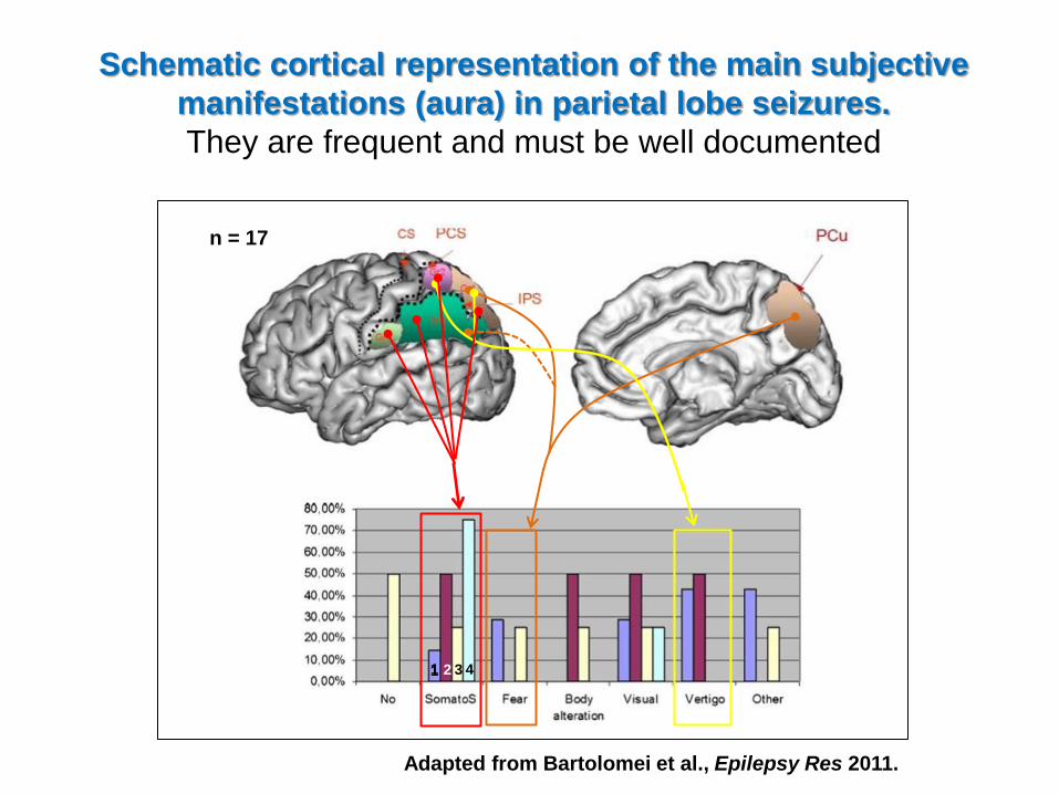
Representation of regions of the parietal lobe
generating seizures (adapted from Bartolomei 2011).
PCC
SS
elementary visual hallucinations
/amaurosis
visual illusions/distortion
complex visual hallucinations
1
1
2
3
2
Visual
elementary somatosensory
pain
sensations of warmth/cold
somatognostic illusions
Somatosensory
4
4
3
5
5
6
6
7
7
Vestibular
The parietal cortex is at the centre of multisensory integration.
It is also highly interconnected to other cortical regions. This
explains the heterogeneity of PL seizures semiology.
2,3
Adapted from AlKawadri, 2013, Montavont, 2013, Enatsu, 2014, Kahane, 2017 and Harroud, 2017.
The parietal lobe and associative cortex is
highly connected to other cortical regions:
Dorsal fronto-parietal network
SPL frontal eye field
Ventral fronto-parietal network
TP junction ventral frontal cortex
outline
Introduction
Semiology of PLE is heterogeneous and mimic seizures
originating from extra-parietal cortex.
Parietal lobe is subdivided in distinct epileptic regions:
A case of precuneal epilepsy
A case of parietal cingulate gyrus epilepsy
A case of inferior parietal lobule epilepsy.
Scalp EEG is also misleading
Diagnostic accuracy of non-invasive modalities in
presurgical evaluation in PLE.
Conclusion and comments.
gr 1. SPL. BA 7.
gr 2. SPL. BA 5.
Scenarios of intrecerebral SEEG schemes in PLE. Adapted from Bartolomei et al., Epilepsy Res 2011.
gr 3. IPL. BA 39,40.
gr 4. parietal operculum. BA 40.
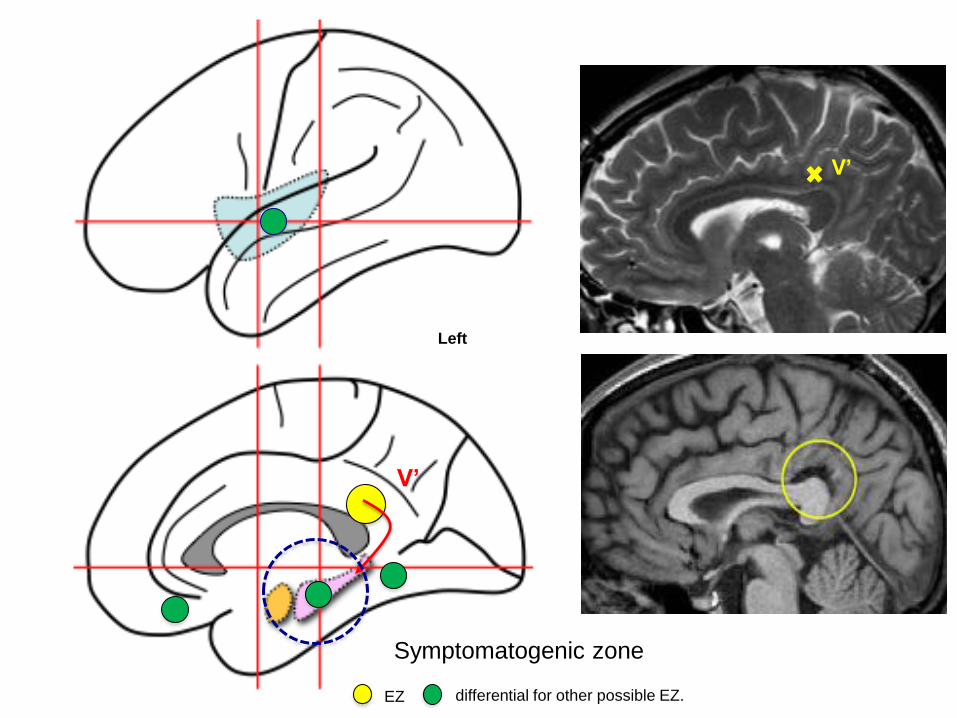
Case 1. Precuneal seizures.
A case-based discussion
21 yo R-handed woman with seizures since age 12:
Uneventful obstetrical history. No antecedents. Family history:
afebrile and febrile seizures.
Seizures started at 12. Typically, daytime, monthly, seizures
with prominent motor features and occasionnal GTC szs.
Low average/borderline intelligence.
Normal examination and phenotype.
Refractory to several AEDs.
Case 1 presentation con’t
Seizure semiology
Aura is present and with vestibular flavor i.e. sudden impression
of unsteadiness or of a movement, not further described, but often
attempted to prevent a fall L UL increased tone and LOC
clonic jerks L arm and inconsistant bilateral eyes blinking.
May fall and occ. 2ary generalization.
Slow recuperation and fatigue. Amnestic or partly amnestic, but
no other apparent deficits.
No triggers. No somatosensory, visual or temporal-like features.
Key point: vestibular aura and early lateralized motor features
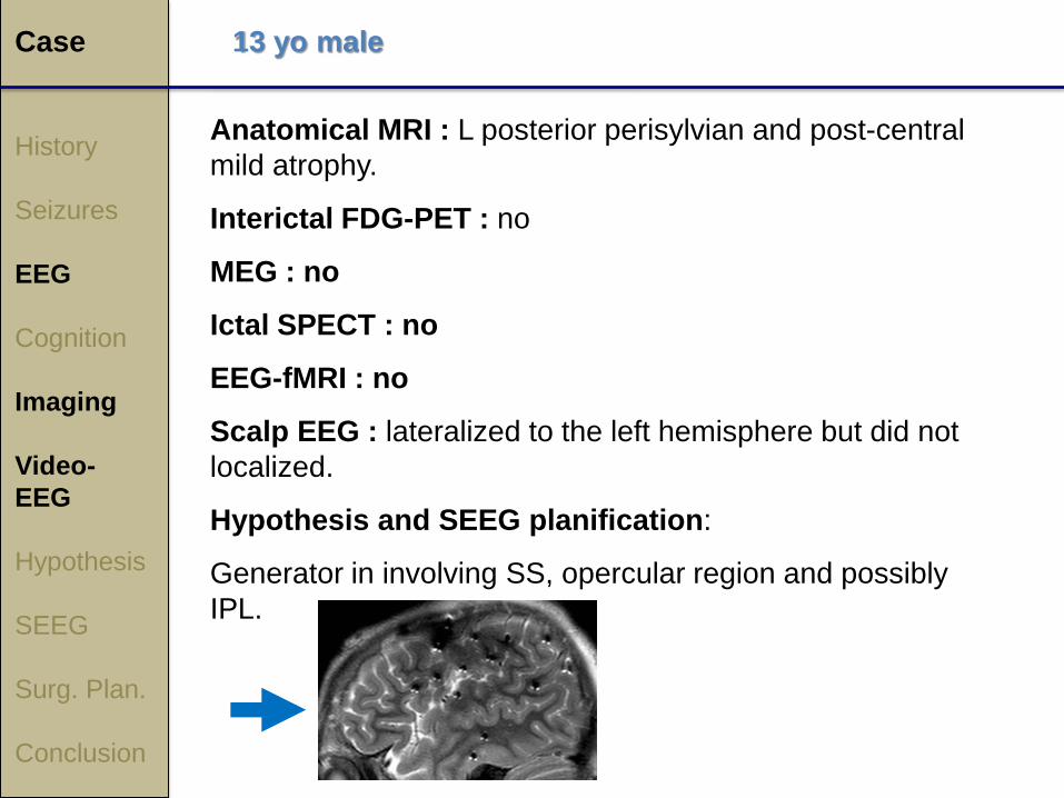
Case
History
Seizures
EEG
Cognition
Imaging
Video-EEG
Hypothesis
SEEG
Surg. Plan.
Conclusion
Anatomical MRI : normal
Interictal FDG-PET : normal
MEG : no
Ictal SPECT : no
EEG-fMRI : no
Summary of neuropsychological evaluation :
R-handed and left hemispheric speech dominance with a
low average IQ.
Neuropsychology profile consistent with bilateral posterior
quadrant dysfunction. Mild impairment of attention, memory
and executive function deficits and mild visuo-spatial
impairments.
21 yo R-handed woman
Case
History
Seizures
EEG
Cognition
Imaging
Video-EEG
Hypothesis
SEEG
Surg. Plan.
Conclusion
Interictal scalp EEG findings (10-20 and 10-10):
bilateral occipital sharp activity (O1, O2)
R centro parietal (C4, P4)
R T (F8, T4, T6)
Ictal scalp EEG findings:
R CP (C4, P4)
21 yo R-handed woman
Key point: EEG not localizing but pointing toward posterior
quadrant generator, probably right.
Case 1 presentation con’t
1st admission
typical seizures were recorded with vestibular aura and
ictal motor manifestations involving predominantly the
L UL.
interictal and ictal scalp discharges suggestive of a
generator in to the right posterior quadrant.
neuropsychology profile consistent with bilateral posterior
quadrant dysfunction.
MRI and PET revealed no anatomical lesion or
hypometabolism.
What’s next?
Precuneal epilepsy
clinical-EEG correlations althought often inacurrate, point to the posterior
quadrant in most cases (n=6). Structural imaging has a better yield
compare to functional imaging (MRI (4/6), SPECT (1/4), PET (1/3) and MEG (2/3)).
Pt AO MRI Ss Vest. visual E+H motor others II EEG I EEG
f,
17 10
L post.
PreCu - + + + - + lat, floc diffuse
F,
21 4 n - + + + + - loc, bil bil
M,
50 16 n - - - - + + loc, floc loc
M,
23 13
R post
PreCu - + + - + + lat, floc lat
M,
59 12
L ant
PreCu - + - - + + none diffuse
F,
21 12
R ant
PreCu - + - - + -
loc, bil,
floc loc
4/6 0 5 3 2 5 4
Harroud et al., Epilepsy & Behavior 2017
Case 2. Posterior cingulate seizures.
A case-based discussion
22 yo L-handed woman with seizures since age 14:
Uneventful obstetrical history . No antecedents.
Seizures started at 14. Typically, daytime monthly
seizures with prominent temporal features.
Low average/borderline intelligence.
Normal examination except discreet R facial paresis.
Refractory to several AEDs.
Case 2 presentation con’t
Seizure semiology
Aura is present, epigastric and +/- gustatory) automotor
behavior with loss of contact and confusion.
Slow recuperation, post-ictal confusion and fatigue.
Amnestic or partly amnestic, but no other apparent deficits.
No triggers. No somatosensory or visual features.
Key point: temporal-like seizures.
Case
History
Seizures
EEG
Cognition
Imaging
Video-EEG
Hypothesis
SEEG
Surg. Plan.
Conclusion
Anatomical MRI : L thalamus hypersignal.
Interictal FDG-PET : L hemispheric hypometabolism,
max temporo-parietal.
MEG : no
Ictal SPECT : no
EEG-fMRI : no
Summary of neuropsychological evaluation :
L-handed and left hemispheric speech dominance with
verbal IQ = 99 and non verbal IQ = 76.
22 yo L-handed woman
Case JP
History
Video
Scalp-EEG
Imaging
SEEG
Conclusion
Case
History
Seizures
EEG
Cognition
Imaging
Video-
EEG
Hypothesis
SEEG
Surg. Plan.
Conclusion
22 yo L-handed woman
T3-T5, F7-T3
Temporal Pattern
Frontal Pattern
Semiology of PCE varies depending upon the seizure spread patterns.
7 patients (Enatsu et al., JNNP 2014)
3/7 with motor manifestations (spread to frontal - premotor area, OF, SMA, ACC - and to parietal
lobe - precuneus, PCC, IPL, SS).
4/7 with dialeptic seizures or automotor seizures (spread to medial temporal or IPL areas).
Garzon & Luders, 2008
Case 3. Inferior parietal lobule seizures.
A case-based discussion
13 yo man with seizures since age 8:
Focal motor seizure involving R side at 2 days of age.
Seizures started at 8. Typically, daytime with R side
somatosensory and prominent motor features.
Refractory to several AEDs.
Case 3 presentation con’t
Seizure semiology
Aura is present, cephalic and somatosensory
manifestations with paresthesia involving R arm and head.
R arm dystonia or tonic posture, unresponsive althought
appears alert. Seizures often triggered during meals. Slow
recuperation and fatigue, but no post-ictal motor deficits.
No triggers except maybe eating. No pain, visual, gustatory
or temporal-like features.
Key point: lateralized somatosensory aura followed by
prominent focal motor manifestations.
Case
History
Seizures
EEG
Cognition
Imaging
Video-
EEG
Hypothesis
SEEG
Surg. Plan.
Conclusion
Anatomical MRI : L posterior perisylvian and post-central
mild atrophy.
Interictal FDG-PET : no
MEG : no
Ictal SPECT : no
EEG-fMRI : no
Scalp EEG : lateralized to the left hemisphere but did not
localized.
Hypothesis and SEEG planification:
Generator in involving SS, opercular region and possibly
IPL.
13 yo male
Et’
B’
D’
W’
P’ O’
S’
N’
U’
L’ R’
Q’
F’
M’
Motor and premotor cortex
FCP operculum
Inferior parietal lobule
Somatosensory cortex
ACG
AMS
preM
preM
PCG
PCG
mesP
preC
IPL
IPL
IPL
P-O
T-O
Fop
Cop
Pop
Pop
STG
Ins
Ins
Ins
Ins
EKG
Myo
Myo
Et’
B’
D’
W’
P’
O’
S’
N’
U’
L’
R’ Q
’
F’
M’
outline
Introduction
Semiology of PLE is heterogeneous and mimic seizures
originating from extra-parietal cortex.
Parietal lobe is subdivided in distinct epileptic regions:
A case of precuneal epilepsy
A case of parietal cingulate gyrus epilepsy
A case of inferior parietal lobule epilepsy.
Scalp EEG is also misleading
Diagnostic accuracy of non-invasive modalities in
presurgical evaluation in PLE.
Conclusion and comments.
Occipital epilepsy (n=25)*
Interictal EEG
occipital only 2
occipital ‘plus’ 4
temporal only 14
frontal bil. synch. 5
Parietal epilepsy (n=11)**
Interictal EEG
• parietal only 1
• parietal ‘plus’ 3
• temporal only 3
• frontal bil. synch. 2
• no spikes 2
*, ** Williamson et al., Ann Neurol 1992
EEG correctly localizes the epileptic generator
in a small proportion of patients with PLE.
Williamson et al., 1992 (11 patients). Sveinbjornsdottir and Duncan, 1993 (review). Foldvary et al., 2001 (x/72 pts). Kim et al., 2004 (40 pts). Ristic et al., 2012 (16 pts). Liava et al., 2014 (11 pts). Asadollahi et al., 2017 (18 pts).
Occipital epilepsy (n=25)*
Interictal EEG
occipital only 2
occipital ‘plus’ 4
temporal only 14
frontal bil. synch. 5
Ictal clinical
TL type 11
FL type 3
2 seizure types 11
Parietal epilepsy (n=11)**
Interictal EEG
parietal only 1
parietal ‘plus’ 3
temporal only 3
frontal bil. synch. 2
No spikes 2
Ictal clinical
temporal lobe type 4
frontal lobe type 4
2 seizure types 3
Temporal Pattern
*, ** Williamson et al. Ann Neurol 1992.
Frontal Pattern
EEG can mimick temporal patterns in patients with PLE.
Occipital epilepsy (n=25)*
Interictal EEG
occipital only 2
occipital ‘plus’ 4
temporal only 14
frontal bil. synch. 5
Ictal clinical
TL type 11
FL type 3
2 seizure types 11
Parietal epilepsy (n=11)**
Interictal EEG
parietal only 1
parietal ‘plus’ 3
temporal only 3
frontal bil. synch. 2
no spikes 2
Ictal clinical
temporal lobe type 4
frontal lobe type 4
2 seizure types 3
Temporal Pattern
*, ** Williamson et al. Ann Neurol 1992.
Frontal Pattern
EEG can mimick frontal patterns in patients with PLE.
Interictal more than ictal activity mis-localizes or mis–
lateralizes epileptic discharges in post Q epilepsy.
62 operated children (mean age 7.9 yrs) Pediatric epilepsy surgery in the posterior cortex.
Liava et al., Epileptic DIsord 2014
PLE
11
4 (36)
4 (36)
2 (18)
1 (9)
0
10
3 (30)
5 (50)
0
1 (10)
0
1 (10)
Ictal EEG 56
localising 21 (37%)
regional 18 (32%)
falsely localising 7 (F) (12.5%)
lateralising only 1 (1.8%)
falsely lateralising 3 (5.35%)
bil hemispheric 6 (10.7%)
Interictal EEG 62 localising 22 (35%) regional 25 (40%) falsely localising 6 (2 F, 3 FT, 1 T) (9.7%) lateralising only 3 (4.8%) falsely lateralising 6 (9.7%)
Maximum electrical field
interictal EEG distribution
in
PLE vs TLE and FLE
PLE patients
• show a more variable scatter of
interictal discharges
• the majority have more than one
spike population
• they also show a lower
localisation value of ictal
recordings.
lobar classification by
electroclinical impression is
least accurate in PLE patients.
Ristic et al., Epileptic Disord 2012
PLE
n=16
TLE
n=17
FLE
n=17
Diagnostic accuracy of pre-surgical evaluation. (n = 26 patients operated and 1 yr f/u)
Diagnostic
modality No. Seizure-free
Persistent
seizure
P
value
Focal lesion
on MRI 26 64% 25% 0,06
Hypometabolism
on PET 26 50% 17% 0,11
Focal hyperperfusion
on SPECT 21 45% 50% 1,00
Localized ictal EEG 26 36% 42% 1,00
Kim et al., Epilepsia 2004
Only structural imaging appears to have a predictive value in terms
of seizure outcome. A good understanding and interpretation of
the semiological features and concordance of different diagnostic
modalities is also associated with higher seizure-free rate
Diagnostic
modality No. localizing lateralizing nonlateral. false loc. false lat.
Interictal EEG 22 3 (14) 6 (27) 8 (36) 5 (23) 0
Ictal EEG 22 8 (36) 5 (23) 1 (4,5) 8 (36) 0
PET 22 8 (36) 5 (23) 4 (18) 4 (18) 1 (4,5)
SPECT 18 9 (50) 0 2 (11) 7 (39) 0
Diagnostic sensitivity of individual modalities in 22 (85%)
operated patients with a favorable (14 seizure-free) surgical
outcome.
Kim et al., Epilepsia 2004
No. in parenthesis are %.
Conclusions
The characterization of adequate diagnostic criteria
is difficult in parietal lobe epilepsy:
• Parietal seizures are rare compare to those observed in
temporal or frontal lobe epilepsy.
• The anatomical boundaries of the parietal lobe are not well
defined and not clearly related to function.
• The cortex, mostly associative, is concerned with higher
sensory, perceptual and cognitive processes or functions. It
is highly interconnected with other brain regions and multiple
systems.
• Seizures are highly variable with inconsistent semiologic
features, often mimicking extra-parietal seizures.
• The yield of the different diagnostic modalities is low,
particularly in MR-negative cases. Scalp EEG for instance is
often poorly localizing and even often mis-localizing.
Comments:
How can we improve diagnostic yield of parietal seizures
and epilepsy?
Subjective phenomena and elementary aura are frequent, often
complex and difficult to characterize, and must be well documented.
Parietal lobe seizures propagate typically to frontal lobe and motor
regions, through a dorsal frontoparietal stream. They may also spread
through ventral pathways toward the ventral frontal regions or to the
temporal lobe. The parietal lobe has also intimate interconnectivity in
the posterior insula.
Studies of the surface epiletic activty with non-invasive modalities such
as EEG-fMRI are scarce. Understanding the hemodynamic responses
of interictal epileptic activity may increase the localisation value of
scalp EEG.
Adapted from Bartolomei et al., Epilepsy Res 2011.
Schematic cortical representation of the main subjective
manifestations (aura) in parietal lobe seizures.
They are frequent and must be well documented
1 3 4 1 2
n = 17
Adapted from Bartolomei et al., Epilepsy Res 2011.
Objective signs (motor) in parietal lobe seizures reflect
the involvement of systems participating in oculomotor
controls and in the involvement of distant cortices.
2 1 3 4
n = 17
Functional organization of the dominant and
non-dominant parietal cortices using neurostimulation.
172 patients using high- (50 Hz) and low- (1 Hz) electrical stimulations
1. Postcentral gyrus, posterior part of paracentral lobule
and parietal operculum, significant association with:
• Somatosensory sensations
• Motor symptoms
• Dysarthria.
2. Superior and inferior parietal lobules associated multiple
types of responses including:
• Somatosensory
• Visual illusions and body scheme alterations
• Motor symptoms
• Eye movements /sensations
• Multimodal.
3. Precuneus associated with visual illusions or hallucinations,
and with vertigo.
4. Intraparietal sulcus associated with visual illusions and
hallucinations and eye movements/sensations.
5. Posterior cingulate gyrus associated with somatosensory
and motor symptoms, with vertigo and with neurovegetative
manifestations.
dominant
non-dominant
Balestrini et al., Brain 2015