1 Supporting Information SI Materials and Methods Plasma IgG adsorptions on SF162 gp120- and SF162 gp120 D368R -coated beads. Plasma adsorptions were performed as previously described (1, 3), with some modifications, and the flow of the adsorptions is outlined in Figure S1. Briefly, total IgG was isolated by protein A chromatography (Pierce, Rockford Il, USA) from VC10042 plasma drawn 22 years post-infection. The purified total IgG was serially adsorbed onto SF162 gp120-coated beads (MyOne Tosylactivated Dynabeads, Invitrogen, Carlsbad, CA, USA), and the flow through collected. The antibodies bound to the gp120 coated beads were eluted by vortexing in increasingly acidic 0.1M glycine solutions, followed by buffer exchange into PBS. The anti-gp120 Ab fraction was then serially adsorbed onto SF162 gp120 D368R -coupled beads to remove Abs that do not bind the CD4-BS. Each fraction described above was tested for residual neutralizing activity against 4 clade B, 3 clade C, and 2 clade A isolates in the TZM-bl neutralization assay. The depleted total IgG and the gp120 D368R depleted anti-gp120 fractions were tested for the presence of anti-CD4-BS antibodies, and the absence of non-CD4-BS gp120 Abs by Luminex assay (Luminex Corporation, Austin, TX, USA) against both wild type SF162 gp120 and SF162 gp120 D368R .
Transcript
1
Supporting Information SI Materials and Methods Plasma IgG adsorptions on SF162 gp120- and SF162 gp120D368R-coated beads. Plasma adsorptions were performed as previously described (1, 3), with some modifications, and the flow of the adsorptions is outlined in Figure S1. Briefly, total IgG was isolated by protein A chromatography (Pierce, Rockford Il, USA) from VC10042 plasma drawn 22 years post-infection. The purified total IgG was serially adsorbed onto SF162 gp120-coated beads (MyOne Tosylactivated Dynabeads, Invitrogen, Carlsbad, CA, USA), and the flow through collected. The antibodies bound to the gp120 coated beads were eluted by vortexing in increasingly acidic 0.1M glycine solutions, followed by buffer exchange into PBS. The anti-gp120 Ab fraction was then serially adsorbed onto SF162 gp120D368R-coupled beads to remove Abs that do not bind the CD4-BS. Each fraction described above was tested for residual neutralizing activity against 4 clade B, 3 clade C, and 2 clade A isolates in the TZM-bl neutralization assay. The depleted total IgG and the gp120D368R depleted anti-gp120 fractions were tested for the presence of anti-CD4-BS antibodies, and the absence of non-CD4-BS gp120 Abs by Luminex assay (Luminex Corporation, Austin, TX, USA) against both wild type SF162 gp120 and SF162 gp120D368R.
ch the plasmad, IC50 titers50 colored inma isolated
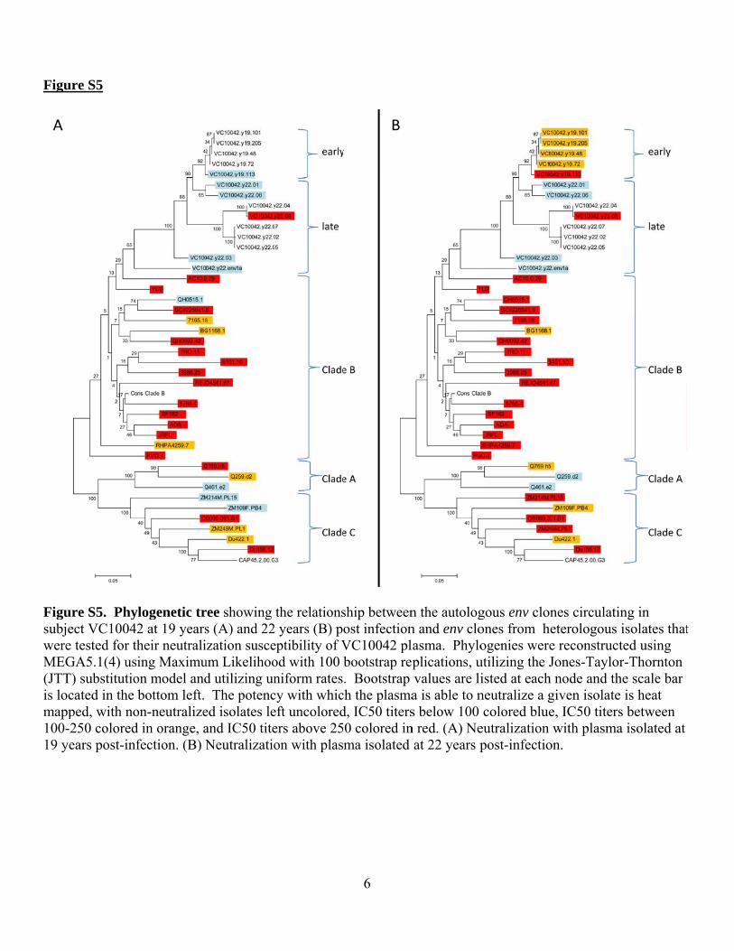
n the autologn and env clolasma. Phyloplications, uvalues are lia is able to ns below 100 n red. (A) Ne
at 22 years p
gous env cloones from hogenies were
utilizing the Jisted at eachneutralize a gcolored blue
eutralization post-infectio
ones circulatiheterologous e reconstrucJones-Taylo
h node and thgiven isolatee, IC50 titerwith plasma
on.
ing in isolates that
cted using or-Thornton he scale bar e is heat s between a isolated at
t
Figu
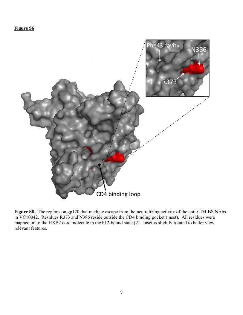
Figuin Vmapprelev
ure S6
ure S6. The C10042. Reped on to thevant features
regions on gesidues R373e HXB2 cors.
gp120 that m3 and N386 re molecule i
mediate escapreside outsidin the b12-bo
7
pe from the de the CD4 bound state (2
neutralizingbinding pock2). Inset is s
g activity of tket (inset). Aslightly rotat
the anti-CD4All residues ted to better
4-BS NAbs were view
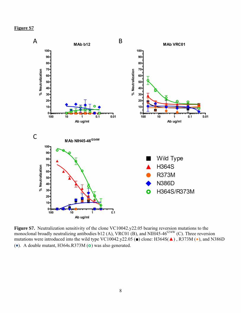
Figu
Figumonmuta(♦).
ure S7
ure S7. Neunoclonal broaations were iA double m
utralization seadly neutraliintroduced in
mutant, H364
ensitivity of zing antibodnto the wild
4s.R373M (o
f the clone Vdies b12 (A)type VC100
o) was also g
8
VC10042.y22
, VRC01 (B042.y22.05 (generated.
2.05 bearing ), and NIH4
(■) clone: H3
reversion m45-46G54W (C364S(▲) , R
mutations to tC). Three revR373M (●), a
the version and N386D
9
References: 1. Li, Y., S. A. Migueles, B. Welcher, K. Svehla, A. Phogat, M. K. Louder, X. Wu, G. M. Shaw, M.
Connors, R. T. Wyatt, and J. R. Mascola. 2007. Broad HIV-1 neutralization mediated by CD4-binding site antibodies. Nat Med 13:1032-4.
2. Liu, J., A. Bartesaghi, M. J. Borgnia, G. Sapiro, and S. Subramaniam. 2008. Molecular architecture of native HIV-1 gp120 trimers. Nature 455:109-13.
3. Sather, D. N., J. Armann, L. K. Ching, A. Mavrantoni, G. Sellhorn, Z. Caldwell, X. Yu, B. Wood, S. Self, S. Kalams, and L. Stamatatos. 2009. Factors associated with the development of cross-reactive neutralizing antibodies during human immunodeficiency virus type 1 infection. J Virol 83:757-69.
4. Tamura, K., D. Peterson, N. Peterson, G. Stecher, M. Nei, and S. Kumar. 2011. MEGA5: molecular evolutionary genetics analysis using maximum likelihood, evolutionary distance, and maximum parsimony methods. Mol Biol Evol 28:2731-9.