Linköping University Medical Dissertations No. 1052 Signs of inflammation in different types of heart valve disease The VOCIN study Lars Wallby Division of Cardiovascular Medicine Department of Medical and Health Sciences Linköping University, Sweden Linköping 2008
Transcript
Linköping University Medical Dissertations No. 1052
Signs of inflammation in different types of heart valve disease
The VOCIN study
Lars Wallby
Division of Cardiovascular Medicine Department of Medical and Health Sciences
1.1 Anatomy and histology of the valves................................................... 5 1.2 Heart valve disease................................................................................... 7
4.2 Role of inflammation in non-rheumatic, regurgitant heart valve disease (II) ........................................................................................................... 24
4.3 Inflammatory characteristics of stenotic aortic valves – a comparison between rheumatic and non-rheumatic aortic stenosis (III)25
4.3.1 Histopathological and immunohistochemical analyses ................ 26 4.4 History and Signs of Rheumatic Disease in Patients with Significant Heart Valve Disease (IV) .................................................................................. 27
4.4.1 Echocardiography................................................................................ 27 4.4.2 Questionnaire ....................................................................................... 28 4.4.3 History and status regarding rheumatic disease ............................ 28 4.4.4 Biochemistry ......................................................................................... 28
5 GENERAL DISCUSSION................................................................................. 30
5.1 Signs of inflammation in different types of heart valve disease.. 30 5.2 The rheumatic aortic valve ................................................................... 33 5.3 The bicuspid aortic valve and the significance of the underlying anomaly................................................................................................................ 34 5.4 Inflammatory signs in heart valve disease and its similarities to the process of atherosclerosis .......................................................................... 38 5.5 The absence of articular symptoms or rheumatic disease in heart valve disease ....................................................................................................... 39 5.6 HLA-B27 ................................................................................................... 40 5.7 The systemic inflammation in patients with heart valve disease 40 5.8 Conclusions ............................................................................................. 43
Heart valve dysfunction is a relatively common condition in the population, whereas significant heart valve disease is more unusual. The cause of different types of heart valve disease depends on which valve is concerned. Rheumatic heart valve disease, has for a long time been considered to constitute a post-inflammatory condition. During the 1990s it was also shown that the so-called non-rheumatic or degenerative tricuspid aortic stenosis, comprised signs of inflammation. In this study, 118 patients (the VOCIN study group) referred to the University Hospital for preoperative investigation due to significant heart valve disease, were examined regarding signs of inflammation. Twenty-nine aortic valves from patients with significant aortic stenosis were divided into tricuspid and bicuspid aortic valves. The bicuspid aortic stenotic valves revealed signs of inflammation to a similar extent as the tricuspid valves. However, the tricuspid and bicuspid valves differed regarding distribution of calcification. In contrast, inflammation was not a predominant feature in 15 aortic and mitral valves from patients with significant heart valve regurgitation. Gross valvular pathology consistent with rheumatic aortic stenosis was found in 10 patients. These valves revealed a somewhat lower degree of inflammatory cell infiltration, but on the whole, there were no substantial differences when compared to non-rheumatic aortic stenotic valves. They did, however, reveal a similar distribution of calcification as the bicuspid, non-rheumatic aortic valves. The VOCIN study group was compared to an age- and gender matched control group with regard to history and signs of rheumatic disease. There was not any increased prevalence of clinical manifestations of non-cardiac inflammatory disease in patients with significant heart valve disease, when compared to healthy control subjects. However, patients with heart valve disease had significantly increased serum levels of inflammatory markers compared to controls. The increase in inflammatory markers remained significant even in the subgroup of non-rheumatic aortic stenosis devoid of coronary artery disease. These results indicate that a systemic inflammatory component is associated with stenotic, non-rheumatic heart valve disease. The similarities between different forms of calcific aortic valve disease indicate a similar pathogenesis. The question is raised whether aortic stenosis is one disease, mainly caused by a general and non-specific response to dynamic tissue stress due to an underlying malformation of the valve.
2
LIST OF PAPERS
3
LIST OF PAPERS
This thesis is based on the following papers, referred to in the text by their Roman numerals. I. Wallby L, Janerot-Sjöberg B, Steffensen T, Broqvist M. T lymphocyte infiltration in non-rheumatic aortic stenosis: a comparative descriptive study between tricuspid and bicuspid aortic valves. Heart 2002;88:348-51. II. Wallby L, Steffensen T, Broqvist M. Role of inflammation in non-rheumatic, regurgitant heart valve disease. A comparative, descriptive study regarding apolipoproteins and inflammatory cells in non-rheumatic heart valve disease. Cardiovasc Pathol 2007;16:171-8. III. Wallby L, Steffensen T, Jonasson L, Broqvist M. Inflammatory characteristics of aortic stenotic valves – a comparison between rheumatic and non-rheumatic aortic stenosis. Submitted. IV. Wallby L, Janerot-Sjöberg B, Jonasson L, Locht H, Broqvist M. History and signs of rheumatic disease in patients with significant heart valve disease. Submitted.
ABBREVIATIONS
4
ABBREVIATIONS
Apo Apolipoprotein AR Aortic regurgitation AS Aortic stenosis AV Aortic valve BAV Bicuspid aortic valve CRP C-reactive protein HLA Human leukocyte antigen ISP’s Inflammation sensitive proteins MHC Major histocompability complex MS Mitral stenosis MR Mitral regurgitation MV Mitral valve NRAR Non-rheumatic aortic regurgitation NRAS Non-rheumatic aortic stenosis NRAS-B Non-rheumatic bicuspid aortic stenosis NRAS-T Non-rheumatic tricuspid aortic stenosis NRMR Non-rheumatic mitral regurgitation RAS Rheumatic aortic stenosis RMR Rheumatic mitral regurgitation RMS Rheumatic mitral stenosis RF Rheumatoid Factor TR Tricuspid regurgitation VOCIN Vitium Organicum Cordis and INflammation WBC White blood cell count
INTRODUCTION
5
1 INTRODUCTION
1.1 Anatomy and histology of the valves
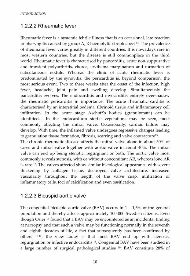
The function of the four cardiac valves (the tricuspid and pulmonary valves in connection to the right atrium and ventricle, the mitral and aortic valves in connection to the left atrium and ventricle) is to maintain unidirectional flow through the heart (Figure 1). The ability of the valves to permit unobstructed forward flow depends on the mobility and pliability of their leaflets.
Figure 1. Schematic picture of the heart showing the atria, chambers and valves. Picture used with permission from the Cleveland Clinic Foundation.
Left ventricle
Left atrium Right atrium
Right ventricle
INTRODUCTION
6
The semilunar valves (aortic and pulmonary) are composed of three, equal sized, crescentic cusps and their respective supporting structures in the aortic and pulmonary roots (Figure 2).
Figure 2.Picture of semilunar valve closed (left) and opened (right) The atrioventricular valves (mitral and tricuspid) are composed of two (mitral) and three (tricuspid) leaflets that together with the chordae tendineae (tendinous cords) and the papillary muscles form the valves (Figure 3).
Figure 3.Picture of atrioventricular valve (mitral) revealing the valve leaflets, annulus (supporting valve ring), chordae (tendinous cords) and the papillary muscles. Valve closed (left) and opened (right). The cardiac valves are lined with endothelium and all have a similar three-layered architecture consisting predominantly of a dense collagenous core, the fibrosa layer, close to the outflow surface and continuous with the valvular supporting structures. Towards the ventricular/atrial cavity is the ventricualis/atrialis layer, rich in elastin. Between these two layers is the centrally located core of loose connective tissue, the spongiosa layer (Figure 4). The collagen of the fibrosa layer is responsible for the mechanical strength of the valve, whereas the connective tissue of the spongiosa layer works as a shock-absorber. Normal leaflets and cusps have only scant blood vessels,
Leaflets
Annulus
Leaflets
Annulus
Chordae
Papillary muscles
INTRODUCTION
7
limited to the proximal portion of the valve and are populated throughout by interstitial cells, but devoid of inflammatory cells and lipid deposition.
Figure 4.Microscopic appearance of normal aortic valve, showing the fibrosa layer (F) with dense collagen and scattered fibroblasts, the spongiosa layer (S) with large amounts of proteoglycan, loosely arranged collagen fibrils, scattered fibroblast and mesenchymal cells and the ventricularis layer (V) rich in elastic fibers.
1.2 Heart valve disease
Heart valve dysfunction is a relatively common condition in the population, whereas significant heart valve disease is more unusual 1,2. As the function of the four cardiac valves is to maintain unidirectional flow through the heart, heart valve dysfunction can be divided into principally two entities; stenotic and regurgitant lesions. The stenotic lesion implies an obstruction of forward flow through the valve and is caused by an abnormal increase in thickness and stiffness of the valve leaflet due to increased amounts of fibrosis and/or calcification, alternatively congenital or acquired fusion of the leaflets, resulting in a decreased effective
F
S
V
INTRODUCTION
8
valve orifice and in consequence, enhanced pressure load on the heart cavity and an increased blood flow velocity across the valve. In the case of the aortic valve one makes a distinction between aortic valve stenosis (AS) and aortic valve sclerosis. The latter implies a mild leaflet thickening without valve obstruction. Aortic valve sclerosis can be diagnosed by echocardiography as focal areas of increased echogenicity on the valve leaflets with normal valve motion and a normal, or only mildly increased antegrade velocity across the valve 3. However, aortic valve sclerosis per se, besides being a possible early stage of AS, comprises an increased risk of morbidity and mortality 3,4. The degree of aortic valve obstruction is generally classified arbitrarily into aortic valve sclerosis, (i.e. mild leaflet thickening without valve obstruction), mild, moderate or severe stenosis. The regurgitant lesions are due to an inability of the valve to stay impenetrable when the pressure in front of the valve exceeds the pressure behind the valve. In such regurgitant cases, the unidirectional flow cannot be maintained, instead the blood flows against the stream and thus increases the amount of blood the respective heart chamber has to cope with to maintain unchanged forward flow. Valve regurgitation can, in general, be caused either by a dilatation of the supporting valve ring making valve leaflet coaptation impossible, or by damages to the valve leaflets or their supporting structures. The degree of valve regurgitation is generally classified arbitrarily into trace, (meaning barely discernable), mild, moderate or severe.
1.2.1 Prevalence
The prevalence of heart valve regurgitation has been described in several large cohort studies. In the Framingham Offspring Study1, mitral valve regurgitation (MR) was detectable in 88% of men and 92% of women, whereas tricuspid valve regurgitation (TR) was detectable in 82% of men and 86% of women. Aortic valve regurgitation (AR) was less prevalent and was observed in 13% of men and 8.5% of women. The prevalence of MR of more than or equal to mild severity was 19% in both men and women and of TR, 15% in men and 18% in women. However, considering MR of more than or equal to moderate severity the prevalence was 2,0% in men and 1,2% in women and of TR, 0,3% in men and 1,2% in women. The prevalence of AR of more than or equal to moderate severity was found to be only 0,5% in men and 0.4% in women. When examined across different age groups, the prevalence of MR and TR of more than or equal to mild severity, and AR of more than or equal to trace severity, increased with age in both sexes.
INTRODUCTION
9
In the Cardiovascular Health Study 2, there was aortic valve sclerosis in 26% and AS in 2% of the entire cohort of 5201 subjects ≥ 65 years of age, but in subjects ≥ 75 years, sclerosis was present in 37% and AS in 2,6%. According to the Swedish Health and Welfare Statistical Database, 1648 (18/100 000 inhabitants) operations on the aortic valve and 688 (8/100 000) on the mitral valve were performed in Swedish hospitals in 2006. Men dominated among the operated subjects, 1033 (23/100 000) men were operated on the aortic valve, versus 615 (13/100 000) women. The numbers for mitral valve operation were 473 (11/100 000) men and 215 (5/100 000) women.
1.2.2 Aetiology The most common cause of severe, pure MR is mitral valve prolaps accounting for about 65% of MR cases. Ischemic heart disease, rheumatic heart disease and endocarditis accounts for most of the remainder 5-7. The predominant aetiology of mitral stenosis (MS) is a postinflammatory condition due to rheumatic fever. In adults undergoing surgery for AS, calcific AS accounts for 51%, bicuspid aortic valve for 36% and rheumatic disease for 9% 8. The aetiology of aortic valve regurgitation (AR) is multifactorial and includes, on the one hand diseases involving the aortic root, e.g. Marfans syndrome, degenerative aortic dilatation and spondyloarthropathies, on the other hand diseases involving the aortic valve, e.g. rheumatic disease, infective endocarditis and bicuspid aortic valve 9.
1.2.2.1 Clinical factors associated with heart valve disease
Age is observed to exert a profound influence on the prevalence of valve regurgitation in the population 1. Hypertension is associated with MR, but not with AR1. AS is the heart valve dysfunction most widely studied regarding risk factor association. In the Cardiovascular Health Study 2, which included 5621 adults ≥ 65 years, clinical factors associated with calcific aortic valve disease included age, male gender, smoking, hypertension and hyperlipidemia, thus overlapping the clinical factors traditionally associated with atherosclerosis 10-
12.
INTRODUCTION
10
1.2.2.2 Rheumatic fever
Rheumatic fever is a systemic febrile illness that is an occasional, late reaction to pharyngitis caused by group A, ß-haemolytic streptococci 13. The prevalence of rheumatic fever varies greatly in different countries. It is nowadays rare in most western countries, but the disease is still commonplace in the third world. Rheumatic fever is characterised by pancarditis, acute non-suppurative and transient polyarthritis, chorea, erythema marginatum and formation of subcutaneous nodule. Whereas the clinic of acute rheumatic fever is predominated by the synovitis, the pericarditis is, beyond comparison, the most serious event. Two to three weeks after the onset of the infection, high fever, headache, joint pain and swelling develop. Simultaneously the pancarditis evolves. The endocarditis and myocarditis entirely overshadow the rheumatic pericarditis in importance. The acute rheumatic carditis is characterised by an interstitial oedema, fibrinoid tissue and inflammatory cell infiltration. In the acute stage Aschoff’s bodies (granulomata) can be identified. In the endocardium sterile vegetations may be seen, most commonly affecting the mitral valve. Occasionally, cardiac failure may develop. With time, the inflamed valve undergoes regressive changes leading to granulation tissue formation, fibrosis, scarring and valve contracture13. The chronic rheumatic disease affects the mitral valve alone in about 50% of cases and mitral valve together with aortic valve in about 40%. The mitral valve can end up being stenotic, regurgitant or both. The aortic valve most commonly reveals stenosis, with or without concomitant AR, whereas lone AR is rare 13. The valves affected show similar histological appearance with severe thickening by collagen tissue, destroyed valve architecture, increased vascularity throughout the length of the valve cusp, infiltration of inflammatory cells, foci of calcification and even ossification.
1.2.2.3 Bicuspid aortic valve
The congenital bicuspid aortic valve (BAV) occurs in 1 – 1,5% of the general population and thereby affects approximately 100 000 Swedish citizens. Even though Osler 14 found that a BAV may be encountered as an incidental finding at necropsy and that such a valve may be functioning normally in the seventh and eighth decades of life, a fact that subsequently has been confirmed by others 15-17, the view today is that most BAV end up with stenosis, regurgitation or infective endocarditis 18. Congenital BAV have been studied in a large number of surgical pathological studies 19. BAV constitute 28% of
INTRODUCTION
11
resected aortic valves in patients undergoing aortic valve replacement worldwide 19. They are 2 - 3 times more frequent in men than in women. In the study of Sabet and co-workers 19 on 542 excised BAV, pure AS occurred in 75% of cases (mean age 65 years, male to female ratio of 1,7:1). The same study showed that pure aortic regurgitation was present in 13% (mean age 46 years, male to female ratio of 17:1) and combined stenosis and regurgitation by 10% (mean age 51 years, male to female ration ratio of 3,8:1). A congenital fibrous band, raphe, can be identified in the midportion of the conjoined cusp in about 75% of all congenital BAV, more often in the 92% of valves with unequal cusp size than in the 5% of valves with equal cusp size or the 2% of valves with a conjoined cusp twice the size of its nonconjoined cusp. The conjoined cusp most frequently consists of the right and left cusps (86%), whereas the right and non-coronary cusps constitute the conjoined cusp in 12% and the left and non-coronary cusps in 3% of all cases 19. Patients with congenital BAV tend to have an inherently weaker ascending aorta compared to normal people. Accordingly, they are prone to develop a dilatation of the valve annulus and a dilatation, aneurysm, dissection or rupture of the ascending aorta, regardless of the functional state of the valve 20-
23. Among 347 patients with aortic dissection in two autopsy studies 21,23, a BAV was present in 32 subjects (9%), about 6 times its prevalence in the population. The inherent character of aortic dilatation was recently shown by Beroukhim et al.24 reporting that children with a functionally normal BAV, revealed significantly greater aortic dimensions and, in addition, significantly greater increase in ascending aortic dimension as compared to age- and BMI-matched controls. BAV and weakened ascending aortas may also coexist with mitral valve prolapse 25 and approxamately 50% of patients with coarctation of the aorta also have a BAV 26.
1.2.2.4 The “Wear and Tear” concept
A large number of publications in the last 100 years have dealt with the pathogenesis of calcific AS. Mönckeberg 27 is usually credited for the first attempt in 1904, concluding that isolated AS was due either to previous inflammation or to atherosclerotic calcification. During the first half of the 20th century numerous studies were published, mostly favouring a rheumatic
INTRODUCTION
12
aetiology, as summarised in the monograph by Karsner and Koletsky 28 in 1947. In spite of the low incidence of previous rheumatic fever, lack of coexisting mitral disease and with non-specific pathological evidence, they summarised that “calcific disease of the aortic valve is the result of previous inflammation, an inflammation of such character as to identify it as rheumatic”. Twenty years later Campbell 29 wrote “these writers were unduly influenced by the dogmatic view that all cases of valvular disease, except syphilitic aortic regurgitation, were rheumatic”. In 1961 Jesse E. Edwards 30 wrote that “Calcific aortic stenosis appears to complicate (1) valves previously affected by rheumatic endocarditis, (2) congenital bicuspid aortic valves, and (3) congenitally stenotic aortic valves”. In another paragraph he incidentally mentions “the type usually seen in elderly people and that is characterised by deposits of fatty and calcific nodules in the leaflets” that “may at times lead to a false diagnosis of significant aortic stenosis”. In the 1970s the significance of an age-related stenosis of the tricuspid aortic valve became more obvious by the work of Roberts and Pomerance 31-33 and in one study Pomerance pointed out that “while aortic stenosis may be due to any of the three pathological processes suggested, the most likely pathogenesis in individual cases is related to age”. Fibrosis and calcification as a consequence of aging or a so-called wear-and-tear process was thus considered to be the aetiology behind stenosis in aortic valves, not harassed by previous rheumatic fever or congenital malformation. However, Roberts also presented the thesis of minor aortic cuspal inequality, as an underlying congenital malformation in adults with tricuspid, valvular AS 33. In 1987, Passik et al 34 showed that among aortic valves removed between 1981 and 1985 due to pure AS, the relative frequencies of postinflammatory disease and bicuspid valves had decreased, whereas the relative frequency of degenerative stenosis in tricuspid aortic valves had increased. These changes were in part explained by improvements in non-invasive diagnostic methods, increased safety and availability of cardiac surgical procedures, but also by change in life expectancy in the general population.
INTRODUCTION
13
1.2.2.5 Chronic inflammation
The development of monoclonal antibodies directed against cellspecific antigens and antigens presented by cells during specific stages of differentiation and activation, has significantly contributed to a pathogenetic understanding of several diseases, among them atherosclerosis 35,36. In the beginning of the 1990s two studies 37,38, independently of each other demonstrated the presence of T lymphocytes in cusps from aortic stenotic valves. Subsequently, different research groups have revealed different signs of chronic inflammation including chronic inflammatory cell infiltration, lipoprotein deposition and active leaflet calcification 39-42. Although the so-called degenerative AS has been associated with chronic inflammation, less is known about the inflammatory component in stenosis or regurgitation of valves with dysfunction due to previous rheumatic fever or congenital, bicuspid malformation.
1.2.2.6 The concept of “HLA-B27-associated cardiac disease”
The principal physiological function of the cell surface histocompatibility molecules is to bind peptide fragments of foreign proteins for presentation to antigen-specific T cells 43. In humans, the genes encoding the most important histocompatibility molecules are clustered on a small segment of chromosome 6, the major histocompatibility complex (MHC), or the human leukocyte antigen (HLA) complex. The HLA system is highly polymorphic, i.e. each individual inherits one, often unique, set of genes. Class I MHC molecules, designated HLA-A, HLA-B and HLA-C, are expressed on all nucleated cells and platelets. On the other hand, class II MHC molecules, such as HLA-DR is mainly restricted to immune cells. A variety of diseases have been found to be associated with certain HLA alleles. A number of inflammatory diseases are associated with HLA-B27. The best known is the association between ankylosing spondylitis and HLA-B27. Individuals who inherit this allele have a 90-fold greater relative risk of developing the disease. Other inflammatory diseases associated with the HLA-B27 allele are several postinfectious arthropathies, acute anterior uveitis and subgroups of intestinal and psoriatic arthropathies. Together these conditions are denominated the seronegative spondyloarthropathies. Common features of these conditions are a strong, but variable, association with HLA-B27, a high frequency of roentgenologic sacroiliitis and the absence
INTRODUCTION
14
of rheumatoid factor (RF) on serologic examination. For more than two decades, this group of rheumatic disorders has been known as HLA-B27-associated rheumatic diseases. Although the distribution of HLA-B27 varies considerably in the general population—6-8% of white people in Europe and North America, around 2% of the Chinese and African-American people and 0,2% of Japanese people—the relation between HLA-B27 and ankylosing spondylitis seems to be equally strong. Some of these HLA-B27-associated rheumatic or inflammatory conditions have long been known to be accompanied by “cardiac complications”, specifically, atrioventricular conduction blocks and lone AR 44. HLA-B27 is also considered an important genetic risk factor for these cardiac conditions, regardless of the presence of the typical extracardiac rheumatic syndromes. Thus an HLA-B27-associated cardiac syndrome that consists of severe cardiac conduction system abnormalities and lone AR, has been identified. The link between the syndrome and HLA-B27 is almost as strong as the link between ankylosing spondylitis and HLA-B27, and a concept of “HLA-B27-associated cardiac disease” has been introduced 44. Overall, AR has been diagnosed in 2% - 10% of patients with ankylosing spondylitis. In a study of 164 patients with Reiters disease 45, 4 cases (2,8%) with AR were identified. Schilder et al 46 found 5 patients with ankylosing spondylitis among 100 patients who were evaluated with respect to surgical correction of AR. In another study 47 4 patients with ankylosing spondylitis and 3 patients with Reiters disease were identified among 100 patients with lone AR. In spite of these associations, rheumatic disease is not considered to be a common cause of heart valve disease. However, the seronegative spondyloarthropathies often presents with symptoms originating from extraarticular organs, and the underlying rheumatic disease may thus be underdiagnosed. According to several studies, approximately 50% of HLA-B27-positive patients with conduction system abnormalities and AR had not previously received a diagnosis of an HLA-B27-related rheumatic disorder 48-
50. In addition, our experience from clinical work has raised the hypothesis that articular symptoms and other features of rheumatic diseases are common in patients with heart valve disease.
AIMS OF THE STUDY
15
2 AIMS OF THE STUDY
• To compare bicuspid and tricuspid, stenotic aortic valves, by means of histopathological and immunohistochemical studies, regarding inflammatory characteristics. (I)
• To investigate, by means of histopathological and immuno-histochemical studies, whether inflammation is involved in the pathogenesis of non-rheumatic regurgitant heart valve disease. (II)
• To compare rheumatic, non-rheumatic bicuspid and tricuspid, stenotic aortic valves, by means of histopathological and immunohistochemical studies, regarding different signs of inflammation. (III)
• To compare a group of patients with significant heart valve disease, irrespective of whether operated or not, and an age- and gender matched control group without significant heart valve disease, regarding history and signs of rheumatic disease. (IV)
MATERIAL & METHODS
16
3 MATERIALS AND METHODS
3.1 Patients
Paper I-IV emanate from patients included in the VOCIN study. The study subjects consisted of patients referred to the University Hospital, Linköping, for preoperative investigation due to significant heart valve disease. Diagnosis was made on preoperative Doppler echocardiography. Patients with Marfan’s syndrome, congenital heart disease other than bicuspid aortic valve, or valve disease due to myocardial infarction, were excluded by study design. 118 patients were included into the study, 62 men and 56 women, mean age 67±11 years. Patient logistics can be seen in Figure 5.
76 patients consecutivelyaccepted for valve surgery
118 patientsincluded into the study
MATERIAL & METHODS
17
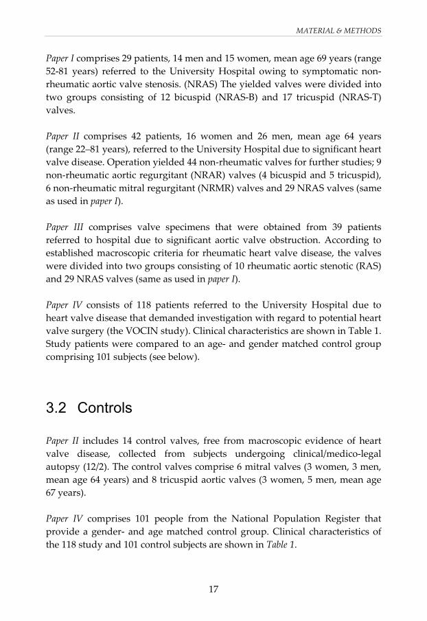
Paper I comprises 29 patients, 14 men and 15 women, mean age 69 years (range 52-81 years) referred to the University Hospital owing to symptomatic non-rheumatic aortic valve stenosis. (NRAS) The yielded valves were divided into two groups consisting of 12 bicuspid (NRAS-B) and 17 tricuspid (NRAS-T) valves. Paper II comprises 42 patients, 16 women and 26 men, mean age 64 years (range 22–81 years), referred to the University Hospital due to significant heart valve disease. Operation yielded 44 non-rheumatic valves for further studies; 9 non-rheumatic aortic regurgitant (NRAR) valves (4 bicuspid and 5 tricuspid), 6 non-rheumatic mitral regurgitant (NRMR) valves and 29 NRAS valves (same as used in paper I). Paper III comprises valve specimens that were obtained from 39 patients referred to hospital due to significant aortic valve obstruction. According to established macroscopic criteria for rheumatic heart valve disease, the valves were divided into two groups consisting of 10 rheumatic aortic stenotic (RAS) and 29 NRAS valves (same as used in paper I). Paper IV consists of 118 patients referred to the University Hospital due to heart valve disease that demanded investigation with regard to potential heart valve surgery (the VOCIN study). Clinical characteristics are shown in Table 1. Study patients were compared to an age- and gender matched control group comprising 101 subjects (see below).
3.2 Controls
Paper II includes 14 control valves, free from macroscopic evidence of heart valve disease, collected from subjects undergoing clinical/medico-legal autopsy (12/2). The control valves comprise 6 mitral valves (3 women, 3 men, mean age 64 years) and 8 tricuspid aortic valves (3 women, 5 men, mean age 67 years). Paper IV comprises 101 people from the National Population Register that provide a gender- and age matched control group. Clinical characteristics of the 118 study and 101 control subjects are shown in Table 1.
MATERIAL & METHODS
18
Ethics
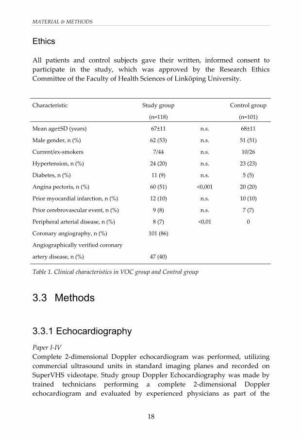
All patients and control subjects gave their written, informed consent to participate in the study, which was approved by the Research Ethics Committee of the Faculty of Health Sciences of Linköping University. Characteristic Study group
(n=118)
Control group
(n=101)
Mean age±SD (years) 67±11 n.s. 68±11
Male gender, n (%) 62 (53) n.s. 51 (51)
Current/ex-smokers 7/44 n.s. 10/26
Hypertension, n (%) 24 (20) n.s. 23 (23)
Diabetes, n (%) 11 (9) n.s. 5 (5)
Angina pectoris, n (%) 60 (51) <0,001 20 (20)
Prior myocardial infarction, n (%) 12 (10) n.s. 10 (10)
Prior cerebrovascular event, n (%) 9 (8) n.s. 7 (7)
Peripheral arterial disease, n (%) 8 (7) <0,01 0
Coronary angiography, n (%) 101 (86)
Angiographically verified coronary
artery disease, n (%)
47 (40)
Table 1. Clinical characteristics in VOC group and Control group
3.3 Methods
3.3.1 Echocardiography Paper I-IV Complete 2-dimensional Doppler echocardiogram was performed, utilizing commercial ultrasound units in standard imaging planes and recorded on SuperVHS videotape. Study group Doppler Echocardiography was made by trained technicians performing a complete 2-dimensional Doppler echocardiogram and evaluated by experienced physicians as part of the
MATERIAL & METHODS
19
clinical everyday routine. Based on the study group Doppler Echocardiography, a decision was made regarding a possible heart valve operation. Paper III and IV Patients with a history of rheumatic fever and/or MS due to a typical funnel-shaped mitral apparatus, were considered to have valve dysfunction of rheumatic origin. Paper IV Control subjects were examined by two experienced technicians to exclude significant heart valve disease. One experienced cardiologist, blinded to all other results, evaluated all the control studies.
3.3.2 Biochemistry Paper I-IV Blood samples for biochemistry including white blood cell count (WBC), haemoglobin, platelet count, erythrocyte sedimentation rate (SR), C-reactive Protein (CRP), Rheumatoid Factor (RF) and HLA-B27 were obtained from study patients as well as control subjects, at the time of enrolment in the study. Serum protein electrophoresis was performed for analysis of inflammation sensiteive proteins (ISP’s) All biochemical analyses were performed at the Department of Clinical Chemistry at the University Hospital, Linköping. CRP was considered positive at a cut-off level of ≥ 10mg/L.
3.3.3 Histopathological analyses Paper I-III After fixation in 10% formalin, the valves were measured and examined macroscopically as described by Schoen 51. A representative sample regarding calcification and fibrous thickening was taken from each valve. One section was taken from each cusp, each representing areas of thickening and calcification, but also including areas with minimal or no changes, as calcification and fibrosis are not uniform processes. In valves appearing macroscopically normal, the sample was taken from the centre of the valve. All grossly calcified valves were decalcified in 10% formic acid solution for 24 hours, then processed and cut in 4-μm sections and stained with haematoxylin–eosin (H&E), van Gieson (VG), von Kossa and Perls stains.
MATERIAL & METHODS
20
Reference samples of all stains, before and after decalcification, were compared, thereby ensuring that the process of decalcification did not interfere with the staining and hence with the results/interpretation of the staining. Calcification was estimated by analysis of the gross specimen and by microscopic analysis of sections stained with the von Kossa stain, which stains calcium and phosphates. The extent of valvular calcification was arbitrarily graded as previously described by Subramanian et al. 52 as 0=absent, 1+=mild, 2+=moderate or 3+=severe. The degree of cusp thickening was arbitrarily graded as 0=absent, 1+=increased valvular thickness only in the apposition area of the valve, 2+=increased valvular thickness beyond the apposition area but not involving the entire valve, 3+=increased thickness of the valve by fibrosis in the entire valve or more limited areas of fibrosis which distorts the cusp shape. The degree of microcalcifications was semiquantitatively and arbitrarily categorized as 0=absent, trace=deposits not clearly visible on low power (25×), mild=scattered loose deposits or dense focal deposits covering <2 high-power fields (HPFs) (400×), moderate=dense deposits in >2 HPFs and <6 HPFs, or severe=dense deposits in 6 or more HPFs. Furthermore, both the localization of the calcification and the localization of the fibrosis were investigated. The valves were evaluated for fresh haemorrhage on the H&E-stained sections and for old haemorrhage (haemosiderin deposits) with the Perls stain. Similar semiquantitative, arbitrary categorisation was used for both, which were graded as 0=absent, (+)=trace=hemosiderin deposits or fresh haemorrhage seen focally in 1 HPF (400×), mild=hemosiderin deposits or fresh haemorrhage seen in 2 HPFs, moderate=deposits seen in >2 HPFs and <6 HPFs, severe=deposits in 6 or more HPFs. One reviewer, blinded as to the type of valve dysfunction and to the clinical history, was used for all the histological and immunohistochemical analyses.
3.3.4 Immunohistochemical studies Paper I-III For determination of types of mononuclear inflammatory cells, additional sections were stained with antibodies for CD3 (pan-T cell antigen, Dako, Carpinteria, CA, USA; 1:400), CD20 (pan-B cell antigen, Dako; 1:400), and CD68 (macrophage antigen, DakoCytomation, Glostrup, Denmark, 1:100). The valves were investigated regarding the presence and localization of mononuclear cell infiltration. The degree of mononuclear cell infiltration was semiquantitatively and arbitrarily determined as previously described by Stratford et al. 53: 0=no inflammatory cells present, 1+=occasional scattered cells or one group of 20 cells in a cusp section, 2+=several groups of 20 cells or more
MATERIAL & METHODS
21
in a cusp section, 3+=many groups of >20 cells or one group of 100 cells or more in a cusp section. For determination of the presence of apolipoprotein (apo) A-I and apo B, sections were stained with goat polyclonal antibody to human apo A-I (Abcam Ltd., UK; 1:800) and goat polyclonal antibody to human apo B (Abcam Ltd.; 1:800) and visualized with LSAB DakoCytomation K0690. Staining was by TechMate 500 according to standard protocol. The degree of apo deposition was semiquantitatively categorized as follows: 0=absent, 1+=deposits in <5% of the cusp, 2+=deposits in 5–25% of the cusp, 3+=deposits in 26–50% of the cusp, 4+=deposits in 51–75% of the cusp, 5+=deposits in >75% of the cusp. If more than one cusp was involved, then the one with the greatest deposits was scored.
3.3.5 Questionnaire Paper IV Each participant in the study was asked to complete a questionnaire regarding the presence of rheumatic symptoms/diseases in themselves and their first grade relatives (Table 2).
Disease/Symptom
Psoriasis and/or pustulosis palmoplantaris Iritis Back and/or neck-pain Discogen back or neck-pain Herniatic disc surgery Arthrosis and/or articular prosthesis Soft tissue rheumatic pain Inflammatory bowel disease Sarcoidosis Rheumatoid arthritis Ankylosing spondylitis Severe pharyngitis, including acute tonsillitis Thyroiditis, asthma, diabetes Appendicitis
Table 2. Symptoms/diseases used in questionnaire regarding the presence of rheumatic disease.
MATERIAL & METHODS
22
The presence or absence of a symptom or disease in the study participant was entered as 1/0 whereas the presence or absence in the first-degree relatives was entered as the number of first-degree relatives revealing the symptom/disease.
3.3.6 History and status regarding rheumatic disease Paper IV Each patient and each control subject underwent a rheumatological examination and their medical records were evaluated regarding the presence of rheumatic symptoms/disease. These examinations were performed by one of two experienced rheumatologists. There was however, no blinding as to whether each person belonged to the study or the control group. The extent of rheumatic disease was arbitrarily graded as; 0=none, 1=arthrosis, 2=arthritis.
3.3.7 Statistical analyses Data were compared on the one hand between the study group and the control group (paper IV), on the other between subgroups of the study group (paper I-III) and between subgroups of the study group and control valves collected from subjects undergoing clinical/medico-legal autopsy (paper II). Comparisons between groups were performed using the t test for parametric values and the Mann–Whitney test for non-parametric, non-dependent samples. Statistical significance was defined as p<0,05.
RESULTS
23
4 RESULTS
4.1 Comparison between tricuspid and bicuspid aortic valves regarding T lymphocyte infiltration in non-rheumatic aortic stenosis (I)
4.1.1 Cusp thickening, fibrosis and calcification All the valves had an increase in cusp thickness although significantly more pronounced in the NRAS-B valves (p<0,05). Fibrosis was seen in all valves without significant differences. NRAS-T and NRAS-B valves showed a moderate to severe degree of calcification although the calcification was significantly more pronounced in NRAS-B valves (p<0,05). The two groups also differed regarding the distribution of calcification. Whereas the NRAS-T valves mainly revealed calcification at the base of the cusps, most of the NRAS-B valves showed a diffuse distribution.
4.1.2 Mononuclear cell infiltration Mononuclear cells were seen in the majority of NRAS-T valves and in all the NRAS-B valves without significant differences between tricuspid and bicuspid valves regarding the degree of lymphocyte infiltration. B cells were present in approximately half of the NRAS-T as well as the NRAS-B valves. However, in a majority of the NRAS-T and in all the NRAS-B valves, T cells dominated the lymphocyte infiltration. In addition, 3/15 NRAS-T and 6/12 NRAS-B valves revealed small numbers of plasma cells. The majority of both NRAS-T and NRAS-B valves showed a diffuse pattern of mononuclear cell infiltration. Only one valve had a mononuclear cell infiltrate exclusively localised to the site of calcification.
RESULTS
24
4.2 Role of inflammation in non-rheumatic, regurgitant heart valve disease (II)
4.2.1 Cusp thickening, fibrosis and calcification Almost all regurgitant valves, and all stenotic aortic valves revealed an increase in cusp thickness. Diffuse distribution of fibrosis was seen in almost all NRAS valves whereas the regurgitant valves were evenly distributed between diffuse and apposition fibrosis. Mineralization was sparse and only detectable in 3 regurgitant valves. Due to our method of decalcification in 10% formic acid solution for 24 hours, we were unable to evaluate the von Kossa stain for mineralization in the stenotic valves. Only a minority of the regurgitant valves showed any calcification.
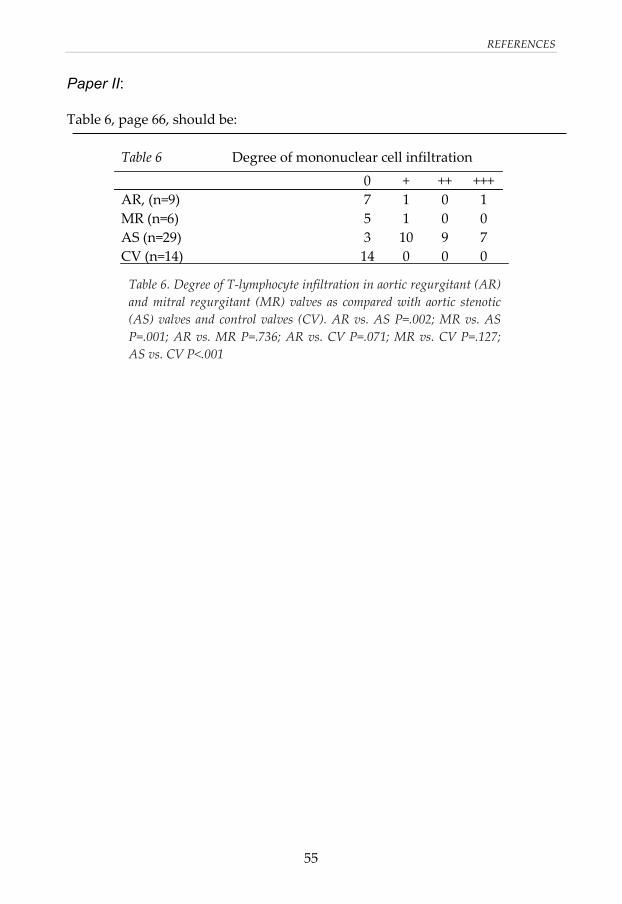
4.2.2 Mononuclear cell infiltration Only 2/9 NRAR and 1/6 NRMR valves revealed lymphocyte infiltration and none of the 14 macroscopically normal control valves. The two NRAR revealed presence of B cells but no plasma cells, whereas the NRMR valve revealed neither B cells nor plasma cells. There was a significantly smaller number of macrophages in regurgitant and control valves as compared to stenotic, aortic valves. The macrophages appeared primarily in the interstitium of the valve, less frequently intermixed in fibrotic areas adjacent to the calcium deposits or in subendocardial location. The two NRAR valves with 3+ macrophage infiltration also revealed lymphocyte infiltration as well as calcification. The regurgitating valves did not differ from the control valves regarding apolipoprotein deposition. However, regurgitant aortic and mitral valves as well as the control valves revealed significant lower degrees of both apo B and apo A-I as compared to the stenotic aortic valves.
RESULTS
25
4.3 Inflammatory characteristics of stenotic aortic valves – a comparison between rheumatic and non-rheumatic aortic stenosis (III) Ten out of 39 valves, 26% (7 men, 3 women, mean age 64±7 years) revealed postinflammatory changes with severely distorted and fused cusp margins, resulting in a central triangular orifice. These valves were considered as RAS while the remaining stenotic aortic valves were judged as NRAS. Seventeen NRAS valves, 43% (7 men, 10 women, mean age 71±7 years) were considered to be tricuspid (NRAS-T) and 12 NRAS valves, 31% (7 men and 5 women, mean age 67±8 years) to be bicuspid (NRAS-B). Thirty percent of the RAS patients and 7% of the NRAS patients had a history of rheumatic fever. Two RAS patients with a history of rheumatic fever also revealed a significant MS, while the third RAS patient had been operated on 12 years earlier because of MS. The remaining 7 RAS patients presented normal mitral valve leaflets with mostly mild MR.
Figure 6. Aortic valve showing a large group of lymphocytes in the center adjacent to thick walled neovessels. Also seen are smaller thin-walled neovessels. (Hematoxylin and Eosin, 40x). α = thickwalled vessels, β = thinwalled vessels, γ = 3+ lymphocyte infiltration.
α
α
α β
γ
RESULTS
26
4.3.1 Histopathological and immunohistochemical analyses All the aortic valves, rheumatic as well as non-rheumatic, revealed mild to moderate cusp thickening. The fibrosis was in most cases localised diffusely and only two valves revealed apposition fibrosis. Complete fibrous destruction of the normal layered architecture was seen in 30% of RAS valves and in 17% of NRAS valves. However, several valves revealed partial destruction of valve architecture, which was seen in 70% of RAS and in 79% of NRAS valves. Thirty percent of the RAS valves and 41% of the NRAS valves revealed neovascularisation characterised by small irregular, thin as well as thick-walled vessels within the basal third of the cusp (Figure 6). Among valves revealing neovessels, these could be seen extending beyond the basal third of the valve in 1/3 RAS valves and 4/12 NRAS valves. Calcification was seen in all valves, ranging from mild to severe and the degree of calcification did not differ significantly between the groups. Whereas the NRAS-T valves revealed calcification predominantly at the base of the cusp, the calcification in the majority of the RAS and NRAS-B valves, was diffusely localised (Figure 7).
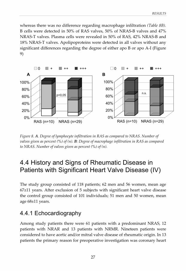
Figure 7.Localisation of calcification, number of valves given as percent (%) of (n).NRAS-T= non-rheumatic tricuspid aortic stenosis, NRAS-B=non-rheumatic bicuspid aortic stenosis, RAS= rheumatic aortic stenosis. T cells were seen in 80% of the RAS and 90% of the NRAS valves. The degree of T cell infiltration was significantly lower in RAS than in NRAS (Figure 8A),
0%
20%
40%
60%
80%
100%
NRAS-T(n=17)
NRAS-B(n=12)
RAS (n=10)
Base Apposition Diffuse
RESULTS
27
whereas there was no difference regarding macrophage infiltration (Table 8B). B cells were detected in 50% of RAS valves, 50% of NRAS-B valves and 47% NRAS-T valves. Plasma cells were revealed in 50% of RAS, 42% NRAS-B and 18% NRAS-T valves. Apolipoproteins were detected in all valves without any significant differences regarding the degree of either apo B or apo A-I (Figure 9)
Figure 8. A. Degree of lymphocyte infiltration in RAS as compared to NRAS. Number of valves given as percent (%) of (n). B: Degree of macrophage infiltration in RAS as compared to NRAS. Number of valves given as percent (%) of (n).
4.4 History and Signs of Rheumatic Disease in Patients with Significant Heart Valve Disease (IV)
The study group consisted of 118 patients; 62 men and 56 women, mean age 67±11 years. After exclusion of 5 subjects with significant heart valve disease the control group consisted of 101 individuals; 51 men and 50 women, mean age 68±11 years.
4.4.1 Echocardiography Among study patients there were 61 patients with a predominant NRAS, 12 patients with NRAR and 13 patients with NRMR. Nineteen patients were considered to have aortic and/or mitral valve disease of rheumatic origin. In 13 patients the primary reason for preoperative investigation was coronary heart
0%
20%
40%
60%
80%
100%
RAS (n=10) NRAS (n=29)
0 + ++ +++
p<0,05
0%
20%
40%
60%
80%
100%
RAS (n=10) NRAS (n=29)
0 + ++ +++
n.s.
BA
RESULTS
28
disease with concomitant valve disease and a possible need for surgical heart valve intervention. The subgroup of NRAS consisted of 61 patients; 27 men and 34 women (mean age 70±8 years) with a mean maximal velocity across the aortic valve of 4,8 m/s and a mean aortic valve cross-sectional area of 0,6 cm2.
4.4.2 Questionnaire Evaluation of the questionnaire did not reveal any differences in listed symptoms/conditions between the study group, the NRAS subgroup and the control group. Neither were there any differences between the groups regarding symptoms/diseases in first-degree relatives.
4.4.3 History and status regarding rheumatic disease The rheumatological examination did not show any differences between study patients and controls regarding the medical record data or physical examination.
4.4.4 Biochemistry WBC and SR levels were significantly increased in the study group when compared to the control group. The proportion of subjects with elevated CRP was also significantly increased in the study group. A similar pattern was seen in the NRAS subgroup (Table 3). Serum protein electrophoresis revealed significantly reduced albumin levels in the study group and in the NRAS subgroup when compared to the controls. Furthermore, the serum protein electrophoresis showed significantly increased values regarding orosomucoid, haptoglobin and immunoglobulin A, in both the study group and NRAS subgroup, when compared to the control group (Table 3). The α-1-antitrypsin levels were significantly lower in the study group and NRAS subgroup when compared to the control group. When analysing the subgroup of heart valve patients devoid of coronary artery disease as judged by normal coronary angiograms, there was still a highly significant increase in WBC and haptoglobin and reduced α-1-antitrypsin in the NRAS group when compared to control subjects. In addition the proportion of subjects with elevated CRP was significantly increased.
RESULTS
29
RF was seen in equal proportions in the study group as in the control group (positive/negative, study group; 10/107, control group; 9/92). The percentage of HLA-B27 positive subjects was significantly increased in patients with non-rheumatic AR or rheumatic mitral valve disease. VOC group
Table 3. Gender, age and biomarkers in the VOC group, control group and NRAS subgroup. Data are given as mean±S.D, CRP as % positive. In a number of patients, as has been described in paper II, the excised valves were examined macro- and microscopically. The number of HLA-B27 positive subjects appeared to be remarkably higher in the group with histologically verified non-rheumatic AR.
DISCUSSION
30
5 GENERAL DISCUSSION
5.1 Signs of inflammation in different types of heart valve disease
In 1904 Mönckeberg published his paper “Der normale histologische bau und die sklerose der aortenklappen” 27 where he concluded that AS was due to either a post-rheumatic condition or atherosclerosis. In the middle of the 20th century the general opinion regarding calcific AS stated that it was either due to a complication of rheumatic endocarditis, congenital bicuspid aortic valves or congenitally stenotic aortic valves 30.
Figure 9. NRAS-B valve. ALP-B deposits staining dark brown on the special stain. The deposits are very prominent and are seen in an area where the architecture of the valve has been completely disrupted by fibrosis. Prominent thin-walled neovessels (*) demonstrating endothelial lining, are seen in close proximity to the ALP-B deposits (100x).
*
***
**
DISCUSSION
31
In the 1970s, by works from Roberts and Pomerance, it became accepted that a third aetiological group existed, namely a degenerative, wear-and-tear, form of AS, preferably affecting the aged part of the population. . In the beginning of the 90s several studies were published revealing signs of inflammation in tricuspid AS, that both in an epidemiological and histopathological view revealed similarities to athero-sclerosis. T cells and macrophages are present in both early stages of the disease process and in valves with severe stenosis removed at surgery 37,38. Focal accumulations of neutral lipids, low density lipoprotein (LDL) cholesterol and lipoprotein(a) have been demonstrated and in addition, there is evidence for local lipid oxidation 41,54. Moreover, several proteins associated with tissue calcification have been localised in sclerotic and stenotic tricuspid aortic valves and in vitro studies have revealed that osteopontin is produced by a subset of lesion macrophages 40. In addition, the bone morphogenetic proteins 2 and 4 are expressed by myofibroblasts 40,55. Altogether, the degenerative AS does not seem to be a passive state due to wear-and-tear, but an active disease process with many similarities to atherosclerosis. Mechanical and/or tensile stress on the leaflets has been proposed as an important initiating factor in the disease process. One major evidence for this hypothesis is that calcific AS appears at an earlier age in patients with a bicuspid aortic valve. However, the disease process at the tissue level in bicuspid aortic valves has not been specifically investigated. In our study comparing bicuspid and tricuspid stenotic aortic valves (I), there were similar infiltrations and distributions of inflammatory cells, i.e. T cells, B cells, macrophages and plasma cells. However there was a significantly increased thickening of the cusps (P<0,05) and degree of calcification (p<0,05) in NRAS-B when compared to NRAS-T. The bicuspid valves differed from the tricuspid valves in another respect as well, namely how calcification was localised in the valve cusps. The tricuspid valves principally revealed calcification at the base of the cusp whereas the majority of the bicuspid valves had a diffusely localised calcification, a difference previously described by Isner 56. The differences in thickening and calcification may be explained by different mechanical and tensile stress in bicuspid and tricuspid valves. Whereas there are relatively few aetiological grounds for AS, the aetiology of the regurgitant lesions is multifactorial. Both the mitral and the aortic valve
DISCUSSION
32
can develop regurgitation due to a variety of changes/diseases in the leaflets, the supporting fibrous ring or, in the case of the mitral valve, the supporting system of chordae tendineae/papillary muscles. There are also non-cardiac, inflammatory conditions that are associated with an increased risk of developing valve regurgitation, e.g. scleroderma, systemic lupus erythema-tosus (mitral valve) and rheumatoid arthritis and ankylosing spondylitis (aortic valve). Leaflet prolapse is, however, considered to be the most frequent cause of MR and prolaps can also occur in AR. Prolapse is usually considered to be associated with myxomatous degeneration but an inflammatory component has also been reported. Tomaru et al. demonstrated postinflammatory changes in 19/33 cases of mitral and/or aortic valve prolapses57,58 including thick-walled vascularisation, round cell infiltration and destruction of architecture, signs usually seen in late post rheumatic fever. Interestingly, only 6 of the 33 patients presented a history of previous rheumatic fever. In our study of non-rheumatic regurgitant valves (II), comprising 6 mitral valves and 9 aortic valves, signs of inflammation were sparse. The inflammatory cell infiltrates were infrequent. Cusp thickening was a regular phenomenon and only two aortic valves were normally thin. The thickening of cusps is however a non-specific phenomenon that has been associated with both myxomatous valve degeneration and normal aging59. Mild calcification was seen in three regurgitant aortic valves and microcalcifications were not seen except for a mild calcium deposition in one regurgitant aortic valve. In addition, the deposition of apolipoproteins was mild in the regurgitant valves not differing from macroscopically normal valves. Although the non-rheumatic regurgitant valve in most cases lack signs of substantial inflammatory features, inflammatory conditions can be found. Quadrivalvular heart disease in patients with rheumatoid arthritis has been described, with aortic and mitral valve regurgitation and on necropsy, findings of rheumatoid nodules in all 4 valves 60,61. In our study was a 48-year-old male with severe rheumatoid arthritis since the age of 32. Echocardiography showed severe AR and perioperatively thickened and shrunken cusps, an antegrade velocity across the valve of 3,2 m/s, and a mean aortic valve gradient of 19 mm Hg. By histological examination, there was a pronounced T cell infiltration, a marked deposition of apolipoproteins, severe thickening of the cusps, but only minor calcification. Thus, even if
DISCUSSION
33
inflammation is a relatively unusual finding in AR, an inflammatory component may be present in occasional cases, mainly in combined aortic valve disease. Thus, non-rheumatic regurgitant valve disease does not present signs of substantial inflammation, although it cannot be ruled out completely, especially not in the presence of other chronic inflammatory conditions, e.g. rheumatoid arthritis. Furthermore, conditions that may be looked upon as non-rheumatic, may reveal postinflammatory elements.
5.2 The rheumatic aortic valve
The rheumatic heart valve disease was for a long time the only heart valve disease that was considered to be associated with inflammation. In 1966, the order of frequency of valve involvement in rheumatic valve disease was 13: Mitral valve alone 48% Mitral and aortic valve 42% Aortic valve alone 2% Mitral, aortic and tricuspid valve 4% Other combinations of valves 4% The low frequency of lone aortic valve involvement was compatible with the emerging view that lone AS represented a non-rheumatic process 13. In cases with rheumatic aortic valve engagement, the macroscopic appearance of the aortic valve is characterised by commissural fusion, cusp thickening and calcification. The histological appearance show extreme thickening due to collagen tissue, destruction of the normal layered architecture, foci of calcification and infiltration of chronic inflammatory cells13. The Aschoff granuloma, the pathognomic feature of the acute rheumatic pancarditis, is not found in chronic rheumatic heart valve disease. During the 1980s two studies 62,63 questioned the diagnostics of rheumatic heart valve disease. Goffin 63 stated that rheumatic heart valve disease was restricted to the mitral valve, not including the aortic valve. Gallo 62 on the other hand, concluded that a postinflammatory valvular scarring of non-rheumatic aetiology must exist.
DISCUSSION
34
In our study (III) we found 10 stenotic aortic valves that revealed on gross valvular pathology thickened and fused cusps, and thus were considered as being of postinflammatory origin. Histology revealed infiltration of chronic inflammatory cells, in much the same manner as in NRAS and there were no significant differences regarding the presence of destruction of valve architecture or neovascularisation. Furthermore, the postinflammatory valves revealed apolipoprotein deposition to the same extent as in NRAS. However, only 3/10 patients revealing commissural fusion had a previous history of rheumatic fever and they also presented a concomitant mitral valve stenosis. The remaining 7/10 patients lacked both history of rheumatic fever and mitral valve engagement. The histology did not differ between the patients with a past history of rheumatic fever and mitral valve stenosis, and those without. The findings raise the question whether other conditions than rheumatic fever may give rise to postinflammatory changes of the aortic valves. As has been mentioned above, Tomaru 57,58 described patients with aortic and/or mitral valve prolapse who histologically revealed postinflammatory marks but presented a history of rheumatic fever in less than 50%. Moreover, herpes simplex virus type 1 has been detected in valvular tissues from 66% of cases in a study 64 on 32 patients with rheumatic heart disease. The postinflammatory heart valve disease is possibly a more multifactorial condition than has previously been thought. If so, this could explain the relatively high, and unexpected, proportion of postinflammatory AS, 26%, in our patient population, despite low incidence of rheumatic fever. One difference with respect to the NRAS valves was noticed. The RAS valves revealed a diffuse distribution of calcification, in much the same way as non-rheumatic bicuspid AS, whereas the tricuspid valves showed a basally located calcification. The difference regarding localisation of calcification between tricuspid and bicuspid valves has previously been described by Isner et al. 56, and the question arises as to what extent the inflammatory changes depend on the underlying valve anomaly.
5.3 The bicuspid aortic valve and the significance of the underlying anomaly
The bicuspid aortic valve is the most common congenital anomaly of the heart65. The population frequency of a bicuspid aortic valve is 0,9 to 1,4% 21,32,66
DISCUSSION
35
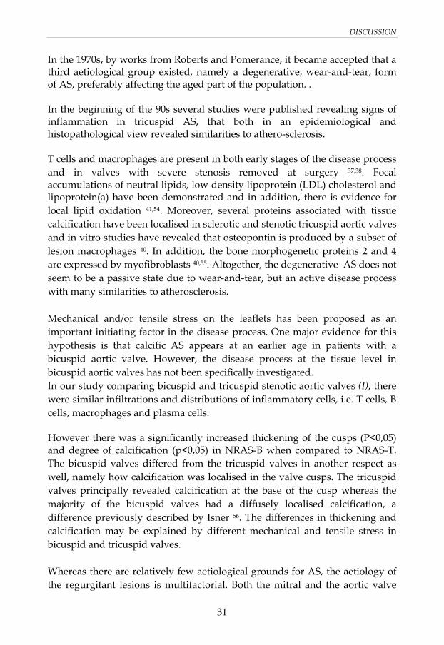
with a 2:1 male:female ratio. The presence of a bicuspid aortic valve may have a genetic basis, with a pattern of transmission in some families suggesting an autosomal dominant pattern of inheritence 67,68. An increased prevalence of a congenital bicuspid aortic valve in the parent or sibling of the proband with any form of left heart obstructive lesion has also been described 69. In the study by Sabet and co-workers 19 on 542 surgically excised congenital bicuspid valves pure regurgitation was revealed in 13% (mean age 46 years), combined stenos and regurgitation in 10% (mean age 51 years) and pure AS in 75% (mean age 65 years). In our studies (I,II,III) there was a total of 48 surgically excised aortic valves, 29 NRAS, 10 RAS and 9 non-rheumatic AR valves. Seventeen (35%) of these were bicuspid; 12 NRAS (mean age 67 years), 4 non-rheumatic AR valves (mean age 35 years), and one bicuspid RAS valve. Thus, if the bicuspid aortic valve is predominantly regurgitant, it tends to need surgical intervention at an earlier age. Those patients that, in spite of AR, remain asymptomatic with normal left ventricular function will subsequently develop valve stenosis 18. In a normal tricuspid aortic valve the distance between the lateral attachments of the cusp along the free margin, is a curved line that is longer than the distance between the lateral attachments (Figure 10A). This anatomic feature allows the cusp to move freely during opening and closure. For a bicuspid aortic valve with two equally sized cusps, the distance along the free margin of the cusp is the same as the distance between the lateral attachments, which makes it almost impossible for the valve to open (Figure 10B). Thus, to permit this type of valve to open, an excessive length of the free margin is necessary, and this fact results in concurrent inability to close properly. Bicuspid valves however often reveals unequal cusp size, with one smaller cusp and one larger cusp, the latter often with a raphe indicating the line of congenital fusion of the cusps (Figure 10C). This type of bicuspid anomaly permits the smaller cusp to open more readily whereas the bigger conjoined cusp cannot open normally, but still better than in the case of equally sized cusps. According to Roberts 17, stenosis of the bicuspid aortic valve is due to the excessive length of one or both cusps, which produces abnormal contact
DISCUSSION
36
between the cusps and consequently focal fibrous thickening and dystrophic calcification. Abnormal mechanical and shear stresses has also been proposed as a general explanation to development of AS, and the more anomalous (e.g. bicuspid aortic valves with equally sized cusps or unicuspid aortic valves) the earlier the leaflet calcification develops.
Figure 10. A: Tricuspid aortic valve where anatomy permits free movement of cusps. Lines of free margin in closed state ( ) and in opened state ( ). Distance between the lateral attachments ( ). B: Bicuspid valve with equally sized cusps, where line of free margin equals distance between the lateral attachments, which permits only limited opening of the cusps. C: Bicuspid valve with bigger cusp, 2:1 the smaller cusp. The smaller cusp anatomy permits normal opening and closure, whereas the bigger cusp will have restricted mobility. Raphe ( ).
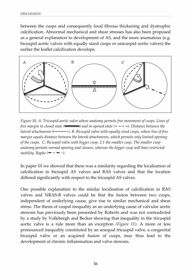
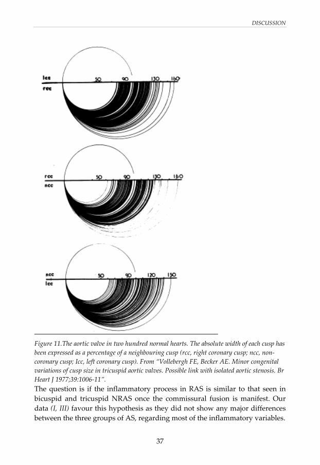
In paper III we showed that there was a similarity regarding the localisation of calcification in bicuspid AS valves and RAS valves and that the location differed significantly with respect to the tricuspid AS valves. One possible explanation to the similar localisation of calcification in RAS valves and NRAS-B valves could be that the fusion between two cusps, independent of underlying cause, give rise to similar mechanical and shear stress. The thesis of cuspal inequality as an underlying cause of valvular aortic stenosis has previously been presented by Roberts and was not contradicted by a study by Vollebergh and Becker showing that inequality in the tricuspid aortic valve is a rule more than an exception (Figure 11). A more or less pronounced inequality constituted by an unequal tricuspid valve, a congenital bicuspid valve or an acquired fusion of cusps, may thus lead to the development of chronic inflammation and valve stenosis.
A B C
DISCUSSION
37
Figure 11.The aortic valve in two hundred normal hearts. The absolute width of each cusp has been expressed as a percentage of a neighbouring cusp (rcc, right coronary cusp; ncc, non-coronary cusp; Icc, left coronary cusp). From “Vollebergh FE, Becker AE. Minor congenital variations of cusp size in tricuspid aortic valves. Possible link with isolated aortic stenosis. Br Heart J 1977;39:1006-11”. The question is if the inflammatory process in RAS is similar to that seen in bicuspid and tricuspid NRAS once the commissural fusion is manifest. Our data (I, III) favour this hypothesis as they did not show any major differences between the three groups of AS, regarding most of the inflammatory variables.
DISCUSSION
38
5.4 Inflammatory signs in heart valve disease and its similarities to the process of atherosclerosis
Research during the last decades 35,36,70,71 have shown that inflammation plays a key role in coronary artery disease and other manifestations of atherosclerosis. The atherosclerotic lesion consists of cells, connective-tissue elements, lipids, and debris. Blood-borne inflammatory and immune cells constitute an important part of an atherosclerotic lesion, as well as endothelial and smooth-muscle cells. T cells, macrophages, and mast cells infiltrate the lesion, and many of the immune cells exhibit signs of activation and produce inflammatory cytokines. The atherosclerotic lesion form at specific arterial regions, where low and oscillatory endothelial shear stress occur 72. Low endothelial shear stress has been shown to attenuate nitric oxide-dependent atheroprotection, promote LDL uptake, synthesis and permeability. Furthermore low endothelial shear stress promotes oxidation of LDL and inflammation. Studies on aortic valve sclerosis and stenosis have shown focal subendothelial plaquelike lesions on the aortic side of the cusp. Such lesions extend to the fibrosa layer. They present accumulation of “atherogenic” lipoproteins, including oxidized LDL and Lp(a), inflammatory cell infiltrates and microscopic calcification 37,38,41,54,73, thus showing similarities with atherosclerosis. The early lesions of valves are considered to be initiated by endothelial disruption due to increased mechanical or decreased endothelial shear stress, similar to that seen in early atherosclerotic lesions. Mechanical stress of the aortic valve is highest on the aortic side of the leaflets, whereas shear stress across the endothelium is lower on the noncoronary cusp then of the left and right coronary cusps, due to the absence of diastolic coronary flow. This fact likely explains why the noncoronary cusp often is first affected. Clinical factors associated with aortic sclerosis and stenosis are similar to those associated with atherosclerosis and include older age, male gender, elevated LDL and Lp(a) levels, hypertension and smoking. The similarities between calcific aortic valve disease and atherosclerosis, indicate a similar pathogenesis in the form of a general response to tissue injury. It also raises the question
DISCUSSION
39
whether AS is one disease, caused by a general and non-specific response to the underlying malformation of the valve.
5.5 The absence of articular symptoms or rheumatic disease in heart valve disease
Arthritis and chronic joint symptoms comprise the leading cause of disability among adults in industrialised countries. In 2001 the estimated prevalence of arthritis/chronic joint symptoms among U.S. adults was 33% 75, including 11% with physician-diagnosed arthritis only, 10% chronic joint symptoms only and 12% with both. Our clinical impression, and the basis for paper IV, was that patients referred to our hospital for evaluation of heart valve disease, reported a high frequency of different inflammatory conditions, mainly from the locomotor system. However, in paper IV, the investigation of 118 patients with significant heart valve disease revealed no differences regarding self-reported inflammatory symptoms/diseases when compared to 101 age- and gender-matched control subjects. Neither was there any evidence for differences in rheumatological status or history in the opinion of the rheumatologist. However, in the literature there are numerous reports on inflammatory conditions with concomitant heart valve disease. Heart valve disease in connection to rheumatoid arthritis 60,61,76-78 is widely reported but several other diseases, e.g. vasculitis disorders, systemic lupus erythematosus, Reiter’s syndrome 79,80, Whipple’s disease81 and carcinoid82, can develop heart valve engagement. However, the valves engaged show, with few exceptions, regurgitant lesions while stenotic heart valves in combination with rheumatic diseases are rare83. In our patient population, only 25 (21%) revealed a predominant regurgitant lesion and of these, only 15 patients (13%) had significant regurgitation that yielded heart valve surgery. This may explain why we did not find any increased presence of rheumatic disease among patients with heart valve disease. However, our results are in line with earlier studies reporting a low prevalence of heart valve disease in unselected materials of rheumatoid arthritis 84,85.
DISCUSSION
40
5.6 HLA-B27
The principal physiologic function of the cell surface histocompability molecules is to bind peptide fragments of foreign proteins for presentation to antigen-specific T lymphocytes. A variety of diseases have been found to be associated with certain HLA alleles. The diseases that show association with the HLA locus can be broadly grouped into three categories 43:
1. Inflammatory diseases, including ankylosing spondylitis, acute anterior uveitis and several postinfectious arthropathies, all associated with HLA-B27.
2. Inherited errors of metabolism, associated with HLA-BW47 and HLA-A. 3. Autoimmune diseases, including rheumatoid arthritis, chronic active
hepatitis, primary Sjögren syndrome and type-1 diabetes, associated mainly with alleles at the HLA-DR locus.
As has been said before, an HLA-B27-associated cardiac syndrome consisting of cardiac conduction system abnormalities and lone AR, has been identified. AR is the most common valve dysfunction associated with HLA-B27, but MR is also described in patients with ankylosing spondylitis 86,87. In paper IV we investigated the presence of HLA-B27 in the study population of 118 patients with significant heart valve disease. There was no increased prevalence of HLA-B27 when the entire study group was compared to the control group. The number of HLA-B27 positive subjects was however remarkably higher in the group with histologically verified non-rheumatic AR, 5/7 patients with a predominant AR were HLA-B27 positive. Among the HLA-B27-positive patients with AR, two revealed sacroiliitis and a wide aortic root, one had a markedly increased width of the aortic root but no signs of spondyloarthropathy whereas two had a normal width of the aortic root but bicuspid aortic valves. However, the cardiac manifestation can be the first symptom of a HLA-B27 associated spondyloarthropathy 44.
5.7 The systemic inflammation in patients with heart valve disease
In paper IV, we showed that patients with heart valve disease had significantly increased levels of inflammatory markers compared to controls. In addition,
DISCUSSION
41
the albumin levels were significantly decreased in the study group, further indicating a chronic systemic inflammation. CRP, one of the major acute-phase proteins, is synthesised in the liver and its main function is to counteract infections by acting as an opsonin for microorganisms activating the classical complement pathway 88. Circulating CRP is related to a number of cardiovascular risk factors, e.g. smoking, blood pressure and obesity 88. Several studies have also consistently reported that elevated CRP is an independent predictor for major cardiovascular events and mortality 89. CRP is the most frequently studied inflammatory biomarker in relation to calcific AS. In a study by Skowasch et al 90, CRP was localised in valve tissue of both calcific AS and degenerative aortic valve prostheses and the deposition of CRP also correlated positively to serum levels. Galante et al. 91 demonstrated elevated CRP levels in 62 patients with severe symptomatic AS when compared to 79 healthy controls. However, in a non-surgical patient group with AS, CRP levels did not correlate with stenosis severity 92. Haptoglobin, α1-antitrypsin (α1-AT), ceruloplasmin, fibrinogen and orosomucoid are all ISP’s, currently used in clinical practice in order to estimate inflammatory activity. In a screening programme aiming to identify individuals with high risk for cardiovascular disease, elevated levels of these ISP’s were related to the risk of developing hypercholesterolemia 93. Moreover, after a latency period of 10 years, they were associated with increased cardiovascular risk in men without traditional risk factors for myocardial infarction 94. Immunoglobulines are secreted from activated B cell and constitute a part of the humoral response to a variety of microorganisms. IgG, IgA and IgM differ as to their time courses and functions in the “immune memory” 88. In a study on ankylosing spondylitis 95, the levels of ESR, ISP’s, IgG, IgA and IgM were compared to clinical measures of disease activity. The levels of ESR and CRP were significantly higher in patients with peripheral joint involvement, whereas IgA and α1-AT revealed a positive correlation with disease activity in general.
DISCUSSION
42
However, in our study we found significant lower levels of α1-AT in the study group when compared to the control group, and the difference persisted in subgroups devoid of coronary artery disease. These differences are difficult to interpret. α1-AT belongs to the antiproteases and its major function is to inhibit different proteases, particularly elastase, cathepsin G and proteinase 3 96, which are normally released from neutrophils at sites of inflammation. α1-AT is most widely studied in connection with emphysema. According to the protease-antiprotease theory the alveolar wall destruction in emphysema results from an imbalance between proteases and antiproteases in the lung. Homozygous subjects with a genetic deficiency of α1-AT have a markedly enhanced tendency to develop emphysema 97. Low levels of α1-AT have also been observed more frequently in patients with spontaneous cervical dissection 98 compared to control subjects, probably due to an increased proteolytic activity against the connective tissue of the arterial wall. Significantly increased levels of inflammatory markers were found in patients with heart valve disease when compared to healthy control subjects. However, significantly elevated levels were also seen when comparing the 61 NRAS patients with the control group, supporting the view that the NRAS disease also represent a condition with inflammatory features. To avoid confounding by concomitant atherosclerosis, we evaluated the group of study patients with normal coronary angiograms. Among patients with NRAS and normal coronary angiograms, the proportion of individuals with elevated CRP levels remained significantly increased compared to the controls. Likewise, the differences in white blood cell counts and albumin remained significant in the subgroup of NRAS devoid of coronary artery disease. The findings thus strongly indicate that a systemic inflammatory component is associated with non-rheumatic heart valve disease per se. So far, we can only speculate whether the systemic inflammation is both a cause and a consequence of the valve disease. It may also be intriguing to suggest that antiinflammatory therapy would be prognostically beneficial in heart valve disease.
DISCUSSION
43
5.8 Conclusions
The results presented in this thesis can be summarised as follows: Signs of inflammation are present in non-rheumatic bicuspid aortic stenosis to the same extent as in non-rheumatic tricuspid aortic stenosis and the two entities share many of the inflammatory features seen in atherosclerosis. Signs of inflammation is not a predominant feature of regurgitant heart valve disease. RAS share many of the histopathological features of NRAS. One can not exclude the existsens of a postinflammatory condition of other cause than rheumatic fever. The similarities between calcific aortic valve disease in its different forms indicates a similar pathogenesis in the form of a general response to tissue injury. It raises the question whether AS is one disease, caused by a general and non-specific response to the underlying malformation of the valve. Clinical manifestations of non-cardiac inflammatory disease is not overrepresented in a cohort of patients with significant heart valve disease, when compared to healthy control subjects Our data suggest that the systemic inflammation in patients with heart valve disease is a consequence of the valve disease rather than a cause of concomitant atherosclerosis or rheumatic disease.
ACKNOWLEDGEMENTS
44
ACKNOWLEDGEMENTS
I wish to thank everyone who, in different ways, have contributed so that I finally could complete these studies and my thesis. I specially want to thank the following people: Mats Broqvist, my assistant supervisor, the originator of the project and my closest colleague (strictly scientific) throughout these years. For teaching me what little I can about clinical research, for your belief in times of despair and for being an exceptional role model. Lena Jonasson, my supervisor, who even when you came into the project late, lifted it to a higher level. Thora Steffenssen, my co-author who decided to take her family and move to USA when I started working in the study. For your helping me to understand a little more about valve pathology and for your fast and willing response with everything I did not understand. Only a shame that you live so far away. Eva Nylander, my mentor and the person who has taught me all I know about Clinical Physiology. For your support and for you belief in me that I could write this thesis. Birgitta Janerot Sjöberg and Henning Locht, my co-authors. Birgitta also in your capacity as Head of the Department of Clinical Physiology with helping me to find the time to finish this work. Christina Andersson for all your help with keeping all papers in order all the time. ”We can ask Christina” has solved many problems during the last six months! Margareta Nordvall and Miriam Johansson, who performed all the echocardiography examinations in the control material. Sally Chesterton for patiently translating my manuscripts to understandable English, but also for giving me lots of sensible advice. Nora Östrup, for checking the language of this thesis.
ACKNOWLEDGEMENTS
45
All the technicians and other personnel at the Department of Clinical Physiology, for making the department the best workplace there is. All my colleagues at the Heart Centre, especially the colleagues at the Department of Clinical Physiology, who have had to draw a heavier workload during the time I have concluded my research. Niels Erik Nielsen and Anders Bergström, for joyous acclamations. Mother and Father. Mother, for endless care and attention. Father, for setting my shining example in every respect of life. Thilda, Britta, Emil, Ola, Marcus and Fredrik, our children. Last but above all others, Helene – without you this work would not have been possible. Without you I am just half a man. This thesis is for you!
Financial support for the present study was obtained from:
The Swedish Heart-Lung Foundation
Östergötland County Council
Linköping University Hospital.
Lions Research Foundation
REFERENCES
46
REFERENCES
1. Singh JP, Evans JC, Levy D, Larson MG, Freed LA, Fuller DL, Lehman B, Benjamin EJ. Prevalence and clinical determinants of mitral, tricuspid, and aortic regurgitation (the Framingham Heart Study). Am J Cardiol 1999;83:897-902. 2. Stewart BF, Siscovick D, Lind BK, Gardin JM, Gottdiener JS, Smith VE, Kitzman DW, Otto CM. Clinical factors associated with calcific aortic valve disease. Cardiovascular Health Study. J Am Coll Cardiol 1997;29:630-4. 3. Otto CM. Why is aortic sclerosis associated with adverse clinical outcomes? J Am Coll Cardiol 2004;43:176-8. 4. Chandra HR, Goldstein JA, Choudhary N, O'Neill CS, George PB, Gangasani SR, Cronin L, Marcovitz PA, Hauser AM, O'Neill WW. Adverse outcome in aortic sclerosis is associated with coronary artery disease and inflammation. J Am Coll Cardiol 2004;43:169-75. 5. Olson LJ, Subramanian R, Ackermann DM, Orszulak TA, Edwards WD. Surgical pathology of the mitral valve: a study of 712 cases spanning 21 years. Mayo Clin Proc 1987;62:22-34. 6. Otto CM. Clinical practice. Evaluation and management of chronic mitral regurgitation. N Engl J Med 2001;345:740-6. 7. Waller BF, Howard J, Fess S. Pathology of mitral valve stenosis and pure mitral regurgitation--Part II. Clin Cardiol 1994;17:395-402. 8. Dare AJ, Veinot JP, Edwards WD, Tazelaar HD, Schaff HV. New observations on the etiology of aortic valve disease: a surgical pathologic study of 236 cases from 1990. Hum Pathol 1993;24:1330-8. 9. Bonow R, Braunwald E. Valvular Heart Disease. In: Zipes D, Libby P, Bonow R, Braunwald E, eds. Braunwald's Heart Disease. A textbook of cardiovascular medicine. Philadelphia: Elsevier Saunders, 2005:1553-1632. 10. Aronow WS, Schwartz KS, Koenigsberg M. Correlation of serum lipids, calcium, and phosphorus, diabetes mellitus and history of systemic hypertension with presence or absence of calcified or thickened aortic cusps or root in elderly patients. Am J Cardiol 1987;59:998-9. 11. Lindroos M, Kupari M, Valvanne J, Strandberg T, Heikkila J, Tilvis R. Factors associated with calcific aortic valve degeneration in the elderly. Eur Heart J 1994;15:865-70.
REFERENCES
47
12. Mohler ER, Sheridan MJ, Nichols R, Harvey WP, Waller BF. Development and progression of aortic valve stenosis: atherosclerosis risk factors--a causal relationship? A clinical morphologic study. Clin Cardiol 1991;14:995-9. 13. Olsen EGJ. The pathology of the heart. London and Basingstoke: The Macmillan Press Ltd, 1980. 14. Osler W. The bicuspid condition of the aortic valves. Transactions of the Association of American Physicians 1886;1:185-192. 15. Edwards JE. The congenital bicuspid aortic valve. Circulation 1961;23:485-88. 16. Fenoglio JJ, McAllister HA, DeCastro CM, Davia JE, Cheitlin MD. Congenital bicuspid aortic valve after age 20. Am J Cardiol 1977;39:164-9. 17. Roberts WC. The congenitally bicuspid aortic valve. Am J Cardiol 1970;26:72-83. 18. Lewin MB, Otto CM. The bicuspid aortic valve: adverse outcomes from infancy to old age. Circulation 2005;111:832-4. 19. Sabet HY, Edwards WD, Tazelaar HD, Daly RC. Congenitally bicuspid aortic valves: a surgical pathology study of 542 cases (1991 through 1996) and a literature review of 2,715 additional cases. Mayo Clin Proc 1999;74:14-26. 20. Hahn RT, Roman MJ, Mogtader AH, Devereux RB. Association of aortic dilation with regurgitant, stenotic and functionally normal bicuspid aortic valves. J Am Coll Cardiol 1992;19:283-8. 21. Larson EW, Edwards WD. Risk factors for aortic dissection: a necropsy study of 161 cases. Am J Cardiol 1984;53:849-55. 22. Pachulski RT, Weinberg AL, Chan KL. Aortic aneurysm in patients with functionally normal or minimally stenotic bicuspid aortic valve. Am J Cardiol 1991;67:781-2. 23. Roberts CS, Roberts WC. Dissection of the aorta associated with congenital malformation of the aortic valve. J Am Coll Cardiol 1991;17:712-6. 24. Beroukhim RS, Kruzick TL, Taylor AL, Gao D, Yetman AT. Progression of aortic dilation in children with a functionally normal bicuspid aortic valve. Am J Cardiol 2006;98:828-30. 25. Olson LJ, Subramanian R, Edwards WD. Surgical pathology of pure aortic insufficiency: a study of 225 cases. Mayo Clin Proc 1984;59:835-41. 26. Becker AE, Becker MJ, Edwards JE. Anomalies associated with coarctation of aorta: particular reference to infancy. Circulation 1970;41:1067-75. 27. Mönckeberg JG. Der normale histologische bau und die sklerose der aortenklappen. Virchows Arch 1904;176:472-514. 28. Karsner HT, Koletsky S. Calcific disease of the aortic valve. Philadelphia: J.B. Lippincott, 1947.
REFERENCES
48
29. Campbell M. Calcific aortic stenosis and congenital bicuspid aortic valves. Br heart J 1968;30:606-616. 30. Edwards JE. Calcific aortic stenosis: Pathologic features. Staaf meetings of the Mayo clinic 1961;36:444-51. 31. Pomerance A. Pathogenesis of aortic stenosis and its relation to age. Br Heart J 1972;34:569-74. 32. Roberts WC. Anatomically isolated aortic valvular disease. The case against its being of rheumatic etiology. Am J Med 1970;49:151-9. 33. Roberts WC. The structure of the aortic valve in clinically isolated aortic stenosis: an autopsy study of 162 patients over 15 years of age. Circulation 1970;42:91-7. 34. Passik CS, Ackermann DM, Pluth JR, Edwards WD. Temporal changes in the causes of aortic stenosis: a surgical pathologic study of 646 cases. Mayo Clin Proc 1987;62:119-23. 35. Jonasson L, Holm J, Skalli O, Bondjers G, Hansson GK. Regional accumulations of T cells, macrophages, and smooth muscle cells in the human atherosclerotic plaque. Arteriosclerosis 1986;6:131-8. 36. Hansson GK, Holm J, Jonasson L. Detection of activated T lymphocytes in the human atherosclerotic plaque. Am J Pathol 1989;135:169-75. 37. Olsson M, Dalsgaard CJ, Haegerstrand A, Rosenqvist M, Ryden L, Nilsson J. Accumulation of T lymphocytes and expression of interleukin-2 receptors in nonrheumatic stenotic aortic valves. J Am Coll Cardiol 1994;23:1162-70. 38. Otto CM, Kuusisto J, Reichenbach DD, Gown AM, O'Brien KD. Characterization of the early lesions of 'degenerative' valvular aortic stenosis. Circulation 1994;90:844-53. 39. Mohler ER, 3rd, Adam LP, McClelland P, Graham L, Hathaway DR. Detection of osteopontin in calcified human aortic valves. Arterioscler Thromb Vasc Biol 1997;17:547-52. 40. O'Brien KD, Kuusisto J, Reichenbach DD, Ferguson M, Giachelli C, Alpers CE, Otto CM. Osteopontin is expressed in human aortic valvular lesions [see comments]. Circulation 1995;92:2163-8. 41. O'Brien KD, Reichenbach DD, Marcovina SM, Kuusisto J, Alpers CE, Otto CM. Apolipoproteins B, (a), and E accumulate in the morphologically early lesion of 'degenerative' valvular aortic stenosis. Arterioscler Thromb Vasc Biol 1996;16:523-32. 42. Olsson M, Rosenqvist M, Nilsson J. Expression of HLA-DR antigen and smooth muscle cell differentiation markers by valvular fibroblasts in degenerative aortic stenosis. J Am Coll Cardiol 1994;24:1664-71.
REFERENCES
49
43. Abbas A. Diseases of Immunity. In: Kumar V, Abbas A, Fausto N, eds. Robbins and Cotran Pathologic Basis of Disease. Philadelphia: Elsevier Saunders, 2005:193-267. 44. Bergfeldt L. HLA-B27-associated cardiac disease. Ann Intern Med 1997;127:621-9. 45. Good AE. Reiter's disease: a review with special attention to cardiovascular and neurologic sequellae. Semin Arthritis Rheum 1974;3:253-86. 46. Schilder DP, Harvey WP, Hufnagel CA. Rheumatoid spondylitis and aortic insufficiency. N Engl J Med 1956;255:11-7. 47. Qaiyumi S, Hassan ZU, Toone E. Seronegative spondyloarthropathies in lone aortic insufficiency. Arch Intern Med 1985;145:822-4. 48. Bergfeldt L. HLA B27-associated rheumatic diseases with severe cardiac bradyarrhythmias. Clinical features and prevalence in 223 men with permanent pacemakers. Am J Med 1983;75:210-5. 49. Bergfeldt L, Edhag O, Vedin L, Vallin H. Ankylosing spondylitis: an important cause of severe disturbances of the cardiac conduction system. Prevalence among 223 pacemaker-treated men. Am J Med 1982;73:187-91. 50. Bergfeldt L, Insulander P, Lindblom D, Moller E, Edhag O. HLA-B27: an important genetic risk factor for lone aortic regurgitation and severe conduction system abnormalities. Am J Med 1988;85:12-8. 51. Schoen FJ. Surgical pathology of removed natural and prosthetic heart valves. Hum Pathol 1987;18:558-67. 52. Subramanian R, Olson LJ, Edwards WD. Surgical pathology of pure aortic stenosis: a study of 374 cases. Mayo Clin Proc 1984;59:683-90. 53. Stratford N, Britten K, Gallagher P. Inflammatory infiltrates in human coronary atherosclerosis. Atherosclerosis 1986;59:271-6. 54. Olsson M, Thyberg J, Nilsson J. Presence of oxidized low density lipoprotein in nonrheumatic stenotic aortic valves. Arterioscler Thromb Vasc Biol 1999;19:1218-22. 55. Mohler ER, 3rd, Gannon F, Reynolds C, Zimmerman R, Keane MG, Kaplan FS. Bone formation and inflammation in cardiac valves. Circulation 2001;103:1522-8. 56. Isner JM, Chokshi SK, DeFranco A, Braimen J, Slovenkai GA. Contrasting histoarchitecture of calcified leaflets from stenotic bicuspid versus stenotic tricuspid aortic valves. J Am Coll Cardiol 1990;15:1104-8. 57. Tomaru T. The multifactorial etiology of mitral valve prolapse, a new entity of postinflammatory mitral valve prolapse. Herz 1988;13:271-6.
REFERENCES
50
58. Tomaru T, Uchida Y, Mohri N, Mori W, Furuse A, Asano K. Postinflammatory mitral and aortic valve prolapse: a clinical and pathological study. Circulation 1987;76:68-76. 59. Schoen F. The Heart. In: Kumar V, Abbas A, Fausto N, eds. Robbins and Cotran Pathologic Basis of Disease. Philadelphia: Elsevier Saunders, 2005:555-618. 60. Carpenter DF, Golden A, Roberts WC. Quadrivalvular rheumatoid heart disease associated with left bundle branch block. Am J Med 1967;43:922-9. 61. Roberts WC, Kehoe JA, Carpenter DF, Golden A. Cardiac valvular lesions in rheumatoid arthritis. Arch Intern Med 1968;122:141-6. 62. Gallo P, Castelvetere M, Papalia U, Tonelli E, Bernucci P, d'Amati G, Marino B. Postinflammatory scarring of cardiac valves of rheumatic and nonrheumatic etiology. Am J Cardiovasc Pathol 1990;3:101-5. 63. Goffin YA, Leclerc JL, Primo GC. Histopathology of the aortic valve in patients with a previous history of acute rheumatic fever. An analysis of 63 surgical specimens. Acta Cardiol 1984;39:329-39. 64. Pan ZG, Wang XN, Li YW, Zhang HY, Archard LC. Detection of herpes simplex virus type 1 in rheumatic valvular tissue. Chin Med J (Engl) 2005;118:370-6. 65. Wauchope G. The clinical importance of variations in the number of cusps forming the aortic and pulmonary valves. . Quarterly Journal of Medicine 1928;21:383-99. 66. Gray GW, Salisbury DA, Gulino AM. Echocardiographic and color flow Doppler findings in military pilot applicants. Aviat Space Environ Med 1995;66:32-4. 67. Clementi M, Notari L, Borghi A, Tenconi R. Familial congenital bicuspid aortic valve: a disorder of uncertain inheritance. Am J Med Genet 1996;62:336-8. 68. Huntington K, Hunter AG, Chan KL. A prospective study to assess the frequency of familial clustering of congenital bicuspid aortic valve. J Am Coll Cardiol 1997;30:1809-12. 69. Lewin MB, McBride KL, Pignatelli R, Fernbach S, Combes A, Menesses A, Lam W, Bezold LI, Kaplan N, Towbin JA, Belmont JW. Echocardiographic evaluation of asymptomatic parental and sibling cardiovascular anomalies associated with congenital left ventricular outflow tract lesions. Pediatrics 2004;114:691-6. 70. Hansson GK. Inflammation, atherosclerosis, and coronary artery disease. N Engl J Med 2005;352:1685-95. 71. Jonasson L, Linderfalk C, Olsson J, Wikby A, Olsson AG. Systemic T-cell activation in stable angina pectoris. Am J Cardiol 2002;89:754-6.
REFERENCES
51
72. Chatzizisis YS, Coskun AU, Jonas M, Edelman ER, Feldman CL, Stone PH. Role of endothelial shear stress in the natural history of coronary atherosclerosis and vascular remodeling: molecular, cellular, and vascular behavior. J Am Coll Cardiol 2007;49:2379-93. 73. Wallby L, Janerot-Sjoberg B, Steffensen T, Broqvist M. T lymphocyte infiltration in non-rheumatic aortic stenosis: a comparative descriptive study between tricuspid and bicuspid aortic valves. Heart 2002;88:348-51. 74. Vollebergh FE, Becker AE. Minor congenital variations of cusp size in tricuspid aortic valves. Possible link with isolated aortic stenosis. Br Heart J 1977;39:1006-11. 75. From the Centers for Disease Control and Prevention. Prevalence of self-reported arthritis or chronic joint symptoms among adults--United States, 2001. JAMA 2002;288:3103-4. 76. Liew M, Wilson D, Horton D, Fleming A. Successful valve replacement for aortic incompetence in rheumatoid arthritis with vasculitis. Ann Rheum Dis 1979;38:483-4. 77. Linch DC, Gillmer DJ, Whimster WF, Keates JR. Rheumatoid aortic valve prolapse requiring emergency aortic valve replacement. Br Heart J 1980;43:237-40. 78. Shimaya K, Kurihashi A, Masago R, Kasanuki H. Rheumatoid arthritis and simultaneous aortic, mitral, and tricuspid valve incompetence [letter; comment]. Int J Cardiol 1999;71:181-3. 79. Paulus HE, Pearson CM, Pitts W, Jr. Aortic insufficiency in five patients with Reiter's syndrome. A detailed clinical and pathologic study. Am J Med 1972;53:464-72. 80. Yates DB, Scott JT. Cardiac valvular disease in chronic inflammatory disorders of connective tissue. Factors influencing survival after surgery. Ann Rheum Dis 1975;34:321-5. 81. McAllister HA, Jr., Fenoglio JJ, Jr. Cardiac involvement in Whipple's disease. Circulation 1975;52:152-6. 82. Pellikka PA, Tajik AJ, Khandheria BK, Seward JB, Callahan JA, Pitot HC, Kvols LK. Carcinoid heart disease. Clinical and echocardiographic spectrum in 74 patients. Circulation 1993;87:1188-96. 83. Lerman BB, Thomas LC, Abrams GD, Pitt B. Aortic stenosis associated with systemic lupus erythematosus. Am J Med 1982;72:707-10. 84. Dawson JK, Goodson NG, Graham DR, Lynch MP. Raised pulmonary artery pressures measured with Doppler echocardiography in rheumatoid arthritis patients. Rheumatology (Oxford) 2000;39:1320-5.
REFERENCES
52
85. Gonzalez-Juanatey C, Testa A, Garcia-Castelo A, Garcia-Porrua C, Llorca J, Ollier WE, Gonzalez-Gay MA. Echocardiographic and Doppler findings in long-term treated rheumatoid arthritis patients without clinically evident cardiovascular disease. Semin Arthritis Rheum 2004;33:231-8. 86. Roldan CA, Chavez J, Wiest PW, Qualls CR, Crawford MH. Aortic root disease and valve disease associated with ankylosing spondylitis. J Am Coll Cardiol 1998;32:1397-404. 87. Vinsonneau U, Brondex A, Mansourati J, Saraux A, Cornily JC, Arles F, Godon P, Quiniou G. Cardiovascular disease in patients with spondyloarthropathies. Joint Bone Spine 2008;75:18-21. 88. Lind L. Circulating markers of inflammation and atherosclerosis. Atherosclerosis 2003;169:203-14. 89. Bisoendial RJ, Kastelein JJ, Stroes ES. C-reactive protein and atherogenesis: from fatty streak to clinical event. Atherosclerosis 2007;195:e10-8. 90. Skowasch D, Schrempf S, Preusse CJ, Likungu JA, Welz A, Luderitz B, Bauriedel G. Tissue resident C reactive protein in degenerative aortic valves: correlation with serum C reactive protein concentrations and modification by statins. Heart 2006;92:495-8. 91. Galante A, Pietroiusti A, Vellini M, Piccolo P, Possati G, De Bonis M, Grillo RL, Fontana C, Favalli C. C-reactive protein is increased in patients with degenerative aortic valvular stenosis. J Am Coll Cardiol 2001;38:1078-82. 92. Gunduz H, Akdemir R, Binak E, Tamer A, Keser N, Uyan C. Can serum lipid and CRP levels predict the "severity" of aortic valve stenosis? Acta Cardiol 2003;58:321-6. 93. Engstrom G, Hedblad B, Janzon L, Lindgarde F. Long-term change in cholesterol in relation to inflammation-sensitive plasma proteins: a longitudinal study. Ann Epidemiol 2007;17:57-63. 94. Engstrom G, Stavenow L, Hedblad B, Lind P, Tyden P, Janzon L, Lindgarde F. Inflammation-sensitive plasma proteins and incidence of myocardial infarction in men with low cardiovascular risk. Arterioscler Thromb Vasc Biol 2003;23:2247-51. 95. Ozgocmen S, Godekmerdan A, Ozkurt-Zengin F. Acute-phase response, clinical measures and disease activity in ankylosing spondylitis. Joint Bone Spine 2007;74:249-53. 96. Crawford J. Liver and Biliary Tract. In: Kumar V, Abbas A, Fausto N, eds. Robbins and Cotran Pathologic Basis of Disease. Philadelphia: Elsevier Saunders, 2005:877-937.
REFERENCES
53
97. Husain A. The Lung. In: Kumar V, Abbas A, Fausto N, eds. Robbins and Cotran Pathologic Basis of Disease. Philadelphia: Elsevier Saunders, 2005:711-72. 98. Vila N, Millan M, Ferrer X, Riutort N, Escudero D. Levels of alpha1-antitrypsin in plasma and risk of spontaneous cervical artery dissections: a case-control study. Stroke 2003;34:E168-9.
54
Errata Paper I:
Table 3, page 58, should be:
Table 3 Localisation of cusp calcification
Base Apposition Diffuse
Bicuspid aortic stenosis (n=12) 2 2 8
Tricuspid aortic stenosis (n=17) 15 0 2
Table 4, page 58, should be:
Table 4 Degree of mononuclear cell infiltration
0 + ++ +++
Bicuspid aortic stenosis (n=12) 0 5 4 3
Tricuspid aortic stenosis (n=17) 3 5 5 4
Table 3 Localisation of calcification in stenotic biscuspid aortic valves compared with stenotic tricuspid valves. Difference between the groups, p<0,05.
Table 4 Degree of mononuclear cell infiltration in stenotic bicuspid aortic valves compared with stenotic tricuspid valves. P=0,56.
Table 6. Degree of T-lymphocyte infiltration in aortic regurgitant (AR) and mitral regurgitant (MR) valves as compared with aortic stenotic (AS) valves and control valves (CV). AR vs. AS P=.002; MR vs. AS P=.001; AR vs. MR P=.736; AR vs. CV P=.071; MR vs. CV P=.127; AS vs. CV P<.001