NASA Technical Memorandum 103890 -/ 7o C Skeletal Responses to Spaceflight Emily Morey-Holton and Sara B. Arnaud (NASA-TM-lO38qO) SKEtETAL $PACE_L[GH[ (NASA) 35 p RESPONSES TO CSCL 06P NQ2-Z3424 Unclas G3152 0077052 December 1991 Quick Release - This Technical Memorandum is an unedited report. It is being released in this format to quickly provide the research community with important information. National Aeronautics and Space Administration https://ntrs.nasa.gov/search.jsp?R=19920014181 2018-05-19T18:27:40+00:00Z
Transcript
NASA Technical Memorandum 103890 -/7o C
Skeletal Responses to Spaceflight
Emily Morey-Holton and Sara B. Arnaud
(NASA-TM-lO38qO) SKEtETAL$PACE_L[GH[ (NASA) 35 p
RESPONSES TOCSCL 06P
NQ2-Z3424
UnclasG3152 0077052
December 1991Quick Release - This Technical Memorandum is
an unedited report. It is being released in this
format to quickly provide the research communitywith important information.
Skeletal Function and Composition ........................................................................................ 2
A. Gravity and Skeletal Development ............................................................................... 2
B. Skeletal Composition and Function .............................................................................. 2
C. The Biomineralization Process ...................................................................................... 3
Skeletal Changes in Humans During Spaceflight and Bedrest ............................................... 4
A. General .......................................................................................................................... 4
B. Results from Spaceflights .............................................................................................. 5
C. Bedrest Studies ............................................................................................................ 10
Space-Induced Skeletal Changes in Growing Rats .............................................................. 10A. General ........................................................................................................................ 10
B. Serum Parameters ........................................................................................................ 11
C. Biomechanics .............................................................................................................. 11
D. Chemical Composition ............................................................................................... 11E. Maturation and Growth ............................................................................................... 13
F. Cancellous Bone and Cell Populations ........................................................................ 15
G. Non-weightbearing Bones ........................................................................................... 16
H. Recovery from Spaceflight ......................................................................................... 16
I. Interpretation ................................................................................................................ 15
Ground-Based Unloading-Induced Skeletal Changes in Growing Rats ............................... 18A. General ........................................................................................................................ 18
B. Serum Parameters ........................................................................................................ 19
C. Biomechanics .............................................................................................................. 20
D. Chemical Composition ................................................................................................ 20
E. Metabolism, Maturation, and Growth ......................................................................... 20
F. Cancellous Bone and Cell Populations ........................................................................ 21
G. Bone Mass Redistribution ........................................................................................... 21
H. Recovery from Suspension ......................................................................................... 21
I. Compariston with Flight Data ...................................................................................... 21
J. Interpretation ................................................................................................................ 22
Conclusion and Summary ..................................................................................................... 22
The r01e of gravity in the determination of bone structure has been elucidated by observations in
adult humans and juvenile animals during and after spaceflight. The response of the skeleton to a
new environment that imposes different biomechanical stresses is complicated by the skeleton's dual
function as a support structure and as a mineral reservoir. The transport of minerals is regulated and
influenced by organ systems (i.e., cardiovascular, muscular, renal, gastrointestinal) whose anatomy
and metabolism are also affected by microgravity. This review deals almost exclusively with the
observed and potential effects of spaceflight on bone, but some findings from other body systems are
also cited. The primary response of bone tissue to mierogravity is at the interface of mineral and
matrix, in the process of biomineralization. This response is manifested by demineralization or
retarded growth in some regions of the skeleton, and hypermineralization in others. The most
pronounced effects are seen in the heel bone and the skull, the most distally located bones relative to
the heart. Whereas quantitative losses of mineral from the whole skeleton are relatively small,
regional or localized areas of change during spaceflight could seriously impair the support function
of the skeleton on return to Earth. Ground-based flight simulation models (head-down bed rest
(human) and the rat tail-traction system) that focus on changes in bone structure at the molecular,
organ, and whole-body levels are described and compared to flight results. On Earth, the
morphologic and compositional changes in the unloaded bones are very similar to changes during
flight; however, the ground-based changes appear to be more transient. In addition, a redistribution
of bone mineral in gravity-dependent bones occurs both in space and during head-down positioning
on Earth. Longitudinal data have provided considerable information on the influence of endocrine
and muscular changes on bone structure after unloading.
I. INTRODUCTION
The skeletal system of vertebrate animals, including man, has been evolving for millions of years
under the constant influence of gravity. Gravitational loading and function have determined the
shape, size, composition, and strength of bones. If gravitational force is decreased, then changes in
the skeleton will occur, but the extent and duration of these changes is not known. Data from
spaceflights suggest that the changes in bone and calcium metabolism begin early in flight and
continue for at least three months. Major concerns for long duration spaceflight include: 1) decreased
skeletal strength which might complicate return to earth as well as human exploration of planets, and
2) deficits in the skeletal mineral pool, particularly calcium and phosphorus, which plays a critical
role in maintaining the function of most cell and organ systems.
This chapter will briefly review gravity and skeletal development, bone composition, and bone
regulation, and focus on spaceflight data and ground studies in both man and growing rats. The
hypothesis that has been formulated states that skeletal adaptation to weightlessness primarily alters
the biomineralization process. The consequence of this alteration is a decrease in mass and strength
of the weight-beating bones. Primarily from ground-based studies, we have become aware of the
gravity-induced relocation of bone mineral from one area of the skeleton to another. The response of
the skeletal system to spaceflight is highly complex with losses and redistribution of bone mineral
operating simultaneously to adapt bone architecture and composition to a new environment.
II. SKELETAL FUNCTION AND COMPOSITION
A. Gravity andSkeletalDevelopment
Earth'sgravitationalforcehasbeenthemostconstantenvironmentalfactorthroughoutth_ co_rseof evolution of species.Gravity isa primary driver in deterr_nln-gt_ type andarn0unt-o-f_ -: _ :, !necessaryfor skeletalstructures.Gravity influencesnot only thesize,shape,andstrengthof bonebutalsothe vascularsupplyandfluid flow to theskeletalsystem.Theimportanceof gravityonskeletalmasshasbeenstudiedby manyinvestigatorsovermanyyears.In the 17thcentury,Galileoexaminedbonesof animalsof increasingsize and noted changesin bone size and shapesimilar to thoseincorporatedby architectsinto load-bearingelementsof structuresof increasingsizeandwroteaboutthis observationin hisPrincipleof Similitude.In the late 19thcentury,Wolffstudied boneform andfunction and contributedwhat is now called Wolff's Iawi_changesin function or ioading causechangesin bonestructurein accordancewith mathematicallaws.His law predictsmechanAc__ectson bone architecturebut doesnot define the mathematics6r the causeof suchchange.D'ArcyThompson(1917) notedthat to build a houseor to constructan animalbeyond a ce_ain s_iz_eonEarthrequiredalteringthedesignor rriaterialsandshowedtha(tl_ siifilbegOforg_s of v_ous _l_ititsandanimalscanbeaccuratelydescribedusinggeometricfiguresdefinedby sirnpleequati0fiS.Frost(1990) reviewed the bonebiomechanicsliterature, redefinedWolff's Law, and providedtestable
theories for mechanical influence on bone modeling, bone remodeling, hyaline cartilage modeling¢
growth, development, and maintenance. However, adeq-u_e-fflef_endocrine mi-i]_U, grow-th_6for_
renal and gut function, blood flow, and blood pressure are necessary for proper translation of thestimulus into an appropriate bone structure.
During growth, the force imposed by gravity causes bone to increase simultaneously in mass and
strength; as a result, larger animals have larger and stronger bones. Gravitational loadlng fi-iay play arole in fusing some of the 350 bones of neonates so that the t6tal number of individual bones in the
adult human skeleton is reduced to about 200. The influence of gravity is also obvious in the skeletal
differences in species from marine vertebrates to large land mammals. In fact, the larger the land
animal, the greater the fraction of the body represented by skeletal mass. For example, a mouse with
a body mass of about 20 g has a skeleton which is about 5% of its body mass, a 5 kg dog's skeleton
is about 13%, a 75 kg man's is about 17%, and a 7000 kg elephant's is about 27% (Smith, 1975).
Marine mammals, on the other hand, do not appear to scale with body mass, but Show constant
skeletal proportions (about 15% of body mass) regardless of size. These mammals spend a
significant portion, if not all, of their lives neutrally buoyant. A whale washed ashore will suffocate
within minutes because its ribs are not designed to resist the force of gravity.
With the advent of long duration spaceflight, function and loading of the skeletal system will
change and these changes will reshape t_e type and amount of skeletal tissue. How dramatic these
changes will be over several generations is not known.
B. Skeletal Composition and Function
Bone is a living, dynamic tissue characterized physically by hardness and rigidity and
histologically by a sparse cell population relative to extracellular substance. Bone is the strongest
2
biological materialonearthon aweightbasis.It is composedof anorganicmatrixor osteoidwhichconveysstrengthandstability andtheinorganicmineralhydroxyapatitewhich contributesstiffness.Bone-formingcells, osteoblasts,synthesizeandsecretetheorganiccomponent(primarily collagen,otherproteins,andpolysaccharides)whichgoesthroughan incompletelydefinedmaturationprocessprior to mineralizationwith hydroxyapatitecrystals.Theactivity of osteoblastsappearsto be bothsystemicallyand self regulated.Factorssuchas transforminggrowth factor, insulin-like growthfactors, prostaglandins,interleukins and interferon are involved in local regulation; systemicregulation is throughhormonesincludingparathyroidhormone,growth hormone,glucocorticoids,thyroid hormones, insulin, estrogen,and possibly the vitamin D hormone (Raisz, 1988). Theosteoblast,in additionto producingosteoid,may alsobe responsiblefor orientingcollagenduringsecretionto appropriatelyalign themoleculefor cross-linkingevents.Ultimately, it is responsibleforbiomineralization. Bone-resorbing cells, osteoclasts,secreteenzymes and provide an acidicenvironment necessaryfor dissolving bone mineral and allowing accessto bone matrix fordegradation(Baron, 1990).Thenumbersandactivitiesof thesetwo cell typesultimatelydeterminebonestructure.
The skeleton has two major functions. In addition to providing support for the body andprotection for the internal organs and bone marrow, the skeletonis a very important mineralreservoir thatworks in concertwith thekidneysandintestineto maintainthecalcium(Ca) levels inextracellularfluids within a narrowrange.All threetargetorgans,regulatedby systemic calciotropic
hormones, control Ca metabolism. This rigid control of Ca is essential for normal functioning of
many processes including muscle contraction, cardiac rhythmicity, blood clotting, nerve function,
and hormone secretion. Bone contributes to the calcium pool with both a readily exchangeable
calcium reservoir which is in equilibrium with plasma calcium and a much larger stable pool of
calcium that is slowly exchangeable. During skeletal unloading, alterations in bone calcium fluxes
may initiate, rather than reflect, changes in calciotropic hormones. However, the interaction of
metabolic processess with gravitational loading is poorly understood.
C. The Biomineralization Process
The primary site of the bone response to unloading appears to be in the mineralization of matrix.
Biologically controlled mineralization is a very complex process (Eanes, 1989; Lowenstam and
Weiner, 1989; Puzas, 1990; Termine, 1990). The process in bone begins with one or more undefined
stimuli. Fluid flow (Reich et al., 1990) and cell deformation induced by cyclic, mechanical tension
(Binderman et al., 1984; Hasegawa et al., 1985; Buckley et al., 1988) or pressure (Ozawa et al.,
1990; Klein-Nulend et al., 1987) are reported to be important mediators of the process. Fluid flow
under hydrostatic pressure would be forced through bone interstices; the fluid movement may
deform cells and displace and separate positive counterions from fLxed negative charges located in
the bone matrix, thus producing an electrical potential gradient in the direction of fluid flow' (Gross
and Williams, 1982; Binderman et al., 1984; Pollack et al., 1984; Erickson, 1976). The stimulus or
stimuli may necessitate differentiation of bone progenitor cells into osteoblasts (Roberts and Morey,
1985) or may directly stimulate these cells to elaborate proteins and polypeptides including collagen
and non-collagenous proteins, enzymes associated with mineralization, and various local growth
factors. The organic matrix is secreted from the cell via a cytoskeletal network and organized
extracellulary where mineralization will occur. Proteoglycan granules which periodically associate
with the cross-banding of collagen fibrils in the matrix may be necessary for normal mineralization
3
(NagomiandOchira, 1988).The cellsform tight junctionsor vesicleswhich actasabarrier to trapions and createa saturatedsolution; calcified cartilage and woven bone have been shown tomineralizeusing matrix vesicles,whereaslamellar bonemay mineralizedirectly onto a localizedsurface.Both processesarethoughtto involve ananionicnon-collagenousproteinwhich binds Ca,hasanactivesurfaceconformationcloselymatchedstructurallyandcomplementedelectricallyto thecontiguoussurfaceof the overlying crystalnucleus,and is securelyanchoredto the surfaceof thecollagen fiber. Crystal growth is initiated, controlled, and terminatedby unknown mechanismsthoughtto involve theosteoblast.Crystalsarealignedwith their crystallographicc-axes parallel to
the fiber axis as long as ion binding is sufficiently strong to keep the growing crystals from
detaching from the collagen surface and rotating into other orientations. The sequence of these
cellular events is being defined in mineralizing bone cell cultures (Gerstenfe!d et al., 1988; Aronow
et al., 1990; Owen et al., 1990). The mineralization process is regulated by local factors and systemic
hormones (Canalis, 1990). Spaceflight and skeletal unloading on Earth appear to interfere with this
process and, hence, cause changes in bone structure. The site(s) and extent of the mineralization
defect in both growing animals and crew members on space missions are unknown.
1Tl. SKELETAL CHANGES IN HUMANS DURING SPACEFLIGHT AND BEDREST
A. General
Loss of skeletal mass is a major medical concern for long duration spaceflight (Nicogossian
et al., 1989). This concern arose primarily from the elegantbiomedical studies conducted aboard the
Skylab missions in 1973. These data showed that urinary calcium increased early and continued
throughout spaceflight (Whedon et aI., i977), that fecal calcium excretion increased-almost linearly
after appro_ximately-bhe-fi]onth in space (R_rnB_ii]t_md Johns_ton, 19_9), that detectable n_'neral lossin the heel bone occurred in some crew members as early as 59 days of flight (Vogel et al., 1977),
and that the lost bone mineral was not replaced flveyears f01Iowing flight (Tilton et al., 1980).
Review of the Skylab data combined with information from recent studies provide new insights
into the potential changes in the skeleton during long term spaceflights. In addition, 1 to 13 week
studies in growing male rats indicate that bone strength may be more severely affected than bone
mineral (Speng_er _t_I., 1983;Patterson-Buckend-_i et ai., 1987; Shaw et al., 1988; and Vailas et
al., 1990) and that skeletal changes may be regional (LeB-lanc et al., 1985; Arnaud and M0rey-
Holton, 1989). Other studies indicate that 1) changes in bone and calcium metabolism begin very
early in flight (Morey-Holton et al., 1988), 2) the amount of skeletal change is dependent upon the
rate of modeling or remodeling of the system and the loading history of the bone (Dalen and
Jacobson, 1974), 3) a redistribution as well as a loss of mineral within the skeletal system may occur
(Arnaud and Morey-Holton, 1989, LeBlanc et al., 1987; Roer and Dillaman, 1990), and 4) the
skeletal changes and adaptive responses to the unique environment of space may vary greatly
between individuals. These findings, to which can be added a body of knowledge documenting
depressed form_atib_fi-afia i0ssof bone in Sl_ace, are leading to the awareness that these phenomena
are only part of a complex physiologic and biomechanical adaptation of the skeleton during
weightlessness.
4
B.Resultsfrom Spaceflights
General
The most elegant human biomedical flights were those of the Skylab series in 1973. The Skylab
satellite was deployed and then visited by three groups of three astronauts; the first group remained
for 28 days, the second for 59 days, and the last for 84 days. Samples were collected preflight,
inflight, and postflight. Of the over 100 experiments and observations conducted during the three
missions, 16 obtained physiological data and three of the 16 studies investigated bone loss, mineral
balance, and hormone status (Johnston and Dietlein, 1977). Metabolic balance techniques were used
for analyzing changes in whole body calcium, phosphorus, and nitrogen (Whedon, 1979). This
technique required 24-hour urine and fecal collections as well as dietary regulation. Dietary intake
was controlled within the constraints of flight packaging, food types allowed, and personal
preferences of the crews. Supplemental mineral tablets were prescribed as needed. Dietary regulation
began 21 to 31 days before flight (depending on the length of the mission) and continued for 17 or
18 days postflight. During flight, 24-hour urines were collected, and 120 ml aliquots were frozen for
analysis. Entire stool samples were dried during flight. Serum samples were taken at different
intervals during the missions and returned to Earth for analysis (Leach and Rambaut, 1977).
An interval of more than 10 years separates the comprehensive work from the Skylab missions
from the only other American flight experiment on calcium metabolism carried out on the 8-day
Spacelab mission (Morey-Holton et al., 1988). Blood samples were collected from four astronauts
for the measurement of calciotropic hormones. A few of the Soviet reports on aspects of bone and
calcium metabolism are noteworthy. There is documentation of the density of the os calcis after 5-
6.5 months in space (Stupakov et al., 1984), an observation of the response of two cosmonauts to an
oral calcium load after 5 months in space (Grigoriev et al., 1982), and a histologic analysis of some
of the bones of three cosmonauts who perished after a month in space (Prokhonchikov et al., 1984).
Samples and measurements taken d.0.!iag a spaceflight are extremely difficult to realize due to
mission constraints. The paucity of inflight data creates critical gaps in our knowledge of potential
factors that might be involved in the response of calcium metabolism to spaceflight, for example
acid-base balance, kidney and intestinal function, fluid flow and pressures, etc.
Calcium metabolism
Modest increases in serum calcium and phosphorus occurred in the nine astronauts during the
Skylab missions (Leach and Rambaut, 1977). Concurrent measurements of immunoreactive
parathyroid hormone, a major regulator of circulating calcium, showed no changes in this parameter.
Indications that the normal negative feedback relationship between serum calcium and parathyroid
hormone is undisturbed during the first week in space were obtained during the SL-2 mission
(Morey-Holton et al., 1988). Biologically active parathyroid hormone and a profile of derivatives of
vitamin D were assayed. The absolute values of serum minerals and hormones were within the
normal range, except for the first day value for serum 1,25-dihydroxyvitamin D which was increased
transiently to 57% of preflight values. Whether the vitamin D hormone is involved in the egress of
calcium from the skeleton during the first 24 hours of flight is entirely speculative, but its well-
known actions in facilitating calcium transport out of cells and in enhancing bone resorption are
consistent with this possibility.
5
Themoregenerallyacceptedconceptsof theregulationof calciumhomeostasisin the long termsituationof disuseosteoporosis,theclinical modelfor spaceflight,are illustratedin Figure 1, whichis basedon observationsduring four monthsof paralysis(Stewartet al., 1982).It suggeststhat theinitial responseto spaceflightis alteredbonecalciumfluxes duringwhich boneresorptionexceedsbone formation. The increasein serum calcium createdby this changecausessuppressionofparathyroid hormone.The renal synthesisof the active derivative of vitamin D is regulatedbyparathyroid hormone, and in turn, is reduced. Depression of the 1,25-dihydroxyvitaminD/parathyroidhormoneaxis maintainscalcium homeostasisby actionson thekidney (reductionofthe renal tubular reabsorptionof calcium) and intestine(reductionof the intestinal absorptionofcalcium). Theseresponsesto inactivity could be the sourceof the calciuria and negativecalciumbalancethat wereobservedin Skylab.
Theresponsivityof thecalciumendocrinesystemfollowing 140daysin spacewasevaluatedbyanoral calciumchallengegivento two cosmonautsa few daysafter landing(Grigorievet al., 1982).Serumionized calciumand-urinarycalciumexcretionwerehigherpostflight thanpreflightaffer thesamecalciumdose.Levelsof parathyroidhormonewereappropriatelylower in responseto thetestdose of calcium postflight than preflight. This study suggestsa normal responseof parathyroidhormonepostflight, but an alteration in calcium metabolism,at the level of kidney, intestine,orbone,thatis difficult to identify.
In spiteof dietscomprisedof recommendedallowances(Harper, 1980),the Skylab astronautsstill exhibitedsomedegl?_-e-0fnegativecalciuifibalaffc-e._ exce-Ilentcriticlue(Parfitt, 1981) of the
fecal and urinary data from Skylab suggested that renal calcium losses accounted for most of the
mineral loss. There is,howe_er, enormous individual v_at_-n in the calcium balance resu_s_Other
endocrine problems in spaceflight such as hypercortisolism, a recognized cause of ma[absorption of
dietary calcium, need to be-considered. In-flight measurements of serum cortisol during Skylab
showed occasional increases while urinary 17-ketosteroids appeared to increase more consistently
throughout the flight ('Leach and Rambaut, 1977).
Photon absorptiometry was used to study the density of the left calcaneus (heel bone) and radius-
ulna complex (wrist) during the Skylab experiment (Vogel et al., 1977; Table 1). Bone losses from
the calcaneus ranging from 4 to 7% were reported in three out of six astronauts who were in Skylab
for more than one month. Decreases in bone mineral ranging from 3 to 10% were found in five out
of six cosmonauts who were in space from 75 to 184 days (Stupakov et al., 1984). The severity of
loss appears to increase with the increase in mission duration. However, no change was found in
bone density in the arrn__0f _y crewmember of Skylab suggesting that bone loss was limited to theweight-bearing bones (Vogel et al., 1977).
.... Similar measurements of the os calcis taken five years after Skylab were published by Tilton et
al. (1980). They compared their data with the Skylab preflight baseline value and suggested that the
data were superficially consistent with a statistically significant long-term loss of bone mineral
following spaceflight, but urged caution in interpretation of the differences. When the Skylab os
calcis density data immediately postflight are compared with the Tilton measurements, it is seen that
the two crewmembers who lost more than 7% bone mass during the flight gained bone mass during
The redistribution of bone mineral combined with histologic studies of bone biopsies of the iliac
crest after 120 days Of bed rest emphasize _the-_adalsfive process ihat the skeleton as a: Whole
undergoes to meet new functional demands. Increases in resorption surfaces and depressed rate of
mineralization Wil;hoot change in bone volume Was reported in three healthy volunteers after 4
months of bed rest (Vico et al., 1987b). This morphology contrasts with similar surface activities
associated with reductions in tissue volume observed in paraplegics. Morphologic measurements on
a two-dirnensional- le_,ei may not be adequate to demonstrate what may be three-dimensional
changes in mic-roarchitecture in the adap-tation t0 altered 10adfng 0i__ne in bed rest. : _ _-_
IV. SPACE-INDUCED SKELETAL CHANGES IN GROWING RATS.
A. General _ _
Indications of acute changes in bonestructure in young, growing, male rats flown on multiple
Soviet Cosmos flights and one Space Shuttle mission have been reported. In the reports cited in this
section, the bones analyzed are specified since the effects of flight are not consistent from bone to
bone and even fr0m_site to site within the same bone_Existing dataare from male rats either 8-9 or
12 wk of age in space for 7 to 19 days. The Soviets also flew pregnant rats for 5d on Cosmos 1514.
The primary affect of spaceflight on bone appears to be delayed bone maturation specifically a
defect in mineralization of matrix. The extent of the defect appears related to the growth rate of the
rat and the duration of the flight.
10
22
B. Serum Parameters
The only opportunity to obtain blood samples for analysis of hormones and markers of bone
metabolism from rats has been immediately postflight. While the short half-lives of these
compounds usually preclude inferences about their status in-flight, some useful information has been
obtained from measurements of osteocalcin and alkaline phosphatase, products of the osteoblast cell,
in postflight serum samplesl Serum osteocalcin was markedly depressed after spaceflight. This may
have been due to the reduced number of osteoblasts in bone, to excess glucocorticosteroids, or to
both (Patterson-Buckendahl et al., 1985). The vitamin D hormone was not changed (Patterson-
Buckendahl et al., 1985) and serum alkaline phosphatase usually was unchanged, while
corticosterone levels were not different (Spacelab 3) or increased (most Cosmos flights) compared to
control levels.
C. Biomechanics
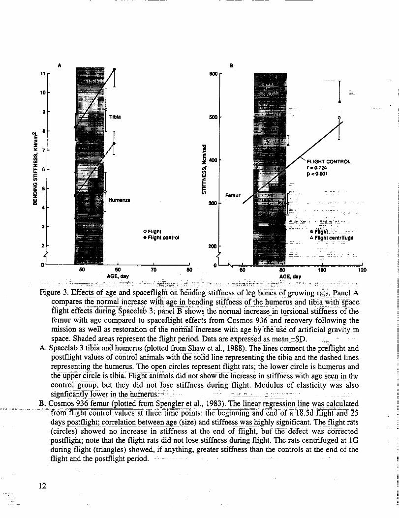
During spaceflight in growing rats, biomechanical parameters do not increase proportional to the
increase in mass in the femur (Spengler et al., 1983), tibia (Shaw et al., 1988) and humerus (Patterson-
Buckendahl et al., 1985; Shaw et al., 1988). The details are shown in Figure 3. Defects in strength are
corrected by inflight centrifugation at 1G (Spengler et al., 1983). The deficit in mechanical strength
could be due to multiple factors including material properties and cross-sectional geometry. A
density decrease was noted in the tibia (Shaw et al., 1988), but not the humerus (Shaw et al., 1988)
or femur (Spengler et al., 1983). Changes in cross-sectional geometry were not implicated in the 7d
SL3 flight or the longer duration Cosmos missions. In fact, the modulus of elasticity in the humerus
was as severly affected as stiffness (Shaw et al., 1988). However, the humeri of rats on Cosmos 1887
(12.5d flight) showed changes in flexural rigidity but not elastic moduli indicating changes in
geometry rather than material properties of cortical bone (Vailas et al., 1990a). This flight landed off
course, requiring 30 hours to transport the rats to the field laboratory and another 18 to 26 hours to
complete the experiment; some recovery from spaceflight effects could have occurred. Vertebrae
from rats on the same flight showed decreased strength and stiffness coupled with a smaller
proportion of mature hydroxypyridinoline cross-links suggesting a delayed maturation (Zernicke
et al., 1990). To understand the changes in bone biomechanics, it is important to review available
data on other skeletal effects of spaceflight.
D. Chemical Composition
In general, the relative concentrations of mineral and matrix determined by analsyis of whole
bone powder were unchanged, but localized regions of long bones showed deficits in mineral and
changes in the composition of the matrix. The lack of dramatic changes in whole bone powder was
not surprising since the majority of bone mass was formed prior to flight. The amount of bone in
cross-section can be used to approximate the percentage of bone accumulated during flight (Table 2).
Flight rats add 3 to 12% of total bone mass while controls add between 5 and 20% depending on rat
age, flight duration, and sampling site. The control groups were killed 2-7 days after the flight group
rather than on the same day allowing additional accumulation of bone; thus, the control area, if
anything, was smaller than suggested by the data. The difference in accumulated mass between flight
and controls during the flight period ranged from <1% to about 7% which is within the range of
11
A B
Tibia 500
8
EEzx7
E
m :_ 400 CONTROLz
6 _ r = 0.724
p.o.oolU.
r3 u.z 5 I::
z Femur -m Humerusm 300
3o Flight _:::_ _- o F_g_ _:- ....
• Flight control z_Flight centrifuge
2 200
I I
50 60 70 80 60 80 100 120
AGE, day AGE, day
Figure 3. Effects0f age-an-d spaceflight:on bending stiffness of]eg-b0_es Of growing rats. Panel A
compares ttie normal increase wi_flf-age in bending stiffness of flae humerus and tibiawith space
flight effects during Spacelab 3; paneI B--sh0ws then0rmal increase in torsional stiffness-6fthe
femur with age compared to spaceflight effects from Cosmos 936 and recovery following the
mission as well as restoration of the normal increase with age by the use of artificial gravity in
space. Shaded areas represent the flight period. Data are expressed asmean _+SD. _ - -- -
A. Spacelab 3 tibia and humerus (plotted from Shaw et al., 1988). The lines connect the preflight and
postflight values of contro] animals with the solid line representing the t_iaand the dashed lines
representing the humerus. The open circles represent flight rats; the lower circle is humerus andthe upper circle is tibia. Flight animals did not show the increase in stiffness with age seen in the
control group, but they did not lose stiffness during flight. Modulus of elasticity was also
signficanfly lower in the humerus,_ ..... _ : ---_ --_- _ .> :: - - -
B. Cosmos 936 femur (plotted from SpengIer et al., 1983). The linear regression line was calculated
from flighYc-rh-trrlVaTue-s at tliree tirne points: ihe_eginning and end--0f ;i- 18.5d flight and 25
days postflight; correlation between age (size) and stiffness was highly significant. The flight rats
(circles) showed no increase in stiffness at the end of flight, but the defect was cisrrected
postflight; note that the flight rats did not lose stiffness during flight. The rats centrifuged at 1G
during flight (triangles) showed, if anything, greater stiffness than the controls at the end of the
flight and the postflight period. ...........
12
I!
Ji
|
E
TABLE 2. PERCENT OF CROSS-SECTIONAL AREA FORMED DURING FLIGHT PERIOD
Cosmos 782 from data collected for Morey and Baylink, 1988.
Cosmos 936 from data collected for Morey,et al., 1978.
Cosmos 1129 from data coUected for Wronski and Morey, 1983a.
SL3 from data collected for Wronski et al., 1987.
biological variability and accuracy of most chemical techniques. If differences in concentration (unit
per gm of bone) are found between flight and flight controls, then the data are suggestive of changes
through-out the total bone rather than solely in the bone formed during flight. Most differences in
bone chemistry from space flight can be attributed to the smaller flight bones rather than changes in
concentration. However, some changes in concentration have been reported. In the younger rats
flown on SL3, phosphorus concentration in the proximal and distal third of the humerus (but not the
middle third or vertebra) decreased and vertebral (but not humeral) hydroxyproline increased while
Ca, magnesium, osteocalcin (a non-collagenous bone matrix protein), and percent nonmineralized
collagen did not change (Patterson-Buckendahl et al., 1987). Decreased accumulation of osteocalcin
was more evident in the rapidly growing vertebra than in the humerus and the amount of this protein
in whole-carcass powder was also reduced reflective of the decreased skeletal growth in the flight
animals rather than a change in concentration. In pooled samples from four Cosmos 1887 flight or
control rats, an increase in reduced collagen cross-links, dihydroxylysinonorleucine (DHLNL) and
hydroxylysinonorleucine (HLNL), and their ratio was noted in flight proximal femur diaphysis
compared to control, but no change was reported in the distal diaphysis or distal end for DHLNL or
in any site in the femur for HLNL and pyridinoline (nonreducible collagen cross-link). In addition,
Ca, phosphorus, and osteocalcin decreased at the distal diaphysis but no other site, while
hydroxyproline (i.e., collagen) decreased only in the proximal metaphysis (Mechanic et al., 1990).
These data suggest that changes in mineral and matrix composition in the whole bone are site
specific.
E. Maturation and Growth
Bone maturation has been studied by means of density gradient fractionation. Bones are ground
into a fine powder which is separated into different density fractions, and mineral and matrix
13
componentsaredeterminedin eachfractionwith the lessdensefractionbeing lessmature(RussellandAvioli, 1972).Delayedbonematurationwasfound in mandible(Simmonset al., 1983),thoracicvertebra,femur shaftand metaphysis(Simmonset al., 1986),andcalvaria (Simmonset al., 1990).The lower incisor andrib did notdiffer from controls(Simmonset al., 1983),but their function didnot changeduring flight. The maturationdefectwasexpresseddifferently in different tissues.Forexample,calcium,phosphorus,andhydroxyprolineincreasedin thethe lower densityfractionsanddecreasedin thehigherdensityfractionsin 18.5dayCosmos1129flight mandibles(Simmonset al.,1983).In the youngerratson the7 day SL3 flight, the low intermediatedensitydecreasedandthehigh intermediatedensityprofiles increasedin the femurwhereasthevertebraehadlessmineral inthe intermediatefractionsanda shift in dry weight towardboth thelow andhigh mineral densities(Simmonset al., 1986).In thecalvariafrom the 12dayCosmos1887missiontheweightdistributionpatternwassimilar to thatof themandiblesof theCosmos1129mission(Simmonsetal., 1990).
Radial growth in thetibia of young adult rats (Wronski et al., I987) and thetibia (Morey andBaylink, 1978;Morey et al., 1978;Wronski andMorey, 1983a)andhumerus(Wronski andMorey,1983a)of rapidly growing ratswasabout40% slowerduring spaceflight.This responsewasveryconsistentin thetibiae from ratsonboardthe Cosmosflights lasting 18.5-19.5days(Table 3). Thevivarium and flight control gi:0ups_d simiiar formht_fi imes--d/ii'lngCosmos782and 936. inCosmos 1129the differencebetweenthesegr0upsprobaS_ resuit_dfr0m_mmobiiiz_i0h in theflight groupssincethe animals,but not the cages,grew dull-rigthe 20d flight delay. A significant
(23%) decrement in bone mineralization rate in the humeruswas also reported in flight rats on
cosmos 1129, while ribs showed a slight decrease _(I8%) _ronski and_orey, 1983a). Mineral
apposition rate in the body of the mandible with contiguoii_:mtiSCle was not aitered, while bone
TABLE 3. DATA FROM COSMOS EXPERIMENTS
;, 2 7 _
COSMOS FLIGHTN..IGI-I_ Di_A_ON
RAT AGE AT LAUNCH
INITIAL BODY MASS" fl :
782 936 1129
19.5di _i8.5d 18.5d
63d 64d 83d
215g 200g 290g>
GROUP PER/OSTEAL BONE MINERALIZATION RATE, mm2/day
FLIGHT CONTROL _ 15.8_+1.5
VIVARIUM CONTROL 16.0-+1.4
FLIGHT CENTRIFUGE (936 only)
FLIGHT 9.4+_2.8
%AFC -40
%AV -41
%AFCent (936 9nly)
Cosmos 782 data from Morey and Bayiink, !988.
Cosmos 936 data from Morey et al., i978.
Cosmos 1 i29 data fromwronski _'et al., i§87.
25.6_+3.7
26.2±3.7
16.0-2_3.2
-37
-39
-9
17.9+__2.7
22.6±4.7
17.6+__3.0
10.0+._2.1
-44
-56
14
without muscleshoweda 30% decrease(Simmonset al., 1983).Variations in mineralizationrateduring flight canalsobe found within the samecross-sectionof a long bone.In the tibia, normalmineralizationrateis highestaroundtheposterioreminencedueto muscleforcesandthisregionwastheleasteffectedby flight (-16%)whereasthegreatestflight effectwasalongtheanterior,medial,andlateralaspectsof thetibia (-48%)(Spectoret al., 1983).
An interestingfinding wastheappearanceof anarrestline in thecorticalboneof ratsallowedtorecover from flight (Cosmos782, 936, and 1129). This cement line was coincident with theperiostealsurfaceat the end of the flight period and extendedaround the boneexcept for theposterioreminence.The arrestline wassuperimposedon thebonemarkergiven3-5 dayspostflightindicatingacessationof boneformationsincelittle or noboneformedimmediatelypostflight.Thisresponsehasbeennotedin the tibia (Morey andBaylink, 1978;Morey et al., 1978;Wronski andMorey, 1983a) andhumerus(Wronski andMorey, 1983a). Thearrestline, which appearedto behypomineralized,wasapproximately3_tmwide and separatedthe boneformedduring spaceflightfrom that formed following spaceflight. Bone matrix at the arrest line was abnormal; fibers(presumablycollagen) were oriented parallel to the arrest line and perpendicularto the radialdirection of growth. The fibers had an irregular convolutedpatternjust within the line along theflight period side;no suchpatternwasnotedin either thepreflight or postflight side.The patternsuggestedthat cytoskeletalelementsin the osteoblasts,alignedto orient collagenvesiclesduringsecretion(Doty, 1988),weredisruptedin space;thismalfunctioncouldaltertheability of collagentoform a stablestructure(Turneret al., 1985a).Lack of anorganizedstructurecould partially explainthe indicationof smallerbonecrystalsin themandible,but not calvaria,of flight rats(Simmonsetal., 1986and 1990).Smallercrystalswould be not be asresistantto abrasionasnormal crystals.Thus,hypomineralizationcouldbeanartifactof tissuepreparation.Thesparsecrystalsorientedtheircrystallographicc-axis along the long axis of the fibrils whereas no extensive regions of preferred
orientation were observed in bone formed before or after flight (Turner et al., 1985a). Focal vascular
occlusion just beneath the outer surface of cortical but not cancellous bone has been noted following
flight and could contribute to localized bone changes (Doty et al., 1990). Normally mineralized
pericanalicular bone in the arrest zone suggested that the defective bone was the result of abnormal
matrix which did not properly mineralize.
F. Cancellous Bone and Cell Populations
Indications of decreased cancellous bone were found in the tibia, femur, and humerus on the 22d
Cosmos 605 mission (Yagodovsky et al., 1976). Cancellous bone volume in the tibia and humerus
was lower and marrow fat was higher in the flight rats in Cosmos 1129; analysis of bone cell
populations in the proximal humerus showed a decrease in osteoblasts immediately adjacent to the
growth cartilage-metaphyseal junction, but osteoclast numbers were unchanged (Jee et al., 1983).
Similar changes were found in the proximal tibia of 105d old male rats following the 7d Cosmos
1667 flight, but no changes were noted in the femur, thoracic or lumbar vertebrae (Vico et al., 1988).
Pregnant rats from the 5d Cosmos 1514 mission showed no change in trabecuiar bone volume in the
tibia, lumbar or thoracic vertebra (Vico et al., 1987c). On SL3, young adult rats showed a
suppression of formation in the tibial shaft, but histomorphometric parameters (trabecular bone
volume, osteoclast surface and number, osteoblast surface and number) in the proximal humerus and
lumbar vertebra were unchanged.
15
Growing rat studieshavefocusedon the biomineralizationdefectsince most datasuggestnochangein osteoclasticactivity (CannandAdachi, 1983[boneresorptionkinetics]; Jeeet al., 1983;Wronski et al., 1987;Vico et al., 1988;Kaplanskyet al., 1987).However, indicationsof increasedosteoclasticactivity havebeenreportedin afew sites(Kaplanskyet al., 1987[Cosmosi 667 primary
spongiosa of tibia]; Vico et al., 1987c [thoracic vertebra but not lumbar vertebra or primary or
secondary spongiosa of tibia in pregnant rats flown on 5d Cosmos 1514]).
G. Non-weightbearing Bones
Although one might anticipate changes in weightbearing bones, there is no a priori reason to
expect alterations in non-weightbearing bones. However, changes in mineralization rates and bone
turnover, maturation, and progenitor populations have been reported in the maxilla and mandible
(Simmons et al., 1983; Roberts et al., 1987; Jackson et al., 1988). Precursor cells which become
osteoblasts may differentiate at a slower rate in the maxilla during spaceflight (Roberts et al., 1987;
Garetto et al., 1990), and fewer osteoblasts would form less bone. The mandibular condyle, which is
loaded during eating but is non-weightbearing, may be exquisitely sensitive to microgravity (Jackson
et al., 1988). The muscles in the jaw may contribute to these changes since the mandible would not
require active muscle tone to stay closed in space and changes in these postural muscles may occur.
In addition to the jaws, maturation of the calvaria was shown to be delayed during Cosmos 1887
(Simmons et al., 1990). Thus, most bone tissues seem to show a response to spaceflight and the
detection of such a change appears to be dependent upon the mineralization rate of the tissue (which
is related to age and function), the length of the flight, and the technique used.
H. Recovery from Spaceflight
With the exception of trabecular bone mass in the proximal tibia, all bone defects noted above
appeared to recover within a month after return from space. Table 4 compares postflight recovery of
tibial mineralization of flight controls with flight rats and rats centrifuged at 1G during Cosmos 936
(Morey et al., 1978). Half of each group of rats was euthanized immediately following flight and the
remaining animals were given a bone marker four days following flight. The difference in bone area
between those animals killed immediately following flight and those receiving the postflight label
calculated to provide an estimate of the amountwas of bone formed during the first four days
postflight for each group. Both groups showed a similar suppression of mineralization during
spa-cefiight. In the first four days following flight, the total amount of bone formed by the flight rats
was formed by control rats in 0.8d suggesting a lag time of approximately 3.2d before the flight rats
began to form bone post-flight. On the other handl the total amount of bone formed by the flightcentrifuged rats in the f'trst four days following flight was slightlygreater than that formed by the
controls suggesting an immediate recove-ryof f0rmation in the centrifugedrats, in addition, the arrest
line was significantly longer in the flight rats than either the flight centrifuged animals or the flight
controls.
I. Interpretation
Data from Cosmos 936 are particularly important in interpreting the skeletal response of growing
rats to spaceflight due to the onboard IG centrifuge control. The arrest line'and the apparent lag time
16
TABLE 4. RAT TIBIA MINERALIZATION ON COSMOS 936
Number of specimens
FLIGHT FLIGHT CENTRIFUGE
5 5
Bone area (mm 2) formed in 4 days
immediately postflight
Time (d) required for controls to form
bone area formed by flight groups
0.04_+0.07 0.12_-20.06
0.8 4.7
Mineralization Rate (mm2/d)
%A flight control
p vs flight control
0.01_+0.02 0.03_+0.02
- 61 + 17
<0.1 n.s.
Length of Arrest Line (mm)
%A flight control
p vs flight control
4.0-&_l.1 2.3-20.4
+ 250 +144
< 0.005 < 0.05
Data from Morey et al., 1978.
for reinitiating bone formation in the flight rats suggest a cessation of formation which would
necessitate stimulation of progenitor populations to recover from the defect. The rapid recovery in
centrifuged animals suggests that mineralization decreased rather than ceased and the decrease may
have been due to reduced activity of the animals in the fixed cage, short-radius, high rotation rate
centrifuge. Both flight groups formed the same amount of bone at the periosteal surface during flight
and had the same bone mass, density, and presumably geometry, yet stiffness was very different
(Spengler et al., 1983). Thus, the changes in stiffness during spaceflight (Spengler et al., 1983; Shaw
et al., 1988) cannot be totally explained by changes in geometry or density and implicate other
alterations in material properties. These studies also imply that techniques measuring solely bone
mass or bone density could significantly underestimate the change in bone biomechanical properties
during spaceflight.
The degree of skeletal change during flight depended on the rate of growth and the site in the
bone. For example, the younger animals on the 7-day SL3 mission exhibited significant bone
changes, while older animals exhibited only similar trends in many bone parameters. Yet all animals
on the 19-day flights showed significant differences whether 63 or 83 days old (Table 2). Regardless
of flight duration, the young animals showed negligible signs of bone loss. Some components of
bone continued to grow, while other features suggested growth suppression or arrest of
biomineralization.
17
The site(s)of the biomineralizationdefect during spaceflightarenot know, but alterationsinbonematrix could alter crystalsizeandimpedeincreasesin bonestrength.The durationandextentof theskeletalchangesin growingratsduringspaceflighthavenotbeendefined.
V. GROUND-BASEDUNLOADING-INDUCED SKELETAL CHANGESIN GROWING RATS.
A. General
In 1975 when the Cosmos cooperation between the US and USSR started, we began
development of a ground-based rat model to simulate some effects of spaceflight. Animal models for
skeletal disuse did exist, but the disuse was produced by decreased activity due to confinement in a
small cage, flacid paralysis due to nerve-section, or unloading through surgical tenotomy. None of
the techniques produced a differential muscle atrophy which primarily affected the postural or
weight-bearing muscles as was noted during spaceflight. Surgical procedures induced a regional
acceleratory phenomena which led to initial, rapid bone loss and the procedures could not be
reversed. Thus, existing models were not appropriate to study the response to and recovery from
spaceflight. The main criteria for development of an acceptable model simulating spaceflight in
growing rats were 1) differential muscle atrophy, 2) a headward fluid shift, 3) the ability to move,
eat, and groom normally using the front paws, 4) unloading of the rear limbs without paralysis so
that the animals could be reloaded and recovery from unloading effects observed, 5) weight gain
throughout the experimental period, and 6) validation using spaceflight data. Such a model would be
invaluable in predicting spaceflight effects on musculoskeletal growth and development, studying
potential mechanisms, and establishing a time course of responses, and would be less expensive and
more accessible than spaceflight. Many ideas for a rat model were generated and some were tested.
The system from which data were initially published used an orthopedic casting material bonded to
the back of the rat (Morey, 1979). A wire thread was incorporated into the harness and the wire was
attached to a freely rotating fishline swivel on an overhanging horizontal aluminum beam. The beam
was fixed to apost by a ball bearing rotating in a horizontal plane. The animals were attached at
about a 30 ° head-down angle so that the rear limbs did not bear weight. The animals could move
freely about a 360 ° arc because the forelimbs were allowed to touch the plexiglass grid floor. Many
synonyms fi_ve been used to describe rats on-ihis model system including ground-based flight
elevation, unweighted, tail-traction, intact unloading (vs surgically-induced), and disuse. In this
document, suspended is used to describe the rat model while unloaded is used to describe skeletalstatus.
Some bone responses to unloading using the back harness appeared to be similar to those noted
in space (decreased periosteal mineralizatiofi in the tibia, reduced cancello_us bone with increased
marrow fat, decreased osteoblast numbers, recovery wiffiin two weeks of reloading after two weeks
of suspension), but some responses were different (increased osteoclastic numbers in the tibia and
humerusand failure to gain weight). Additionally, a decrease in the rate Of longitudinal bone growth
in both tibia and humerus was reported (Wronski and Morey, 1982, 1983b). Unfortunately, the
suspended animals did not gain weight while controls grew, and the harness had to be rebonded at
about two-week intervals. The lack of weight gain using this harness system implied a stressful
situation which could inh_it bone growth. To minimize discomfort to the young male rats, the
harness device was changed first to a plaster of Pads cast at the base of the tail (Morey-Holton and
18
Wronski, 1981) and finally to orthopedic traction tape along the tail (Wronski and Morey-Holton,
1987). The head-down angle, suspension of the hindquarters, the cephalad fluid shifts, and the use of
the forelimbs for locomotion remained the same but the overhead beam was changed to an X-Y
pulley system which allowed the animal free movement throughout the cage. The tail-traction system
fulfilled most of the criteria for an adequate model system and evolved into the technique of choice
for studying bone changes mimicking spaceflight in growing rats. The tail-traction system rather
than the back harness or body harness (Musacchia et al., 1980) is better for developmental studies
because rats gain weight similar to controls when controls are fed daily the mean food consumption
of the experimental rats. If one wants to study growth and development, then the animals must grow;
if growth is inhibited in experimental animals while the controls continue to grow, then
differentiating between the effects of growth retardation and the effects of unloading on the skeleton
is difficult, if not impossible.
B. Serum Parameters
The initial period of inhibited bone formation during skeletal unloading was marked by a small
but significant increase (-10%) in the serum concentration of Ca, a dramatic decrease (60%) in the
serum concentration of 1,25-dihydroxyvitaminD (1,25(OH)2D), a small and insignificant increase in
the serum of 24,25-dihydroxyvitaminD (24,25(OH)2D), and no change in the serum concentration of
25-hydroxyvitaminD (25-OHD)(Halloran et al., 1986). Between days 5-15 of suspension, the serum
concentrations of Ca, 1,25(OH)2D and 24,25(OH)2D all returned to normal and presumably
remained so indefinitely, although measurements in animals suspended for periods longer than 15
days were not made (Halloran et al., 1986). The bone changes still occurred when the transitory fall
in serum 1,25(OH)2D was prevented by continuous infusion of 1,25(OH)2D (Halloran et al., 1986),
or when the serum concentrations of 1,25(OH)2D were manipulated by changing dietary Ca (Globus
et al., 1986a). These facts suggest that the transitory decrease in serum 1,25(OH)2D associated with
acute skeletal unloading was probably not the cause of the defect in bone formation, but rather the
result of changes in bone cell activity and demand for Ca. It is important to note that increasing
dietary Ca did not prevent the decreased bone growth induced by suspension, but it did increase bone
Ca and thereby provided some protection against unloading (Globus et al., 1986a).
The serum concentration of parathyroid hormone (PTH) was normal after 15 days of skeletal
unloading (Globus et al., 1986a) as were the serum concentrations of Ca, inorganic phosphate,
1,25(OH)2D, 24,25(OH)2D, 25-OHD. Osteocalcin decreased in serum within five days of
suspension, but returned to normal within 15 days reflecting changes in bone growth (Patterson-
Buckendahl et al., 1989).
To determine if glucocorticoids, potent inhibitors of bone formation, might be involved in the
suppressed bone mineralization, adrenalectomized rats were suspended. Also, the circadian
periodicity of glucocorticoids was measured in intact rats following suspension. The results
suggested that the response was not due to increased plasma glucocorticoids or an increase in bone
sensitivity to the glucocorticoids (Halloran et al., 1988).
19
C. Biomechanics
A study investigatingfemurbiomechanicalpropertiesin young ratssuspendedfor one, two, orthreeweeksuseda modification of the backharnessand reportedalteredgeometricandmaterialpropertiescomparedto controls (Abramet al., 1988).Most parameterscontinued to decrease with
time of suspension, but the changes may have been due to failure of the suspended rats to grow
normally. Using _taii harness tdsuspend_6fing, femaie rats for fourweeks, Shaw it al (1987)
reported that the only cross-sectional morphology change was the anterior cortical thickness of the
femur. All mechanical characteristics were decreased in the femur but only bending stiffness was
altered in the tibia. Vailas et al. (1990b) reported that young male rats suspended for two weeks
showed decreased mechanical properties of the femur/tibia medial collateral ligament while cross-
sectional area of the tissue did not change. These studies suggest that suspending the hindquarters
induced changes in mechanical properties of tibia, femur, tendons, and ligaments.
D. Chemical Composition
Ca concentration of multiple bones at different time periods was not affected by suspension.
Shaw et al (1987) reported that tibia and femur concentrations of collagen, phosphorus, and Ca in
suspended rats did not differ from controls. Vailas et al. (1988) reported no differences between
suspended and control rats in femur concentration of Ca, phosphorus, DNA, hydroxyproline, or
uronic acid. However, hydroxyproline and uronic acid, but not DNA, were significantly lower in the
suspended patellar tendon. Changes in osteocalcin and Ca in the lumbar vertebra and femoral
diaphysis with time of suspension paralleled the changes in bone weight and few changes in
concentration were found (Patterson-Buckendahl et al., 1989). Total bone Ca in tibia and lumbar
vertebra decreased 10-40% within one week of unloading and remained supressed until reloaded,
reflecting the smaller bones in the suspended rats (Globus et al., 1986b; Halloran et al., 1986).
E. Metabolism, Maturation, and Growth
Bone metabolism changed during suspension. Unloaded bones (tibia, femur, and lumbar
vertebra) showed decreased metabolism, while metabolism in loaded bones (mandible, humerus, or
cervical vertebra) did not change (Globus et al., 1984; Globus et al., 1986b). Within 5-7 days of
suspension, a significant inhibition of Ca metabolism and collagen production (as indicated by
[45Ca] and [3H]-proline uptake) occurred in unloaded bones. Between days 7-15 of suspension,
uptake of [45Ca] and [3H]-proline returned to normal. Retarded mineralization resulted in a decrease
in mature bone; the effects were maximal in young, male rats after 10 days of suspension, then
returned toward control values (Bikle et al., 1987).
Bone formation rate at the tibiofibular junction was reduced by 50% within one week of
suspension (Globus et al., 1986b). Between days 7-15 of suspension, bone formation rate at the
tibiofibular junction returned toward normal, but was still significantly suppressed at the end of two
weeksl Til_ial longitudinal bone growth was reduced by _within 5 days (I-Iall0ran et al,,_986),
but returned to normal within two weeks (Wronski and Morey, 1987). Vailas et al. (1988) reported
n0: differences between suspended a_dcontroi rats in femur length or weight and cortical or
endosteal parameters (area, circumference, density, maximum diameter), but did find an increase in
minimum diameter in the suspended group. In another experiment, rats were suspended for 14, 30,
20
60, or 90 days (LeBlanc et al., 1985). Technetium-labeledmethylene diphosphonateuptake(normalizedto Caconcentration)in tibia, femur,andhumerusindicateda suppressionof formationin thetwo-weeksuspendedgroupwith indicationsof increaseduptakein thefemurat60d.Regionaldensitometryof the femurshowedprogressivelylessbonemineralcomparedto controlsbeginningwith cancellousboneat 30daysandextendingto the shaftby 60dayswith thedefectbeingsimilarin magnitudein bothregionsby 90daysof suspension.Regionalblooddistributiondid notchangeinany long bone,but did changein muscle.Although no differencein anybone length wasnoted,decreasesin femurtotal width, medullarycanalwidth,andcorticalthicknesswerenotedat 90d.Nomorphometricevidencewasfoundfor anincreasedmedullaryresorptionrate in theboneshaft.
F. Cancellousboneandcell populations
Trabecularbonesurfacelined with osteoblastswasreducedby 32%,but percentbonesurfacelined with osteoclastswas unchangedwithin 5 days of suspension(Halloran et al., 1986).Theosteoblastpopulationreturnedto normalwithin two weeksof continuedsuspension(Wronski andMorey, 1987).Osteoclasticactivity appearedelevatedby 60din themetaphysealregionof thefemursuggestingincreasedturnoverin thisbone(LeBlancetal., 1985).
G. Bone Mass Redistribution
A recent three-week study using weanling rats showed a redistribution of bone mass during
suspension with a decrease in the femur and tibia, a moderate increase in the humerus and
radius/ulna, and a significant increase in the skull and mandible, and implicates fluid shifts in the
response (Roer and Dillaman, 1990). A similar finding has been reported in bed-rested human
subjects (LeBlanc et al., 1990).
H. Recovery from Suspension
If animals were reloaded after two weeks of suspension, recovery of the bone mass deficit and
strength appeared to require somewhat more than two weeks (Sessions et al., 1989; Abram et al.,
1988). These data are consistent with the hypothesis that skeletal unloading in growing animal
produces inhibition of bone formation. With time and continued suspension, bone metabolism and
growth return toward normal. Total bone mass remains low because of the initial reduction in boneformation and subtle defects in mineralization (Bikle et al., 1987). Recovery of the defect required
longer for some bone parameters than others.
I. Comparison with Flight Data
Simulation of Cosmos 1667 suggested that 7d of suspension decreased tibial metaphyseal bone
less in suspended than in flight rats. No change was noted in flight femoral trabecular bone measured
under muscular insertions, but a reduction was noted in suspended rats. Vertebral bodies were not
affected by flight but suspended rats showed a trend of decreasing mass (Vico et al, 1987a). Turner
(1985b) compared bone mineralization in anterior, posterior, medial, and lateral aspects of tibial
cross-sections in flight (19d), suspended (14d), and nerve-sectioned (21d) rats and found a greater
inhibition of mineralization at all sites in flight rats. Simulation of SL3 reported hindlimb muscle
21
atrophy(Morey-Holtonet al., 1988)anddecreasedfemoralbonebiomechanics(Martin et al., 1988)similar to spaceflight;unlikespaceflight,humeralparametersdid not change.
J.Interpretation
The rat modelhasprovedinvaluable,particularlyin predictingtheearlyresponseto spaceflight.Data from the first weekof suspension,whetheron Earthorin space,aresimilar. However,bonemetabolismandmineralizationappearto returnto normal levelswith continuedsuspensiononEarthwhile spaceflightmayrequirelongerfor stabilizationof boneparameters.A spaceflightdefectwhichhasnot yet beenreproducedin ground-basedstudiesis thearrestline in cortical bone.Total skeletalunloadingoccursduring flight, but only partial unloadingis possiblein the ground model asthehumerusand cervical vertebracontinue to bear someweight. In fact, the humerusservesas aninternal controlfor thegroundexperiments(resultsshouldbe thesamein humerusof suspendedandcontrol groups)but this bonedoeschangeduringspaceflight.Also, thestressof reentry,andthetimedelay betweenreentry (with reloading)and samplingof flight specimens,cancreatedifficulty incomparing datawith model experimentswhereanimalsare sampledwhile still suspended.Themodelhasalsobeenusedto studythetime courseof changesin boneandtheendocrinestatusat theend of the suspensionperiod which has not beenpossibleon a single SpaceShuttle mission orunmannedflight.
VI. CONCLUSIONAND SUMMARY
Observationsin adulthumansandjuvenileanimalsduringandafterspaceflighthaveprovideduswith basicconceptsregardingtherole of gravity in thedeterminationof bonestructure.Theresponseof theskeletonto anewenvironmentthat imposesdifferentbiomechanicalstressesis complicatedby
its dual function as a support structure and as a mineral reservoir. The transport of the minerals is
regulated and influenced by organ systems (i.e. cardiovascular, muscular, renal, gastrointestinal)
whose anatomy and metabolism are also affected by microgravity. This review deals almost
exclusively with the observed and potential effects of spaceflight on bone, but some findings are
cited from other body systems where spaceflight seems to have a major impact on the adaptation of
bone to microgravity.
The primary site of the response of bone tissue to microgravity is at the interface of mineral and
matrix in the process of biomineralization. This is manifested in the adult by demineralization of
some regions of the skeleton. The most pronounced effects are seen in os calcis, the most distally
located bone relative to the heart. In-flight biochemical and endocrine studies from the Skylab
missions, primarily, suggest an hypothetical scheme for the pathogenesis of the two major
consequences of space flight, negative calcium balance and demineralization_ The extent of these
effects varies greatly between individual astronauts and cosmonauts. Diet, glucocorticoid excess, and
activity level prior to and during spaceflight are influences that may account_fqr theyariation indensity of bones measured. While quantitative losses of mineral from the whole skeleton are
relatively small, regional or localized areas of demineralization during spaceflight could seriously
impair the support functionof the skeleton on return to Earth.
In depth study of the biomechanics, morphology, chemistry and development of young rat bones
have revealed the highly selective and regional nature of the response of bone structure to
22
|
i
spaceflight.Thereis eitherdepressedor arrestedmaturationof newbone.Thereis alsoadecreaseinstrengthcomparedto controls, which may be relatedto the failure of mineralization and to animpaireddifferentiationof boneformingcells.However,from observationof animalsmaintainedatartificial gravity during spaceflight,we have learnedthat otherelementsin the compositionandstructureof bone,independentof mineral content,geometry,and growth rates,are important forbonestrength.
A ground-basedflight simulationmodel for extendedstudyof bonestructureat the molecular,organ and whole body levels is describedand comparedto the flight results. On Earth, themorphologicandcompositionalchangesin theunloadedbonesarevery similar to changesin flight;however, the ground-basedchangesappearto be more transient than those reported duringspaceflight.In addition,thereis a redistributionof bonemineral to gravity dependentbonesof themodel.Longitudinaldatahaveprovidedconsiderableinformationon theinfluenceof endocrineandmuscularchangesonbonestructurefollowing unloading.
Theseinvestigationshave defined important areasof future investigationfor resolving basicbiology andbiomedicalquestionsonthemechanismof structuraladaptationof theskeletonto anewenvironment.Thereis clearlyaneedfor moreinterdisciplinaryphysiologicalstudies.Basicquestionsregardingthe mechanismof revisionof bonearchitectureare likely to be answeredby integratedwork in muscleandbonethatis directedto uncoveringthesignalsfor localcell activity anda betterknowledgeof themolecularstructureof boneandits regulation.
23
VII. REFERENCES
Abram, A. C., T. S. Keller, andD. M. Spengler.The effectsof simulated weightlessness on bone
biomechanical and biochemical properties in the maturing rat. J. Biomechanics. 21: 755-767,1988.
Aranow, M. A., L. C. Gerstenfeld, T. A. Owen, M. S. Tassinari, G. S. Stein, and J. B. Lian. Factors
that promote progressive development of the osteoblast phenotype in cultured fetal rat calvaria
cells. J. Cell PhysioL 143: 213-221, 1990.
Arnaud, S. B. and E. Morey-Holton. Gravity, calcium, and bone: Update, 1989. The Physiologist.33: $65-67, 1990.
Amaud, S. B., Powell, M. R., Whalen, R. T. and Vernikos-Danellis, J. Bone mineral redistribution
during head down tilt bed rest. Am. Soc. Gravit. Space Biol. Bull 2: 54, 1989.
Arnaud, S. B., V. S. Schneider, and E. Morey-Holton. Effects of inactivity on bone and calcium
metabolism. In:Inactivity: Physiological Effects, H. Sandier and J. Vernikos, eds., Academic
Press, Inc. Orlando, FL, 1986, pp. 49-76.
Baron, R. Anatomy and ultrastructure of bone. In: Primer on the Metabolic Bone Diseases and
Disorders of Mineral Metabolism, M. J. Favus, ed., Am. Soc. Bone Mineral Res., Kelseyville,CA, First Edition, 1990, pp. 3-7.
Bikle, D. D., B. P. Halloran, C. C. Cone, R. K. Globus, and E. Morey-Holton. The effects of
simulated weightlessness on bone maturation. Endocrinology. 120: 678-684, 1987.
Binderman, I., Z. Shimshoni, and D. Somjen. Biochemical pathways involved in the translation of
Jee, W. S. S., T. J. Wronski, E. R. Morey, and D. B. Kimmel. Effects of spaceflight on trabecular
bone in rats. Am. J. Physiol. 244: R310-R314, 1983.
Johnston. R. S. and L. F. Dietlein, eds.Biomedical Results from Skylab, NASA SP-377, Washington,DC, 1977, pp. 491.
Kaplansky, A. S., G. N. Durnova, Z. F. Sakharova, and Y. I. Ilyina-Kakuyeva. Histomorphometric
analysis of rat bones after spaceflight aboard Cosmos-1667 biosatellite. Space Biol. Med. 21:33-40, 1987.
Klein-Nulend, J., J. P. Veldhuijzen, M. deJong, and E. H. Burger. Increased bone formation and
decreased bone resorption in fetal mouse calvaria as a result of intermittent compressive forcein vitro. Bone Mineral. 2: 441-448, 1987.
Leach, C. S. and P. C. Rambaut. Biochemical responses of the Skylab crewmen: an overview.
In:Biomedical Results from Skylab, R. S. Johnston and L. F. Dietlein, eds., NASA SP-377,
Washington, DC, 1977, pp. 204-216.
LeBlanc, A., E. Marsh, H. Evans, P. Johnson, V. Schneider, and S. Jhingran. Bone and muscle
atrophy with suspension of the rat. J. Appl. Physiol. 58: I669-1675, 1985.
LeBlanc, A., Schneider, V., Krebs, J., Evans, H., Jhingran, S. and Johnson, P. Spinal bone mineral
after five weeks of bed rest. Calcif. Tissue Int. 41: 259-261, 1987.
LeBlanc, A. D., V. S. Schneider, H. J. Evans, D. A. Engelbretson, and J. M. Krebs. Bone mineral
i0ss andrecovery after 17 weeks of bed rest. J. Bone Mineral Res. 5: 843-850, 1990.
26
Lowenstam,H. A. andS.Weiner.OnBiomineralization. Oxford University Press, New York, NY,
1989, pp. 25-49.
Martin, R. B., E. R. Morey-Holton, N. A. Sharkey, and A. C. Maese. Spacelab 3 simulation: bone
strength study. ASGSB Bulletin. 1: 38, 1988.
Mechanic, G. L., S. B. Amaud, A. B. Boyde, T. G. Bromage, P. Buckendahl, J. C. Elliott, E. P. Katz,
and G. N. Durnova. Regional distribution of mineral and matrix in the femurs of rats flown on
Cosmos 1887 biosatellite. FASEB J. 4: 34-40, 1990.
Morey, E. R. Space flight and bone turnover: correlation with a new rat model of weightlessness.
BioScience 29: 168-172, 1979.
Morey, E. R. and D. J. Baylink. Inhibition of bone formation during spaceflight.Science 201:
1138-1141, 1978.
Morey, E. R., R. D. Turner, and D. J. Baylink. Quantitative Analysis of Selected Bone Parameters.
In: Final Reports of U.S. Experiments Flown on the Soviet Satellite Cosmos 936, S. N.
Rosenzweig and K. A. Souza, eds., NASA TM 78526, Moffett Field, CA, 1978, pp. 135-178
Morey-Holton, E. R., A. C. Maese, L. M. Kraft, and T. N. Fast. Spacelab 3 simulation. ASGSB
Bulletin. 1: 38, 1988.
Morey-Holton, E. R., and T. J. Wronski. Animal models simulating weightlessness. The Physiologist
24: $45-$48, 1981.
Morey-Holton, E. R., H. K. Schnoes, H. F. DeLuca, et al. Vitamin D metabolites and bioactive
parathyroid hormone levels during Spacelab 2. Aviat. Space Environ. Med. 59: 1038-1041,1988.
Musacchia, X. J., D. R. Deavers, G. A. Meininger, and T. P. Davis. A model for hypokinesia: effects
on muscle atrophy in the rat. J. Appl. Physiol. 48: 479-486, 1980.
Nicogossian, A. E., C. L. Huntoon, and S. L. Pool, eds.Space Physiology and Medicine, Lea and
Febiger, Philadelphia, PA, Second edition, 1989, pp. 401.
Nogami, H. and A. Oohira. Defective association between collagen fibrils and proteogtycans in
fragile bone of osteogenesis imperfecta. Clin. Orthopaed. Rel. Res. 232: 284-291, 1988.
Oganov, V. S., C. Cann, A. S. Rakhmanov, and S. K. Ternovoy. A computer tomographic
investigation of the musculoskeletal system of the spine in humans after long-term space flight.
Space Biol. Med. 24(4) 20-21, 1990 (Translated in USSR Space Life Sciences Digest, Issue 29,
pp. 76-78, 1991).
27
Owen,T. A., M. Aronow, V. Shalhoub,L. M. Barone,L. Wilming, M. S.Tassinari,M. B. Kennedy,S. Pockwinse,J. B. Lian, and G. S. Stein. Progressivedevelopmentof the rat osteoblastphenotypein vitro: reciprocalrelationshipsin expressionof genesassociatedwith osteoblastproliferation and differentiation during formation of the boneextracellular matrix. J. Cell.
Ozawa, H., K. imamura, E. Abe, N. Takahashi,. T. Hiraide, Y. Shibasaki, T. Fukuhara, and T. Suda.
Effect of a continuously applied compressive pressure on mouse osteoblast-like cells
(MC3T3-E1) in vitro. J. Ceil Physiol. 142: 177-185, 1990.
Parfltt, A. M. Bone effects of space flight: analysis by quantum concept of bone remodelling.ActaAstronautica 8 (9-10): 1083-1090, 1981.
Patterson-Buckendahl, P., R. K. Globus, D. D. Bikle, C. E. Cann, and E. Morey-Holton. The effects
of simulated weightlessness on rat osteocalcin and bone calcium concentrations. Am. J.
Physiol. 257:R1103-1109, 1989.
Patterson-Buckendahl, P., R. E. Grindeland, R. B. Martin, C. E. Cann and S. B. Arnaud. Osteocalcin
as an indicator of bone metabolism during spaceflight.The Physiologist 28: $227-228, 1985.
Patterson-Buckendahl, P., S. B. Arnaud, G. L. Mechanic, R. B. Martin, R. E. Grindeland, and C. E.
Cann. Fragility and composition of growing rat bone after one week in spaceflight. Am. J.Physiol. 252: R240-246, 1987.
Pollack, S. R., R. Salzstein, and D. Pienkowski. The electric double layer in bone and its influence
on stress-generated potentials. CaIcif Tissue Int. 36: $77-$81,1984.
Prokhonchikov, A. A., N. A. Zhizina, and V. Tigrnayan. Homeostasis of bone tissue under normal
and extreme action. In: Problems in Space Biology, ed., P. D. Gorizontov, Nauka Press,
Moscow, 1984, pp. 152-165. (English translation NASA TM 77614).
Puzas, J. E. The osteoblast. In: Primer on the Metabolic Bone Diseases and Disorders of Mineral
Metabolism, M. J. Favus, ed., Am. Soc. Bone Mineral Res., Kelseyville, CA, First Edition,
1990, pp. 11-15. _
Raisz, L. G. Local and systemic factors in the pathogenesis of osteoporosis. New England J. Med.
318: 818-828, 1988 ....
Rambaut, P. C. and R. S. Johnston. Prolonged weightlessness and calcium loss in man. Acta
Astronautica 4:1113-1122, 1979.
Reich, K. M., C. V. Gay and J. A. Frangos. Fluid shear stress as a mediator of osteoblast cyclic
adenosine monophosphate production. J. Cell. Physiol. 143:100-104, 1990.
Roberts, W. E. and E. R. Morey. Proliferation and differentiation sequence of osteoblast histogenesis
under physiological conditions in rat periodontal ligament. Am. J. Anat. 174:105-118, 1985.
28
Roberts,W. E., P. J. Fielder,L. M. L. Rosenoer,A. C. Maese,M. R. Gonsalves,and E.R. Morey.Nuclearmorphometricanalysisof osteoblastprecursorcellsin periodontalligament,SL-3 rats.Am. J. Physiol. 252: R247-R251, 1987.
Roer, R. D. and R. M. Dillaman. Bone growth and calcium balance during simulated weightlessness
in the rat. J. Appl. Physiol. 68:13-20, 1990.
Russell, J. E. and L. V. Avioli. Effect of experimental chronic renal insufficiency on bone mineraland collagen maturation. J. Clin. Invest. 51: 3072-3079, 1972.
Sessions, N. D., B. P. Halloran, D. D. Bikle, T. J. Wronski, C. M. Cone, and E. Morey-Holton. Bone
response to normal weightbearing after a period of skeletal unloading. Am. J. Physiol. 257:E606-610, 1989.
Shaw, S. R., A. C. Vailas, R. E. Grindeland, and R. F. Zernicke. Effects of a 1-wk spaceflight on
morphological and mechanical properties of growing bone. Am. J. Physiol. 254: R78-83, 1988.
Shaw, S. R., R. F. Zernicke, A. C. Vailas, D. DeLuna, D. B. Thomason, and K. M. Baldwin.
Mechanical, morphological and biochemical adaptations of bone and muscle to hindlimb
suspension and exercise. J. Biomechanics 20: 225-234, 1987.
Simmons, D. J., J. E. Russell, and M. D. Grynpas. Bone maturation and quality of bone mineral in
rats flown on the Space Shuttle "Spacelab-3 mission". Bone Mineral 1: 485-493, 1986.
Simmons, D. J., M. D. Grynpas, and G. D.'Rosenberg. Maturation of bone and dentin matrices in
rats flown on the Soviet biosatellite Cosmos 1887. FASEB J. 4: 29-33, 1990.
Simmons, D. J., J. E. Russell, F. Winter, P. T. Van, A. Vignery, R. Baron, G. D. Rosenberg, and
W. V. Walker. Effect of spaceflight on the non-weight-bearing bones of rat skeleton. Am. J.
Physiol. 244: R319-326, 1983.
Smith, A. H. Principles of Gravitational Biology. In: Foundations of Space Biology and Medicine,
M. Calvin and O. G. Gazenko, Eds., NASA, Washington, DC, 1975, Vol. II, Book I,
pp. 129-162.
Spector, M., R. T. Turner, E. Morey-Holton, D. J. Bayiink, and N. H. Bell. Arrested bone formation
during space flight results in a hypomineralized skeletal defect. The Physiologist 26:Sll0-Slll, 1983.
Spengler, D. M., E. R. Morey, D. R. Carter, R. T. Turner, and D. J. Baylink. Effects of spaceflight on
structural and material strength of growing bone. Proc. Soc. ExptL Biol. Med. 174: 224-228,1983.
Stewart, A. F., M. Adler, C. M. Byers, G. V. Segre, and A. E. Broadus. Calcium homeostasis in
immobilization: an example of resorptive hypercalciuria. N. EngI. J. Med. 306:1136-1140,1982.
29
Stupakov,G. P., V. S.Kazeykin, A. P. Kozlovskiy, and V. V. Korolev. Evaluationof changesinhumanaxial skeletalbonestructuresduring long-termspaceflights.Space Biol. Med. 18: 42-
47, 1984.
Termine, J. D. Bone matrix proteins and the mineralization process. In: Primer on the Metabolic
Bone Diseases and Disorders of Mineral Metabolism, M. J. Favus, ed., Am. Soc. Bone Mineral
Res., Kelseyville, CA, First Edition, 1990, pp. 16-22.
Thompson, D. W. On Growth and Form. Cambridge University Press, New York, 1917 (Revised,
Bonner, J.T., Ed., 1961).
Thornton, W. E. and J. Ord. Physiological mass measurements in Skylab. In: Biomedical Results
from SkyIab, R. S. Johnston and L. F. Dietlein, eds., NASA SP-377, Washington, DC, 1977,
pp. 175-182.
Tilton, F. E., J. J. C. Degiganni, and V. S. Schneider. Long-term follow-up of Skylab bone
demineralization. Aviat. Space Environ. Med. 51: 1209-1213, 1980.
Turner, R. T., N. H. Bell, P. Duvall, J. D. Bobyn, M. Spector, E. Morey-Holton, and D. J. Baylink.
Spaceflight results in formation of defective bone. Proc. Soc. Exptl. Biol. Med. 180: 544-549,1985a.
Turner, R. T., G. K. Wakley, and B. W. Szukalski. Effects of gravitational and muscular loading on
bone formation in growing rats.The Physiologist 28: $67-$68; 1985b.
Vailas, A. C., D. M. DeLuna, L. L. Lewis, S. L. Curwin, R. R. Roy, and E. K. Alford. Adaptation of
bone and tendon to prolonged hindlimb suspension in rats. J. Appl. Physiol. 65: 373-376, 1988.
Vailas, A. C., R. F. Zernicke, R. E. Grindeland, A. Kaplansky, G. N. Durnova, K. C. Li, and D. A.
Martinez. Effects of spaceflight on rat humerus geometry, biomechanics, and biochemistry.FASEB J. 4: 47-54, 1990a.
Vailas, A. C., R. F. Zernicke, R. E. Grindeland, and K.-C. Li. Suspension effects on rat femur-medial
collateral ligament-tibia unit. Am. J. Physiol. 258: R724-R728, 1990b.
Vico, L., A. V. Bakulin, and C. Alexandre. Does 7-day hindquarters unloading simulate 7-day of
_weigiatiessness _exposure in rat trabecular bone? Proc. 3rd European Symposium on Life
Sciences Research in space, Graz, Austria, i987a (ESA SP-271), pp. 179-182.
Vico, L., Chappard, D., Alexandre, C., Palle, S., Minaire, P., Riffat, G., Morukov, B. and
Rakhmanov, S. Effects of a 120-day period of bed rest on bone mass and bone cell activities in
man: Attempts at countermeasure. Bone and Mineral 2: 383-394, 1987b.
Vico, L., D. Chappard, C. Alexandre, S. Palle, P. Minaire, G. Riffat, V. E. Novikov, and A. V. :
B_ulin.EffeCts 0f weightiessness on bone mass and osteociast number in pregnant rats after a
five-day Spaceflight (cosmos 15 i4). Bone 8:95-103,' i987c.
30
Vico, L., D. Chappard,S.Palle,A. V. Bakulin, V. E. Novikov, andC. Alexandre.Trabecularboneremodelingaftersevendaysof weightlessnessexposure(BIOCOSMOS1667).Am. J. Physiol.
255: R243-R247, 1988.
Vogel, J. M., M. W. Whittle, M. C. Smith, Jr., and P. C. Rambaut. Bone mineral measurement-
experiment M078. In:Biomedical Results from SkyIab, R. S. Johnston and L. F. Dietlein, eds.,
NASA SP-377, Washington, DC, 1977, pp. 183-190.
Whedon, G. D., L. Lutwak, P. C. Rambaut, M. W. Whittle, M. C. Smith, J. Reid, C. S. Leach, C. R.
Stadler, D. D. Sanford. Mineral and nitrogen metabolic studies, experiment M071. In:
Biomedical Results from Skylab, R. S. Johnston and L. F. Dietlein, eds., NASA SP-377,
Washington, DC, 1977, pp. 164-174.
Whedon, G. D. Metabolic and endocrine hormone studies in manned space flight. In:Molecular
Endocrinology, I. MacIntyre and T. Szelke, eds., Elsevier/North Holland Biomedical Press,
New York, 1979, pp. 229-250.
Wolff, J. The Classic: Concerning the Interrelationship Between Form and Function of the
Individual Parts of the Organism. Clin. Orthopaed. 228:2-11, 1988 (translated by Max Scheck
from Wolff, J. Ober die Wechselbeziehungen Zwischen der Form und der Function tier
Einzelnen Gebilde des Organismus. Leipzig, FCW. Vogel, 1901).
Wronski, T. J. and E. R. Morey-Holton. Skeletal response to simulated weightlessness: a comparison
of suspension techniques. Aviat. Space Environ. Med. 58: 63-68, 1987.
Wronski, T. J. and E. R. Morey. Effect of spaceflight on periosteal bone formation in rats. Am. J.
Physiol. 244: R305-R309, 1983a.
Wronski, T. J. and E. R. Morey. Recovery of the rat skeleton from the adverse effects of simulated
weightlessness. Metab. Bone Dis. & Rel.Res. 4: 347-352, 1983b.
Wronski, T. J., and E. R. Morey. Skeletal abnormalities in rats induced by simulated weightlessness.
Metab. Bone Dis. & ReI. Res. 4: 69-75, 1982.
Wronski, T. J., E. R. Morey-Holton, S. B. Doty, A. C. Maese, and C. C. Walsh. Spacelab 3:
Histomorphometric analysis of rat skeleton following spaceflight. Am. J. Physiol. 252:
R252-R255, 1987.
Yagodovsky, V. A., L. A. Tfiftanidi, and G. P. Gorokhova. Space flight effects on skeletal bones of
rats (light and electron microscopic examination). Aviat. Space Environ. Med. 47: 734-738,1976.
Zernicke, R. F., A. C. Vailas, R. E. Grindeland, A. Kaplansky, G. J. Salem, and D. A. Martinez.
Spaceflight effects on biomechanical and biochemical properties of rat vertebrae. Am. J.
Physiol. 258: R1327-1332,1990.
31
Form ApprovedREPORT DOCUMENTATION PAGE OMSNoozo4-o188
Public reporting burden for this coJlection of inforTnatlon is estimated to average 1 hour per responso, including the tams for reviewing instructions, searching existing data sources,
gethednge.n¢lmaintaining,_a dila needed,andcomp,latingandreviewingthecollectionof Information.Sendcommentsregardingthisburdenestimateoranyotheraspectofthisconecltonot mtormation,,nc_uo,ngauggaslionstot reoucingthisburpen,toWashingtonHse(_:lUartarsServices.DirectorateforInformationOperationsandReports,1215JeffersonDavis Highway, Suits 1204. Adinglon. VA 222024302, and to ths Office of Management end Budget, Paperwork Reduction Pro iect {0704-0188), Washinglon, DC 20503.
1. AGENCY USE ONLY (Leave blank) 1 2. REPORT DATE 3.
I December 19914. TITLE AND SUBTITLE
Skeletal Responses to Spaceflight
6. AUTHOR(S)
Emily Morey-Holton and Sara B. Arnaud
7. PERFORMING ORGANIZATION NAME(S) AND ADDRESS(EB)
National Aeronautics and Space AdministrationWashington, DC 20546-(X)01
REPORT TYPE AND DATES COVERED
Technical Memorandum
S. FUNDING NUMBERS
199-40-42-01
!8. PERFORMING ORGANIZATIONREPORT NUMBER
A-91232
10. SPONSORING/MONITORINGAGENCY REPORT NUMBER
NASA TM-103890
11. SUPPLEMENTARY NOTES
Point of Contact: Emily M(xey-Holton, Ames Research Center, MS 236-7, Moffett Held, CA 94035-1000;(415) 604-5471 0¢ FI'S 464-5471
12e. DISTRIBUTION/AVAILABILITY BTATEMENT
Unclassified u Unlimited
Subject Category 52
12b. DISTRIBUTION CODE
muscular chan[es on bone structureafter unloadin[.14. SUBJECT TERMS
Serum biomineralization, Biomechanics, Bed rest, Bone mineral redistribution
17. SECURITY CLASSIFICATIONOF REPORT
Unclassified
NSN 7540-01-280-5500
18. SECURITY CLASSIFICATIONOF THIS PAGE
Unclassified
119. SECURITY CLASSIFICATIONOF ABSTRACT
15. NUMBER OF PAGES
3416. PRICE CODE
A0320. LIMITATION OF ABSTRAC'T
Standard Form 298 (Rev 2-80)
13. ABSTRACT (Maximum 200 words)
The roleof gravity in the determinationof bone structurehas beenelucidated by observations in adult humans andjuvenile animalsduring and after spaceflight. The response of the skeleton to a new environment that imposes different biomechanical stresses iscomplicated by the skeleton's dual function as a supportstructureand as a mineral reservoir. The transport of minerals is regulatedand influenced by organ systems (i.e., cardiovascular,muscular, renal, gastrointestinal) whose anatomy and metabolism are also
affected by microgravity. Tiffs review deals almost exclusively with the observed and potential effects of spaceflight on bone, butsome findings from other body systems are also cited. The primary response of bone tissue to microgravity is at the interface ofmineral and matrix, in the process ofbiomineralization. This response is manifested by demineralization or retarded growth in someregions of the skeleton, and hypermineralization in others. The most pronounced effects are seen in the heel bone and the skull, themost distally located bones relative to the heart. Whereas quantitative losses of mineral from the whole skeleton are relatively small,regional or localized areas of change during spaceflight could seriously impair thesupport function of the skeleton on return to Earth.Ground-based flight simulation models (head-down bed rest (human) and the rat tail-motion system) that focus on changes in bonestructureat the molecular, organ, and whole-body levels are described and compared to flight results. On Earth, the morphologicand compositional changes in the unloaded bones are very similar to changes during flight; however, the ground-hased changes
appear to be moretransient.In addition, aredislribution of bone mineral in gravity-dependent bones occurs both in space and duringhead-down positioning on Earth. Longitudinal data have provided considerable information on the influence of endocrine and