P2 will be very different from P1. It will be in the practicum room down the hall, and you will be rotating to different stations. The Department will try to set-up a practice practicum the week before so you can go through to give you an idea of what to expect. - PowerPoint PPT Presentation
27
• P2 will be very different from P1. It will be in the practicum room down the hall, and you will be rotating to different stations. The Department will try to set-up a practice practicum the week before so you can go through to give you an idea of what to expect. • There will be 100 questions, 1 pt each. • There are 3 types of questions: – 1) name the bone (bold print words on the unit 2 handout) – 2) name the marking (regular font words below each bone) – 3) side (right or left for appendicular skeleton). • You CANNOT touch the bone on the practicum since the sticker/pipe cleaners can easily be displaced. • There are 32 stations w/ 3 questions each and 2 stations w/ 2 questions each = 100 questions. • You will have 1.5 minutes per station.
Transcript
• P2 will be very different from P1. It will be in the practicum room down the hall, and you will be rotating to different stations. The Department will try to set-up a practice practicum the week before so you can go through to give you an idea of what to expect.
• There will be 100 questions, 1 pt each.
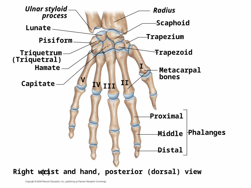
• There are 3 types of questions: – 1) name the bone (bold print words on the unit 2 handout)
– 2) name the marking (regular font words below each bone)
– 3) side (right or left for appendicular skeleton).
• You CANNOT touch the bone on the practicum since the sticker/pipe cleaners can easily be displaced.
• There are 32 stations w/ 3 questions each and 2 stations w/ 2 questions each = 100 questions.
• You will have 1.5 minutes per station.
• Everything you need to know for the next exam is on the bone flashcards posted on my website, plus you need to know right from left on some of the bones. I posted a document on my website about that as well.Print the flashcards out and fold them lengthwise to hide the answers on the left. Be able to write each answer out, and then you will be ready for the test.
• It will take about 10 hours a week to learn all that material, so you need to spend 2 hours per day M-F plus another 5 hours on the weekend. After you learn several pages of the flashcards on the first day, go over them again the second day before you start to learn the next group of pages. On the third day, go over the first two batches before learning the next batch. You need to go over and over what you have learned so it will go into long term memory. Otherwise, if you don't frequently go over it, you will forget during the stress of the test.
• We will cover upper extremity bones today, so by the next lab period, you should have ALL of today’s material memorized. We will also have a 2 point quiz on that material. If you are not ready for the quiz, you are behind. The key to success in an Anatomy class is to spend at least 10 hours a week studying the lab material, plus a similar number of hours studying the lecture material.
• For the rest of this semester, every Sat 12:15-3:30pm, the lab will be open for all 10A students to study the bones, and later on, the cats. Dr. Nguyen will be there to answer questions.
• The Bio Resource Room in building 61 also has 2 bone boxes that students can use outside of lab. The hours and location are posted on the doors of the lab.
• Just a reminder that on the day of the bone exam, you need to bring a lab coat, goggles or safety glasses, a box of gloves, and a dissection kit. You also must have closed toe shoes. If you do not have those items, you cannot enter the lab. I have some items that I can loan you, but I have been instructed to deduct one point from your grade for each item you have to borrow, each time you borrow something. Make sure you have your supplies with you each week from now on, and remember to wear closed toe shoes or you cannot come in.
• I recommend http://www.blangenberg.com/protected/pal2/index.html to study bone. I also remind you of the open lab on Sat 12:30-3:30pm and the Bio Resource Room in 61-3318 (various hours which I do not know) to get more time w/ the bones.
• I am working on uploading a practice bone practicum on You Tube and it should be up later this week.
• You are REQUIRED to take the practicum w/ the section that you have been attending. If you don't take it at this time, then you MUST contact me ASAP with one of these choices: 1) take it with another section BEFORE the scheduled time w/ the section you have been attending with NO penalty OR 2) take it with another section AFTER the scheduled time but you will have a PENALTY of 10 points DEDUCTION from your score IF YOU DO NOT HAVE A VERIFIABLE EXCUSE, like a doctor's note or hospital paperwork for being sick, funeral paperwork if you have a death in the family, police report if you were in an accident, etc.
• If you have other circumstances, please talk to me individually.
• Some of you have NOT been attending regularly. If you do not want to continue, then please drop yourself from this class.