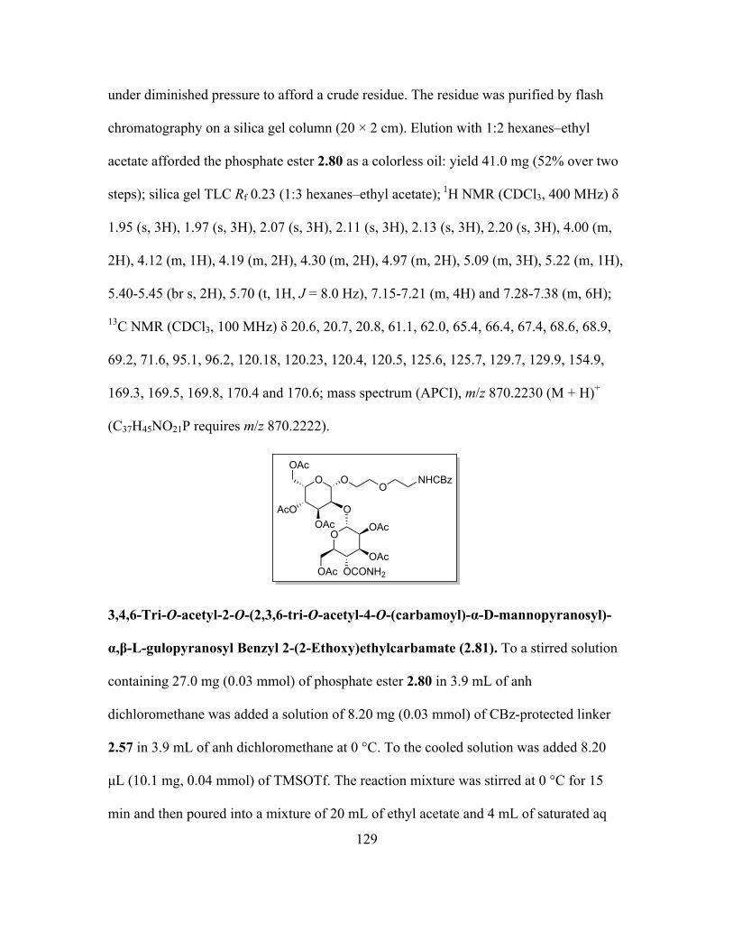
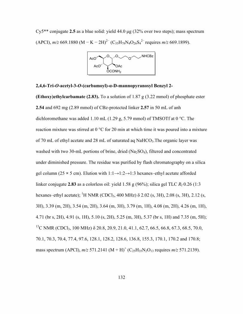
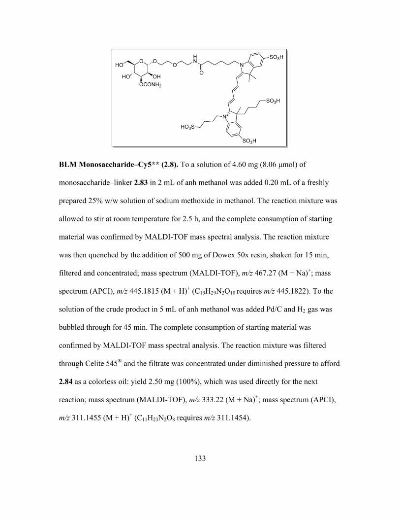
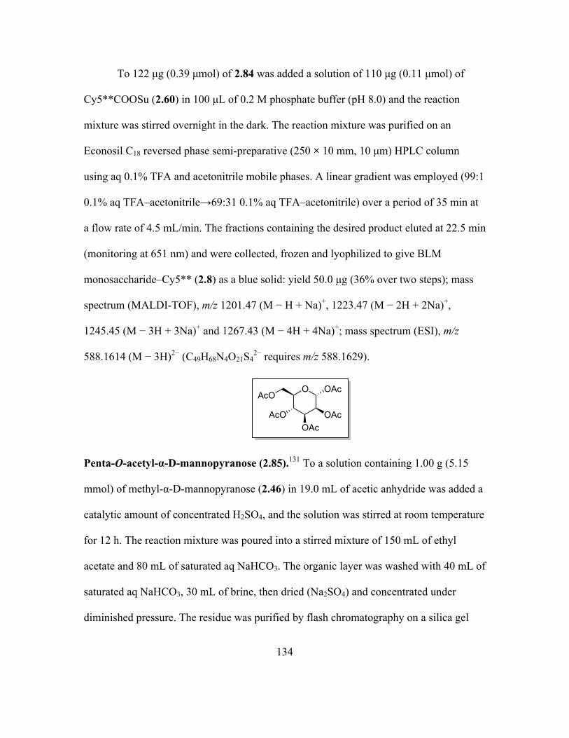
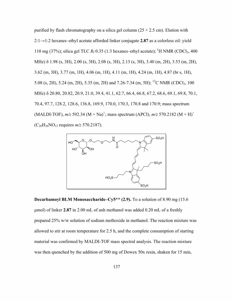
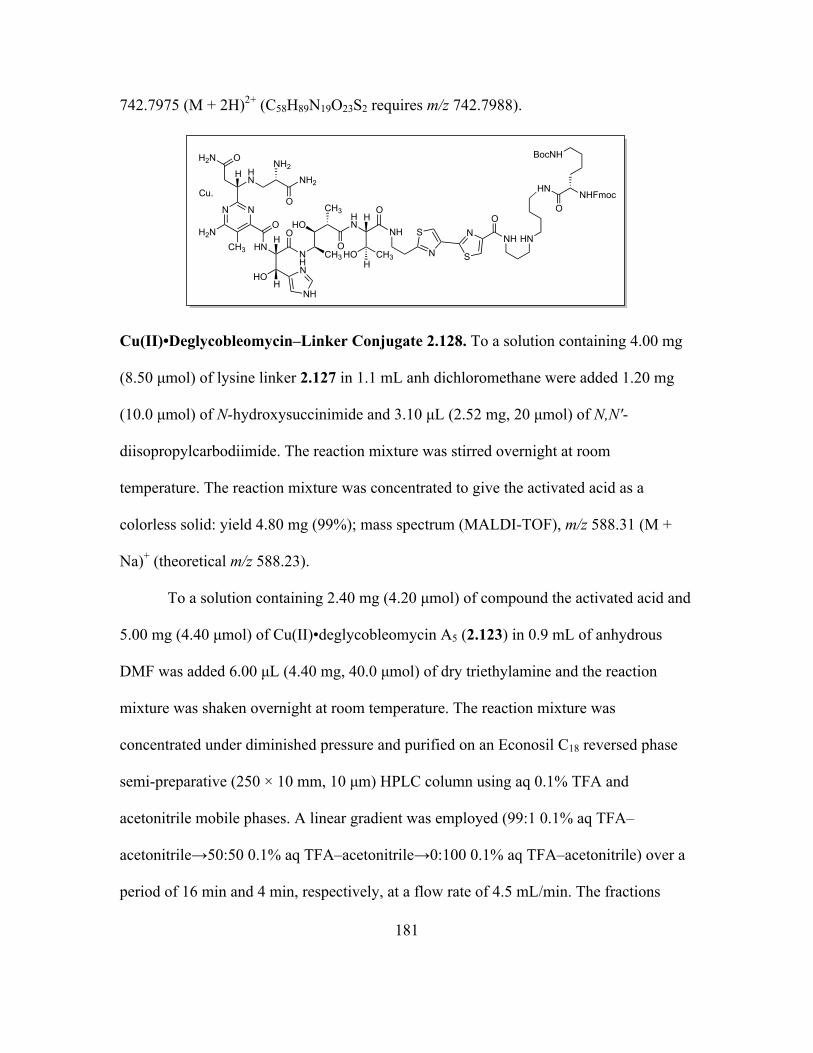
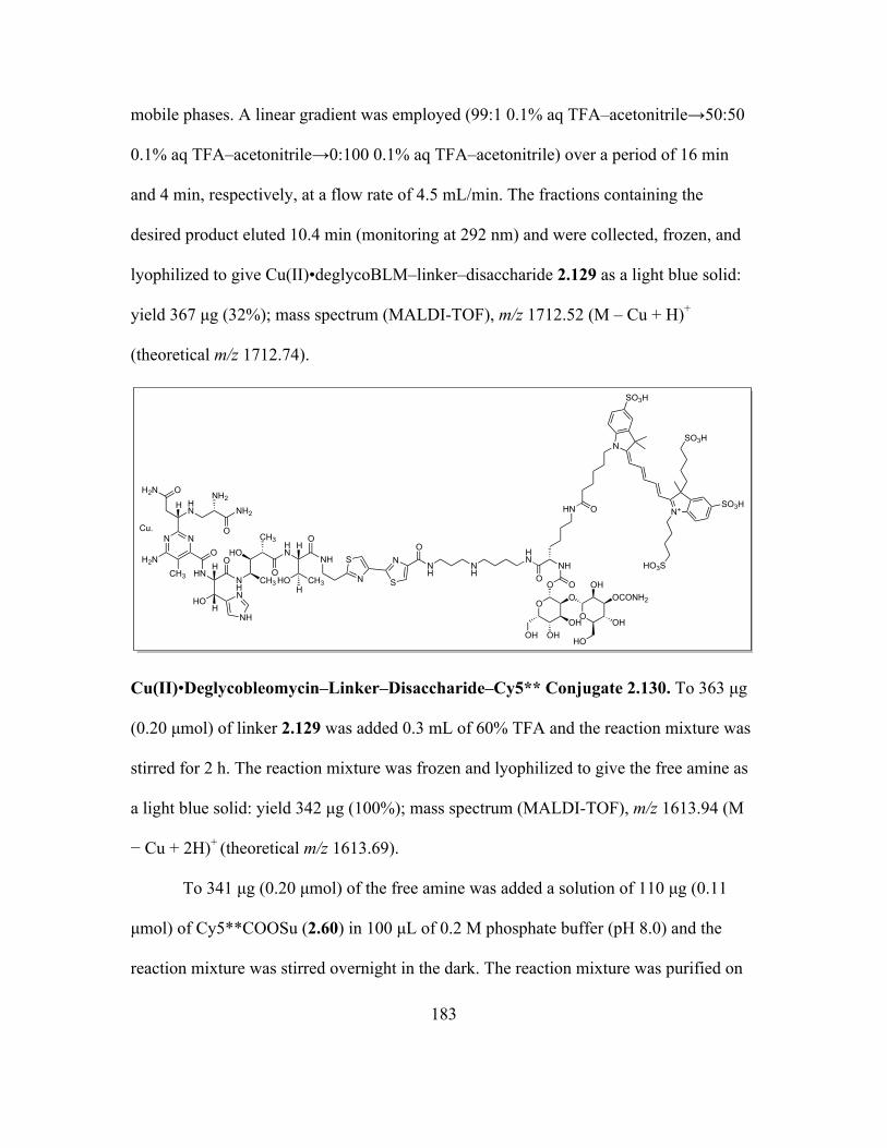
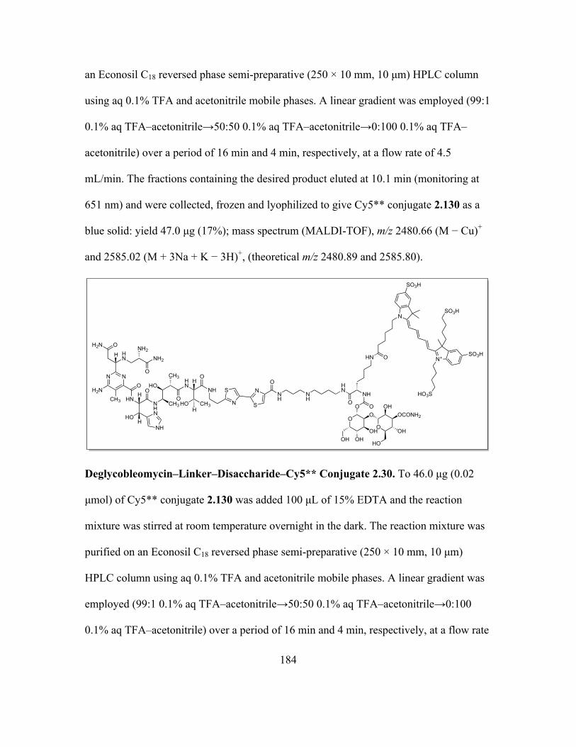
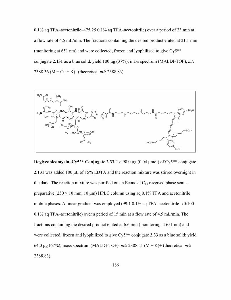
Page 1
Small Molecule Probes for Studying Cellular Receptors and Enzymes
by
Chandrabali Bhattacharya
A Dissertation Presented in Partial Fulfillment of the Requirements for the Degree
Doctor of Philosophy
Approved November 2014 by the Graduate Supervisory Committee:
Sidney M. Hecht, Chair
Ana Moore Ian R. Gould
ARIZONA STATE UNIVERSITY
May 2015
Page 2
i
ABSTRACT
Small molecules have proven to be very important tools for exploration of
biological systems including diagnosis and treatment of lethal diseases like cancer.
Fluorescent probes have been extensively used to further amplify the utilization of small
molecules. The manipulation of naturally occurring biological targets with the help of
synthetic compounds is the focus of the work described in this thesis.
Bleomycins (BLMs) are a class of water soluble, glycopeptide-derived antitumor
antibiotics consisting of a structurally complicated unnatural hexapeptide and a
disaccharide, clinically used as an anticancer chemotherapeutic agent at an exceptionally
low therapeutic dose. The efficiency of BLM is likely achieved both by selective
localization within tumor cells and selective binding to DNA followed by efficient
double-strand cleavage. The disaccharide moiety is responsible for the tumor cell
targeting properties of BLM. A recent study showed that both BLM and its disaccharide,
conjugated to the cyanine dye Cy5**, bound selectively to cancer cells. Thus, the
disaccharide moiety alone recapitulates the tumor cell targeting properties of BLM. Work
presented here describes the synthesis of the fluorescent carbohydrate conjugates. A
number of dye-labeled modified disaccharides and monosaccharides were synthesized to
study the nature of the participation of the carbamoyl moiety in the mechanism of tumor
cell recognition and uptake by BLM saccharides. It was demonstrated that the
carbamoylmannose moiety of BLM is the smallest structural entity capable for the
cellular targeting and internalization, and the carbamoyl functionality is indispensible for
tumor cell targeting. It was also confirmed that BLM is a modular molecule, composed of
Page 3
ii
a tumor cell targeting moiety (the saccharide) attached to a cytotoxic DNA cleaving
domain (the BLM aglycone). These finding encouraged us to further synthesize
carbohydrate probes for PET imaging and to conjugate the saccharide moiety with
cytotoxins for targeted delivery to tumor cells.
The misacylated suppressor tRNA technique has enabled the site-specific
incorporation of noncanonical amino acids into proteins. The focus of the present work
was the synthesis of unnatural lysine analogues with nucleophilic properties for
incorporation at position 72 of the lyase domain of human DNA polymerase beta, a
multifunctional enzyme with dRP lyase and polymerase activity.
Page 4
iii
To Maa and Bapi……….
Page 5
iv
ACKNOWLEDGEMENTS
It has been a gratifying experience to pursue my graduate studies under the
supervision of Professor Sidney Hecht. His enthusiasm to discuss every problem together
with an insistence on independent thought and action has helped me to understand the
subject at a deeper level. I would like to thank him for his guidance, perseverance,
incessant support, incredible dedication and strict discipline. I would also like to express
my gratitude to my committee members, Professor Ana Moore and Professor Ian Gould
and my previous advisor, Professor Dipak Ranjan Mal for their support. I am extremely
grateful to my teachers, Professor Achintya Kumar Sarkar and Professor Amit Bask for
increasing my interest and love for organic chemistry through their extraordinary
teaching skills and helping me to reach the level of pursuing graduate research. I am
indebted to Arizona State University for the awards of Graduate Teaching Assistantships
and tuition scholarships for the years 2009-2011.
During the course of my research, the constant association with the scientists in
the Hecht laboratory has been very valuable; without their support it would have been
incredibly more difficult to succeed in my graduate studies. They include Dr. Damien
Duveau, Dr. Manikandadas M. M., Dr. Pablo Arce, Mohammad Parvez Alam, Poulami
Talukder, Dr. Rakesh Paul and Dr. Omar Khdour. I am also thankful to Dr. Ryan
Nangreave and Mohammad Parvez Alam for teaching me high-performance liquid
chromatography. I would also like to acknowledge my collaborators, Dr. Shengxi Chen,
Dr. Zhiqiang Yu, Justin Kaye, Dr. Sasha Daskalova, and all the other members of the
Hecht lab who have contributed to my education in their own ways. I would also like to
Page 6
v
give special thanks to dearest friends Mohammad Parvez Alam and Poulami Talukder for
helping me and supporting me during every difficult time of my graduate life without
which I could not have completed my studies. I also want to extend my gratitude to my
lab partners Sandipan Roy Chowdhury and Viswanath Arutla for helping me through lab
related issues and sharing research ideas with me. Gina Dunphy has been an amazing
friend and has offered great help during my time in the Hecht lab.
It is also a pleasure to thank all my friends in Tempe who have made my stay in
this city pleasant and memorable. I also wanted to thank my loving grandparents who
embedded the passion of never giving up in me and taught me to believe in myself. And
finally, this thesis is fondly dedicated to my parents for their love, blessings, relentless
support and teaching me science, without which this thesis would not have been possible.
Page 7
vi
TABLE OF CONTENTS
Page
LIST OF FIGURES ......................................................................................................... viii
LIST OF ABBREVIATIONS .......................................................................................... xiv
LIST OF SCHEMES .........................................................................................................xx
CHAPTER
1. INTRODUCTION ................................................................................................1
1.1 Cancer .............................................................................................................1
1.2 Bleomycin .......................................................................................................2
1.3 Carbohydrates .................................................................................................8
1.4 Protein Biosynthesis .....................................................................................12
1.5 Site-specific Incorporation of Unnatural Amino Acids into Proteins ...........15
2. SYNTHESIS OF BLEOMYCIN SACCHARIDE CONJUGATES FOR
IMAGING STUDIES .........................................................................................18
2.1 Introduction ...................................................................................................18
2.1.1 Synthesis of Bleomycin Saccharide–dye Conjugates for Selective
Targeting of Cancer Cells ...................................................................18
2.1.2 Synthesis of Fluorinated Saccharides for Imaging Studies ...............31
2.2 Results ...........................................................................................................32
2.2.1 Synthesis of Fluorescent Dye Conjugates .........................................32
2.2.2 Biological Evaluation of Fluorescent Carbohydrate Conjugates ......56
2.2.3 Synthesis of Fluorinated Saccharides ...............................................71
Page 8
vii
CHAPTER Page
2.3 Discussion .....................................................................................................73
2.4 Experimental Procedures ..............................................................................89
3. SYNTHESIS OF SACCHARIDE–DRUG CONJUGATES FOR TARGETED
DELIVERY TO CANCER CELLS ..................................................................196
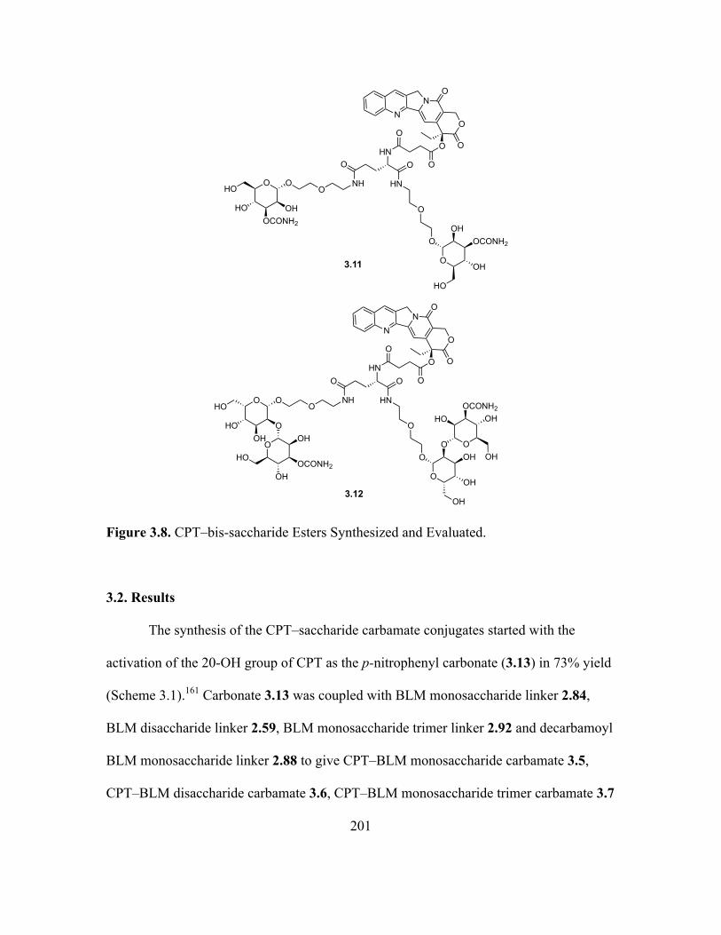
3.1 Introduction .................................................................................................196
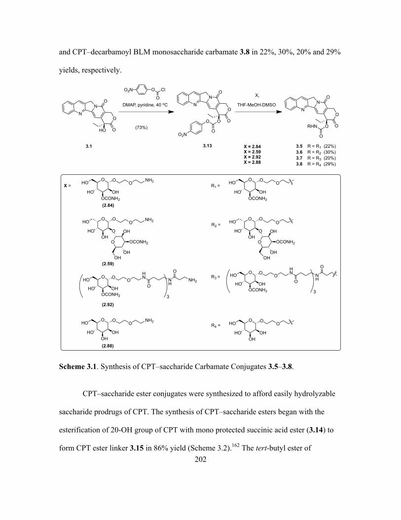
3.2 Results .........................................................................................................201
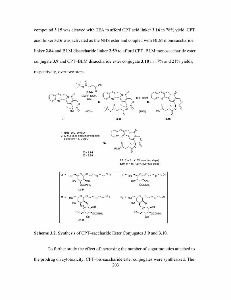
3.3 Discussion ...................................................................................................205
3.4 Experimental Procedures ............................................................................206
4. SYNTHESIS OF LYSINE ANALOGUES FOR MODIFICATION OF
HUMAN DNA POLYMERASE BETA ...........................................................218
4.1 Introduction .................................................................................................218
4.2 Results .........................................................................................................224
4.3 Discussion ...................................................................................................231
4.4 Experimental Procedures ............................................................................232
REFERENCES ................................................................................................................246
APPENDIX
A. COPYRIGHT PERMISSION ............................................................................257
Page 9
viii
LIST OF FIGURES
Figure Page
1.1 Overview of Oncogenesis ..........................................................................................1
1.2 Core Structure of BLM, Illustrating its Various Domains .........................................3
1.3 Proposed Catalytic Cycle for Activation of Bleomycin .............................................5
1.4 Bleomycin-mediated DNA Degradation by the Frank Strand Scission and
Alkali-labile Lesion Pathways ..................................................................................5
1.5 Proposed Mechanism for Double-strand Cleavage of DNA ......................................7
1.6 Catabolism of Bleomycin to Deamido Bleomycin ....................................................8
1.7 The Major Monomeric Monosaccharide Constituents Found in
Glycoconjugates: Glycoproteins, Glycolipids and Proteoglycans .............................9
1.8 ABO Blood Group System Showing the Glycan Chains That Determine the Blood
Type ..........................................................................................................................11
1.9 Peptide Bond Formation During Protein Translation ..............................................14
1.10 Strategy for the Site-specific Incorporation of Unnatural Amino Acids Into
Proteins In Vitro .......................................................................................................16
2.1 Proposed Model for Metal Coordination of Fe(II)•BLM .........................................18
2.2 Magnified Picture of the Surface of Microbubble Derivatized BLM A5 .................19
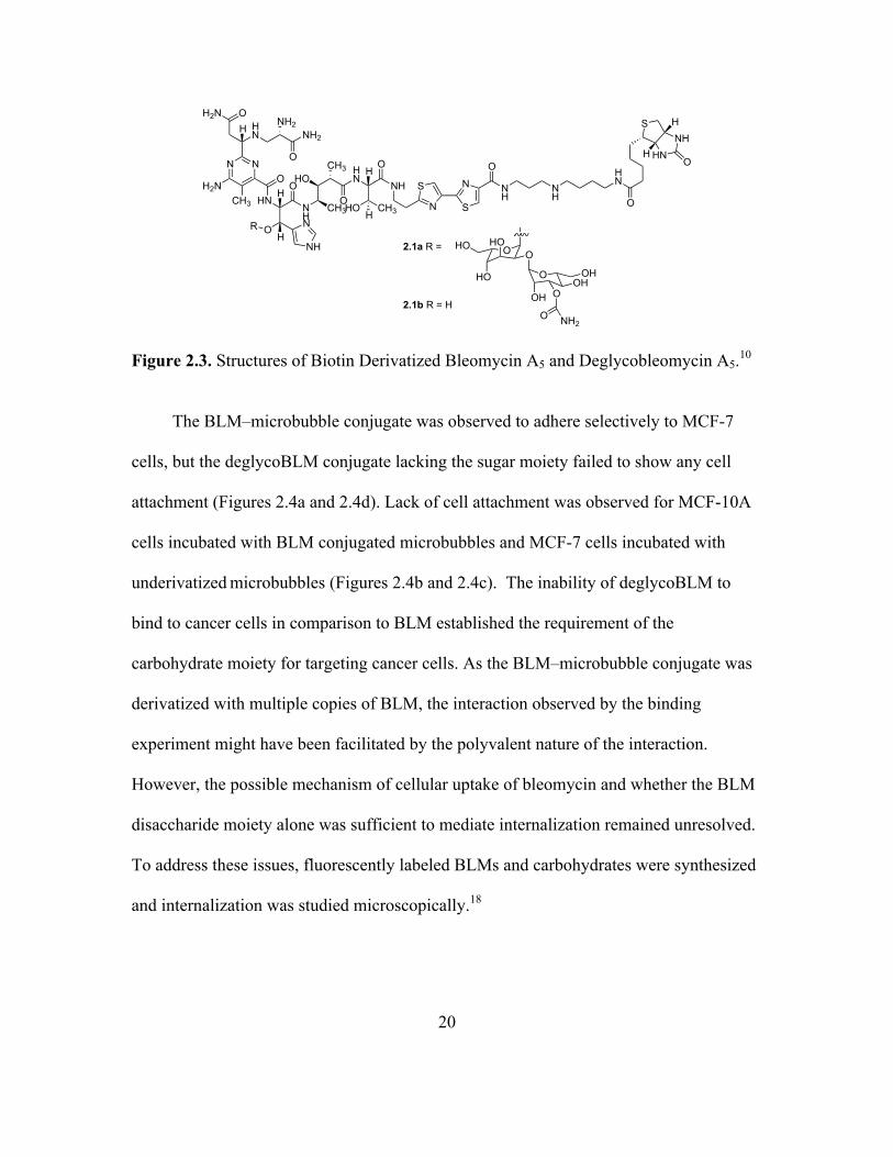
2.3 Structures of Biotin Derivatized Bleomycin A5 and Deglycobleomycin A5 ...........20
2.4 Microscopic Images of Microbubble Experiment ....................................................21
2.5 Structures of BLM–Cy5** (2.2a), DeglycoBLM–Cy5** (2.2b) and BLM
Disaccharide–Cy5** (2.3) ........................................................................................22
Page 10
ix
Figure Page
2.6 Comparison of the Binding/Uptake of BLM–Cy5** (2.2a), DeglycoBLM–Cy5**
(2.2b) and BLM Disaccharide–Cy5** (2.3) in MCF-7 Breast Cancer Cells and
Matched Normal Breast Cells ..................................................................................22
2.7 Quantification of the Binding/Uptake of BLM–Cy5** (2.2a), DeglycoBLM–Cy5**
(2.2b) and BLM Disaccharide–Cy5** (2.3) in MCF-7 Breast Cancer Cells and
Matched Normal Breast Cells ..................................................................................22
2.8 Comparison of the Binding/Uptake of BLM–Cy5** (2.2a), DeglycoBLM–Cy5**
(2.2b) and BLM Disaccharide–Cy5** (2.3) in DU-145 Prostate Cancer Cells and
Matched Normal Prostate Cells ................................................................................23
2.9 Quantification of the Binding/Uptake of BLM–Cy5** (2.2a), DeglycoBLM–Cy5**
(2.2b) and BLM Disaccharide–Cy5** (2.3) in DU-145 Prostate Cancer Cells and
Matched Normal Prostate Cells ................................................................................23
2.10 Library of Disaccharide–dye Conjugates 2.3–2.7 Synthesized and Evaluated ........24
2.11 Structures of BLM Monosaccharide–Cy5** (2.8), Decarbamoyl BLM
Monosaccharide–Cy5** (2.9) and BLM Monosaccharide–Cy5** Trimer (2.10)
Synthesized and Evaluated .......................................................................................25
2.12 Library of Monosaccharide–dye Conjugates 2.11–2.16 Synthesized and
Evaluated ..................................................................................................................26
2.13 Structures of Different Monosaccharide–dye Conjugates 2.17–2.22 Synthesized for
Selection of a Dye to Replace Cy5** .......................................................................27
2.14 Series of Additional Dye Conjugates 2.23–2.28 Synthesized and Evaluated ..........28
Page 11
x
Figure Page
2.15 Structures of a Modified BLM With a C-terminal Disaccharide 2.29 and its
Conjugate With Cy5** 2.30 Synthesized and Evaluated .........................................29
2.16 Structures of Modified BLMs With the Disaccharide Attached to Threonine Moiety
2.31 and 2.32, and Their Fluorescent Dye Conjugates 2.33 and 2.34 Synthesized
and Evaluated ...........................................................................................................29
2.17 PET-CT Image of Breast Ductal Carcinoma Treated With 18F-FDG ......................30
2.18 Structures of Fluorinated BLM Disaccharide 2.35 and Fluorinated Decarbamoyl
BLM Disaccharide 2.36 Synthesized .......................................................................31
2.19 Comparison of the Binding/Uptake of BLM Disaccharide–Cy5** Conjugate (2.3)
and Modified Disaccharide–Cy5** Library Conjugates 2.4−2.7 in Four Cancer
Cell Lines .................................................................................................................56
2.20 Comparison of the Binding/Uptake of BLM Monosaccharide–Cy5** (2.8) and
BLM Disaccharide–Cy5** (2.3) Conjugates in Six Cancer Cell Lines ...................58
2.21 Comparison of the Binding/Uptake of Decarbamoyl BLM Monosaccharide–Cy5**
(2.9) and BLM Monosaccharide–Cy5** (2.8) Conjugates in Four Cancer Cell
Lines .........................................................................................................................59
2.22 Comparison of the Binding/Uptake of BLM Monosaccharide–Cy5** (2.8) and
BLM Monosaccharide–Cy5** Trimer (2.10) Conjugates in Six Cancer Cell
Lines .........................................................................................................................60
2.23 Effect of the Incubation Temperature on the Internalization of BLM
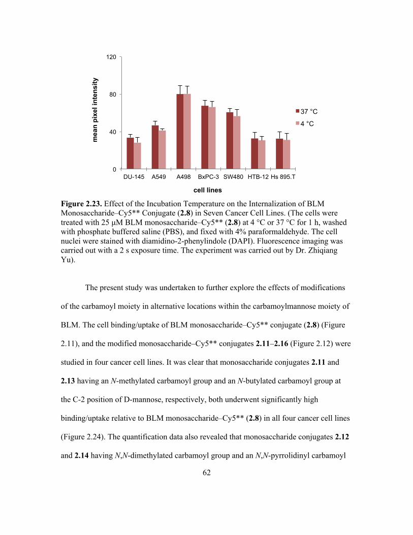
Monosaccharide–Cy5** Conjugate (2.8) in Seven Cancer Cell Lines ....................61
Page 12
xi
Figure Page
2.24 Comparison of the Binding/Uptake of Modified Monosaccharide–Cy5** Library
Conjugates 2.11–2.16 in Four Cancer Cell Lines ....................................................63
2.25 Comparison of the Binding/Uptake of BLM Monosaccharide–dye Conjugates
2.17–2.22 in Three Cancer Cell Lines ......................................................................64
2.26 Comparison of the Binding/Uptake of BLM Monosaccharide–Cy5 (2.18) and the
Free Dye Cy5 in A549 Lung Cancer Cells, A498 Kidney Cancer Cells and
DU-145 Prostate Cells ..............................................................................................65
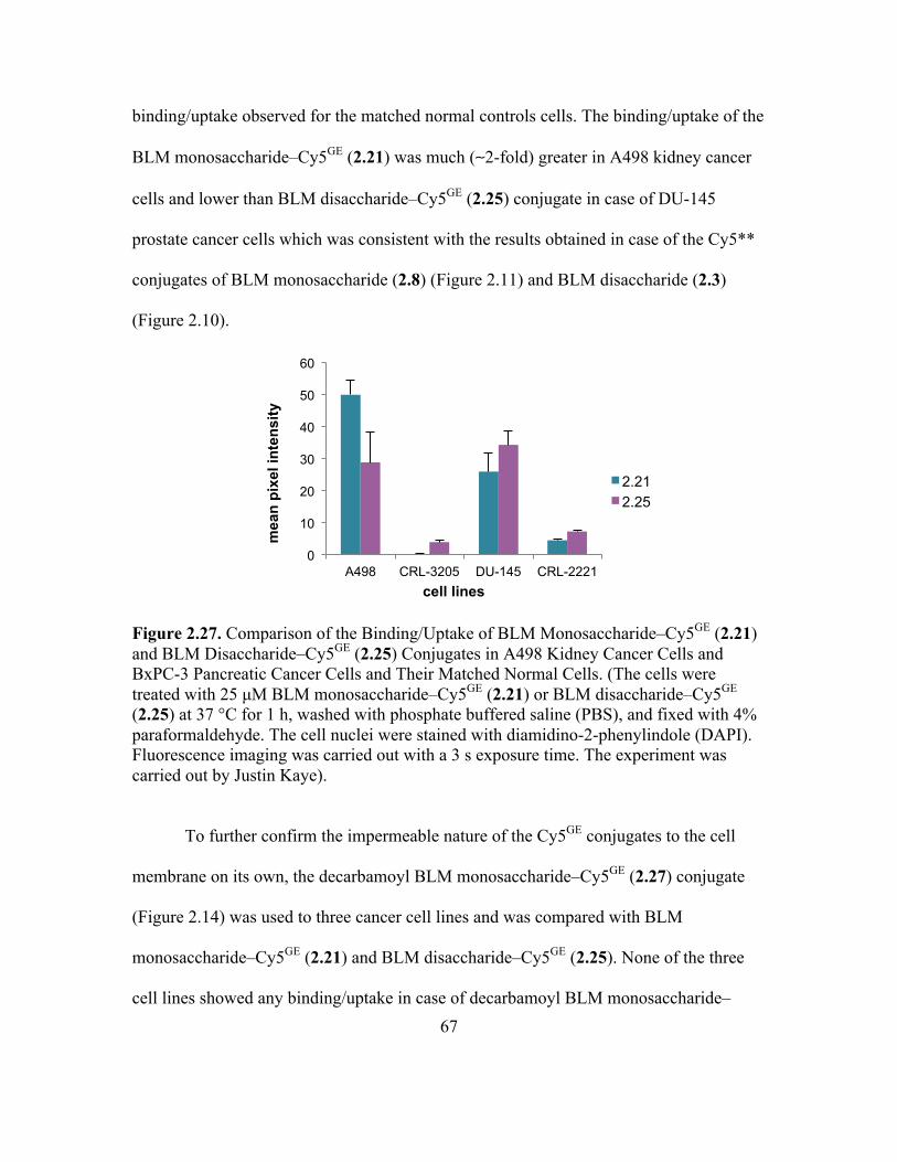
2.27 Comparison of the Binding/Uptake of BLM Monosaccharide–Cy5GE (2.21) and
BLM Disaccharide–Cy5GE (2.25) Conjugates in A498 Kidney Cancer Cells and
BxPC-3 Pancreatic Cancer Cells and Their Matched Normal Cells ........................66
2.28 Comparison of the Binding/Uptake of BLM Monosaccharide–Cy5GE (2.21), BLM
Disaccharide–Cy5GE (2.25) and Decarbamoyl BLM Disaccharide–Cy5GE (2.27)
Conjugates in A498 Kidney Cancer Cells, DU-145 Prostate Cancer Cells and
A549 Lung Cells .....................................................................................................67
2.29 Dose-dependent Effects of Modified BLM Analogues 2.29, 2.31 and 2.32 on the
Viability of Cultured DU-145 Prostate Cancer Cells ...............................................69
2.30 Comparison of the Binding/Uptake of BLM–Cy5** (2.2a), DeglycoBLM–Cy5**
(2.2b), and Modified BLM–Cy5** Conjugates 2.30 and 2.34 in DU-145 Prostate
Cancer Cells and PZ-HPV-7 Normal Prostate Cells ................................................69
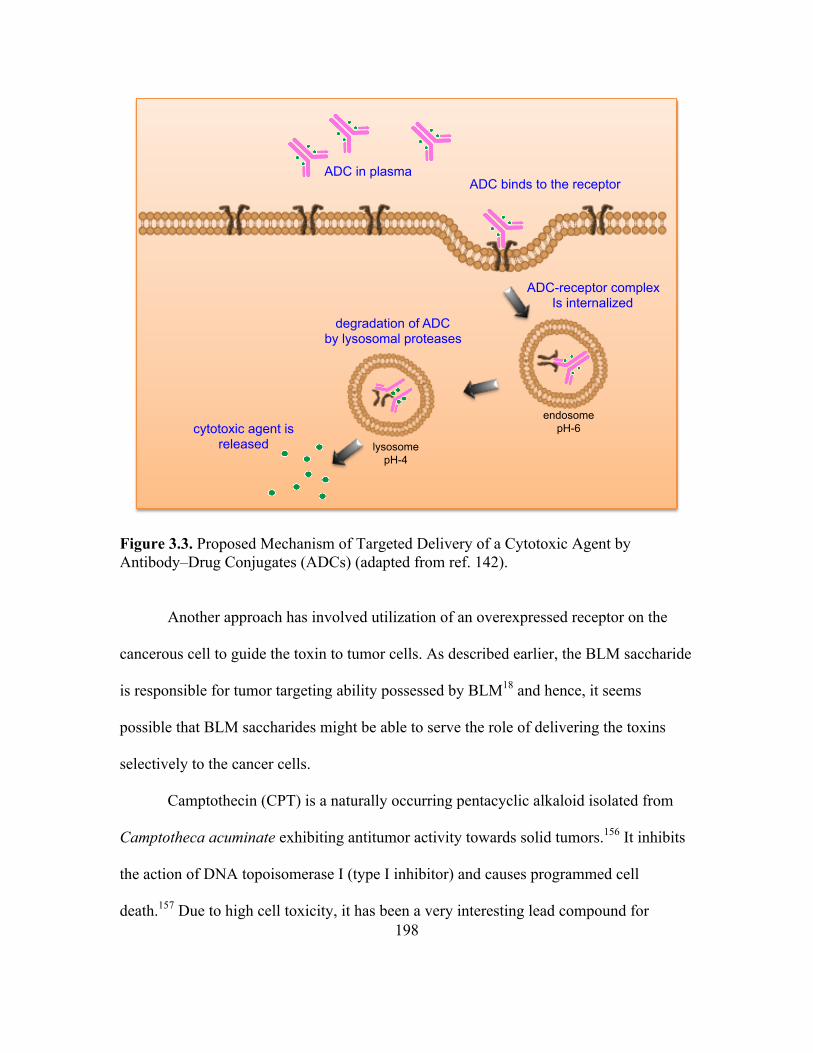
3.1 Elements of an Antibody–Drug Conjugate (ADC) ................................................195
3.2 Structure of Antibody–Drug Conjugate: Trastuzumab-DM1 ................................195
Page 13
xii
Figure Page
3.3 Proposed Mechanism of Targeted Delivery of a Cytotoxic Agent by Antibody–
Drug Conjugates (ADCs) .......................................................................................196
3.4 Structures of CPT Congeners: Topotecan and Irinotecan ......................................197
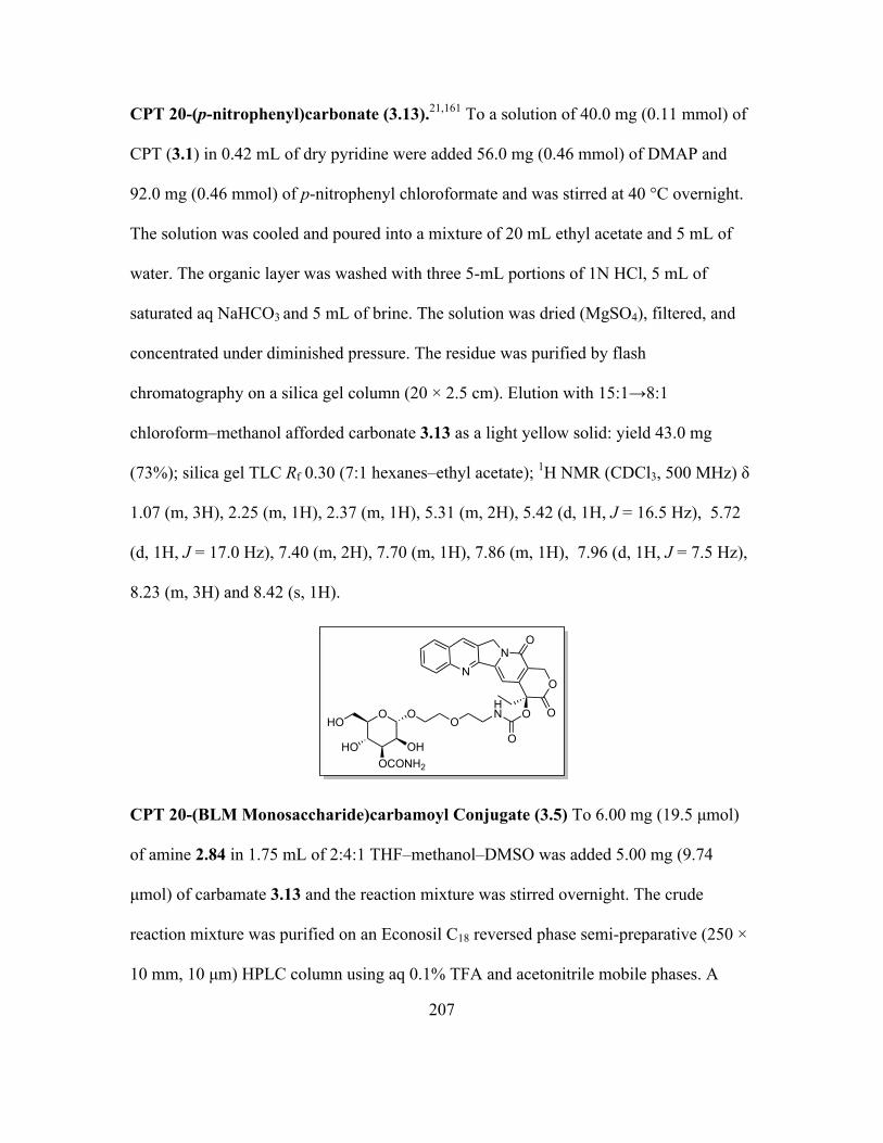
3.5 Equilibrium Between the Closed and Open Lactone Ring of CPT ........................197
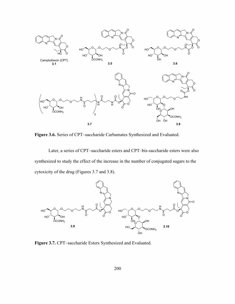
3.6 Series of CPT–saccharide Carbamates Synthesized and Evaluated .......................198
3.7 CPT–saccharide Esters Synthesized and Evaluated ...............................................198
3.8 CPT–bis-saccharide Esters Synthesized and Evaluated .........................................199
4.1 Amino Acid Sequence of Human DNA Polymerase Beta, Highlighted Red Letters
Include the Residues Present in the Active Site of dRP Lyase Domain ................217
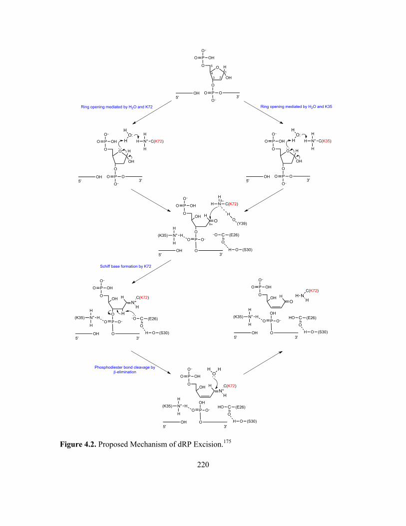
4.2 Proposed Mechanism of dRP Excision ..................................................................218
4.3 Active Site of dRP Lyase Domain of Human DNA Polymerase Beta Including
Lys35, Tyr39, Lys60, Lys68, Lys72 And Lys84 ...................................................219
4.4 Amino Acids Used for Modification of Lys72 of Rat DNA Polymerase
Beta .........................................................................................................................219
4.5 Summary of dRP Excision Activity of Rat Polymerases Beta Modified at
Position 72 With Lysine Analogues .......................................................................220
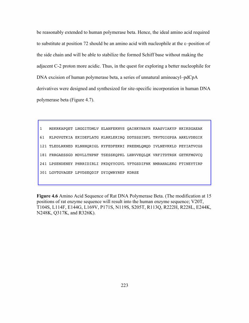
4.6 Amino Acid Sequence of Rat DNA Polymerase Beta ...........................................221
4.7 Series of Aminoacylated pdCpA Derivatives Synthesized for Site Directed
Incorporation at Position 72 of Human DNA Polymerase Beta ............................222
Page 14
xiii
Figure Page
4.8 Strategy Employed for Incorporation of Lysine Analogues Into Position 72 of
Human DNA Polymerase Beta (EchDPB), the Gene for Which had Been
Optimized for Expression in E. coli .......................................................................227
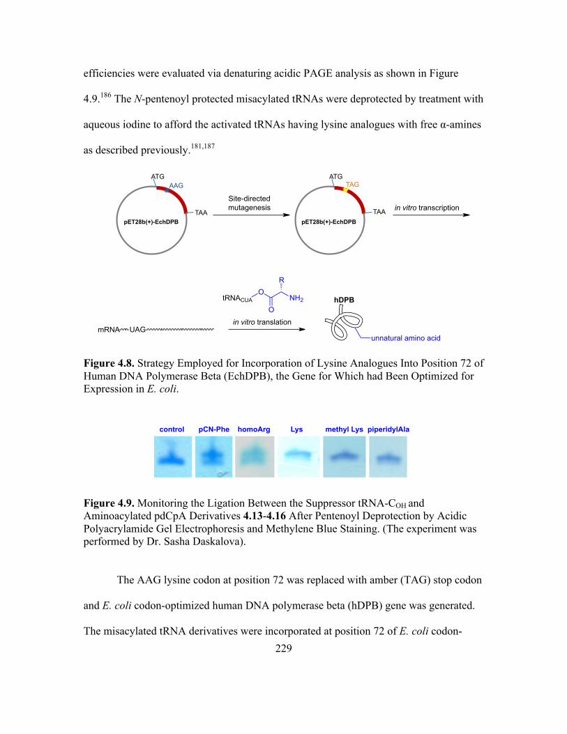
4.9 Monitoring the Ligation Between the Suppressor tRNA-COH and Aminoacylated
pdCpA Derivatives 4.13-4.16 After Pentenoyl Deprotection by Acidic
Polyacrylamide Gel Electrophoresis and Methylene Blue Staining ......................228
4.10 (I) In Vitro Translation of Wild Type Protein From E. coli Codon-optimized
(EchDPB) and Non-optimized (hDPB) Template and In Vitro Translation of
Modified Proteins With Incorporated pCN-Phe or Homoarginine at Position 72
From E. coli Codon-optimized Template or Non-optimized template Having an
Amber Stop Codon at Position 72; (II) In vitro Translation of Modified Proteins
With Incorporated Lysine or Lysine Analogues at Position 72 From E. coli Codon-
optimized Template Having an Amber Stop Codon at Position 72 .......................228
Page 15
xiv
LIST OF ABBREVIATIONS
AcOH Acetic Acid
Ac2O Acetic Anhydride
APCI Atmospheric Pressure Chemical Ionization
anh Anhydrous
aq Aqueous
atm Atmosphere
BH3 Borane
BLM Bleomycin
BLEDTA Bleomycin–ethylenedinitrilotetraacetic Acid Conjugate
Bn Benzyl
BnBr Benzyl Bromide
Boc t-Butoxycarbonyl
br s Broad Singlet
Bu2SnO Dibutyltin Oxide
oC Degrees Celsius
13CNMR Carbon Nuclear Magnetic Resonance Spectroscopy
cat Catalytic
CBr4 Tetrabromomethane
CBz Carboxybenzyl
CDCl3 Deuterated Chloroform
cm Centimeter
Page 16
xv
conc. Concentrated
CoCl2 Coballt(II) Chloride
CSA Camphorsulfonic Acid
CuCl2 Copper(II) Chloride
CuO Copper(II) Oxide
Cy5 Cyanine 5 (Lumiprobe Co.)
Cy5GE Cyanine 5 (General Electric Co.)
Cy5** Cyanine 5** (General Electric Co.)
Cy5.5GE Cyanine 5 (General Electric Co.)
Cy7 Cyanine 7 (Lumiprobe Co.)
δ Chemical Shift (ppm)
d Doublet
dd Doublet of Doublet
ddd Doublet of Doublet of Doublet
dRP Deoxyribosephosphate
DAPI 4′,6-Diamino-2-phenylindole
DBU 1,8-Diazabiocyclo[5.4.0]undec-7-ene
DCM Dichloromethane
DIBAL-H Diisobutylaluminium Hydride
DIC Diisopropylcarbodiimide
DIPEA Diisopropylethylamine
DMAP Dimethylaminopyridine
Page 17
xvi
DMF Dimethylformamide
DMSO Dimethylsulfoxide
DNA Deoxyribonucleic Acid
EDTA Ethylenedinitrilotetraacetic Acid
EtOAc Ethyl Acetate
Et3N Triethylamine
EtSH Ethanethiol
ESI Electrospray Ionization
FAB Fast Atom Bombardment
FDG 2-Fluoro-2-deoxy-D-glucose
Fmoc 9-Fluorenylmethoxycarbonyl
g Gram(s)
h Hours
H2 Hydrogen Gas
H2O Water
H2SO4 Sulfuric Acid
HCl Hydrochloric Acid
HgCl2 Mercury(II) Chloride
HgO Mercury(II) Oxide
1HNMR Proton Nuclear Magnetic Resonance Spectroscopy
HPLC High-performance Liquid Chromatography
Hz Hertz
Page 18
xvii
J Coupling Constant
L Liter
LiOH Lithium Hydroxide
m Multiplet
M Molar
M+ Molecular Ion
MALDI-TOF Matrix Assisted Laser Desorption Ionization Time of Flight
MeOH Methanol
MeOTf Methyl Triflate
mg Milligram(s)
µm Microgram(s)
MgSO4 Magnesium Sulfate (Anhydrous)
MHz Mega Hertz
min Minutes
mL Milliliter
mM Millimolar
mmol Millimole(s)
µmol Micromole(s)
MTT 3-(4,5-Dimethylthiazol-2-yl)-2,5-diphenyltetrazolium Bromide
N Normal
N2 Nitrogen Gas
NaBH4 Sodium Borohydride
Page 19
xviii
NaHCO3 Sodium Bicarbonate
Na2CO3 Sodium Carbonate
Na2SO4 Sodium Sulfate (Anhydrous)
NaOMe Sodium Methoxide
NH3 Ammonia
NH4OAc Ammonium Acetate
NHS N-Hydroxysuccinimide
nm Nanometer
NMR Nuclear Magnetic Resonance
PBS Phosphate Buffer Saline
pCN-Phe p-Cyanophenylalanine
p-NO2C6H4-OCOCl p-Nitrophenyl Chloroformate
p-TsOH p-Toluenesulfonic Acid
Pd Palladium
Pd/C Palladium on Carbon
Pd(OH)2 Palladium Hydroxide on Carbon (Pearlman’s catalyst)
pdCpA 5ʹ′-O-Phosphoryl-2ʹ′-deoxycytidylyl(3ʹ′→5ʹ′)adenosine
(PhO)2P(O)Cl Diphenyl Phosphoryl Chloride
PhCH(OMe)2 Benzaldehyde Dimethyl Acetal
ppm Parts Per Million
PPh3 Triphenylphosphine
PTC Peptidyltransferase Center
Page 20
xix
q Quartet
quin Quintet
Rf Ratio of Fronts
RNA Ribonucleic Acid
s Singlet
SDS-PAGE Sodium Dodecyl Sulfate Polyacrylamide Gel Electrophoresis
sulphoCy5 Sulphocyanine 5 (Lumiprobe Co.)
t Triplet
TBA Tetrabutylammonium
TFA Trifluoroacetic acid
THF Tetrahydrofuran
TLC Thin Layer Chromatography
tRNA Transfer RNA
TMSOTf Trimethylsilyl Triflate
TSTU O-(N-succinimidyl)-1,1,3,3-tetramethyluronium tetrafluoroborate
UV Ultraviolet
v Volume
Page 21
xx
LIST OF SCHEMES
Schemes Page
2.1 Synthesis of the Gulose Glycosyl Acceptor 2.45 .....................................................33
2.2 Synthesis of the Activated C-3-carbamoylmannose Glycosyl Donor 2.54 ..............34
2.3 Synthesis of the BLM Disaccharide–Cy5** (2.3) ...................................................36
2.4 Synthesis of the Activated C-2-O-benzylmannose Glycosyl Donor 2.64 ................37
2.5 Synthesis of the C-2-carbamoylmannose Modified Disaccharide–Cy5** 2.4 ........39
2.6 Synthesis of the Activated C-4-carbamoylmannose Glycosyl Donor 2.78 ..............40
2.7 Synthesis of the C-4-carbamoylmannose Modified Disaccharide–Cy5** 2.5 ........41
2.8 Synthesis of the BLM Monosaccharide–Cy5** (2.8) ..............................................42
2.9 Synthesis of Decarbamoyl BLM Monosaccharide–Cy5** (2.9) .............................43
2.10 Synthesis of the BLM Monosaccharide–Cy5** Trimer (2.10) ................................44
2.11 Synthesis of the C-2-carbamoyl Modified Monosaccharide–Cy5**
2.11–2.15 ..................................................................................................................46
2.12 Synthesis of the C-3-methylcarbamoyl Modified Monosaccharide–
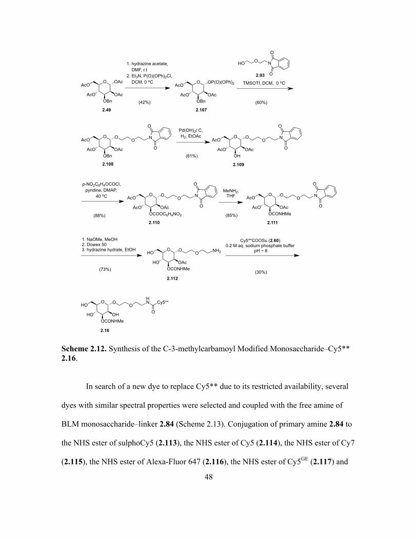
Cy5** 2.16 ...............................................................................................................48
2.13 Synthesis of BLM Monosaccharide–dye Conjugates 2.17–2.22 .............................49
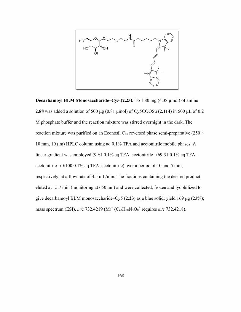
2.14 Synthesis of Decarbmoyl BLM Monosaccharide–Cy5 (2.23) .................................50
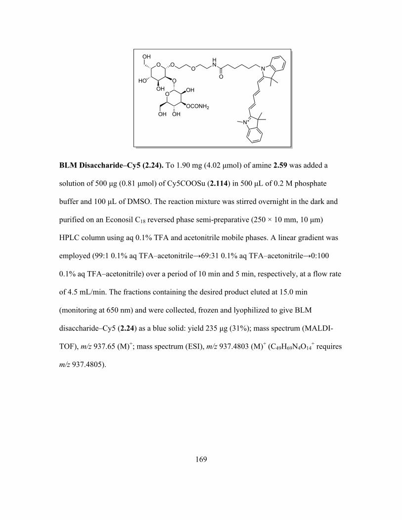
2.15 Synthesis of BLM Disaccharide–dye Conjugates 2.24 and 2.25 .............................50
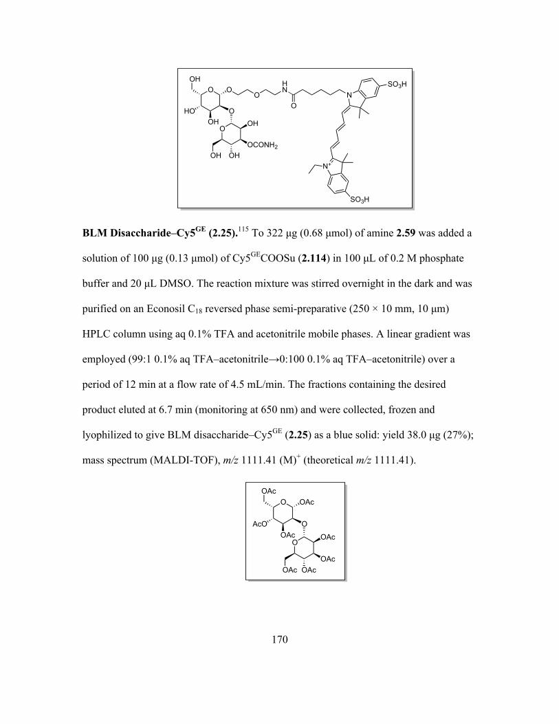
2.16 Synthesis of Decarbamoyl BLM Disaccharide–dye Conjugates 2.26 and 2.27 .......51
2.17 Synthesis of BLM–Cy5GE (2.28) ..............................................................................52
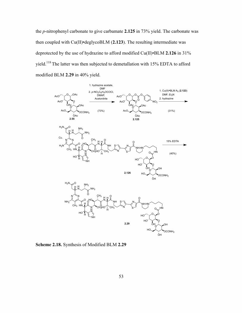
2.18 Synthesis of Modified BLM 2.29 .............................................................................53
Page 22
xxi
Schemes Page
2.19 Synthesis of Modified BLM–Cy5** Conjugate 2.30 ...............................................54
2.20 Synthesis of Modified BLM–Cy5** Conjugates 2.33 and 2.34 ..............................55
2.21 Synthesis of Fluorinated BLM Disaccharide 2.35 ...................................................72
2.22 Synthesis of Fluorinated Decarbamoyl BLM Disaccharide 2.36 .............................73
3.1 Synthesis of CPT–saccharide Carbamate Conjugates 3.5–3.8 ...............................202
3.2 Synthesis of CPT–saccharide Ester Conjugates 3.9 and 3.10 ................................203
3.3 Synthesis of CPT–bis-saccharide Ester Conjugates 3.11 and 3.12 ........................204
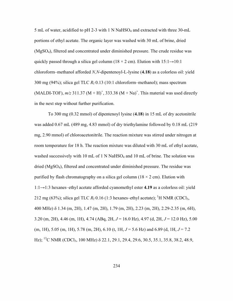
4.1 Synthesis of Lysyl–pdCpA 4.13 .............................................................................225
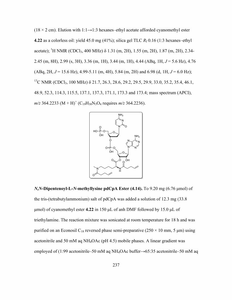
4.2 Synthesis of N-methyllysyl–pdCpA 4.14 ...............................................................226
4.3 Synthesis of Homoargininyl–pdCpA 4.15 .............................................................226
4.4 Synthesis of Piperidylalanyl–pdCpA 4.16 .............................................................228
Page 23
1
CHAPTER 1
INTRODUCTION
1.1. Cancer
The term "cancer" was derived from the Greek word carcinos, which means crab.
It was used by Hippocrates to describe the ulcerous diseases causing finger-spreading
projections.1 The disruption of the balance between cell division and cell death results in
the transition of a normal cell to a cancerous cell. This results in faster multiplication of
the cells leading to augmented uncontrolled proliferation and loss of cellular
differentiation, producing a growing mass of tissue called a ‘tumor’ or ‘malignancy’
(Figure 1.1). Biologically, cancer is a disease which involves a cycle of dynamic genetic
and epigenetic changes in a normal cell.2 For several decades, studies have been ongoing
to discover the specific alterations that result in a malignancy.
Figure 1.1. Overview of Oncogenesis.
Page 24
2
There are more than 100 clinically different types of cancer, which are recognized
by their unique properties and symptoms, and require specific treatment strategies.3
However, they still can be categorized into four major subdivisions: leukemias,
lymphomas, sarcomas and carcinomas. A leukemia involves an abnormality in white
blood cells or leukocytes, which are produced by the bone marrow. A lymphoma results
from an abnormality in lymphocytes, which are produced by spleen and lymph nodes;
these are considered similar to a leukemia. A sarcoma is solid tumor of mesodermal
origin such as connective tissues, bone, and muscles. Finally, a carcinoma is a solid
tumor of epithelial origin and constitutes the major form of cancer, accounting for about
of all malignant neoplasms.3 The hallmarks of cancer mainly involves six biological traits
acquired during the various stages of cancer development.4 They include hyperplasia
(uncontrolled proliferation), growth factor abnormalities, suppression of programmed cell
death or apoptosis, replication of immortality in the cells, angiogenesis (expansion of
blood vessels), and metastasis (the ability of cells to dissociate from a tumor mass and
create a new tumor at an anatomically distant site).4
1.2. Bleomycins
Bleomycins (BLMs) are a class of water soluble, glycopeptide-derived antitumor
antibiotics consisting of a structurally complicated unnatural hexapeptide and a
disaccharide (Figure 1.2).5 It was first isolated from a culture broth of a bacterium,
Streptomyces verticillus, as a Cu(II) chelate by Umezawa and co-workers in 1966.6
Clinically, bleomycin is used for the treatment of various kinds of tumors of soft tissues,
Page 25
3
especially squamous cell carcinomas and malignant lymphomas.7 In the United States, it
is sold under the tradename Blenoxane as a mixture consisting mainly bleomycin A2 and
B2, whereas bleomycin A5 is used clinically in Japan, Russia and China.8 Bleomycin
mediates oxidative cleavage of DNA selectively at 5ʹ-GPy-3ʹ sequences in the presence
of a metal co-factor and oxygen.5 The exceptionally low therapeutic dose of BLM (~5
µmol) implies extremely high therapeutic efficiency in comparison to other drugs. Such
efficiency of BLM is likely achieved both by selective localization within tumor cells9,10
and selective binding to DNA followed by efficient double-strand cleavage.11
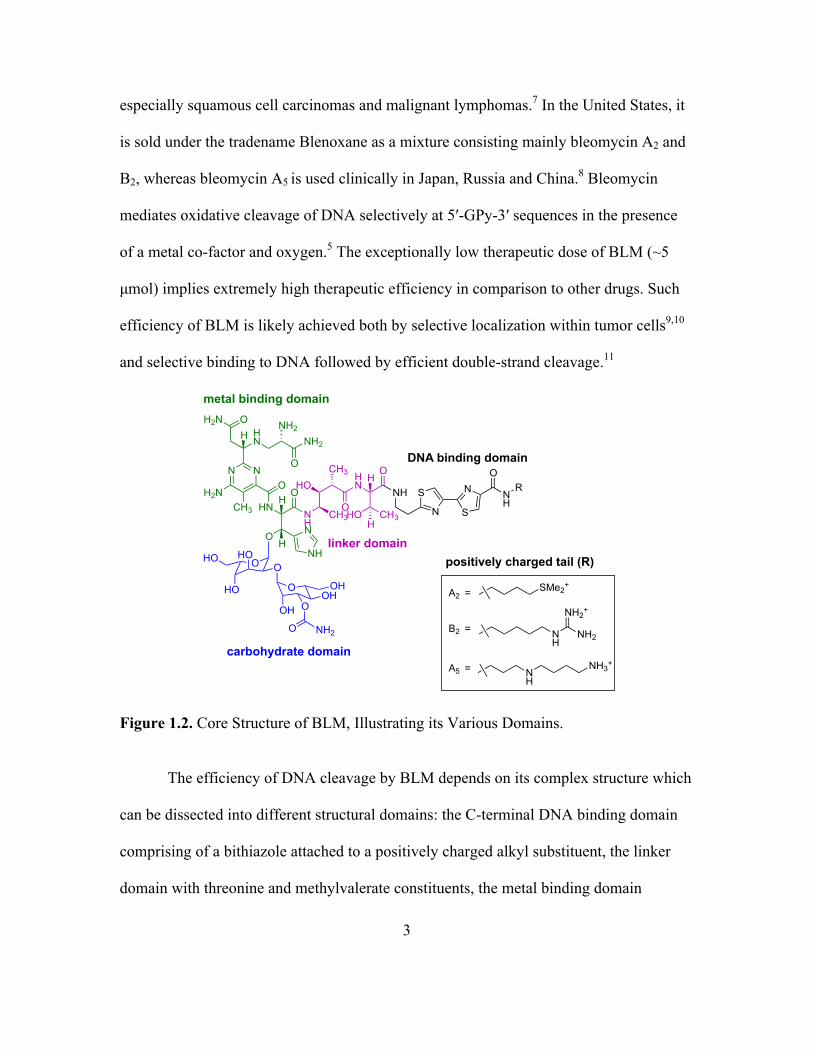
Figure 1.2. Core Structure of BLM, Illustrating its Various Domains.
The efficiency of DNA cleavage by BLM depends on its complex structure which
can be dissected into different structural domains: the C-terminal DNA binding domain
comprising of a bithiazole attached to a positively charged alkyl substituent, the linker
domain with threonine and methylvalerate constituents, the metal binding domain
N N
H2NCH3
OH2NHN NH2
O
HN
OH
NH
CH3
HOHN
NH
N
S
S
N NH
O
OOHO CH3
H
O
O
OHOHO
HO
O
O
OH
OHOH
O
NH2O
NH
NH
H
H
CH3
metal binding domain
linker domain
DNA binding domain
carbohydrate domain
R
NH2
NH
NH3+
NH
NH2
SMe2+
NH2+
A2 =
B2 =
A5 =
positively charged tail (R)
Page 26
4
containing the pyrimidoblamic acid and β-hydroxyhistidine moieties, and the
disaccharide moiety. The metal binding domain is the primary determinant of the
sequence selectivity of DNA, which is responsible for chelation to metal and oxygen
required for the activation of BLM.12-15 The DNA binding domain binds to DNA through
specific interactions. The linker domain maintains the essential compact folded structure
required for the cleavage.16,17 The disaccharide sugar moiety is essential for cancer cell
targeting.10,18 The first total synthesis of bleomycin was accomplished by the Umezawa19
and Hecht20 laboratories in 1982. In 1994, Boger and co-workers also reported a total
synthesis of BLM A2.21
Though bleomycin is not an enzyme, the interactions of BLM with oxygen
possess some similarities to the activation mediated by the metalloenzyme cytochrome
P450.22-24 There are two notable pathways for DNA strand cleavage both of which
require activated Fe(II)•BLM (Figure 1.3). The specific mechanism involved in the
degradation depends on the availability of oxygen (Figure 1.4). The frank strand scission
pathway is favored by a high concentration of oxygen, whereas the mechanism shifts
toward the alkali labile lesion pathway in a low oxygen environment.25-27 As the ratio of
products depends on concentration of oxygen, both the pathways are believed to involve
the common radical intermediate28,29 produced by abstraction of the C-4ʹ hydrogen atom
from the deoxyribose of the susceptible nucleotide (Figure 1.4).
Page 27
5
Figure 1.3. Proposed Catalytic Cycle for Activation of Bleomycin.30
Figure 1.4. Bleomycin-mediated DNA Degradation by the Frank Strand Scission and Alkali-labile Lesion Pathways.5
CpGpO OH
pathway leading to alkali labile lesion
Criegee rearrangement
pathway leading to frank strand scission
CGCTAGCGGCGATCGC
5'
3'
FeII-BLM
anti-elimination
O−PO
O
OHO C
N
NH
NH2
O
+
OH-
O−PO
O
OO G
OO
OPOOTAGCG
O−
OHHO
O−PO
O
OHO C
O−PO
O
OO G
OO
OPOOTAGCG
O−
C.
O-PO
O
OHO C
O-PO
O
OO G
OCH2COOH
N
N
NH2
O
+
CHO
+ pTAGCG
N
N
NH2
O
CHO
+ pCGCTAGpOCH2COOH + 5'-GMP
product formed by cleavage of C3
product formed by cleavage of C7
O
+ pTAGCG
product formed by cleavage of C3
Page 28
6
The ability of BLM to mediate double-strand cleavage of DNA depends on the binding
interactions between DNA and BLM. Double-strand cleavage of DNA by bleomycin was
first studied systematically by the Povirk laboratory.31-34 Accordingly, a single molecule
of BLM effects double-strand cleavage of DNA. After the first lesion generated on the
DNA, the BLM is reactivated and the specificity of the DNA gets altered in the
complementary strand which results in the second break at one of the two nucleotide
positions opposite to the primary site of cleavage.35 However, recently Hecht and co-
workers reported a new mechanism of double-strand cleavage not previously described,
which showed that many of the double-strand cleavages associated with the strongly
bound hairpin DNAs results from two independent events rather than the coupled event
(Figure 1.5).11
The high valence iron complex is regenerated from sequestered hydroxyl radical
analogous to the propagation in lipid peroxidation. Double-strand cleavages are
refractory to repair by DNA repair enzymes and, hence, is highly cytotoxic, thereby
making bleomycin selective and cytotoxic.35
Page 29
7
Figure 1.5. Proposed Mechanism for Double-strand Cleavage of DNA.11,35
In spite of its therapeutic efficiency, a part of the drug undergoes catabolism to
deamido bleomycin by cysteine proteinase bleomycin hydrolase36-39 (Figure 1.6). Though
in earlier reports, deamido bleomycin was asserted to possess no antitumor activity, it
was later found to retain one-half of its ability to mediate single strand scission and one-
eighth of its ability to mediate double-strand scission.40
O
T
O O
C
O
G
O O
A
O
ACT
5'
T
3'
O
A
OO
T
O
G
OO
G
O
A C TA
3' 5'
.
O
T
O O
C
O
G
O O
A
O
AOHT
5'
T
3'
O
A
OO
T
O
G
OO
G
O
A C TA
3' 5'
O
T
O O
C
O
CH2COO−
G
HO O
A
O
AT
5'
T
3'
O
A
OO
T
O
G
OO
G
O
A C TA
3' 5'
O
T
O O
C
O
CH2COO−
G
HO O
A
O
AT
5'
T
3'
O
A
OO
CH2COO−
T
OHOO
G
O
A C TA
3' 5'
O
T
O O
C
O
CH2COO−
G
HO O
A
O
AT
5'
T
3'
O
A
OO
T
O
OH
OO
G
O
A C TA
3' 5'
O
T
O O
C
O
CH2COO−
G
HO O
A
O
AT
5'
T
3'
O
A
OHO
T
OHOO
G
O
A C TA
3' 5'
primary attack by bleomycin
primary strand break AP site formation
secondary attack by bleomycin
secondary strand break AP site formation at secondary site
double strand cleavage product
n-butylamine treatment
Page 30
8
Figure 1.6. Catabolism of Bleomycin to Deamido Bleomycin.40
Though bleomycin has proved to be an excellent therapeutic agent for cancer
chemotherapy, a significant barrier to its widespread clinical use is a side effect involving
lung fibrosis.41 This raises the question of the requirement for a greater understanding of
the chemistry associated with BLM.
1.3. Carbohydrates
Carbohydrates are a class of oxygen containing biomolecules, which play
numerous crucial roles in living systems. In principle, they are hydrates of carbon and can
be classified as monosaccharides, disaccharides, oligosaccharides, and polysaccharides.
The various roles of carbohydrates in the biological processes, apart from being a primary
source of energy (starch and glycogen), were not discovered until recently due to the
presence of a relatively constrained number of monomers constituting the biologically
important polymers.42 With time, the detailed and closer examination of cellular
functions and structure revealed the complexities associated with carbohydrates.
N N
H2NCH3
OH2NHN NH2
O
HN
OH
NH
CH3
HOHN
NH
N
S
S
N NH
O
OOHO CH3
H
O
O
OHOHO
HO
O
O
OH
OHOH
O
NH2O
NH
NH
H
H
CH3
S+(CH3)2X−
NH2
bleomycin A2
BLM hydrolaseOH2N
HN OH
O
HNH2
deamido bleomycin A2
Page 31
9
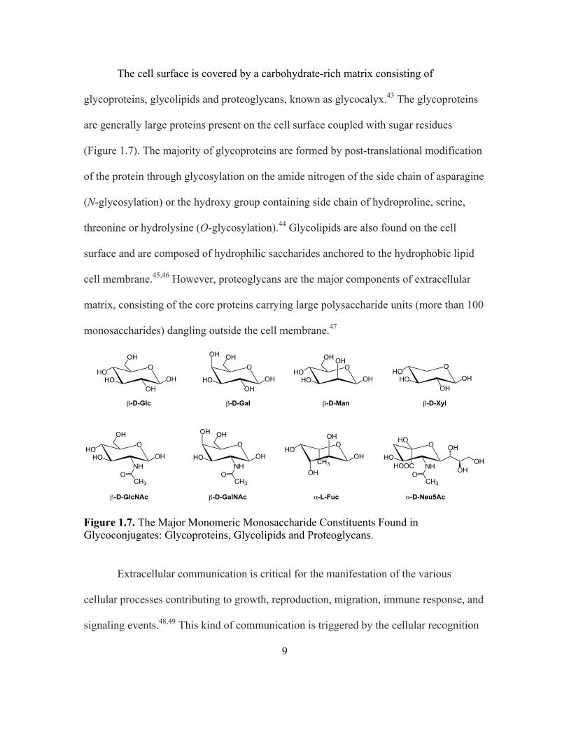
The cell surface is covered by a carbohydrate-rich matrix consisting of
glycoproteins, glycolipids and proteoglycans, known as glycocalyx.43 The glycoproteins
are generally large proteins present on the cell surface coupled with sugar residues
(Figure 1.7). The majority of glycoproteins are formed by post-translational modification
of the protein through glycosylation on the amide nitrogen of the side chain of asparagine
(N-glycosylation) or the hydroxy group containing side chain of hydroproline, serine,
threonine or hydrolysine (O-glycosylation).44 Glycolipids are also found on the cell
surface and are composed of hydrophilic saccharides anchored to the hydrophobic lipid
cell membrane.45,46 However, proteoglycans are the major components of extracellular
matrix, consisting of the core proteins carrying large polysaccharide units (more than 100
monosaccharides) dangling outside the cell membrane.47
Figure 1.7. The Major Monomeric Monosaccharide Constituents Found in Glycoconjugates: Glycoproteins, Glycolipids and Proteoglycans.
Extracellular communication is critical for the manifestation of the various
cellular processes contributing to growth, reproduction, migration, immune response, and
signaling events.48,49 This kind of communication is triggered by the cellular recognition
O
OHOH
HOHO
OHO
OHOH
HO
OH OHO
OH
OH
HOHO
OH
O
OHNH
HOHO
OH
CH3O
O
OHNH
HO
OH OH
CH3O
O
OH
OH
OH
HO
CH3
O
NHHO
HO
CH3O
HOOC OHOH
OH
β-D-Glc β-D-Gal β-D-Man
O
OHOH
HOHO
β-D-Xyl
β-D-GlcNAc β-D-GalNAc α-L-Fuc α-D-Neu5Ac
Page 32
10
that involves the interaction of the receptors on the cell surface with their ligands, which
was first described as “lock-and-key” hypothesis by Emil Fischer in 1894.50 Receptor–
ligand interaction is effected by the organization and activity of the receptor, which is
influenced by the bulky constituents of the glycocalyx. In addition, the larger
glycoproteins can influence protein sorting, endocytosis, and neurological and
immunological developments.51-55
Due to the diverse biological functions associated with the glycan, a
comprehensive understanding of protein–carbohydrate interactions is vital in order to
study the biological processes.56,57 In nature, there are several carbohydrate-binding
proteins which are mainly classified as lectins and antibodies. The lectins have high
affinity towards carbohydrates and bind them reversibly through their carbohydrate
recognition domains. They have evolved to recognize the specific carbohydrate at the
surface of the cell to mediate cell–cell adhesion, trafficking, triggering immune responses
and cell signaling.58-61 Antibodies are glycoproteins secreted by the plasma cells, which
are used by the immune system and are directed towards cell surface glycans. Different
glycan signatures are expressed by different cells and have a vital role clinically.62 One
such example for antibody–carbohydrate interaction is represented by the blood group
ABO antigen–antibody interaction. The different structure of glycan chains is responsible
for the generation of different antigens which forms the ABO blood group system (Figure
1.8) and failure of unmatched blood transfusions.63,64
Page 33
11
Figure 1.8. ABO Blood Group System Showing the Glycan Chains That Determine the Blood Type.63
Many proteins bind to the glycan ligands through polyvalent interactions and
possess multiple binding sites.65 The binding efficiency in the case of polyvalent
interaction increases with an increase in the concentration of the monovalent ligands near
the site of binding. On the basis of these interactions, many scaffolds were developed for
drug delivery, diagnosis and other therapeutic purposes such as cyclodextrins, chitins,
chitosans and calixerenes.66,67
O-linked-β-N-acetylglucosamine (O-GlcNAc), a post-translational modification
of the glycoproteins, plays a crucial regulatory role in cellular processes and is termed a
nutritional sensor.68 UDP-GlcNAc, the end product in the hexosamine biosynthetic
pathway (HBP), is the donor substrate for the enzymatic addition of O-GlcNAc used by
O-GlcNAc transferase (OGT). The removal of O-GlcNAc from the proteins is catalyzed
A
AB
B
O
`"`"
`"`"
`"`"
`"`"
`"`"
Red blood cell
N-acetylgalactosamine Fucose Galactose N-acetylglucosamine
Page 34
12
by O-GlcNAcase (OGA). The cycling of O-GlcNAc in proteins depends on the balance
of activity of OGT and OGA, and plays a crucial role in control of cellular processes.69-73
Excessive O-GlcNAc substitution is a general feature associated with cancer.74-78 As a
result of cancer, the energy metabolism of the cell shifts from oxidative phosphorylation
to a less efficient energy pathway involving glycolysis; this shift is known as the
Warburg effect.79-81 This results in an increase in the demand for glucose in the cell and
eventually increases the uptake of glucose and glucosamine. Further, the levels of the
glucose transporters and glycolytic enzymes can be upregulated by Kras oncogene. As a
result of the increase in HBP flux, the level of UDP-GlcNAc increases and results in
hyper-O-GlcNAcylation.82 The reduction of hyper-O-GlcNAcylation, however, inhibits
cellular proliferation and results in apoptotic cell death.73
Recently, it has been reported that the surface of the glycocalyx in the cancer cells
are covered with bulky glycoproteins. These bulky substituents help to facilitate the
metastatic spread of cancer cells and to regulate cell survival.55,83
1.4. Protein Biosynthesis
Proteins are the biochemical polymeric macromolecules made up of amino acid
residues that play a crucial role in a vast array of cellular processes including DNA
replication, catalyzing metabolic reactions and the internalization of molecules in the cell.
Each protein contains a signature sequence of amino acid residues that arranges itself into
a specific three-dimensional structure and thereby contributes to the structure and activity
of the protein. In nature, the genetic information encoded in a messenger RNA (mRNA)
Page 35
13
is translated into the protein by the cellular organelle called a ribosome. The ribosome is
a large nucleoprotein complex made up of a complex array of ribosomal RNAs (rRNAs)
and proteins. The ribosome is comprised of two subunits, denoted as 30S and 50S in
bacteria.84,85 Each subunit in the ribosome has three sites for binding transfer RNA
(tRNA), namely the A-site (aminoacyl-tRNA binding site), P-site (peptidyl-tRNA
transfer site) and E-site (exit site for deacylated tRNA). Each tRNA decodes the genetic
information by recognizing a set of three nucleotides known as a codon (triplet
sequence), which is complementary to tRNA anticodon, and linking the nucleotide
sequence with amino acid residues found at the opposite end of the tRNA. The high
fidelity of protein synthesis is due to accurate codon–anticodon base pairing among the
different RNAs during the decoding process.86
The translation of mRNA proceeds through three distinct steps, namely initiation,
elongation and termination. The process begins with activation of tRNA with its cognate
amino acid by its aminoacyl-tRNA synthetase. Initiation of protein translation involves
the binding of the 30S ribosomal subunit with the mRNA and aminoacylated tRNA with
the rRNA having the base pairs complementary to the Shine–Dalgarno sequence of the
bound mRNA.84 This event is followed by binding of the 50S ribosomal subunit to the
30S subunit, affording the 70S ribosomal complex. The formation of initiation complex
requires initiation factors (IF1, IF2 and IF3). Generally, the initiation codon translated is
AUG, which codes for methionine. Only the activated form N-formylmethionine is able
to form the initiation complex and occupies the P-site of ribosomal subunit. After
initiation, the empty A-site of the ribosomal subunit is charged with aminoacyl-tRNA
Page 36
14
having anticodon corresponding to the next codon on mRNA. This event is conducted
with the help of GTP and elongation factor EF-Tu, the latter of which is recharged by
another elongation factor EF-Ts. The α-amino group of the aminoacyl-tRNA present in
the A-site mediates a nucleophilic attack on the carbonyl carbon of the adjacent nascent
peptide in P-site and results in transfer of the amino group to the tRNA in the A site
thereby elongating the peptide chain (Figure 1.9). Subsequently, the tRNAs in the A-site
and P-site are translocated to the P-site and E-site, respectively, with the help of
elongation factor EF-G.
Figure 1.9. Peptide Bond Formation During Protein Translation (R1, R2 and R3 are the side chain substituents in the amino acids).84
AAA UCUUAGmRNA AGA
OO
HNpeptide R1
O
OH2N
R2
peptidyl transfer
AAA UCUUAGmRNA AGA
OH O
O
NH
R2
O
HNpeptide R1
AAA UCUUAGmRNA AGA
O
OH2N
R3 dissociation of tRNA from E-site
followed by binding of new aminoacyl-tRNA
at A-site
AAA UCUUAGmRNA AGA
OH O
O
NH
R2
O
HNpeptide R1
translocation
Peptidyl Site Aminoacyl Site Peptidyl Site Aminoacyl Site
Peptidyl Site Aminoacyl Site Exit Site Peptidyl Site
OO
HN R2
ONH
peptide
R1
Page 37
15
This event is followed by release of the tRNA from E-site and binding of the next
aminoacylated-tRNA, having anticodon corresponding to the next codon on mRNA, to
the vacant A-site.84,87 The process of elongation continues until the termination is
signaled by the presence of one of the stop codons (UAG, UAA or UGA) on mRNA. The
presence of the stop codon in the A-site results in the binding of the release factors (RF1,
RF2 and RF3) and eventually triggers the release of the nascent polypeptide and
dissociation of ribosomal complex into 50S and 30S subunits.87
1.5. Site-specific Incorporation of Unnatural Amino Acids into Proteins
In nature, there are 20 proteinogenic amino acids and the different order in
arrangement of these resides in proteins gives rise to an array of different proteins with a
variety of biological roles. To study protein structure, function, dynamics and
intermolecular interactions, unnatural amino acids can be introduced into the proteins
using misacylated suppressor tRNAs.88-91 This technique has developed into a very
powerful tool and involves site-directed mutagenesis of DNA to replace the codon for a
specific amino acid of interest with a nonsense codon (Figure 1.10).
Page 38
16
Figure 1.10. Strategy for the Site-specific Incorporation of Unnatural Amino Acids Into Proteins In Vitro.84
The translation of a stop codon by circumventing the effects of the release factors
responsible for the termination of protein synthesis is referred to as suppression event.
The nonsense codons used for this purpose are mainly UAG (amber)88, UAA (opal)92 and
UGA (ochre).93 Hecht and co-workers first developed the general technique for
misacylation of the suppressor tRNA comprising the anticodon complementary to the
nonsense codon by ligating a tRNA, lacking the 3ʹ-terminal cytidine and adenosine
moieties, to the aminoacylated dinucleotide pCpA with the help of T4 RNA ligase.94-96
Later, Schultz and co-workers modified the method by replacing the aminoacylated
dinucleotide pCpA with dinucleotide pdCpA that is now extensively used for
misacylation of tRNA.97,98 This technology has proven revolutionary in the field of
in vitro transcription
in vitro translation TAG
promoter T7
pET28b(+)-protein(TAG)plasmid bearing a stop
codon
AUC
COH
ACC
unnatural amino acid
AUC
1. T4 RNA ligase, pdCpA-unnatural amino
acid
2. Deprotection
abbreviated supressor RNA
unnatural amino acid
modified protein
mRNA UAG
Page 39
17
protein engineering and helped in the incorporation of a wide variety of unnatural amino
acids in proteins with novel properties. Further, Hecht and co-workers demonstrated that
the modification of the 23S rRNA in bacteria can dramatically changes the ribosomal
architecture and permit the incorporation of D-amino acids and β-L-amino acids.99-102
Page 40
18
CHAPTER 2
SYNTHESIS OF BLEOMYCIN SACCHARIDE CONJUGATES FOR IMAGING
STUDIES
2.1. Introduction
2.1.1. Synthesis of Bleomycin Saccharide–dye Conjugates for Selective Targeting of
Cancer Cells
For many years the role of the disaccharide moiety in bleomycin was a matter of
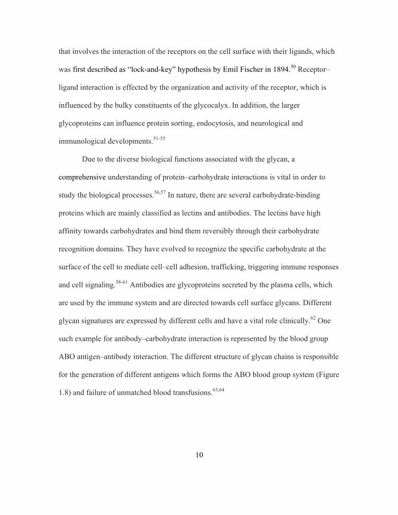
debate. It was postulated that the C-3 carbamoyl group attached to the mannose is
involved in metal binding as the sixth ligand (Figure 2.1) and that the sterically hindered
disaccharide forms a protective pocket to shield the reactive radical intermediates.103-106
Figure 2.1. Proposed Model for Metal Coordination of Fe(II)•BLM.104
However, the documented DNA cleavage activity of deglycoBLM in a cell free
system rendered the involvement of the sugar in metal binding moot since deglycoBLM
has no sugar.107,108 In parallel, extensive in vivo imaging studies were carried out with
57Co-BLM and 111In-BLM complexes.109,110 Later, a new modified imaging agent
Page 41
19
BLEDTA, containing an ethylenedinitrilotetraacetic acid (EDTA) moiety attached to the
C-terminal end of BLM A2 was developed and 111In-BLEDTA was used successfully to
image several carcinomas in vivo. These studies failed to show comparably effective
imaging for 111In-deglycoBLEDTA.111-113 Thus, these results strongly implied that the
carbohydrate moiety in bleomycin could possibly play a role in cellular recognition and
internalization of the drug. In order to further study the role of the carbohydrate moiety,
our laboratory designed a targeting experiment in which cancer cells were treated with
microbubbles that had been derivatized with bleomycin.10
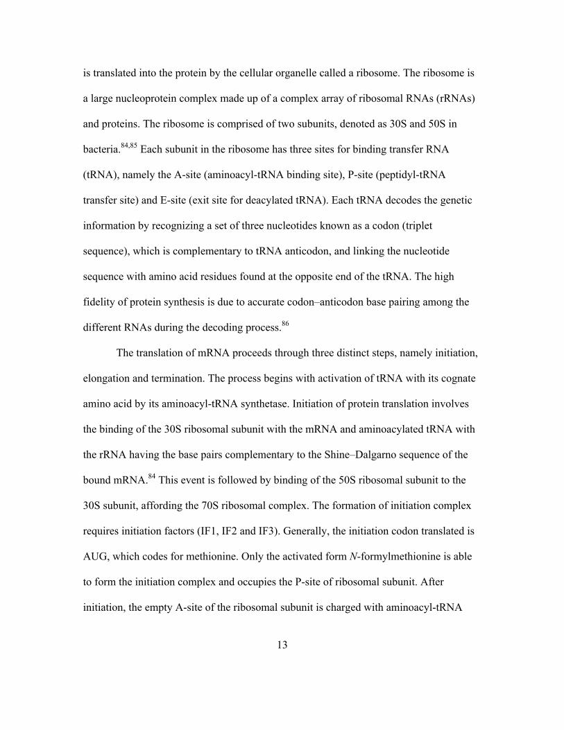
Microbubbles are lipid membranes made up of albumin or other materials, and
enclosing a gaseous core such as air, nitrogen or a perfluorocarbon (Figure 2.2). They are
used as contrast agents in ultrasonography.114 The surface of microbubbles that has been
modified with streptavidin was used to bind biotin–BLM A5 and biotin–deglycoBLM A5
(Figure 2.3) through a biotin–streptavidin specific interaction. The derivatized
microbubbles were then incubated with cultured MCF-7 breast cancer cells and MCF-
10A normal breast cells and imaged using a Zeiss Axiovert 200M microscope.10
Figure 2.2. Magnified Picture of the Surface of Microbubble Derivatized BLM A5.10
gaseous core
S
BLM-A5B
SB B
microbubble
lipid shell
lipid monolayer
bleomycin-A5
biotin
steptavidin
magnified view of microbubble surface
PEG
Page 42
20
Figure 2.3. Structures of Biotin Derivatized Bleomycin A5 and Deglycobleomycin A5.10
The BLM–microbubble conjugate was observed to adhere selectively to MCF-7
cells, but the deglycoBLM conjugate lacking the sugar moiety failed to show any cell
attachment (Figures 2.4a and 2.4d). Lack of cell attachment was observed for MCF-10A
cells incubated with BLM conjugated microbubbles and MCF-7 cells incubated with
underivatized microbubbles (Figures 2.4b and 2.4c). The inability of deglycoBLM to
bind to cancer cells in comparison to BLM established the requirement of the
carbohydrate moiety for targeting cancer cells. As the BLM–microbubble conjugate was
derivatized with multiple copies of BLM, the interaction observed by the binding
experiment might have been facilitated by the polyvalent nature of the interaction.
However, the possible mechanism of cellular uptake of bleomycin and whether the BLM
disaccharide moiety alone was sufficient to mediate internalization remained unresolved.
To address these issues, fluorescently labeled BLMs and carbohydrates were synthesized
and internalization was studied microscopically.18
N N
H2NCH3
OH2NHN NH2
O
HN
OH
NH
CH3
HOHN
NH
N
S
S
N
O
OOHO CH3H
O
OR
NH
NH
H
HNH2
CH3
NH
NH
HN
2.1a R =
2.1b R = H
OHOHO
HO
O
O
OH
OHOH
O
NH2O
O
S
HNNH
O
H
H
Page 43
21
Figure 2.4. Microscopic Images of Microbubble Experiment: (a) Microscopic image of MCF-7 breast cancer cells treated with BLM A5 derivatized microbubbles. (b) Microscopic image showing MCF-10A normal breast cells treated with BLM A5 derivatized microbubbles. (c) Microscopic image of MCF-7 breast cancer cells treated with underivatized microbubbles. (d) Microscopic image showing MCF-7 breast cancer cells treated deglycoBLM A5 derivatized microbubbles.10
Classically, in a fluorescence microscopy experiment, the fluorescent tag or label is
attached chemically to the molecule of interest to enable its detection following
incubation with the target cell lines. Fluorescence microscopy is then used to image the
chemical tag localized in the cell. In order to choose the correct reporter, the fluorescent
dye should not bind to the cell on its own. Also, it should have sufficient fluorescence
signal strength to be detectable above the autofluorescence of the cells studied. Cy5**
was chosen as the reporter after many trials115 and was attached to BLM A5,
deglycoBLM A5 and BLM disaccharide (Figure 2.5).18 The conjugates were incubated
a b
c d
Page 44
22
with breast, prostate, colon and lung cell lines, as well as their matched normal cell lines,
and were imaged using a Zeiss Axiovert microscope. Both BLM–Cy5** (2.2a) and BLM
disaccharide–Cy5** (2.3) showed selective uptake in the MCF-7 breast cancer cells
relative to matched normal cells (Figure 2.6 and Figure 2.7). Similar results were also
obtained in the case of DU-145 prostate cells and matched normal prostate cells (Figure
2.8 and Figure 2.9).18
Figure 2.5. Structures of BLM–Cy5** (2.2a), DeglycoBLM–Cy5** (2.2b) and BLM Disaccharide–Cy5** (2.3).18
N N
H2NCH3
OH2NHN NH2
O
HN
OH
NH
CH3
HOHN
NH
N
S
S
N
O
OO
HO CH3H
O
OR
NH
NH
H
HNH2
CH3
NH
NH
HN
BLM–Cy5** 2.2a R =
deglycoBLM–Cy5** 2.2b R = H
OHOHO
HO
O
O
OH
OHOH
O
NH2O
Cy5**
O
Cy5**
O
OOH
OCONH2OHOH
O O
OOH
HO
OH
OHN
BLM disaccharide–Cy5**(2.3)
O
N+
N
SO3H
SO3H
HO3S
SO3H
HOCy5**COOH =
Page 45
23
Figure 2.6. Comparison of the Binding/Uptake of BLM–Cy5** (2.2a), DeglycoBLM–Cy5** (2.2b) and BLM Disaccharide–Cy5** (2.3) in MCF-7 Breast Cancer Cells and Matched Normal Breast Cells.18
Figure 2.7. Quantification of the Binding/Uptake of BLM–Cy5** (2.2a), DeglycoBLM–Cy5** (2.2b) and BLM Disaccharide–Cy5** (2.3) in MCF-7 Breast Cancer Cells and Matched Normal Breast Cells.18
MCF-7 breast carcinoma cells
MCF-10A normal breast cells
BLM−Cy5**
deglycoBLM BLM−Cy5**
BLM disaccharide−Cy5**
Cy5**
0
20
40
60
Cy5** BLM-Cy5**
deglycoBLM-Cy5**
disaccharide-Cy5**
mea
n pi
xel i
nten
sity
MCF-7 MCF-10A
Page 46
24
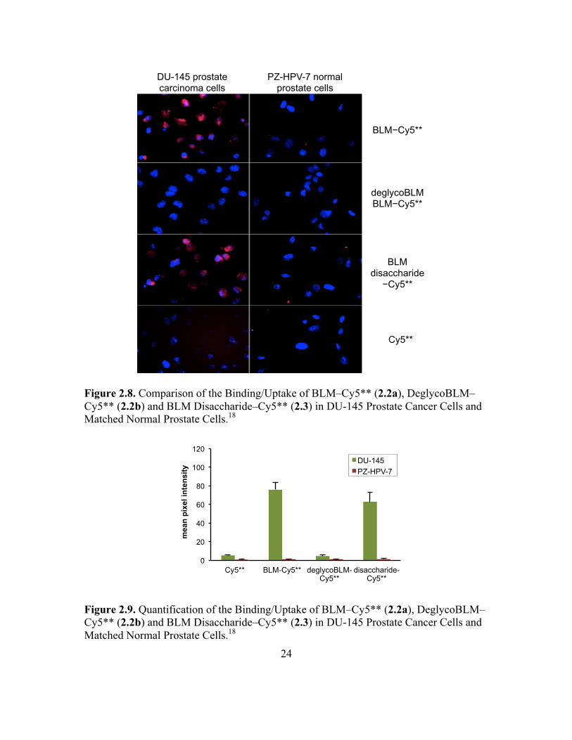
Figure 2.8. Comparison of the Binding/Uptake of BLM–Cy5** (2.2a), DeglycoBLM–Cy5** (2.2b) and BLM Disaccharide–Cy5** (2.3) in DU-145 Prostate Cancer Cells and Matched Normal Prostate Cells.18
Figure 2.9. Quantification of the Binding/Uptake of BLM–Cy5** (2.2a), DeglycoBLM–Cy5** (2.2b) and BLM Disaccharide–Cy5** (2.3) in DU-145 Prostate Cancer Cells and Matched Normal Prostate Cells.18
DU-145 prostate carcinoma cells
PZ-HPV-7 normal prostate cells
BLM−Cy5**
deglycoBLM BLM−Cy5**
BLM disaccharide−Cy5**
Cy5**
0
20
40
60
80
100
120
Cy5** BLM-Cy5** deglycoBLM-Cy5**
disaccharide-Cy5**
mea
n pi
xel i
nten
sity
DU-145 PZ-HPV-7
Page 47
25
DeglycoBLM–Cy5** (2.2b), lacking the disaccharide moiety showed no uptake
in any cell line, which was consistent with the results obtained from the microbubble
experiment.18 Hence, the targeting ability of the carbohydrate domain of BLM along with
the sufficiency of the sugar to internalize the reporter molecule can be potentially utilized
to develop a better therapeutic agent or drug delivery vehicle for chemotherapy. To
further understand the importance of the carbohydrate domain of BLM and explore the
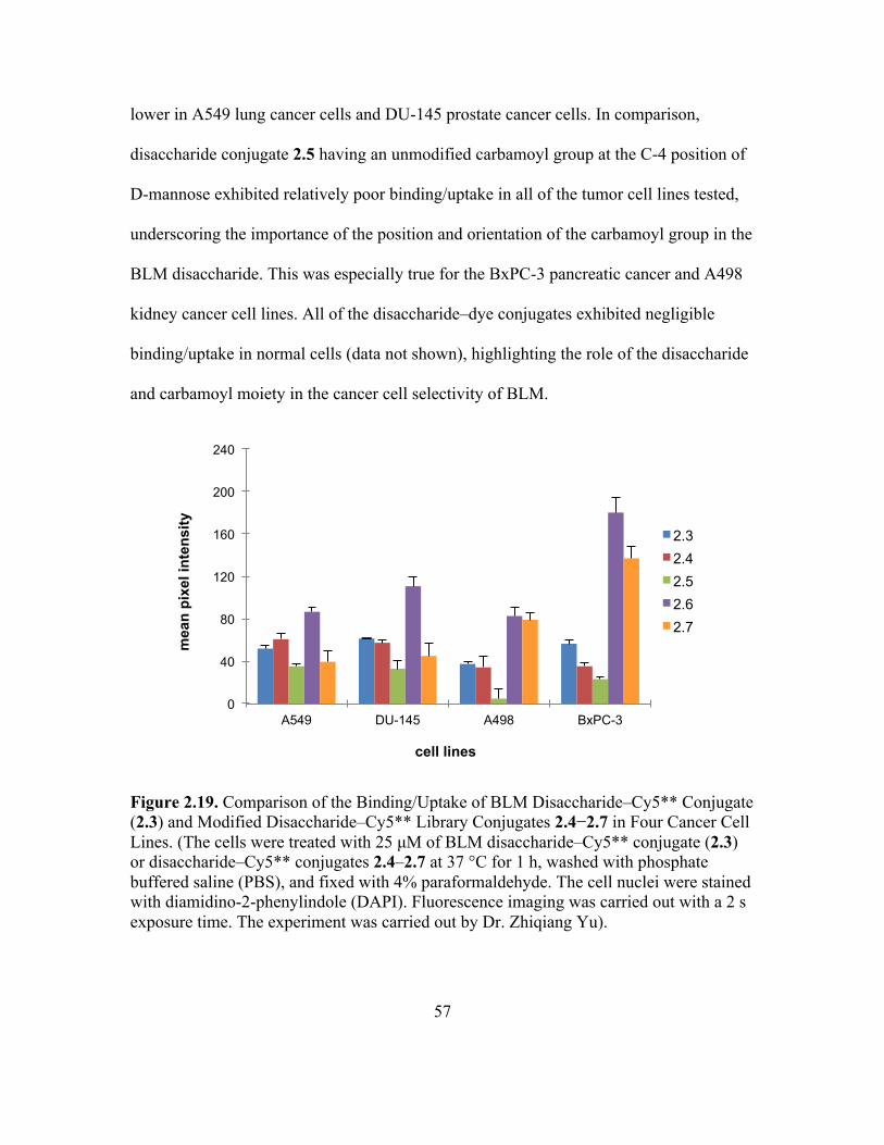
participation of the carbamoyl moiety, a library of disaccharide–Cy5** conjugates was
synthesized (Figure 2.10).
Figure 2.10. Library of Disaccharide–dye Conjugates 2.3–2.7 Synthesized and Evaluated. (The synthesis of 2.6 and 2.7 were carried out by Dr. Manikandadas Mathilakathu Madathil).116
To address the issue of whether the BLM disaccharide, while small and
uncomplicated relative to the natural product itself, actually represents the simplest
structural entity capable of selective tumor cell targeting, C3-carbamoyl mannose (BLM
monosaccharide) conjugated to Cy5** (2.8) was also synthesized. Further, to
BLM disaccharide–Cy5**(2.3) 2.4
OOH
OCONH2
OH
HO
O
O
OHHO
HO
2.5
Cy5**HN
OO
O
OOCONH2
OH
OH
HO
O
O
OHHO
HOCy5**
HN
OO
O
OOH
OH
OCONH2
HO
O
O
OHHO
HOCy5**
HN
OO
O
2.6 2.7
OOCONHMe
OH
OH
HO
O
O
OHHO
HOCy5**
HN
OO
O
OOH
OCONHMe
OH
HO
O
O
OHHO
HOCy5**
HN
OO
O
Page 48
26
demonstrate the importance of carbamoyl moiety in selective tumor cell targeting and the
effect of trimeric carbohydrate cluster of BLM monosaccharide on cell targeting,
mannose (decarbamoyl BLM monosaccharide) conjugated to Cy5** (2.9) and a trimer
cluster of BLM monosaccharide conjugated to Cy5** (2.10) were synthesized (Figure
2.11) and evaluated for their tumor cell targeting ability.
Figure 2.11. Structures of BLM Monosaccharide–Cy5** (2.8), Decarbamoyl BLM Monosaccharide–Cy5** (2.9) and BLM Monosaccharide–Cy5** Trimer (2.10) Synthesized and Evaluated.117
To further explore the importance of the carbamoyl moiety and its positioning in
the monosaccharide subunit, efforts were then extended to synthesize a library of
monosaccharides with modifications of the carbamoyl group and alteration of its position
on the mannose ring (Figure 2.12).
O
OHOCONH2
HO
HOO
OHN
NH
NHO
O
Cy5**
O
OH
O
OHHO
HOCy5**
HN
OO
OOH
O
OCONH2
HO
HOCy5**
HN
OO
O
3
BLM monosaccharide–Cy5**(2.8) 2.9
2.10
Page 49
27
Figure 2.12. Library of Monosaccharide–dye Conjugates 2.11–2.16 Synthesized and Evaluated.
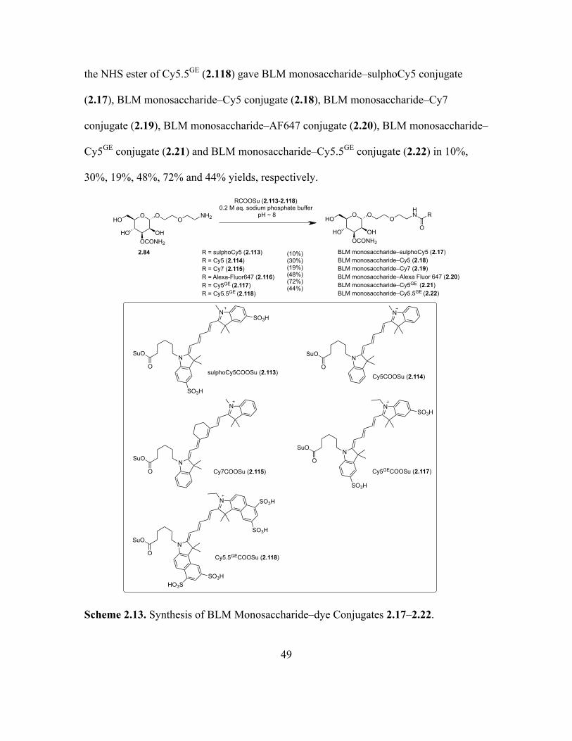
Though the reporter dye Cy5** was affording excellent results, the lack of
availability of the dye resulted in a search for a replacement dye with similar properties.
Hence, a number of commercially available dyes with similar extinction coefficients, and
absorption and emission spectra to Cy5** were selected and conjugated with BLM
monosaccharide (Figure 2.13) and later with other saccharides (Figure 2.14).
2.11 2.12 2.13
2.14
Cy5**
O
O O
OCONHBuOH
HO
HO OHNCy5**
O
O O
OCONHMeOH
HO
HO OHN Cy5**
O
O O
OCONMe2OH
HO
HO OHN
Cy5**
O
O O
OCONOH
HO
HO OHN
2.15
Cy5**
O
O O
OCONHHexOH
HO
OH
OHN
2.16
Cy5**
O
O O
OHOCONHMe
HO
OH
OHN
Page 50
28
Figure 2.13. Structures of Different Monosaccharide–dye Conjugates 2.17–2.22 Synthesized for Selection of a Dye to Replace Cy5**.
N
NO
SO3H
SO3HBLM monosaccharide–sulphoCy5
2.17
N
NO
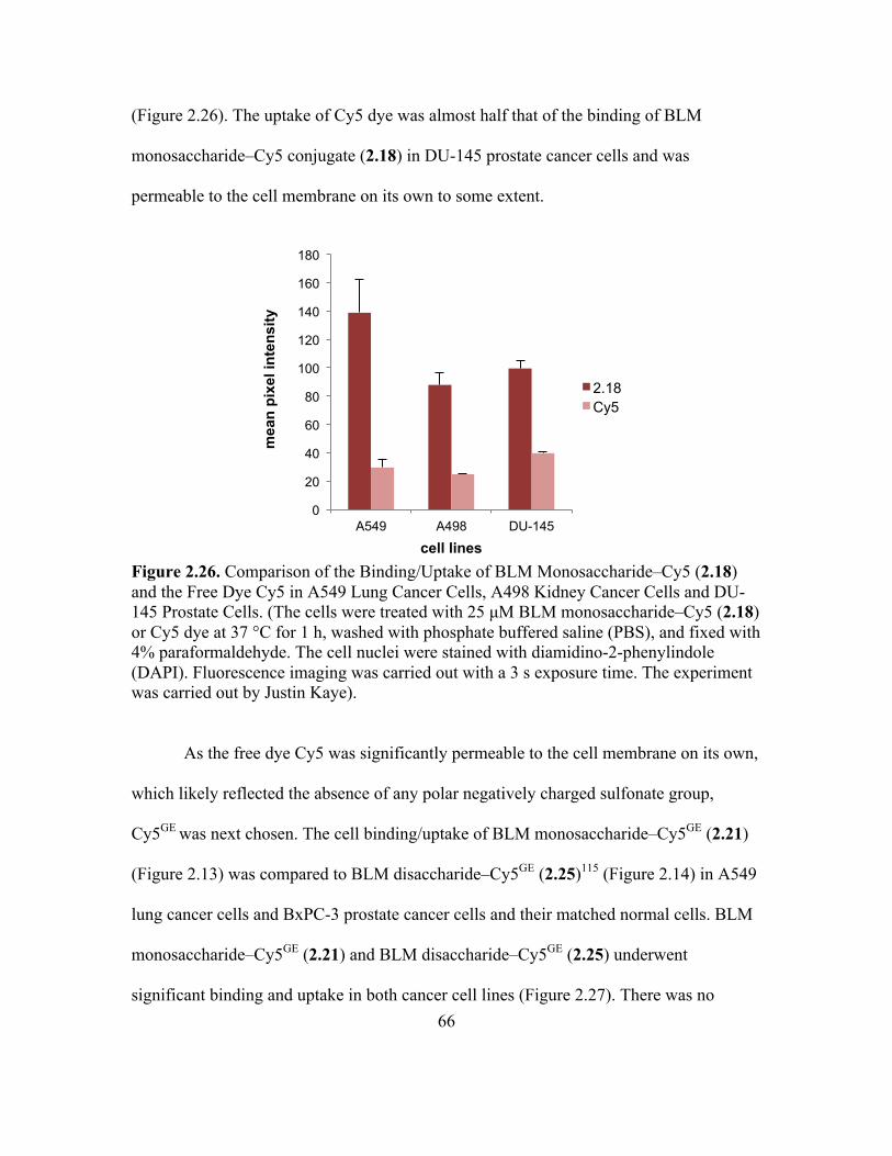
BLM monosaccharide–Cy5 2.18
NO
N
BLM monosaccharide–Cy7 2.19
N
NO
SO3H
SO3H N
NO
BLM monosaccharide–Cy5.5GE 2.22
SO3HHO3S
SO3H
SO3H
BLM monosaccharide–Alexa Fluor 6472.20
BLM monosaccharide–Cy5GE 2.21
O
Alexa-Fluor 647
O
OHOCONH2
HO
HOO
OHN O
OHOCONH2
HO
HOO
OHN
O
OHOCONH2
HO
HOO
OHN
O
OHOCONH2
HO
HOO
OHN
O
OHOCONH2
HO
HOO
OHN
O
OHOCONH2
HO
HOO
OHN
Page 51
29
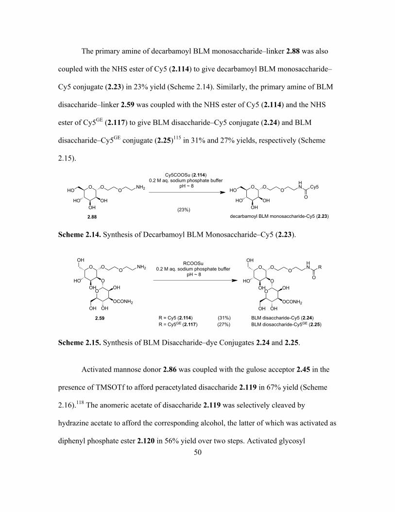
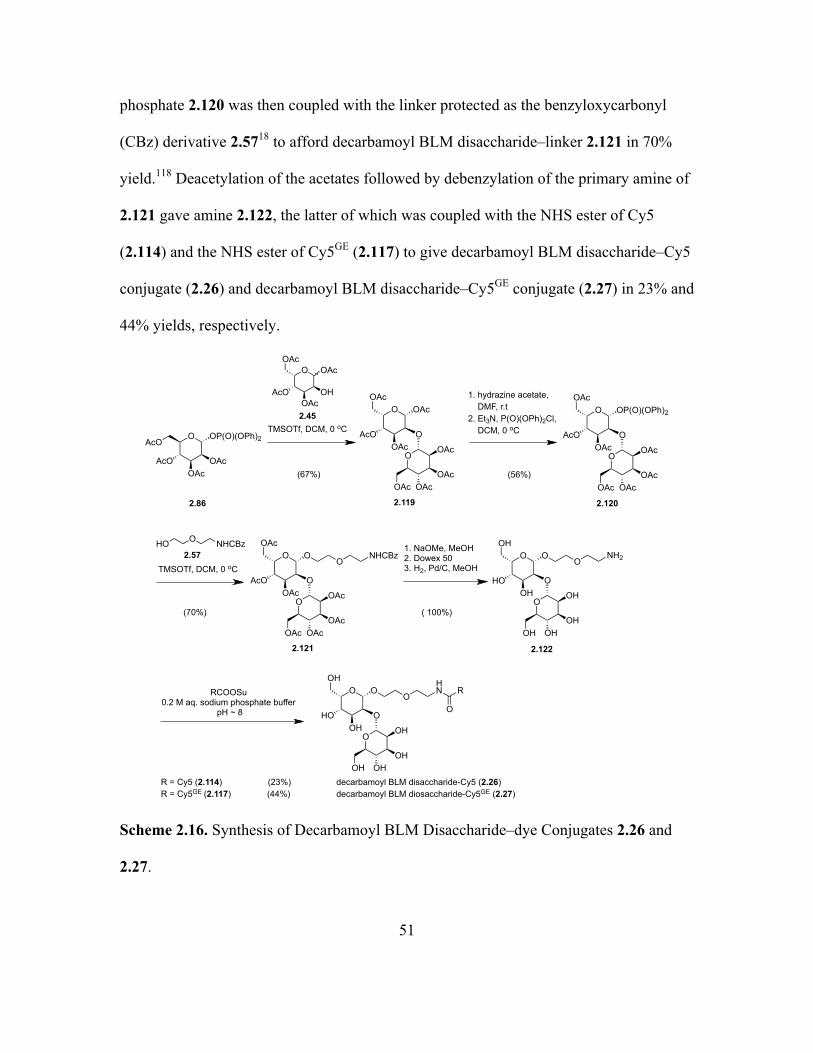
Figure 2.14. Series of Additional Dye Conjugates 2.23–2.28 Synthesized and Evaluated.
To explore the modular nature of BLM suggested by diminished cytotoxicity of
deglycoBLM in comparison to BLM, different BLM analogues were prepared in which
the disaccharide moiety was attached to novel positions of deglycobleomycin, mainly via
the C-terminal substituent (2.29)118 and the threonine moiety (2.31 and 2.32). Compounds
2.31 and 2.32 were synthesized by Dr. Benjamin R. Schroeder.119 Later, to study the
cellular uptake of the modified BLMs, their dye conjugates 2.30, 2.33 and 2.34 were
synthesized and evaluated (Figures 2.15 and 2.16).
Cy5
O
OOH
OCONH2OHOH
O O
OOH
HO
HO OHN
Cy5
O
OOH
OHOHOH
O O
OOH
HO
HO OHN
Cy5GE
O
OOH
OCONH2OHOH
O O
OOH
HO
HO OHN
Cy5GE
O
OOH
OHOHOH
O O
OOH
HO
HO OHN
2.24
2.26
2.25
2.27
O
Cy5O
OHOH
HO
HOO
OHN
2.23
2.28
N N
H2NCH3
OH2NHN NH2
O
HN
OH
NH
CH3
HOHN
NH
N
S
S
N
O
OOHO CH3H
O
O
OHOHO
HO
O
O
OH
OHOH
O
NH2O
NH
NH
H
HNH2
CH3
NH HNNH
Cy5GEO
Page 52
30
Figure 2.15. Structures of a Modified BLM With a C-terminal Disaccharide 2.29 and its Conjugate With Cy5** 2.30 Synthesized and Evaluated.
Figure 2.16. Structures of Modified BLMs With the Disaccharide Attached to Threonine Moiety 2.31 and 2.32 (synthesized by Dr. Benjamin R. Schroeder),119 and Their Fluorescent Dye Conjugates 2.33 and 2.34 Synthesized and Evaluated.
2.302.29
N N
H2NCH3
OH2NHN NH2
O
HN
OH
NH
CH3
HOHN
NH
N
S
S
NNH
OO
OHO CH3
H
O
HONH
NH
H
H
CH3
HN
NH2
= R
O
OHOHO
HO
O
O
OH
OHOH
O
NH2O
NHR
O
O
OHOHO
HO
O
O
OH
OHOH
O
NH2O
O
Cy5**
ONHR
NH
O
NH
2.31
2.32
OHOHO
HO
O
OHO OHOH
O
NH2O
HN NH
N
S
S
N NH
O
O CH3H
OH
NH
HN
H2N
N N
H2NCH3
OH2NHN NH2
O
HN
OH
NH
CH3
HOO
O
HNN
H
CH3
NH2
OHOHO
HO
O
O
OH
OHOH
O
NH2O
HN NH
N
S
S
N NH
O
O CH3H
OH
NH
HN
H2N
N N
H2NCH3
OH2NHN NH2
O
HN
OH
NH
CH3
HOO
O
HNN
H
CH3
NH2
2.33
2.34
OHOHO
HO
O
OHO OHOH
O
NH2O
HN NH
N
S
S
N NH
O
O CH3H
OH
NH
HN
HN
Cy5**
O
N N
H2NCH3
OH2NHN NH2
O
HN
OH
NH
CH3
HOO
O
HNN
H
CH3
NH2
OHOHO
HO
O
O
OH
OHOH
O
NH2O
HN NH
N
S
S
N NH
O
O CH3H
OH
NH
HN
HN
Cy5**
O
N N
H2NCH3
OH2NHN NH2
O
HN
OH
NH
CH3
HOO
O
HNN
H
CH3
NH2
Page 53
31
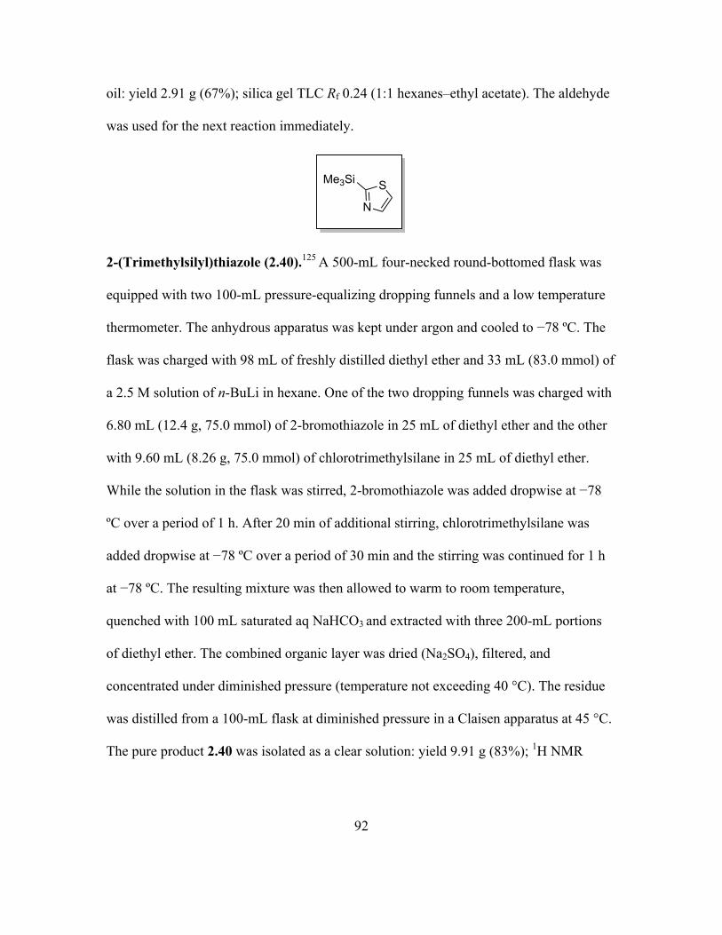
2.1.2. Synthesis of Fluorinated Saccharides for Imaging Studies
In the field of nuclear medicine, the radioisotopically labeled drugs are used for
diagnosis of many diseases like cancer.120 The molecular imaging technique in which
compounds labeled with positron emitting radioisotopes are used to produce three-
dimensional images of the functional processes in the body is known as PET. The
radiolabeled molecules are retained in the tissues and the tomographic images of the
biodistribution within the body are generated by detection of the gamma rays.121
Clinically, 18F-FDG (2-[18F]-fluoro-2-deoxy-D-glucose) has been extensively used for
various PET studies. 18F-FDG is a glucose analogue in which C-2 carbon has a fluoride
atom in place of the hydroxyl group present in glucose. It undergoes receptor mediated
transport and phosphorylation but cannot undergo further metabolism like glucose.122 The
higher uptake in tumor tissues than in normal tissues because of increased glycolysis
helps in strong signal intensity in PET imaging (Figure 2.17).123
Figure 2.17. PET-CT Image of Breast Ductal Carcinoma Treated With 18F-FDG.123
Page 54
32
As described earlier, due to the unique ability of BLM disaccharide to target
tumors,18 it represents a potentially attractive PET biomarker enabling possible cancer
localization, their biodistribution, in vivo quantification of metastatic cells and eventual
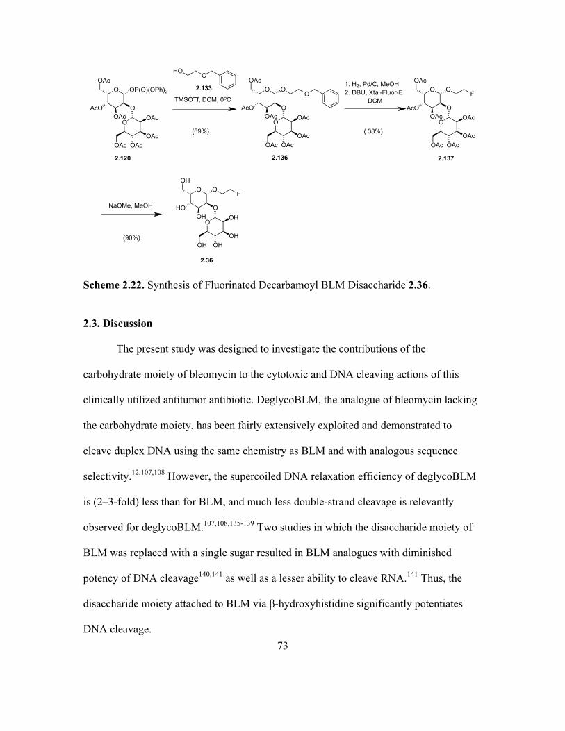
elimination. Hence, the non-radioactive fluorinated disaccharides were synthesized as
non-radioactive reference compounds for disaccharide PET biomarkers (Figure 2.18).
Figure 2.18. Structures of Fluorinated BLM Disaccharide 2.35 and Fluorinated Decarbamoyl BLM Disaccharide 2.36 Synthesized.
2.2. Results
2.2.1. Synthesis of Fluorescent Dye Conjugates
The fluorescent BLM disaccharide–Cy5** conjugate was synthesized from the
natural BLM disaccharide according to a reported procedure.18 The preparation of
different disaccharide–dye conjugates began with the synthesis of the corresponding
disaccharides attached to a protected amine linker. The syntheses of dye conjugates 2.3,
2.4 and 2.5 were effected by the coupling of gulose glycosyl acceptor with corresponding
mannose donors. The synthesis of gulose glycosyl acceptor was achieved by
homologation of commercially available L-xylose following Dondoni’s method (Scheme
2.1).124 L-xylose (2.37) was fully protected as dithioacetal 2.38 in 65% yield, the latter of
which was selectively hydrolyzed to afford aldehyde 2.39 in the presence of Hg(II) in
OOH
OHOHOH
O O
OOH
HO
OH
F
2.36
OOH
OCONH2OHOH
O O
OOH
HO
OH
F
2.35
Page 55
33
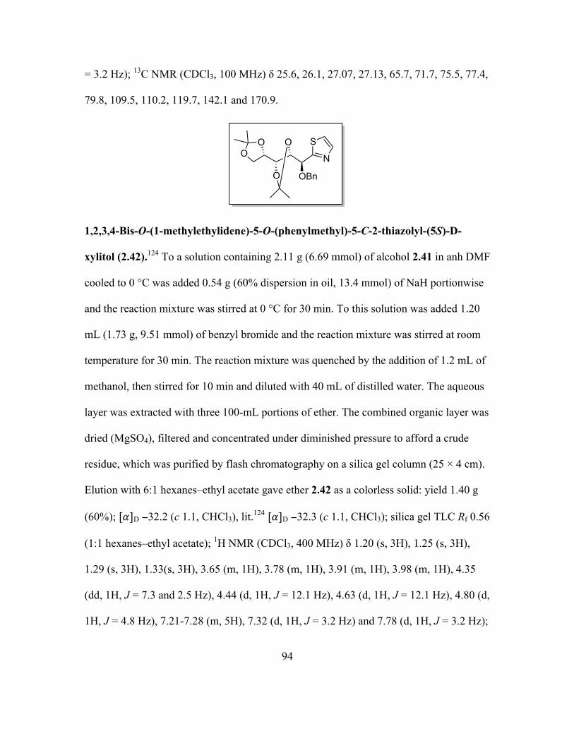
67% yield. Aldehyde 2.39 was immediately coupled with 2-(trimethylsilyl)thiazole
(2.40)125 followed by desilylation to afford the alcohol 2.41 in 60% yield. The free
hydroxyl group of 2.41 was protected as benzyl ether 2.42 in 60% yield. The thiazole ring
of 2.42 was then converted into aldehyde 2.43 in 94% yield through a series of
transformations. Acetal deprotection followed by exhaustive acetylation of aldehyde 2.43
afforded tetra-O-acetyl-2-O-benzyl-L-gulopyranoside (2.44) in 60% yield. Reductive
debenzylation of gulopyranoside 2.44 gave the gulose acceptor 2.45 in 98% yield.
Scheme 2.1. Synthesis of the Gulose Glycosyl Acceptor 2.45.
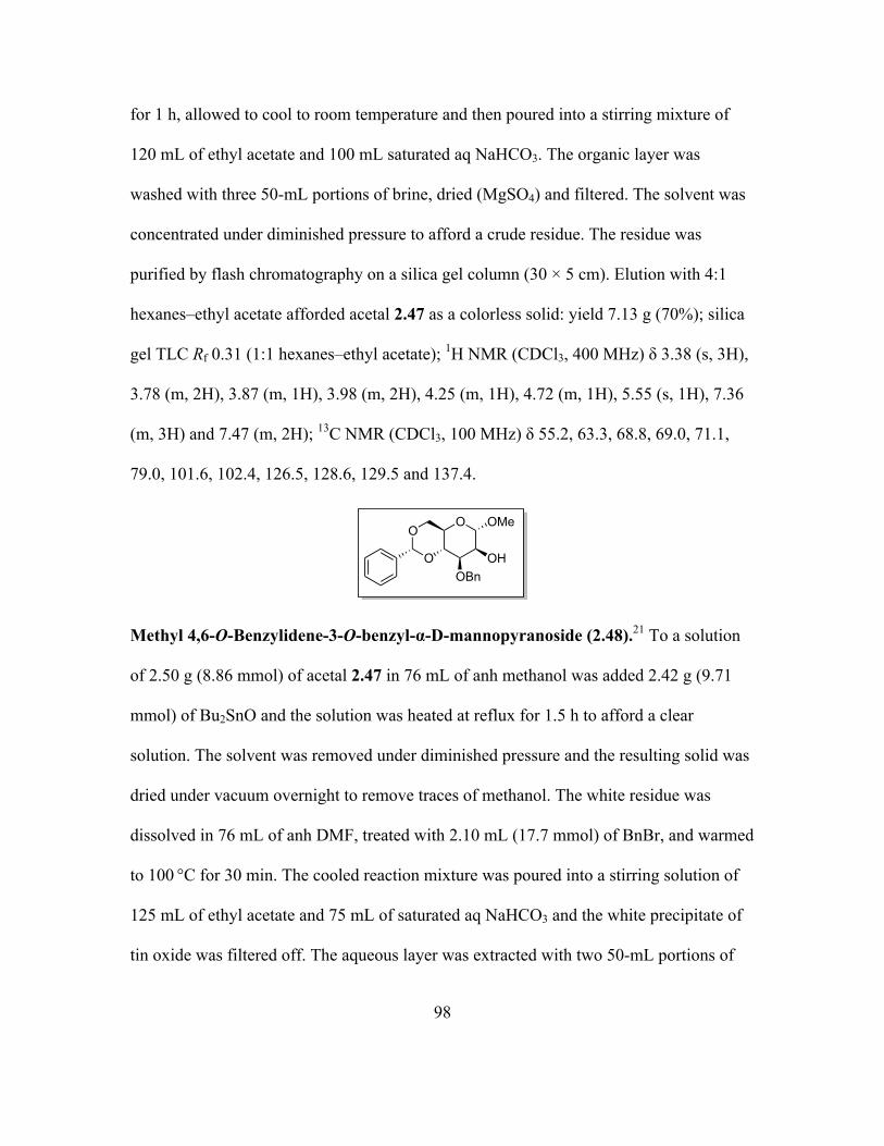
Mannose donor 2.54 was prepared from commercially available α-D-
methylmannopyranoside (2.46) following reported procedures with slight modifications
(Scheme 2.2).21 The synthesis began with protection of α-D-methylmannopyranoside
O OH
OH
EtSH, conc. HClAcetone
(65%)
HgCl2, HgOAcetone-H2O
(67%)
1. MeOTf, Acetonitrile2. NaBH4, MeOH3. CuO, CuCl2.2H2O, Acetonitrile, H2O
2.37
BnBr, NaHDMF
(60%)
2.41 2.42
2.45
OCH(SEt)2
O
O
O
HOOH
OCHO
O
O
O
2.38 2.39
1. DCM,
(60%)
OO
O
O
N
S
OH
OO
O
O
N
S
OBn
2.43
OO
O
OCHO
OBn
O
OBn
2.44
OAcAcO
OAcO
OHOAc
AcO
OAc1. AcOH, H2O2. Ac2O, pyridine
Me3Si
N
S
2. THF, Bu4NF•3H2O
2.40
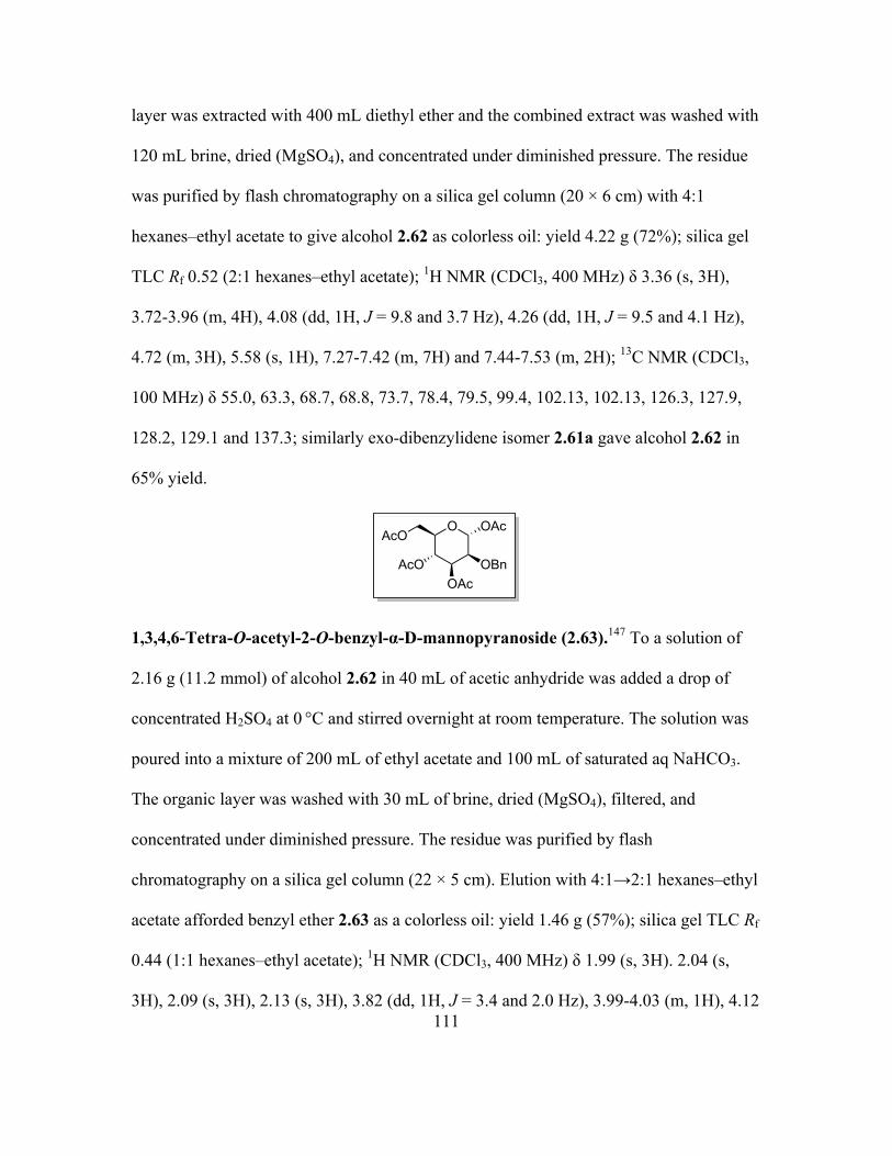
H2, Pd/COAc OAc
(94%)
(60%) (98%)
Page 56
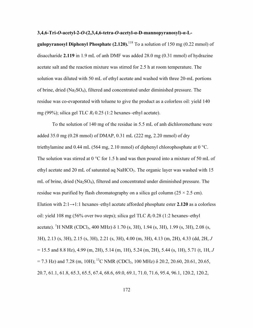
34
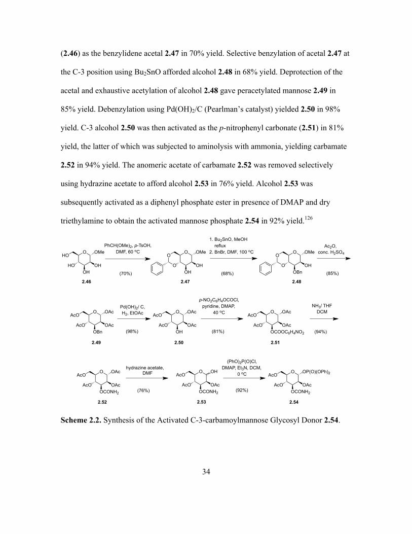
(2.46) as the benzylidene acetal 2.47 in 70% yield. Selective benzylation of acetal 2.47 at
the C-3 position using Bu2SnO afforded alcohol 2.48 in 68% yield. Deprotection of the
acetal and exhaustive acetylation of alcohol 2.48 gave peracetylated mannose 2.49 in
85% yield. Debenzylation using Pd(OH)2/C (Pearlman’s catalyst) yielded 2.50 in 98%
yield. C-3 alcohol 2.50 was then activated as the p-nitrophenyl carbonate (2.51) in 81%
yield, the latter of which was subjected to aminolysis with ammonia, yielding carbamate
2.52 in 94% yield. The anomeric acetate of carbamate 2.52 was removed selectively
using hydrazine acetate to afford alcohol 2.53 in 76% yield. Alcohol 2.53 was
subsequently activated as a diphenyl phosphate ester in presence of DMAP and dry
triethylamine to obtain the activated mannose phosphate 2.54 in 92% yield.126
Scheme 2.2. Synthesis of the Activated C-3-carbamoylmannose Glycosyl Donor 2.54.
O OAc
OAcOBn
AcO
AcO
2.49
Pd(OH)2/ C,H2, EtOAc
(PhO)2P(O)Cl, DMAP, Et3N, DCM,
0 oC O OP(O)(OPh)2
OAcOCONH2
AcO
AcO
(98%)
(92%)
NH3/ THFDCM
(94%)
O OMe
OHOH
HO
HO
PhCH(OMe)2, p-TsOH, DMF, 60 oC
2.46
O OMe
OHOH
O
O
2.47
Ac2O, conc. H2SO4
(85%)
1. Bu2SnO, MeOH reflux2. BnBr, DMF, 100 oC
(70%) (68%)
O OMe
OHOBn
O
O
2.48
O OAc
OAcOH
AcO
AcO
2.50
p-NO2C6H4OCOCl, pyridine, DMAP,
40 oC
(81%)
O OAc
OAcOCOOC6H4NO2
AcO
AcO
2.51
O OAc
OAcOCONH2
AcO
AcO
2.52
hydrazine acetate, DMF O OH
OAcOCONH2
AcO
AcO
(76%)
2.53 2.54
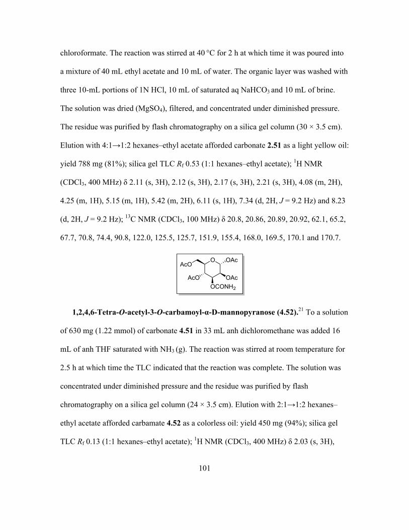
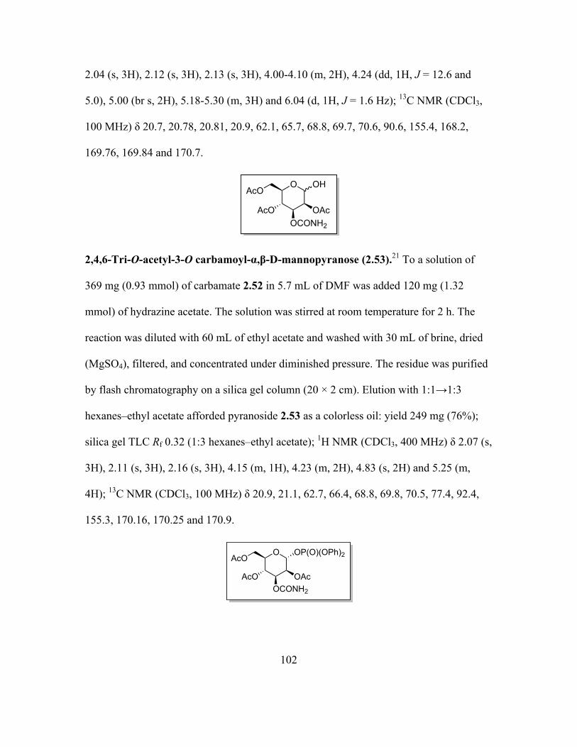
Page 57
35
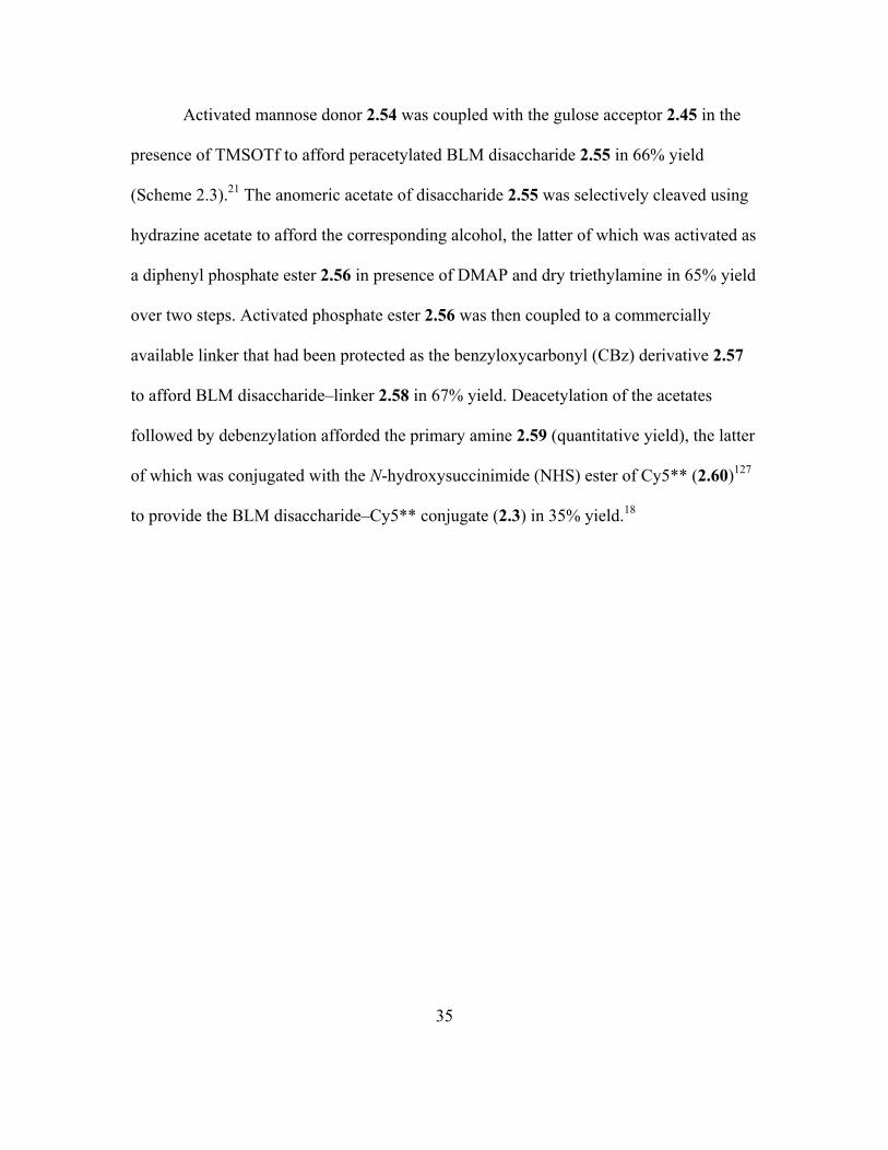
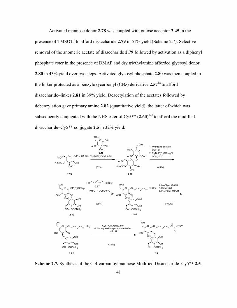
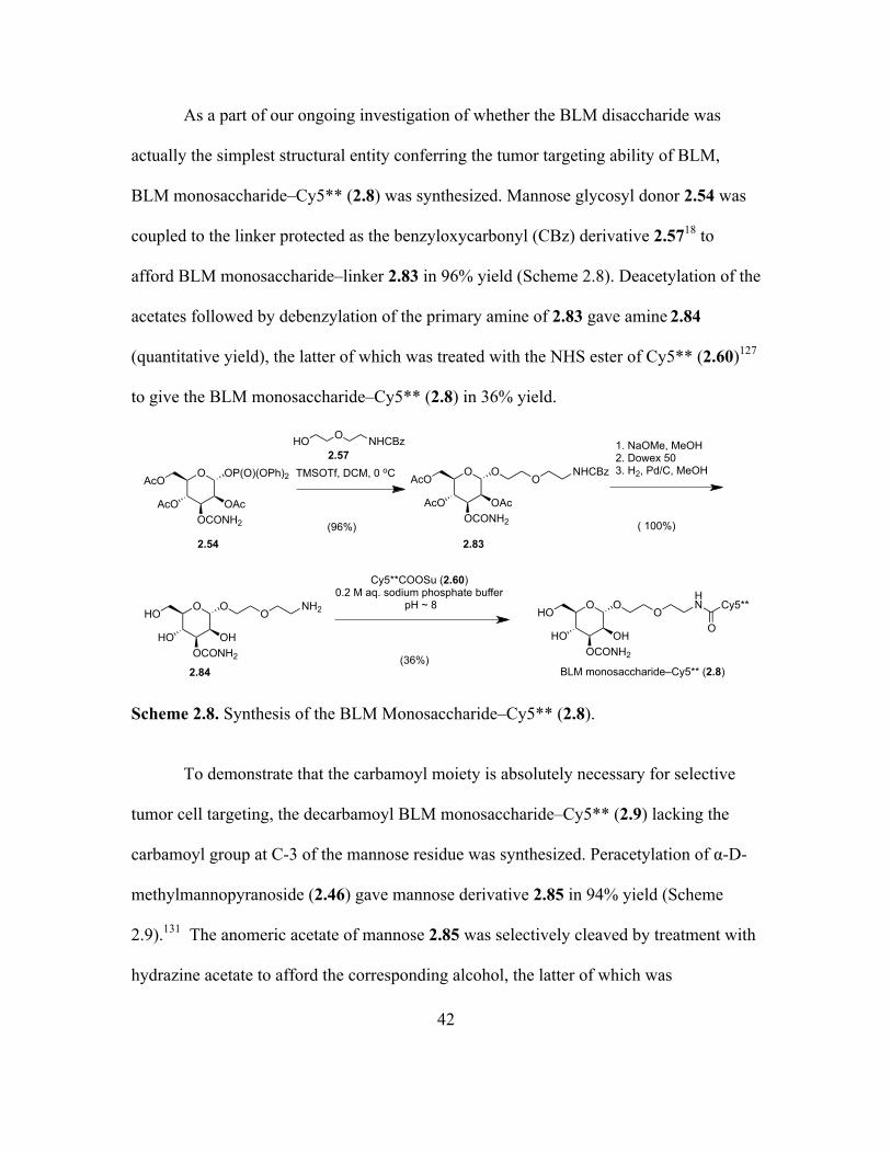
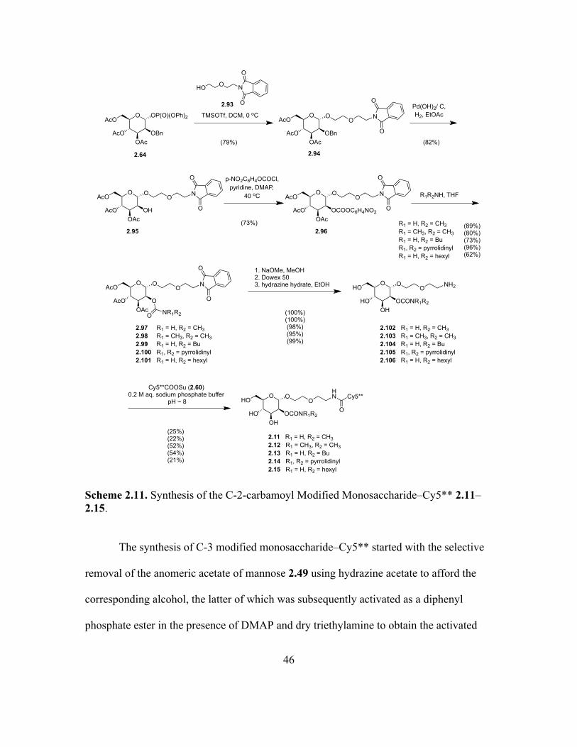
Activated mannose donor 2.54 was coupled with the gulose acceptor 2.45 in the
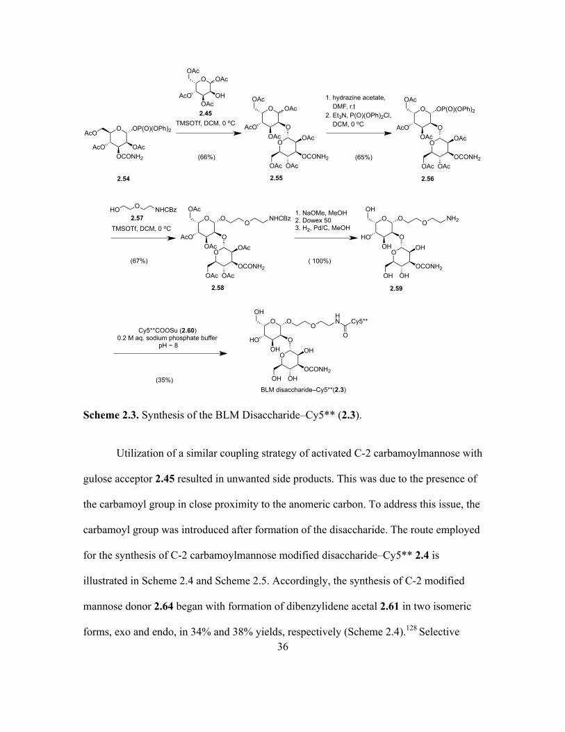
presence of TMSOTf to afford peracetylated BLM disaccharide 2.55 in 66% yield
(Scheme 2.3).21 The anomeric acetate of disaccharide 2.55 was selectively cleaved using
hydrazine acetate to afford the corresponding alcohol, the latter of which was activated as
a diphenyl phosphate ester 2.56 in presence of DMAP and dry triethylamine in 65% yield
over two steps. Activated phosphate ester 2.56 was then coupled to a commercially
available linker that had been protected as the benzyloxycarbonyl (CBz) derivative 2.57
to afford BLM disaccharide–linker 2.58 in 67% yield. Deacetylation of the acetates
followed by debenzylation afforded the primary amine 2.59 (quantitative yield), the latter
of which was conjugated with the N-hydroxysuccinimide (NHS) ester of Cy5** (2.60)127
to provide the BLM disaccharide–Cy5** conjugate (2.3) in 35% yield.18
Page 58
36
Scheme 2.3. Synthesis of the BLM Disaccharide–Cy5** (2.3).
Utilization of a similar coupling strategy of activated C-2 carbamoylmannose with
gulose acceptor 2.45 resulted in unwanted side products. This was due to the presence of
the carbamoyl group in close proximity to the anomeric carbon. To address this issue, the
carbamoyl group was introduced after formation of the disaccharide. The route employed
for the synthesis of C-2 carbamoylmannose modified disaccharide–Cy5** 2.4 is
illustrated in Scheme 2.4 and Scheme 2.5. Accordingly, the synthesis of C-2 modified
mannose donor 2.64 began with formation of dibenzylidene acetal 2.61 in two isomeric
forms, exo and endo, in 34% and 38% yields, respectively (Scheme 2.4).128 Selective
Cy5**COOSu (2.60)0.2 M aq. sodium phosphate buffer
pH ~ 8
O
OAcOCONH2
AcO
AcOOP(O)(OPh)2
OOAc
OCONH2OAcOAc
O OAc
OOAc
AcO
OAc
OOAc
OCONH2OAcOAc
O OP(O)(OPh)2
OOAc
AcO
OAc
OOAc
OCONH2OAcOAc
O O
OOAc
AcO
OAc
ONHCBz
OOH
OCONH2OHOH
O O
OOH
HO
OH
ONH2
(66%)
1. hydrazine acetate, DMF, r.t2. Et3N, P(O)(OPh)2Cl, DCM, 0 oC
(65%)
TMSOTf, DCM, 0 oC
(67%)
O NHCBzHO2.57
( 100%)
1. NaOMe, MeOH2. Dowex 503. H2, Pd/C, MeOH
2.54 2.55 2.56
2.58 2.59
Cy5**
O
OOH
OCONH2OHOH
O O
OOH
HO
OH
OHN
BLM disaccharide–Cy5**(2.3)
O OAc
OHOAc
AcO
OAc
2.45TMSOTf, DCM, 0 oC
(35%)
Page 59
37
reduction of the acetal at the C-2 and C-3 positions of both exo and endo isomers of
acetal 2.61 with DIBAL-H in presence of 5% aluminum chloride afforded alcohol 2.62 in
65% and 72% yield, respectively.129 Deprotection of the acetal followed by exhaustive
acetylation of alcohol 2.62 gave peracetylated mannose 2.63 in 57% yield. The anomeric
acetate of mannose 2.63 was selectively cleaved by hydrazine acetate to afford the
alcohol, the latter of which was subsequently activated as a diphenyl phosphate ester in
the presence of DMAP and dry triethylamine to obtain the activated mannose phosphate
2.64 in 55% yield over two steps.
Scheme 2.4. Synthesis of the Activated C-2-O-benzylmannose Glycosyl Donor 2.64.
Coupling of activated mannose donor 2.64 with the gulose acceptor 2.45 in the
presence of TMSOTf afforded disaccharide 2.65 in 56% yield (Scheme 2.5).
Debenzylation using Pd(OH)2/C (Pearlman’s catalyst) yielded alcohol 2.66 in 98% yield.
O OAc
OBnOAc
AcO
AcO
2.63
O OP(O)(OPh)2
OBnOAc
AcO
AcO
2.64
O OMe
OHOH
HO
HO
O OMe
OO
O
O
PhCH(OMe)2CSA, DMF
60 oC, 250 mbar
2.46
2.61b (38%, endo)
O OMe
OO
O
O
+
2.61a (34%, exo)
DIBAL-H in toluene, AlCl3, Toluene,
0 oC
O OMe
OBnOH
O
O
Ph
Ac2O, conc. H2SO4
(72%)
(57%)
1. hydrazine acetate, DMF, r.t2. Et3N, P(O)(OPh)2Cl, DCM, 0 oC
(55%)2.62
(65%)
Page 60
38
C-2 alcohol 2.66 was then activated as the p-nitrophenyl carbonate (2.67) in 86% yield,
the latter of which was subjected to aminolysis with ammonia yielding carbamate 2.68
with 92% yield. The anomeric acetate of disaccharide 2.68 was selectively cleaved by
hydrazine acetate to afford the corresponding alcohol, the latter of which was activated as
a diphenyl phosphate ester 2.69 in the presence of DMAP and dry triethylamine in 43%
yield over two steps. Activated phosphate ester 2.69 was then coupled to the linker
protected as the benzyloxycarbonyl (CBz) derivative 2.5718 to afford BLM disaccharide–
linker 2.70 in 39% yield. Deacetylation of the acetates followed by debenzylation gave
the primary amine 2.71 (quantitative yield), the latter of which was treated with the NHS
ester of Cy5** (2.60)127 to provide the modified disaccharide–Cy5** conjugate 2.4 in
35% yield.
Page 61
39
Scheme 2.5. Synthesis of the C-2-carbamoylmannose Modified Disaccharide–Cy5** 2.4.
The route employed for the synthesis of C-4 carbamoylmannose modified
disaccharide–Cy5** 2.5 was similar to that used for the natural disaccharide–Cy5**
conjugate. The synthesis of C-4 carbamoylmannose modified disaccharide–Cy5** was
achieved by coupling of activated C-4 carbamoylmannose donor 2.78 with gulose
O OAc
OHOAc
AcO
OAc
O
OAcO
AcO
AcO
OAc
O
OBnOAc
AcO
AcO
2.65
(56%)
Pd(OH)2/ C, H2, EtOAc,
O
OAcO
AcO
AcO
OAc
O
OHOAc
AcO
AcO
2.66
(98%)
p-NO2C6H4OCOCl, pyridine, DMAP,
40 oCNH3/ THF
DCMO
OAcO
AcO
AcO
OAc
O
OCONH2OAc
AcO
AcO
2.68
O
OAcO
AcO
AcO
OAc
O
OCOOC6H4NO2OAc
AcO
AcO
2.67
(86%) (92%)
2.70
OOCONH2
OAcOAcOAc
O
OOAc
AcO
OAcO
ONHCBz
OOCONH2
OAcOAcOAc
O
OOAc
AcO
OAcOP(O)(OPh2)
2.69
1. NaOMe, MeOH2. Dowex 503. H2, Pd/C, MeOH
OOCONH2
OHOHOH
O
OOH
HO
OH
2.71
OO
NH2
(100%)
2.45
1. hydrazine acetate, DMF, r.t2. Et3N, P(O)(OPh)2Cl, DCM, 0 oC
(43%)
(39%)
TMSOTf, DCM, 0 oC
O NHCBzHO2.57
TMSOTf, DCM, 0 oCO OP(O)(OPh)2
OBnOAc
AcO
AcO
2.64
OOCONH2
OHOHOH
O
OOH
HO
OH
2.4
OO
HNCy5**COOSu (2.60)
0.2 M aq. sodium phosphate buffer pH ~ 8
(35%)
Cy5**
O
Page 62
40
acceptor 2.45. The synthesis began with regioselective reductive ring opening of acetal
2.47 in presence of CoCl2 and BH3•THF to afford alcohol 2.72 in 99% yield (Scheme
2.6).130 Exhaustive acetylation of alcohol 2.72 in acidic medium gave peracetylated
mannose 2.73 in 79% yield. Debenzylation using Pd(OH)2/C (Pearlman’s catalyst)
yielded alcohol 2.74 in 84% yield. C-4 alcohol 2.74 was then activated as the p-
nitrophenyl carbonate (2.75) in 74% yield, the latter of which was subjected to
aminolysis with ammonia yielding carbamate 2.76 in 87% yield. The anomeric acetate of
carbamate 2.76 was removed selectively using hydrazine acetate to afford alcohol 2.77 in
100% yield, the latter of which was subsequently activated as a diphenyl phosphate ester
in the presence of DMAP and dry triethylamine to obtain the activated mannose
phosphate 2.78 in 56% yield.
Scheme 2.6. Synthesis of the Activated C-4-carbamoylmannose Glycosyl Donor 2.78.
O OAc
OAcOAc
BnO
AcO
2.73
Pd(OH)2/ C,H2, EtOAc
(PhO)2P(O)Cl, DMAP, Et3N, DCM,
0 oC O OP(O)(OPh)2
OAcOAc
H2NOCO
AcO
(84%)
(56%)
NH3/ THFDCM
(87%)
O OMe
OHOH
O
O
2.47
Ac2O, conc. H2SO4
(79%)
CoCl2, BH3•THF
(99%)
O OMe
OHOH
BnO
HO
2.72
O OAc
OAcOAc
HO
AcO
2.74
p-NO2C6H4OCOCl, pyridine, DMAP,
40 oC
(74%)
O OAc
OAcOAc
O2NC6H4OOCO
AcO
2.75
O OAc
OAcOAc
H2NOCO
AcO
2.76
hydrazine acetate, DMF O OH
OAcOAc
H2NOCO
AcO
(100%)
2.77 2.78
Page 63
41
Activated mannose donor 2.78 was coupled with gulose acceptor 2.45 in the
presence of TMSOTf to afford disaccharide 2.79 in 51% yield (Scheme 2.7). Selective
removal of the anomeric acetate of disaccharide 2.79 followed by activation as a diphenyl
phosphate ester in the presence of DMAP and dry triethylamine afforded glycosyl donor
2.80 in 43% yield over two steps. Activated glycosyl phosphate 2.80 was then coupled to
the linker protected as a benzyloxycarbonyl (CBz) derivative 2.5718 to afford
disaccharide–linker 2.81 in 39% yield. Deacetylation of the acetates followed by
debenzylation gave primary amine 2.82 (quantitative yield), the latter of which was
subsequently conjugated with the NHS ester of Cy5** (2.60)127 to afford the modified
disaccharide–Cy5** conjugate 2.5 in 32% yield.
Scheme 2.7. Synthesis of the C-4-carbamoylmannose Modified Disaccharide–Cy5** 2.5.
O OAc
OHOAc
AcO
OAc
O
OAcO
AcO
AcO
OAc
O
OAcOAc
H2NOCO
AcO
2.79
(51%)
2.81
OOAc
OAcOCONH2OAc
O
OOAc
AcO
OAcO
ONHCbz
OOAc
OAcOCONH2OAc
O
OOAc
AcO
OAcOP(O)(OPh2)
2.80
1. NaOMe, MeOH2. Dowex 503. H2, Pd/C, MeOH
OOH
OHOCONH2OH
O
OOH
HO
OH
2.82
OO
NH2
(100%)
2.45
1. hydrazine acetate, DMF, r.t2. Et3N, P(O)(OPh)2Cl, DCM, 0 oC
(43%)
(39%)
TMSOTf, DCM, 0 oC
O NHCBzHO2.57
TMSOTf, DCM, 0 oCO OP(O)(OPh)2
OAcOAc
H2NOCO
AcO
2.78
OOH
OHOCONH2OH
O
OOH
HO
OH
2.5
OO
HNCy5**COOSu (2.60)
0.2 M aq. sodium phosphate buffer pH ~ 8
(32%)
Cy5**
O
Page 64
42
As a part of our ongoing investigation of whether the BLM disaccharide was
actually the simplest structural entity conferring the tumor targeting ability of BLM,
BLM monosaccharide–Cy5** (2.8) was synthesized. Mannose glycosyl donor 2.54 was
coupled to the linker protected as the benzyloxycarbonyl (CBz) derivative 2.5718 to
afford BLM monosaccharide–linker 2.83 in 96% yield (Scheme 2.8). Deacetylation of the
acetates followed by debenzylation of the primary amine of 2.83 gave amine 2.84
(quantitative yield), the latter of which was treated with the NHS ester of Cy5** (2.60)127
to give the BLM monosaccharide–Cy5** (2.8) in 36% yield.
Scheme 2.8. Synthesis of the BLM Monosaccharide–Cy5** (2.8).
To demonstrate that the carbamoyl moiety is absolutely necessary for selective
tumor cell targeting, the decarbamoyl BLM monosaccharide–Cy5** (2.9) lacking the
carbamoyl group at C-3 of the mannose residue was synthesized. Peracetylation of α-D-
methylmannopyranoside (2.46) gave mannose derivative 2.85 in 94% yield (Scheme
2.9).131 The anomeric acetate of mannose 2.85 was selectively cleaved by treatment with
hydrazine acetate to afford the corresponding alcohol, the latter of which was
O
OAcOCONH2
AcO
AcOOP(O)(OPh)2 TMSOTf, DCM, 0 oC
(96%)
O NHCBzHO
O
OAcOCONH2
AcO
AcOO
ONHCBz
O
OHOCONH2
HO
HOO
ONH2
( 100%)
2.54 2.83
2.84
1. NaOMe, MeOH2. Dowex 503. H2, Pd/C, MeOH
Cy5**COOSu (2.60)0.2 M aq. sodium phosphate buffer
pH ~ 8
(36%)
2.57
BLM monosaccharide–Cy5** (2.8)
O
OHOCONH2
HO
HOO
OHN Cy5**
O
Page 65
43
subsequently activated as a diphenyl phosphate ester in the presence of DMAP and dry
triethylamine to obtain the activated mannose phosphate 2.86 in 80% yield over two
steps.132 Mannose glycosyl donor 2.86 was coupled with the linker protected as the
benzyloxycarbonyl (CBz) derivative 2.5718 to afford decarbamoyl BLM
monosaccharide–linker 2.87 in 37% yield. Deacetylation followed by debenzylation of
the primary amine of 2.87 gave amine 2.88 (quantitative yield), the latter of which was
then conjugated with the N-hydroxysuccinimide (NHS) ester of Cy5** (2.60)127 to give
decarbamoyl BLM monosaccharide–Cy5** (2.9) in 36% yield.
Scheme 2.9. Synthesis of Decarbamoyl BLM Monosaccharide–Cy5** (2.9).
To study the nature of the receptor that might be responsible for the process of
internalization, the dye-labeled trimeric carbohydrate cluster 2.10 was prepared.
O OP(O)(OPh)2
OAcOAc
AcO
AcOO OMe
OHOH
HO
HOAc2O,
conc. H2SO4
2.46(94%)
O OAc
OAcOAc
AcO
AcO
2.85
1. hydrazine acetate, DMF, r.t2. Et3N, P(O)(OPh)2Cl, DCM, 0 oC
(80%)
2.86
(37%)
TMSOTf, DCM, 0 oC
O NHCBzHO2.57
O
OAcOAc
AcO
AcOO
ONHCBz O
OHOH
HO
HOO
ONH2
( 100%)2.87 2.88
1. NaOMe, MeOH2. Dowex 503. H2, Pd/C, MeOH
Cy5**COOSu (2.60)0.2 M aq. sodium phosphate buffer
pH ~ 8
(36%)decarbamoyl BLM monosaccharide–Cy5** (2.9)
O
OHOH
HO
HOO
OHN Cy5**
O
Page 66
44
The synthesis commenced with debenzylation of the primary amine of 2.83 to give amine
2.89 in 100% yield. Compound 2.89 was conjugated to the NHS ester of protected
trimer–linker 2.90118 to afford BLM monosaccharide trimer linker 2.91 in 40% yield
(Scheme 2.10). Complete deacetylation followed by debenzylation of the primary amine
of 2.91 gave trimer cluster linker amine 2.92 quantitatively. Coupling of trimer linker
amine 2.92 with the NHS ester of Cy5** (2.60)127 provided the BLM monosaccharide–
Cy5** trimer conjugate 2.10 in 33% yield.
Scheme 2.10. Synthesis of the BLM Monosaccharide–Cy5** Trimer (2.10).
In an effort to improve the targeting ability of the natural monosaccharide moiety
of the bleomycin, a number of modified monosaccharide–Cy5** conjugates were
R =
OOC COO
HN
COO
NHCBzO
N
N
NO O
O
O
O
O
2.90
R1NHOC CONHR1
HN
CONHR1
NHCBz
O
(100%)
1. NaOMe, MeOH2. Dowex 503. H2, Pd/C, MeOH
2.91
RNHOC CONHR
HN
CONHR
NH2
O
Cy5**COOSu (2.60)0.2 M aq. sodium phosphate buffer
pH ~ 8
(33%)
RNHOC CONHR
HN
CONHR
NH
Cy5**
O O
2.92
R1 =
trimer BLM monosaccharide–Cy5** (2.10)
O
OAcOCONH2
AcO
AcOO
ONHCBz
( 100%)
2.83
H2, Pd/C, MeOH O
OAcOCONH2
AcO
AcOO
ONH2
2.89
Et3N, DMF
(40%)
O
OAcOCONH2
AcO
AcOO
OO
OHOCONH2
HO
HOO
O
Page 67
45
synthesized. The synthesis of C-2 modified monosaccharide–Cy5** began with the
coupling of mannose donor 2.64 with the linker protected as the phthalimide derivative
2.93133 to afford monosaccharide–linker 2.94 in 79% yield (Scheme 2.11). Debenzylation
using Pd(OH)2/C (Pearlman’s catalyst) yielded alcohol 2.95 in 82% yield. C-2 alcohol
2.95 was then activated as the p-nitrophenyl carbonate (2.96) in 73% yield. Subsequently,
carbonate 2.96 was subjected to aminolysis with methylamine, dimethylamine,
butylamine, pyrrolidine and hexylamine to afford C-2 methylcarbamate 2.97 in 89%
yield, C-2 dimethylcarbamate 2.98 in 80% yield, C-2 butylcarbamate 2.99 in 73% yield,
C-2 pyrrolidinylcarbamate 2.100 in 96% yield and C-2 hexylcarbamate 2.101 in 62%
yield. Deacetylation of the acetates followed by deprotection of the phthalimide group
using hydrazine hydrate afforded amine–linkers 2.102, 2.103, 2.104, 2.105 and 2.106 in
100%, 100%, 98%, 95% and 99% yields, respectively. Amines 2.102, 2.103, 2.104, 2.105
and 2.106 were conjugated with NHS ester of Cy5** (2.60)127 to give modified
monosaccharide–Cy5** conjugates 2.11, 2.12, 2.13, 2.14 and 2.15 in 25%, 22%, 52%,
54% and 21% yields, respectively.
Page 68
46
Scheme 2.11. Synthesis of the C-2-carbamoyl Modified Monosaccharide–Cy5** 2.11–2.15.
The synthesis of C-3 modified monosaccharide–Cy5** started with the selective
removal of the anomeric acetate of mannose 2.49 using hydrazine acetate to afford the
corresponding alcohol, the latter of which was subsequently activated as a diphenyl
phosphate ester in the presence of DMAP and dry triethylamine to obtain the activated
OHO N
O
O
TMSOTf, DCM, 0 oC O
OBnOAc
AcO
AcO OO N
O
O
O
OBnOAc
AcO
AcOOP(O)(OPh)2
(79%)
O
OHOAc
AcO
AcO OO N
O
O
Pd(OH)2/ C,H2, EtOAc
O
OCOOC6H4NO2OAc
AcO
AcO OO N
O
Op-NO2C6H4OCOCl, pyridine, DMAP,
40 oC
O
OOAc
AcO
AcO OO N
O
O
O NR1R2
(82%)
(73%)
1. NaOMe, MeOH2. Dowex 503. hydrazine hydrate, EtOH O
OCONR1R2OH
HO
HO OO NH2
Cy5**COOSu (2.60)0.2 M aq. sodium phosphate buffer
pH ~ 8O
OCONR1R2OH
HO
HO OO
HN Cy5**
O
(100%)(100%)(98%)(95%)(99%)
(25%)(22%)(52%)(54%)(21%)
2.11 R1 = H, R2 = CH32.12 R1 = CH3, R2 = CH32.13 R1 = H, R2 = Bu2.14 R1, R2 = pyrrolidinyl2.15 R1 = H, R2 = hexyl
2.102 R1 = H, R2 = CH32.103 R1 = CH3, R2 = CH32.104 R1 = H, R2 = Bu2.105 R1, R2 = pyrrolidinyl2.106 R1 = H, R2 = hexyl
2.97 R1 = H, R2 = CH3 2.98 R1 = CH3, R2 = CH3 2.99 R1 = H, R2 = Bu 2.100 R1, R2 = pyrrolidinyl 2.101 R1 = H, R2 = hexyl
2.64 2.94
2.95 2.96
2.93
R1 = H, R2 = CH3R1 = CH3, R2 = CH3R1 = H, R2 = BuR1, R2 = pyrrolidinylR1 = H, R2 = hexyl
R1R2NH, THF
(89%)(80%)(73%)(96%)(62%)
Page 69
47
mannose phosphate 2.107 in 42% yield over two steps. Mannose donor 2.107 was then
coupled with the linker protected as the phthalimide derivative 2.93133 to afford
monosaccharide–linker 2.108 in 60% yield (Scheme 2.12). Debenzylation using
Pd(OH)2/C (Pearlman’s catalyst) yielded the alcohol 2.109 in 61% conversion, the latter
of which was activated as the p-nitrophenyl carbonate (2.110) in 88% yield. Carbonate
2.110 was subjected to aminolysis with methylamine to afford C-2 methylcarbamate
2.111 in 85% yield. Deacetylation followed by deprotection of the phthalimide group
using hydrazine hydrate afforded amine–linker 2.112 in 73% yield, the latter of which
was conjugated with the NHS ester of Cy5** (2.60)127 to give modified monosaccharide–
Cy5** conjugate 2.16 in 30% yield.
Page 70
48
Scheme 2.12. Synthesis of the C-3-methylcarbamoyl Modified Monosaccharide–Cy5** 2.16.
In search of a new dye to replace Cy5** due to its restricted availability, several
dyes with similar spectral properties were selected and coupled with the free amine of
BLM monosaccharide–linker 2.84 (Scheme 2.13). Conjugation of primary amine 2.84 to
the NHS ester of sulphoCy5 (2.113), the NHS ester of Cy5 (2.114), the NHS ester of Cy7
(2.115), the NHS ester of Alexa-Fluor 647 (2.116), the NHS ester of Cy5GE (2.117) and
OHO N
O
O
TMSOTf, DCM, 0 oC
O
OAcOBn
AcO
AcO OO N
O
O
(60%)
O
OAcOH
AcO
AcO OO N
O
OPd(OH)2/ C,H2, EtOAc
O
OAcOCOOC6H4NO2
AcO
AcO OO N
O
Op-NO2C6H4OCOCl, pyridine, DMAP,
40 oC O
OAcOCONHMe
AcO
AcO OO N
O
OMeNH2,
THF
(61%)
(88%) (85%)
1. NaOMe, MeOH2. Dowex 503. hydrazine hydrate, EtOH O
OAcOCONHMe
HO
HO OO NH2
Cy5**COOSu (2.60)0.2 M aq. sodium phosphate buffer
pH ~ 8
O
OHOCONHMe
HO
HO OO
HN Cy5**
O
(73%) (30%)
2.16
2.112
2.111
2.108 2.109
2.110
2.93O OAc
OAcOBn
AcO
AcO
2.49
O OP(O)(OPh)2
OAcOBn
AcO
AcO
2.107
1. hydrazine acetate, DMF, r.t2. Et3N, P(O)(OPh)2Cl, DCM, 0 oC
(42%)
Page 71
49
the NHS ester of Cy5.5GE (2.118) gave BLM monosaccharide–sulphoCy5 conjugate
(2.17), BLM monosaccharide–Cy5 conjugate (2.18), BLM monosaccharide–Cy7
conjugate (2.19), BLM monosaccharide–AF647 conjugate (2.20), BLM monosaccharide–
Cy5GE conjugate (2.21) and BLM monosaccharide–Cy5.5GE conjugate (2.22) in 10%,
30%, 19%, 48%, 72% and 44% yields, respectively.
Scheme 2.13. Synthesis of BLM Monosaccharide–dye Conjugates 2.17–2.22.
O
OHOCONH2
HO
HOO
ONH2
2.84
RCOOSu (2.113-2.118)0.2 M aq. sodium phosphate buffer
pH ~ 8
BLM monosaccharide–sulphoCy5 (2.17)BLM monosaccharide–Cy5 (2.18)BLM monosaccharide–Cy7 (2.19)BLM monosaccharide–Alexa Fluor 647 (2.20)BLM monosaccharide–Cy5GE (2.21)BLM monosaccharide–Cy5.5GE (2.22)
O
OHOCONH2
HO
HOO
OHN R
O
R = sulphoCy5 (2.113)R = Cy5 (2.114)R = Cy7 (2.115)R = Alexa-Fluor647 (2.116)R = Cy5GE (2.117)R = Cy5.5GE (2.118)
(10%)(30%)(19%)(48%)(72%)(44%)
sulphoCy5COOSu (2.113)Cy5COOSu (2.114)
Cy7COOSu (2.115) Cy5GECOOSu (2.117)
Cy5.5GECOOSu (2.118)
N
NO
SO3H
SO3H
N
NO
NO
N N
N
SO3H
SO3H
N
NO
SO3HHO3S
SO3H
SO3H
SuO
SuO
SuO
O
SuO
SuO
Page 72
50
The primary amine of decarbamoyl BLM monosaccharide–linker 2.88 was also
coupled with the NHS ester of Cy5 (2.114) to give decarbamoyl BLM monosaccharide–
Cy5 conjugate (2.23) in 23% yield (Scheme 2.14). Similarly, the primary amine of BLM
disaccharide–linker 2.59 was coupled with the NHS ester of Cy5 (2.114) and the NHS
ester of Cy5GE (2.117) to give BLM disaccharide–Cy5 conjugate (2.24) and BLM
disaccharide–Cy5GE conjugate (2.25)115 in 31% and 27% yields, respectively (Scheme
2.15).
Scheme 2.14. Synthesis of Decarbamoyl BLM Monosaccharide–Cy5 (2.23).
Scheme 2.15. Synthesis of BLM Disaccharide–dye Conjugates 2.24 and 2.25.
Activated mannose donor 2.86 was coupled with the gulose acceptor 2.45 in the
presence of TMSOTf to afford peracetylated disaccharide 2.119 in 67% yield (Scheme
2.16).118 The anomeric acetate of disaccharide 2.119 was selectively cleaved by
hydrazine acetate to afford the corresponding alcohol, the latter of which was activated as
diphenyl phosphate ester 2.120 in 56% yield over two steps. Activated glycosyl
O
OHOH
HO
HOO
ONH2
2.88
Cy5COOSu (2.114)0.2 M aq. sodium phosphate buffer
pH ~ 8
(23%)decarbamoyl BLM monosaccharide-Cy5 (2.23)
O
OHOH
HO
HOO
OHN Cy5
O
RCOOSu 0.2 M aq. sodium phosphate buffer
pH ~ 8
OOH
OCONH2OHOH
O O
OOH
HO
OH
ONH2
2.59
R
O
OOH
OCONH2OHOH
O O
OOH
HO
OH
OHN
BLM disaccharide-Cy5 (2.24)BLM diosaccharide-Cy5GE (2.25)
R = Cy5 (2.114) (31%) R = Cy5GE (2.117) (27%)
Page 73
51
phosphate 2.120 was then coupled with the linker protected as the benzyloxycarbonyl
(CBz) derivative 2.5718 to afford decarbamoyl BLM disaccharide–linker 2.121 in 70%
yield.118 Deacetylation of the acetates followed by debenzylation of the primary amine of
2.121 gave amine 2.122, the latter of which was coupled with the NHS ester of Cy5
(2.114) and the NHS ester of Cy5GE (2.117) to give decarbamoyl BLM disaccharide–Cy5
conjugate (2.26) and decarbamoyl BLM disaccharide–Cy5GE conjugate (2.27) in 23% and
44% yields, respectively.
Scheme 2.16. Synthesis of Decarbamoyl BLM Disaccharide–dye Conjugates 2.26 and
2.27.
RCOOSu 0.2 M aq. sodium phosphate buffer
pH ~ 8
O
OAcOAc
AcO
AcOOP(O)(OPh)2
OOAc
OAcOAcOAc
O OAc
OOAc
AcO
OAc
OOAc
OAcOAcOAc
O OP(O)(OPh)2
OOAc
AcO
OAc
OOAc
OAcOAcOAc
O O
OOAc
AcO
OAc
ONHCBz
OOH
OHOHOH
O O
OOH
HO
OH
ONH2
(67%)
1. hydrazine acetate, DMF, r.t2. Et3N, P(O)(OPh)2Cl, DCM, 0 oC
(56%)
TMSOTf, DCM, 0 oC
(70%)
O NHCBzHO2.57
( 100%)
1. NaOMe, MeOH2. Dowex 503. H2, Pd/C, MeOH
2.86 2.119 2.120
2.121 2.122
R
O
OOH
OHOHOH
O O
OOH
HO
OH
OHN
decarbamoyl BLM disaccharide-Cy5 (2.26)decarbamoyl BLM diosaccharide-Cy5GE (2.27)
O OAc
OHOAc
AcO
OAc
2.45TMSOTf, DCM, 0 oC
R = Cy5 (2.114) (23%) R = Cy5GE (2.117) (44%)
Page 74
52
The primary amine in the C-substituent of Cu(II)•BLM A5 (2.123) was coupled
with the NHS ester of Cy5GE (2.117) to give Cu(II)•BLM–Cy5GE conjugate 2.124 in 22%
yield (Scheme 2.17), the latter of which was demetallated using 15% EDTA to afford
BLM–Cy5GE (2.28) in 94% yield.115
Scheme 2.17. Synthesis of BLM–Cy5GE (2.28).
To gain improved insight into the nature of BLM and the role of the BLM
disaccharide moiety in cell targeting, different BLM analogues (2.29, 2.31 and 2.32) were
prepared as probes. BLM analogues 2.31 and 2.32 were synthesized by Dr. Benjamin R.
Schroeder.119 The bleomycin analogue 2.29 was prepared following previously reported
procedures (Scheme 2.18).118 The synthesis began with the selective deacetylation of the
anomeric acetate of disaccharide 2.55 using hydrazine acetate followed by activation as
Cu(II)-BLM A52.123
Cy5GECOOSu (2.117)0.2 M aq. sodium phosphate buffer
pH ~ 8
15% EDTA
2.124
(22%)
(94%)
N N
H2NCH3
OH2NHN NH2
O
HN
OH
NH
CH3
HOHN
NH
N
S
S
N
O
OOHO CH3H
O
O
OHOHO
HO
O
O
OH
OHOH
O
NH2O
NH
NH
H
HNH2
CH3
NH HNNH
Cy5GEO
Cu.
2.28
N N
H2NCH3
OH2NHN NH2
O
HN
OH
NH
CH3
HOHN
NH
N
S
S
N
O
OOHO CH3H
O
O
OHOHO
HO
O
O
OH
OHOH
O
NH2O
NH
NH
H
HNH2
CH3
NH HNNH
Cy5GEO
Page 75
53
the p-nitrophenyl carbonate to give carbamate 2.125 in 73% yield. The carbonate was
then coupled with Cu(II)•deglycoBLM (2.123). The resulting intermediate was
deprotected by the use of hydrazine to afford modified Cu(II)•BLM 2.126 in 31%
yield.118 The latter was then subjected to demetallation with 15% EDTA to afford
modified BLM 2.29 in 40% yield.
Scheme 2.18. Synthesis of Modified BLM 2.29
OAc
OOAc
OCONH2OAc
AcO
O
O
OAcAcO
AcO O
OOAc
OCONH2OAc
AcO
O
O
OAcAcO
AcOO
NO2O
1. Cu(II)•BLM A5 (2.123) DMF, Et3N2. hydrazine
N N
NH2
CH3
OH2NHN NH2
O
HN
OH
NH
CH3
HOHN
NH
N
S
S
NNH
OO
OHO CH3
H
O
HO
NH
NH
H
H
CH3
HNHN
NH2
O
O
OOH
OCONH2OH
HO
O
O
OHHO
HO
15% EDTAN N
H2NCH3
OH2NHN NH2
O
HN
OH
NH
CH3
HOHN
NH
N
S
S
NNH
OO
OHO CH3
H
O
HO
NH
NH
H
H
CH3
HNHN
NH2
O
O
OOH
OCONH2OH
HO
O
O
OHHO
HO
1. hydrazine acetate, DMF
2. p-NO2C6H4OCOCl, DMAP,
Acetonitrile
(31%)
(40%)
2.55 2.125
2.126
2.29
(73%)
Cu.
Page 76
54
To study the cellular uptake of modified BLMs, the dye-labeled analogues were
prepared. The synthesis of dye-labeled analogue 2.30 began with the coupling of the
activated NHS ester of acid 2.127 with Cu(II)•deglycoBLM (2.123) to afford modified
linker BLM 2.128 in 15% yield over two steps (Scheme 2.19). The Fmoc group of linker
BLM 2.128 was removed using 20% piperidine in DMF followed by coupling with
carbonate 2.125 to provide modified linker BLM–disaccharide 2.129 in 32% yield over
two steps. The deprotection of Boc group followed by coupling with the NHS ester of
Cy5** (2.60)127 gave conjugate 2.130 in 17% yield over two steps. The demetallation of
conjugate 2.130 with 15% EDTA afforded modified BLM–Cy5** 2.29 in 40% yield.
Scheme 2.19. Synthesis of Modified BLM–Cy5** Conjugate 2.30.
HONHFmoc
O
BocNH
RHNNHFmoc
O
BocNH1. NHS, DIC. DCM2. Cu(II)•deglycoBLM A5 (2.123), DMF, NEt3
2.127
(15%)
1. 20% piperidine2. 2.125, DMF, NEt33. hydrazine
(32%)
1. 60% TFA 2. Cy5**COOSu (2.60) 0.2 M aq. sodium phosphate buffer pH ~ 8
(17%)
N N
H2NCH3
OH2NHN NH2
O
HN
OH
NH
CH3
HOHN
NH
N
S
S
NNH
OO
OHO CH3
H
O
HO
NH
NH
H
H
CH3
HN
NH2
= R
15% EDTA
(40%)
NHR
NHO
NHBoc
OOH
OCONH2
OH
HO
O O
OOH
HO
HOO
Cu.
2.128 2.129
NHR
NHO
HN
OOH
OCONH2
OH
HO
O O
OOH
HO
HOO
2.130
Cy5**
O
NHR
NHO
HN
OOH
OCONH2
OH
HO
O O
OOH
HO
HOO
2.30
Cy5**
O
Cu.
Cu.
Page 77
55
The syntheses of dye labeled analogues of modified BLMs 2.31119 and 2.32119
started by metallation of each with CuCl2, followed by coupling with the NHS ester of
Cy5** (2.60),127 the latter of which afforded conjugates 2.131 and 2.132 in 37% and 36%
yields, respectively (Scheme 2.20). Successive demetallation of 2.131 and 2.132 using
15% EDTA afforded modified BLM–Cy5** conjugates 2.33 and 2.34 in 67% and 42%
yields, respectively.
Scheme 2.20. Synthesis of Modified BLM–Cy5** Conjugates 2.33 and 2.34.
OOH
OCONH2OH
HO
O
O
OHHO
HO
OOH
OCONH2OH
HO
O
O
OHHO
HO
HN
NH
N
S
S
N NH
O
OR CH3H
OH
HNNH
NH2
N N
H2NCH3
OH2NHN NH2
O
HN
OH
NH
CH3
HOO
O
HNN
H
CH3
NH2
R1 = R2 =
1. CuCl22. Cy5**COOSu (2.60) 0.2 M aq. sodium phosphate buffer pH ~ 8
15% EDTA
(37%)(36%)
(67%)(42%)
HN
NH
N
S
S
N NH
O
OR CH3H
OH
HNNH
NH
N N
H2NCH3
OH2NHN NH2
O
HN
OH
NH
CH3
HOO
O
HNN
H
CH3
NH2
O Cy5**
Cu.
HN
NH
N
S
S
N NH
O
OR CH3H
OH
HNNH
NH
N N
H2NCH3
OH2NHN NH2
O
HN
OH
NH
CH3
HOO
O
HNN
H
CH3
NH2
O Cy5**
2.31 R = R1 2.32 R = R2
2.131 R = R1 2.132 R = R2
2.33 R = R1 2.34 R = R2
Page 78
56
2.2.2. Biological Evaluation of Fluorescent Carbohydrate Conjugates
The natural disaccharide moiety in BLM is comprised of D-mannose and L-
gulose monosaccharide subunits. The importance of the disaccharide moiety of the
bleomycin in the tumor cell selectivity has been well documented by the microbubble10
and fluorescent microscopy experiments.18 To better study the importance of the
carbamoyl moiety, a library of disaccharide–dye conjugates was synthesized (Figure
2.10). Cy5** was used as the fluorescent probe; it was the perfect dye candidate for the
study as it has negligible cell surface affinity and is soluble in aqueous media due to the
presence of four sulfonate groups. The cell binding/uptake of BLM disaccharide–Cy5**
conjugate (2.3), and the newly synthesized disaccharide–Cy5** conjugates 2.4–2.7
(Figure 2.10) by A549 lung carcinoma cells, DU-145 prostate carcinoma cells, A498
kidney carcinoma cells, and BxPC-3 pancreatic carcinoma cells were quantified by
fluorescence imaging.
Disaccharide conjugate 2.4, having a unmodified carbamoyl group at the C-2
position of D-mannose (Figure 2.10) showed comparable binding/uptake relative to the
natural disaccharide conjugate 2.3 in all the cancer cell lines tested except BxPC-3
pancreatic cancer cells (Figure 2.19). Disaccharide conjugate 2.6 having an N-methylated
carbamoyl group at the C-2 position of D-mannose exhibited best binding/uptake in all
tested cancer cell lines, especially in BxPC-3 pancreatic cancer cells. Disaccharide
conjugate 2.7 having an N-methylated carbamoyl group at the C-3 position of D-mannose
also showed excellent binding/uptake characteristics, almost comparable to disaccharide
conjugate 2.6 in A498 kidney cancer cells and BxPC-3 pancreatic cancer cells, but much
Page 79
57
lower in A549 lung cancer cells and DU-145 prostate cancer cells. In comparison,
disaccharide conjugate 2.5 having an unmodified carbamoyl group at the C-4 position of
D-mannose exhibited relatively poor binding/uptake in all of the tumor cell lines tested,
underscoring the importance of the position and orientation of the carbamoyl group in the
BLM disaccharide. This was especially true for the BxPC-3 pancreatic cancer and A498
kidney cancer cell lines. All of the disaccharide–dye conjugates exhibited negligible
binding/uptake in normal cells (data not shown), highlighting the role of the disaccharide
and carbamoyl moiety in the cancer cell selectivity of BLM.
Figure 2.19. Comparison of the Binding/Uptake of BLM Disaccharide–Cy5** Conjugate (2.3) and Modified Disaccharide–Cy5** Library Conjugates 2.4−2.7 in Four Cancer Cell Lines. (The cells were treated with 25 µM of BLM disaccharide–Cy5** conjugate (2.3) or disaccharide–Cy5** conjugates 2.4–2.7 at 37 °C for 1 h, washed with phosphate buffered saline (PBS), and fixed with 4% paraformaldehyde. The cell nuclei were stained with diamidino-2-phenylindole (DAPI). Fluorescence imaging was carried out with a 2 s exposure time. The experiment was carried out by Dr. Zhiqiang Yu).
0
40
80
120
160
200
240
A549 DU-145 A498 BxPC-3
mea
n pi
xel i
nten
sity
cell lines
2.3 2.4 2.5 2.6 2.7
Page 80
58
Carbamoylmannose conjugated to Cy5** was synthesized (Figure 2.11) to
evaluate its effect on cellular internalization. Six cancer cell lines were cultured on 16-
well glass chamber slides for 48 h and the cell binding/uptake of BLM monosaccharide–
Cy5** (2.8) was compared to BLM disaccharide–Cy5** (2.3) conjugate (Figure 2.10).
The cells were stained with DAPI to permit evaluation of localization of the conjugates
relative to the cell nuclei. It was clear that both BLM monosaccharide–Cy5** (2.8) and
BLM disaccharide–Cy5** (2.3) conjugates underwent significant binding and uptake in
all six cell lines (Figure 2.20). The binding/uptake was found to be specific for the cancer
cell lines, as compared with matched normal controls (data not shown). Quantification of
the data revealed that the binding/uptake of the BLM monosaccharide–Cy5** (2.8) was
much (~2-fold) greater than that of BLM disaccharide–Cy5** (2.3) in A549 lung cancer
cells, A498 kidney cancer cells, and MCF-7 breast carcinoma cells. The binding/uptake
was almost identical for BT-474 breast ductal carcinoma cells and BxPC-3 pancreas
cells. In comparison, the uptake exhibited by the BLM disaccharide–Cy5** (2.3) was
much greater than that of monosaccharide 2.8 in DU-145 prostate cancer cells.
Page 81
59
Figure 2.20. Comparison of the Binding/Uptake of BLM Monosaccharide–Cy5** (2.8) and BLM Disaccharide–Cy5** (2.3) Conjugates in Six Cancer Cell Lines. (The cells were treated with 25 µM BLM monosaccharide–Cy5** (2.8) or BLM disaccharide–Cy5** (2.3) at 37 °C for 1 h, washed with phosphate buffered saline (PBS), and fixed with 4% paraformaldehyde. The cell nuclei were stained with diamidino-2-phenylindole (DAPI). Fluorescence imaging was carried out with a 3 s exposure time. The experiment was carried out by Dr. Zhiqiang Yu).
To better define the importance of the carbamoyl group in cellular recognition,
binding, and internalization, an experiment was conducted in which BLM
monosaccharide–Cy5** (2.8) and decarbamoyl BLM monosaccharide–Cy5** (2.9)
conjugates (Figure 2.11) were treated with four cancer cell lines and evaluated for
cellular localization using fluorescent microscopy. None of the four cancer cell lines
bound decarbamoyl BLM monosaccharide–Cy5** (2.9) to a significant extent, indicating
that the carbamoyl group is essential for cell surface receptor interaction and uptake in
these tumor cell lines (Figure 2.21).
0
40
80
120
160
200
A549 A498 DU-145 BxPC-3 BT474 MCF-7
mea
n pi
xel i
nten
sity
cell lines
2.8 2.3
Page 82
60
Figure 2.21. Comparison of the Binding/Uptake of Decarbamoyl BLM Monosaccharide–Cy5** (2.9) and BLM Monosaccharide–Cy5** (2.8) Conjugates in Four Cancer Cell Lines. (The cells were treated with 25 µM decarbamoyl BLM monosaccharide–Cy5** (2.9) or BLM monosaccharide–Cy5** (2.8) at 37 °C for 1 h, washed with phosphate buffered saline (PBS), and fixed with 4% paraformaldehyde. The cell nuclei were stained with diamidino-2-phenylindole (DAPI). Fluorescence imaging was carried out with a 3 s exposure time. The experiment was carried out by Dr. Zhiqiang Yu).
Cellular receptors responsible for carbohydrate binding frequently exhibit
multivalency. Accordingly, the cellular targeting and uptake of the Cy5** conjugate
containing a cluster of three carbamoylmannose molecules (2.10) was studied. The BLM
monosaccharide–Cy5** conjugate (2.8) and the BLM monosaccharide–Cy5** trimer
(2.10) (Figure 2.11) were studied in six cancer cell lines. The quantified data reflect the
(1.6–2.3-fold) greater binding/uptake of the BLM monosaccharide–Cy5** trimer (2.10)
compared to the BLM monosaccharide–Cy5** conjugate (2.8) in all six cancer cell lines,
consistent with multivalent targeting of the putative cell surface carbohydrate receptors
(Figure 2.22).
0
40
80
120
160
A549 BxPC-3 DU-145 MCF-7
mea
n pi
xel i
nten
sity
cell lines
2.8 2.9
Page 83
61
Figure 2.22. Comparison of the Binding/Uptake of BLM Monosaccharide–Cy5** (2.8) and BLM Monosaccharide–Cy5** Trimer (2.10) Conjugates in Six Cancer Cell Lines. (The cells were treated with 25 µM BLM monosaccharide–Cy5** (2.8) or BLM monosaccharide–Cy5** trimer (2.10) at 37 °C for 1 h, washed with phosphate buffered saline (PBS), and fixed with 4% paraformaldehyde. The cell nuclei were stained with diamidino-2-phenylindole (DAPI). Fluorescence imaging was carried out with a 3 s exposure time. The experiment was carried out by Dr. Zhiqiang Yu).
One interesting aspect of the cellular interaction of the BLM disaccharide–Cy5**
conjugate (2.3) with cancer cells was found to be its temperature-dependent
binding/uptake by cultured MCF-7 breast cancer cells.18 In order to test the mechanism of
uptake of the BLM monosaccharide–Cy5** conjugate (2.8), the uptake was measured in
seven cell lines at both 4 °C and 37 °C. The uptake measured at 4 °C after 1 h was similar
to that observed at 37 °C (Figure 2.23), suggesting that the mechanism of internalization
of the BLM monosaccharide–Cy5** conjugate (2.8) apparently differs from that of the
BLM disaccharide–Cy5** conjugate (2.3), at least in this regard.
0
40
80
120
160
200
A498 A549 BxPC-3 DU-145 BT-474 MCF-7
mea
n pi
xel i
nten
sity
cell lines
2.8 2.10
Page 84
62
Figure 2.23. Effect of the Incubation Temperature on the Internalization of BLM Monosaccharide–Cy5** Conjugate (2.8) in Seven Cancer Cell Lines. (The cells were treated with 25 µM BLM monosaccharide–Cy5** (2.8) at 4 °C or 37 °C for 1 h, washed with phosphate buffered saline (PBS), and fixed with 4% paraformaldehyde. The cell nuclei were stained with diamidino-2-phenylindole (DAPI). Fluorescence imaging was carried out with a 2 s exposure time. The experiment was carried out by Dr. Zhiqiang Yu).
The present study was undertaken to further explore the effects of modifications
of the carbamoyl moiety in alternative locations within the carbamoylmannose moiety of
BLM. The cell binding/uptake of BLM monosaccharide–Cy5** conjugate (2.8) (Figure
2.11), and the modified monosaccharide–Cy5** conjugates 2.11–2.16 (Figure 2.12) were
studied in four cancer cell lines. It was clear that monosaccharide conjugates 2.11 and
2.13 having an N-methylated carbamoyl group and an N-butylated carbamoyl group at
the C-2 position of D-mannose, respectively, both underwent significantly high
binding/uptake relative to BLM monosaccharide–Cy5** (2.8) in all four cancer cell lines
(Figure 2.24). The quantification data also revealed that monosaccharide conjugates 2.12
and 2.14 having N,N-dimethylated carbamoyl group and an N,N-pyrrolidinyl carbamoyl
0
40
80
120
DU-145 A549 A498 BxPC-3 SW480 HTB-12 Hs 895.T
mea
n pi
xel i
nten
sity
cell lines
37 °C
4 °C
Page 85
63
group at the C-2 position of D-mannose, respectively, exhibited relatively poor
binding/uptake in all of the tumor cell lines tested, underscoring the importance of the
substitution and electron density on the cell binding/uptake. However, monosaccharide
conjugate 2.15, having an N-hexylated carbamoyl group at the C-2 position of D-
mannose showed high binding/uptake in BxPC-3 pancreatic cancer lines and much lower
binding/uptake in A549 lung cancer cells and DU-145 prostate cancer cells. In the case of
MCF-7 breast cancer cells, it showed moderate binding/uptake. Monosaccharide
conjugate 2.16, having an N-methylated carbamoyl group at the C-3 of D-mannose
exhibited considerably lower binding/uptake relative to BLM monosaccharide–Cy5**
conjugate 2.8 and monosaccharide conjugate 2.11 in A549 lung cancer cells, DU-145
prostate cancer cells and MCF-7 breast cancer cells which is consistent with the results
obtained in case of the disaccharide conjugates 2.6 and 2.7 (Figure 2.12) having an N-
methylated carbamoyl group at the C-2 and C-3 positions of D-mannose subunit,
respectively. The binding/uptake was almost identical to the uptake exhibited by
monosaccharide conjugate 2.11 in BxPC-3 pancreatic cancer cells.
Page 86
64
Figure 2.24. Comparison of the Binding/Uptake of Modified Monosaccharide–Cy5** Library Conjugates 2.11–2.16 in Four Cancer Cell Lines. (The cells were treated with 25 µM of monosaccharide–Cy5** conjugates 2.11–2.16 at 37 °C for 1 h, washed with phosphate buffered saline (PBS), and fixed with 4% paraformaldehyde. The cell nuclei were stained with diamidino-2-phenylindole (DAPI). Fluorescence imaging was carried out with a 3 s exposure time. The experiment was carried out by Dr. Zhiqiang Yu).
Cyanine dye Cy5** produced excellent reproducible results, but due to the lack of
availability of Cy5** dye several commercially available dyes with similar extinction
coefficients, absorption and emission spectra to Cy5** were coupled with the BLM
monosaccharide and tested for binding/uptake in the cancer cells. BLM monosaccharide–
Cy5 (2.18) and BLM monosaccharide–Cy5GE (2.21) conjugates (Figure 2.13) showed
significant intensity of binding/uptake in all of the tested cancer cell lines (Figure 2.25).
The binding/uptake observed was very low for BLM monosaccharide–sulphoCy5 (2.17),
BLM monosaccharide–Cy7 (2.19) and BLM monosaccharide–Cy5.5GE (2.22) in all of the
0
50
100
150
200
250
300
A549 BxPC-3 DU-145 MCF-7
mea
n pi
xel i
nten
sity
cell lines
2.8 2.11 2.12 2.13 2.14 2.15 2.16
Page 87
65
tested cancer cell lines. However, the binding/uptake of the BLM monosaccharide–
AF647 (2.20) was high in A549 lung cancer cells and A498 kidney cancer cells, but the
uptake was non evenly distributed throughout the cells. Hence, Cy5 and Cy5GE were
selected as promising candidates for the future experiments.
Figure 2.25. Comparison of the Binding/Uptake of BLM Monosaccharide–dye Conjugates 2.17–2.22 in Three Cancer Cell Lines. (The cells were treated with 25 µM BLM monosaccharide–dye conjugates 2.17–2.22 at 37 °C for 1 h, washed with phosphate buffered saline (PBS), and fixed with 4% paraformaldehyde. The cell nuclei were stained with diamidino-2-phenylindole (DAPI). Fluorescence imaging was carried out with a 3 s exposure time. The experiment was carried out by Justin Kaye).
Due to significantly higher uptake of the BLM monosaccharide–Cy5 conjugate
2.18 observed in comparison to the other conjugates, the cell binding/uptake of BLM
monosaccharide–Cy5 conjugate (2.18) and free dye Cy5, used as a control, were studied
in A549 lung cancer cells, A498 kidney cancer cells and DU-145 prostate cancer cells.
The free dye Cy5 showed significant background binding/uptake in all of the cell lines
0
20
40
60
80
100
120
A549 A498 DU-145
mea
n pi
xel i
nten
sity
cell lines
2.17 2.18 2.19 2.20 2.21 2.22
Page 88
66
(Figure 2.26). The uptake of Cy5 dye was almost half that of the binding of BLM
monosaccharide–Cy5 conjugate (2.18) in DU-145 prostate cancer cells and was
permeable to the cell membrane on its own to some extent.
Figure 2.26. Comparison of the Binding/Uptake of BLM Monosaccharide–Cy5 (2.18) and the Free Dye Cy5 in A549 Lung Cancer Cells, A498 Kidney Cancer Cells and DU-145 Prostate Cells. (The cells were treated with 25 µM BLM monosaccharide–Cy5 (2.18) or Cy5 dye at 37 °C for 1 h, washed with phosphate buffered saline (PBS), and fixed with 4% paraformaldehyde. The cell nuclei were stained with diamidino-2-phenylindole (DAPI). Fluorescence imaging was carried out with a 3 s exposure time. The experiment was carried out by Justin Kaye).
As the free dye Cy5 was significantly permeable to the cell membrane on its own,
which likely reflected the absence of any polar negatively charged sulfonate group,
Cy5GE was next chosen. The cell binding/uptake of BLM monosaccharide–Cy5GE (2.21)
(Figure 2.13) was compared to BLM disaccharide–Cy5GE (2.25)115 (Figure 2.14) in A549
lung cancer cells and BxPC-3 prostate cancer cells and their matched normal cells. BLM
monosaccharide–Cy5GE (2.21) and BLM disaccharide–Cy5GE (2.25) underwent
significant binding and uptake in both cancer cell lines (Figure 2.27). There was no
0
20
40
60
80
100
120
140
160
180
A549 A498 DU-145
mea
n pi
xel i
nten
sity
cell lines
2.18 Cy5
Page 89
67
binding/uptake observed for the matched normal controls cells. The binding/uptake of the
BLM monosaccharide–Cy5GE (2.21) was much (~2-fold) greater in A498 kidney cancer
cells and lower than BLM disaccharide–Cy5GE (2.25) conjugate in case of DU-145
prostate cancer cells which was consistent with the results obtained in case of the Cy5**
conjugates of BLM monosaccharide (2.8) (Figure 2.11) and BLM disaccharide (2.3)
(Figure 2.10).
Figure 2.27. Comparison of the Binding/Uptake of BLM Monosaccharide–Cy5GE (2.21) and BLM Disaccharide–Cy5GE (2.25) Conjugates in A498 Kidney Cancer Cells and BxPC-3 Pancreatic Cancer Cells and Their Matched Normal Cells. (The cells were treated with 25 µM BLM monosaccharide–Cy5GE (2.21) or BLM disaccharide–Cy5GE
(2.25) at 37 °C for 1 h, washed with phosphate buffered saline (PBS), and fixed with 4% paraformaldehyde. The cell nuclei were stained with diamidino-2-phenylindole (DAPI). Fluorescence imaging was carried out with a 3 s exposure time. The experiment was carried out by Justin Kaye).
To further confirm the impermeable nature of the Cy5GE conjugates to the cell
membrane on its own, the decarbamoyl BLM monosaccharide–Cy5GE (2.27) conjugate
(Figure 2.14) was used to three cancer cell lines and was compared with BLM
monosaccharide–Cy5GE (2.21) and BLM disaccharide–Cy5GE (2.25). None of the three
cell lines showed any binding/uptake in case of decarbamoyl BLM monosaccharide–
0
10
20
30
40
50
60
A498 CRL-3205 DU-145 CRL-2221
mea
n pi
xel i
nten
sity
cell lines
2.21 2.25
Page 90
68
Cy5GE (2.27), while both BLM monosaccharide–Cy5GE (2.21) and BLM disaccharide–
Cy5GE (2.25) conjugates showed significant binding/uptake in all tested cancer cell lines
(Figure 2.28). These results are consistent with the results obtained in case of the
respective Cy5** conjugates of BLM monosaccharide (2.8), BLM disaccharide (2.3) and
decarbamoyl BLM disaccharide (2.9).
Figure 2.28. Comparison of the Binding/Uptake of BLM Monosaccharide–Cy5GE (2.21), BLM Disaccharide–Cy5GE (2.25) and Decarbamoyl BLM Disaccharide–Cy5GE (2.27) Conjugates in A498 Kidney Cancer Cells, DU-145 Prostate Cancer Cells and A549 Lung Cells. (The cells were treated with 25 µM BLM monosaccharide–Cy5GE (2.21) or BLM disaccharide–Cy5GE (2.25) or decarbamoyl BLM disaccharide–Cy5GE (2.27) at 37 °C for 1 h, washed with phosphate buffered saline (PBS), and fixed with 4% paraformaldehyde. The cell nuclei were stained with diamidino-2-phenylindole (DAPI). Fluorescence imaging was carried out with a 3 s exposure time. The experiment was carried out by Justin Kaye).
To better define the possible modular nature of bleomycin as an antitumor agent,
derivatives of deglycoBLM in which the BLM disaccharide is attached to the C-terminal
substituent (2.29)118 (Figure 2.15) or to the threonine moiety of deglycoBLM (2.32)119
were prepared (Figure 2.16). The cytotoxicity of the BLM analogues was determined by
0
10
20
30
40
50
60
70
A498 DU-144 A549
mea
n pi
xel i
nten
sity
cell lines
2.21 2.25 2.27
Page 91
69
use of 3-(4,5-dimethylthiazol-2-yl)-2,5-diphenyltetrazolium bromide (MTT) as a vital
dye in DU-145 prostate cancer cells. It was clear from the assay that BLM A5 was the
most potent of the derivatives, whereas BLM analogue 2.29, containing the BLM
disaccharide attached to the C-terminal substituent, was less cytotoxic than BLM A5 but
significantly more cytotoxic than deglycoBLM or BLM analogue 2.32, containing the
BLM disaccharide attached to the threonine moiety (Figure 2.29). DeglycoBLM A5,
lacking the tumor-targeting disaccharide, was cytotoxic to DU-145 cells but much less
than BLM A5 or BLM analogue 2.32.
Figure 2.29. Dose-dependent Effects of Modified BLM Analogues 2.29, 2.31 and 2.32 on the Viability of Cultured DU-145 Prostate Cancer Cells. (The experiment was carried out by Imran Ghare).
0
20
40
60
80
100
120
0 10 20 30 40 50 60
cell
viab
ility
(%)
[drug] (µM)
BLM deglycoBLM 2.29 2.32
Page 92
70
To provide additional evidence in support of the basis for expression of
cytotoxicity by glycosylated deglycoBLMs, the cytotoxic derivatives 2.29 and 2.32 were
conjugated to the cyanine dye Cy5**. The modified BLM–Cy5** conjugates 2.30
(Figure 2.15) and 2.34 (Figure 2.16) were studied in DU-145 prostate cancer cells and
PZ-HPV-7 normal prostate cells. It was clear from the quantification data that BLM–
Cy5** conjugate (2.2a) was internalized most efficiently in DU-145 prostate cancer cells,
while uptake of deglycoBLM–Cy5** (2.2b) was the least efficient (Figure 2.30). The
modified BLM–Cy5** conjugate 2.30 was internalized more efficiently than the
modified BLM–Cy5** conjugate 2.34 but less efficiently than that of BLM–Cy5**
(2.2a). None of the conjugates showed any binding/uptake in PZ-HPV-7 normal prostate
cells. Thus, the rank order of uptake of these conjugates was precisely the same as the
rank order of observed cytotoxicities toward DU-145 prostate cancer cells.
Page 93
71
Figure 2.30. Comparison of the Binding/Uptake of BLM–Cy5** (2.2a), DeglycoBLM–Cy5** (2.2b), and Modified BLM–Cy5** Conjugates 2.30 and 2.34 in DU-145 Prostate Cancer Cells and PZ-HPV-7 Normal Prostate Cells. (The cells were treated with 25 µM Cy5** conjugates (2.2a, 2.2b, 2.30 and 2.34) at 37 °C for 1 h, washed with phosphate buffered saline (PBS), and fixed with 4% paraformaldehyde. The cell nuclei were stained with diamidino-2-phenylindole (DAPI). Fluorescence imaging was carried out after a 3 s exposure. The experiment was carried out by Dr. Zhiqiang Yu).
2.2.3. Synthesis of Fluorinated Saccharides
To extend the application of tumor targeting by BLM saccharides, the fluorinated
disaccharides were synthesized as references for the preparation of the radiolabeled 18F-
saccharide PET biomarkers for tumor diagnostic studies. The synthesis of fluorinated
BLM disaccharide started with the coupling of activated disaccharide phosphate ester
2.56 with the commercially available linker 2.133 to afford BLM disaccharide–linker
2.134 in 68% yield (Scheme 2.21). Debenzylation of disaccharide 2.134 gave the free
alcohol, the latter of which was fluorinated with XtalFluor-E134 to give fluorinated
disaccharide 2.135 in 54% yield over two steps. Complete deacetylation with sodium
methoxide in methanol afforded fluorinated BLM disaccharide 2.35 in quantitative yield.
0
10
20
30
40
50
60
70
80
90
DU-145 PZ-HPV-7
mea
n pi
xel i
nten
sity
cell lines
2.2a 2.2b 2.30 2.34
Page 94
72
Scheme 2.21. Synthesis of Fluorinated BLM Disaccharide 2.35.
Similarly, the fluorinated decarbamoyl BLM disaccharide was synthesized by
coupling phosphate ester 2.120 with the commercially available linker 2.133 to afford
decarbamoyl BLM disaccharide–linker 2.136 in 69% yield (Scheme 2.22). Debenzylation
of disaccharide 2.136 followed by fluorination with XtalFluor-E134 afforded fluorinated
disaccharide 2.137 in 38% yield over two steps. Complete deacetylation with sodium
methoxide in methanol afforded fluorinated decarbamoyl BLM disaccharide 2.36 in 90%
yield.
OOAc
OCONH2OAcOAc
O OP(O)(OPh)2
OOAc
AcO
OAc
OOAc
OCONH2OAcOAc
O O
OOAc
AcO
OAc
O
OOAc
OCONH2OAcOAc
O O
OOAc
AcO
OAc
FTMSOTf, DCM, 0oC
(68%) ( 54%)
1. H2, Pd/C, MeOH2. DBU, Xtal-Fluor-E DCM
2.56 2.134 2.135
HOO
2.133
(100%)
NaOMe, MeOH
OOH
OCONH2OHOH
O O
OOH
HO
OH
F
2.35
Page 95
73
Scheme 2.22. Synthesis of Fluorinated Decarbamoyl BLM Disaccharide 2.36.
2.3. Discussion
The present study was designed to investigate the contributions of the
carbohydrate moiety of bleomycin to the cytotoxic and DNA cleaving actions of this
clinically utilized antitumor antibiotic. DeglycoBLM, the analogue of bleomycin lacking
the carbohydrate moiety, has been fairly extensively exploited and demonstrated to
cleave duplex DNA using the same chemistry as BLM and with analogous sequence
selectivity.12,107,108 However, the supercoiled DNA relaxation efficiency of deglycoBLM
is (2–3-fold) less than for BLM, and much less double-strand cleavage is relevantly
observed for deglycoBLM.107,108,135-139 Two studies in which the disaccharide moiety of
BLM was replaced with a single sugar resulted in BLM analogues with diminished
potency of DNA cleavage140,141 as well as a lesser ability to cleave RNA.141 Thus, the
disaccharide moiety attached to BLM via β-hydroxyhistidine significantly potentiates
DNA cleavage.
OOAc
OAcOAcOAc
O OP(O)(OPh)2
OOAc
AcO
OAc
OOAc
OAcOAcOAc
O O
OOAc
AcO
OAc
O
OOAc
OAcOAcOAc
O O
OOAc
AcO
OAc
FTMSOTf, DCM, 0oC
(69%) ( 38%)
1. H2, Pd/C, MeOH2. DBU, Xtal-Fluor-E DCM
2.120 2.136 2.137
HOO
2.133
(90%)
NaOMe, MeOH
OOH
OHOHOH
O O
OOH
HO
OH
F
2.36
Page 96
74
It was initially uncertain whether the diminished cytotoxic potential of
deglycoBLM107,108,135-139 was due to diminished potency of DNA cleavage, lack of
significant double-strand cleavage, or lack of tumor cell targeting and uptake. The recent
study from our laboratory documenting tumor targeting and uptake by the disaccharide
moiety itself18 highlights the ambiguity intrinsic to defining the role(s) of the BLM
disaccharide. In order to gain insights into possible roles of the disaccharide, a library of
disaccharide–Cy5** conjugates was synthesized.
The synthesis of different disaccharide–dye conjugates started with the coupling
of the gulose glycosyl acceptor with the corresponding mannose donor. The gulose
glycosyl acceptor was obtained from L-xylose following Dondoni’s method (Scheme
2.1).124 At first, L-xylose (2.37) was fully protected as dithioacetal 2.38, the latter of
which was selectively converted into aldehyde 2.39 in presence of Hg(II). Due to the
unstable nature of aldehyde 2.39, it was immediately coupled with 2-
(trimethylsilyl)thiazole (2.40)125 and desilylated to afford alcohol 2.41. The protection of
the free hydroxyl group as benzyl ether 2.42 and subsequent cleavage of the thiazole ring
to provide a formyl group gave aldehyde 2.43. Acetal deprotection followed by
peracetylation of aldehyde 2.43 gave pyranoside 2.44, the latter of which was
debenzylated to give gulose acceptor 2.45.
The synthesis of mannose donor 2.54 was achieved following reported procedures
with slight modifications (Scheme 2.2).21 The synthesis began with protection of
commercially available α-D-methylmannopyranoside (2.46) as its benzylidene acetal
2.47 followed by selective benzylation at C-3 position using Bu2SnO to give alcohol
Page 97
75
2.48. Acetal deprotection of alcohol 2.48 followed by exhaustive acetylation gave
peracetylated mannose 2.49, the latter of which was debenzylated using Pd(OH)2/C
(Pearlman’s catalyst) to yield C-3 alcohol 2.50. Activation of the alcohol as its p-
nitrophenyl carbonate (2.51) followed by aminolysis with ammonia gave carbamate 2.52.
Selective deacetylation at the anomeric position of carbamate 2.52 with hydrazine acetate
afforded alcohol 2.53. Subsequent activation of alcohol 2.53 as a diphenyl phosphate
ester in the presence of DMAP and dry triethylamine gave the activated mannose
phosphate 2.54,126 the latter of which was then coupled with gulose acceptor 2.45 in the
presence of TMSOTf to afford peracetylated BLM disaccharide 2.55 (Scheme 2.3).21
Selective deacetylation at anomeric position of disaccharide 2.55 with hydrazine acetate,
followed by activation as a diphenyl phosphate ester in presence of DMAP and dry
triethylamine, gave activated ester 2.56. Activated phosphate ester 2.56 was then
conjugated with the benzyloxycarbonyl (CBz) protected commercially available linker
derivative 2.57 to afford BLM disaccharide–linker 2.58. Complete deacetylation of the
acetates followed by debenzylation afforded primary amine 2.59, the latter of which was
conjugated with the NHS ester of Cy5** (2.60)127 to provide the BLM
disaccharide−Cy5** conjugate (2.3).18
During the synthesis of C-2 carbamoylmannose modified disaccharide–Cy5**, a
similar coupling strategy of activated C-2 carbamoylmannose with gulose acceptor 2.45
was tried. Several attempts were made to isolate the coupled C-2 disaccharide but each
time a cyclic carbamate by-product was formed due to attack of the C-2 carbamoyl amine
on the activated C-1 carbon. Finally, a new strategy was developed, involving the
Page 98
76
introduction of the carbamoyl group later in synthesis after formation of the disaccharide.
The synthesis of C-2 modified mannose donor 2.64 began with protection of α-D-
methylmannopyranoside (2.46) as dibenzylidene acetal 2.61 in two isomeric forms, exo
and endo, the latter of which were easily separable by crystallization (Scheme 2.4).128
Selective reduction of the acetal at the C-2 and C-3 positions of both the isomers of 2.61
with DIBAL-H in the presence of aluminium chloride afforded the C-3 alcohol 2.62,129
the latter of which was acetylated in presence of concentrated H2SO4 to gave
peracetylated mannose 2.63. The selective cleavage of anomeric acetate of 2.63 with
hydrazine acetate followed by activation as a diphenyl phosphate ester in presence of
DMAP and dry triethylamine gave the activated mannose phosphate 2.64. Coupling of
activated mannose donor 2.64 with gulose acceptor 2.45 in the presence of TMSOTf
afforded disaccharide 2.65 (Scheme 2.5). The latter was debenzylated using Pd(OH)2/C
(Pearlman’s catalyst) to give alcohol 2.66. C-2 alcohol 2.66 was then activated as the p-
nitrophenyl carbonate (2.67) and subjected to aminolysis with ammonia to give
carbamate 2.68. The selective removal of the anomeric acetate of disaccharide 2.68,
followed by activation as a diphenyl phosphate ester in presence of DMAP and dry
triethylamine, gave activated ester 2.69. Compound 2.69 was coupled to
benzyloxycarbonyl (CBz) protected linker 2.5718 to afford BLM disaccharide–linker 2.70.
Complete deacetylation of the acetates followed by debenzylation gave primary amine
2.71, the latter of which was treated with the NHS ester of Cy5** (2.60)127 to provide the
modified disaccharide–Cy5** conjugate 2.4.
The synthesis of C-4 carbamoylmannose modified disaccharide–Cy5** was
Page 99
77
achieved using the same strategy as for the natural disaccharide. The synthesis started
with regioselective reductive ring opening of acetal 2.47 in presence of CoCl2 and
BH3•THF to afford alcohol 2.72 (Scheme 2.6).130 Exhaustive acetylation of alcohol 2.72
in acidic medium gave peracetylated mannose 2.73, the latter of which was debenzylated
using Pd(OH)2/C (Pearlman’s catalyst) to give the alcohol 2.74. C-4 alcohol 2.74 was
then activated as the p-nitrophenyl carbonate (2.75) and subjected to aminolysis with
ammonia to form the carbamate 2.76. Selective deprotection of the anomeric acetate with
hydrazine acetate followed by activation as a diphenyl phosphate ester in presence of
DMAP and dry triethylamine gave the activated mannose phosphate 2.78. This activated
mannose donor was then coupled with gulose acceptor 2.45 in the presence of TMSOTf
to afford disaccharide 2.79 (Scheme 2.7). Selective removal of the anomeric acetate of
disaccharide 2.79, followed by activation as a diphenyl phosphate ester, afforded glycosyl
donor 2.80, the latter of which was coupled to the benzyloxycarbonyl (CBz) protected
linker 2.5718 to afford disaccharide–linker 2.81. Complete deacetylation of the acetates
followed by debenzylation gave primary amine 2.82, the latter of which was subsequently
conjugated with the N-hydroxysuccinimide (NHS) ester of Cy5** (2.60)127 to afford the
modified disaccharide–Cy5** conjugate 2.5.
In order to find the simplest structural entity capable of mediating tumor cell
targeting, the BLM monosaccharide–Cy5** (2.8) was synthesized. Mannose glycosyl
donor 2.54 was coupled to the benzyloxycarbonyl (CBz) protected linker 2.5718 to afford
BLM monosaccharide–linker 2.83 (Scheme 2.8). Complete deacetylation of the acetates
followed by debenzylation of primary amine of 2.83 gave amine 2.84, the latter of which
Page 100
78
was treated with the NHS ester of Cy5** (2.60)127 to generate the BLM monosaccharide–
Cy5** conjugate (2.8).
To confirm the necessity of the carbamoyl moiety for selective tumor cell
targeting, the decarbamoyl BLM monosaccharide–Cy5** (2.9) lacking the carbamoyl
group at C-3 of the mannose residue was also synthesized. Exhaustive acetylation of α-D-
methylmannopyranoside (2.46) gave peracetylated mannose 2.85131 (Scheme 2.9), the
latter of which was converted into activated mannosyl phosphate 2.86 by selective
removal of anomeric acetate and subsequent activation as a diphenyl phosphate ester in
presence of DMAP and dry triethylamine.132 This mannose glycosyl donor 2.86 was
coupled with the benzyloxycarbonyl (CBz) protected linker 2.5718 to afford decarbamoyl
BLM monosaccharide–linker 2.87. Complete deacetylation of the acetates followed by
debenzylation of the primary amine of 2.87 gave amine 2.88, the latter of which was
conjugated to the NHS ester of Cy5** (2.60)127 to afford decarbamoyl BLM
monosaccharide–Cy5** conjugate (2.9).
To study the nature of the cell receptor–saccharide interaction, the fluorescently
labeled trimeric carbohydrate cluster 2.10 was prepared. The synthesis commenced with
debenzylation of the primary amine of 2.83 to give amine 2.89. Coupling of amine 2.89
with the NHS ester of protected trimer–linker 2.90118 was very challenging. Several
attempts were made to isolate the trimeric sugar cluster but very low yields were
obtained. Finally, the reaction was optimized using three equivalents of amine 2.89
relative to NHS ester of protected trimer–linker 2.90 affording BLM monosaccharide
trimer linker 2.91 in better yield (Scheme 2.10). Complete deacetylation followed by
Page 101
79
debenzylation of the primary amine in 2.91 gave trimer cluster linker amine 2.92.
Coupling of trimer linker amine 2.92 with the NHS ester of Cy5** (2.60)127 provided the
BLM monosaccharide–Cy5** trimer conjugate (2.10).
In order to further explore the possible roles of the carbamoyl moiety of BLM in
tumor cell targeting and modify the properties of the carbamoylmannose moiety, a library
of monosaccharide–Cy5** conjugates was synthesized. The synthesis of the C-2
modified monosaccharide conjugates started with the coupling of mannose donor 2.64
with the phthalimide protected linker 2.93133 to afford monosaccharide–linker 2.94
(Scheme 2.11). Debenzylation using Pd(OH)2/C (Pearlman’s catalyst) yielded the alcohol
2.95, the latter of which was then activated as the p-nitrophenyl carbonate (2.96).
Carbonate 2.96 was subjected to aminolysis with methylamine, dimethylamine,
butylamine, pyrrolidine and hexylamine to afford C-2 methylcarbamate 2.97, C-2
dimethylcarbamate 2.98, C-2 butylcarbamate 2.99, C-2 pyrrolidinylcarbamate 2.100 and
C-2 hexylcarbamate 2.101, respectively. Complete deacetylation of the acetates followed
by deprotection of the phthalimide group using hydrazine hydrate afforded amine–linkers
2.102, 2.103, 2.104, 2.105 and 2.106, respectively, the latter of which were conjugated to
the NHS ester of Cy5** (2.60)127 to give modified monosaccharide–Cy5** conjugates
2.11-2.15, respectively.
The synthesis of the C-3 modified monosaccharide conjugate 2.16 started with the
selective deacetylation of the anomeric acetate of mannose 2.49 followed by activation as
a diphenyl phosphate ester in presence of DMAP and dry triethylamine, giving the
activated mannose phosphate 2.107 (Scheme 2.12). Mannose donor 2.107 was then
Page 102
80
coupled to the phthalimide protected linker 2.93133 to afford monosaccharide–linker
2.108. Debenzylation of 2.108 using Pd(OH)2/C (Pearlman’s catalyst) yielded alcohol
2.109, the latter of which was then activated as the p-nitrophenyl carbonate (2.110).
Aminolysis with methylamine then afforded C-2 methylcarbamate 2.111. Complete
deacetylation followed by deprotection of the phthalimide group using hydrazine hydrate
afforded amine–linker 2.112, the latter of which was conjugated with the NHS ester of
Cy5** (2.60)127 to give modified monosaccharide–Cy5** conjugate 2.16.
In order to find a new dye with properties similar to Cy5**, several dyes were
coupled to the free amine of BLM monosaccharide–linker 2.84 (Scheme 2.13).
Conjugation of the primary amine in 2.84 to the NHS ester of sulphoCy5 (2.113), the
NHS ester of Cy5 (2.114), the NHS ester of Cy7 (2.115), the NHS ester of Alexa Fluor
647 (2.116), the NHS ester of Cy5GE (2.117) and the NHS ester of Cy5.5GE (2.118) gave
BLM monosaccharide–sulphoCy5 conjugate (2.17), BLM monosaccharide–Cy5
conjugate (2.18), BLM monosaccharide–Cy7 conjugate (2.19), BLM monosaccharide–
Alexa Fluor 647 conjugate (2.20), BLM monosaccharide–Cy5GE conjugate (2.21) and
BLM monosaccharide–Cy5.5GE conjugate (2.22), respectively. Later, additional
conjugates of the selected dyes (Cy5 and Cy5GE) were also synthesized. The primary
amine of decarbamoyl BLM monosaccharide–linker 2.88 was also coupled with the NHS
ester of Cy5 (2.114) to give decarbamoyl BLM monosaccharide–Cy5 conjugate (2.23)
(Scheme 2.14). Similarly, the primary amine of BLM disaccharide–linker 2.59 was
coupled with the NHS ester of Cy5 (2.114) and the NHS ester of Cy5GE (2.117) to give
Page 103
81
BLM disaccharide–Cy5 conjugate (2.24) and BLM disaccharide–Cy5GE conjugate (2.25),
115 respectively (Scheme 2.15).
In order to obtain controls lacking uptake in any of the cancer cells, decarbamoyl
BLM disaccharide conjugates were synthesized.130 Activated mannosyl donor 2.86 was
coupled with gulose acceptor 2.45 in the presence of TMSOTf to afford peracetylated
disaccharide 2.119 (Scheme 2.16).115 Selective deacetylation of the anomeric acetate of
disaccharide 2.119 with hydrazine acetate afforded the corresponding alcohol, the latter
of which was activated as a diphenyl phosphate ester 2.120 in presence of DMAP and dry
triethylamine. Activated glycosyl phosphate 2.120 was then coupled with the
benzyloxycarbonyl (CBz) protected linker 2.5718 to afford protected decarbamoyl BLM
disaccharide–linker 2.121.118 Complete deacetylation followed by debenzylation of the
primary amine of 2.121 gave amine 2.122, the latter of which was coupled with the NHS
ester of Cy5 (2.114) and the NHS ester of Cy5GE (2.117) to give decarbamoyl BLM
disaccharide–Cy5 conjugate (2.26) and decarbamoyl BLM disaccharide–Cy5GE conjugate
(2.27), respectively. The primary amine of Cu•BLM A5 (2.123) was coupled with the
NHS ester of Cy5GE (2.117) to give Cu•BLM–Cy5GE conjugate 2.124 (Scheme 2.17).
This intermediate was demetallated using 15% EDTA to afford BLM–Cy5GE (2.28).115
To test the hypothesis that BLM may be modular in nature, composed of a
carbohydrate moiety responsible for tumor targeting and an aglycone that mediates DNA
strand scission, different BLM analogues 2.29,118 2.31 and 2.32 were prepared containing
BLM disaccharide at altered positions.119 The BLM analogues 2.31 and 2.32 were
synthesized by Dr. Benjamin R. Schroeder.119 The C-terminal disaccharide substituted
Page 104
82
bleomycin 2.29 was prepared following previously reported procedures (Scheme 2.18).118
The synthesis began with the selective deacetylation of the anomeric acetate of
disaccharide 2.55 using hydrazine acetate, followed by activation as the p-nitrophenyl
carbonate (2.125). Carbonate 2.125 was then coupled with Cu(II)•deglycoBLMA5
(2.123) and was deprotected by the use of hydrazine to afford C-terminal modified
Cu(II)•BLM 2.126,118 the latter of which was then subjected to demetallation with 15%
EDTA to afford modified BLM 2.29. The synthesis of the dye labeled analogue of C-
terminal disaccharide substituted bleomycin 2.30 started with the coupling of the
activated NHS ester of acid 2.127 with Cu(II)•deglycoBLMA5 (2.123) to afford modified
linker BLM 2.128 (Scheme 2.19). The Fmoc group of linker BLM 2.128 was removed
using 20% piperidine in DMF, followed by coupling with activated carbonate 2.125 to
provide modified linker BLM–disaccharide 2.129. The butyloxycarbonyl group of
disaccharide 2.129 was removed using TFA and then coupled with the NHS ester of
Cy5** (2.60)127 to give conjugate 2.130. Demetallation of conjugate 2.130 with 15%
EDTA afforded modified BLM–Cy5** 2.30. The synthesis of dye labeled analogues of
modified BLMs 2.31119 and 2.32119 started with metallation with CuCl2 followed by
coupling with the NHS ester of Cy5** (2.60),127 which afforded conjugates 2.131 and
2.132 (Scheme 2.20). Successive demetallation of 2.131 and 2.132 using 15% EDTA
afforded modified BLM–Cy5** conjugates 2.33 and 2.34, respectively.
The cell binding/uptake of the various dye–conjugates was analyzed using a Zeiss
Axiovert 200M inverted microscope, having a 40× oil objective. The binding/cellular
uptake of BLM disaccharide–Cy5** conjugate (2.3), and disaccharide–Cy5** conjugates
Page 105
83
2.4–2.7 (Figure 2.10) in four cancer cell lines were quantified by fluorescence imaging. It
was observed that disaccharide conjugate 2.6 having an N-carbamoyl group at the C-2
position of D-mannose exhibited the best binding/uptake profile in all four cancer cell
lines studied (Figure 2.19). The binding/uptake of disaccharide conjugate 2.4 having an
unmodified carbamoyl group at the C-2 position of D-mannose was found to be
comparable to the binding/uptake of natural disaccharide conjugate 2.3 in all of the
cancer cell lines studied (Figure 2.19), but disaccharide conjugate 2.7 having an N-
carbamoyl group at the C-3 position of D-mannose showed significant binding in A498
kidney cancer cells and BxPC-3 pancreatic cancer cells and rather poor binding in A549
lung cancer cells and DU-145 prostate cancer cells. The binding/uptake of disaccharide
conjugate 2.5 having an unmodified carbamoyl group at the C-4 position of D-mannose
gave very poor results. It is obvious from the results that the position and orientation of
the carbamoyl group in the BLM disaccharide is crucial for cell targeting. In common
with the results of earlier studies,18 little dye was observed to be associated with the cell
surface of any of the tumor cell lines studied. The lack of cell surface fluorescence is
consistent with the interpretation that internalization/uptake of the disaccharide–dye
conjugate is rapid relative to cell surface binding. This interpretation is supported by the
observation that microbubbles whose surface had been derivatized covalently with
multiple BLM or BLM disaccharide molecules bound selectively to cultured cancer cells
but could not be internalized due to their large sizes. Hence, only cell surface binding
with no uptake was observed in case of the microbubbles derivatized BLM or BLM
disaccharide.18
Page 106
84
The cell binding/uptake of BLM monosaccharide–Cy5** (2.8) (Figure 2.11) was
compared to the BLM disaccharide–Cy5** (2.3) conjugate (Figure 2.10). The
binding/uptake of BLM monosaccharide–Cy5** (2.8) was much (~2-fold) greater in
A549 lung cancer cells, A498 kidney cancer cells and MCF-7 breast carcinoma cells,
essentially identical for BT-474 breast ductal carcinoma cells and BxPC-3 pancreas cells
and much lower in DU-145 prostate cancer cells (Figure 2.20). The cell binding/uptake
of decarbamoyl BLM monosaccharide−Cy5** conjugate (2.9) revealed that there was no
uptake in any of the four cell lines tested. This confirmed that the carbamoyl group is
indispensible for cell surface receptor interaction, and for facilitating selective cell uptake
in tumor cells (Figure 2.21).
The cellular targeting and uptake of the Cy5** conjugate containing a cluster of
three carbamoylmannose molecules (2.10) (Figure 2.11) indicated that the binding/uptake
of the BLM monosaccharide–Cy5** trimer (2.10) was (1.6–2.3-fold) greater than that of
the BLM monosaccharide–Cy5** conjugate (2.8) in each of six cancer cell lines tested
(Figure 2.22), suggesting the involvement of a multivalent cell surface carbohydrate
receptor. As for the BLM disaccharide–Cy5** conjugate (2.3), the BLM
monosaccharide–Cy5** conjugate (2.8) presumably first binds to a cell surface receptor
followed by internalization. This model is supported by the results obtained with the
BLM monosaccharide–Cy5** trimer (2.10), which showed enhanced uptake in each of
six cell lines. The majority of fluorescence within the cells was found to colocalize with
DAPI, suggesting that the conjugates were present within the nucleus. The increase in
binding/uptake observed for the BLM monosaccharide−Cy5** trimer provides evidence
Page 107
85
of the nature of the receptor, and a tool for further studying and exploiting the tumor
targeting properties of 3-O-carbamoylmannose.
The cell binding/uptake study of BLM monosaccharide–Cy5** (2.8) at different
incubation temperatures (4 and 37 °C) was found to be quite similar (Figure 2.23),
suggesting that the mechanism of cellular internalization of the BLM
disaccharide−Cy5** conjugate (2.3), which exhibits temperature-dependent
binding/uptake, may be different from the mechanism of cellular internalization of BLM
monosaccharide−Cy5** conjugate (2.8), at least in this regard.
The cell binding/uptake of BLM monosaccharide–Cy5** conjugate (2.8), and the
modified monosaccharide–Cy5** conjugates 2.11–2.16 (Figure 2.12) showed that the
monosaccharide conjugates 2.11 and 2.13 having N-methylated and N-butylated
carbamoyl groups at the C-2 position of D-mannose exhibited best binding/uptake
profiles in all six cancer cell lines (Figure 2.24). It was also evident that monosaccharide
conjugates 2.12 and 2.14 having an N,N-dimethylated and N,N-pyrrolidinylated
carbamoyl groups at the C-2 position of D-mannose, respectively, exhibitted almost no
binding/uptake in any of the tumor cell lines tested. This result clearly establishes the
involvement of steric factors in the cell surface binding interaction and internalization.
Binding/uptake exhibited by the sterically hindered monosaccharide conjugate 2.15
having an N-hexylated carbamoyl group at the C-2 position of D-mannose was very low
in A549 lung cancer cells, DU-145 prostate cancer and MCF-7 breast cancer cells, but
comparable to BLM monosaccharide–Cy5** (2.8) in the BxPC-3 pancreatic cancer line.
The monosaccharide conjugate 2.16, having an N-methylated carbamoyl group at the C-3
Page 108
86
position of D-mannose, exhibited considerably lower uptake than either the BLM
monosaccharide–Cy5** conjugate (2.8) or monosaccharide conjugates 2.11 having an N-
methylated carbamoyl group at the C-2 position of D-mannose in A549 lung cancer cells
and MCF-7 breast cancer cells. This was consistent with the results obtained for
disaccharide conjugates 2.6 and 2.7 (Figure 2.10) having an N-methylated carbamoyl
group at the C-2 and C-3 positions of D-mannose, respectively. The efficient cellular
uptake of monosaccharide conjugates 2.11 and 2.13 make them potentially good
candidates for targeted drug delivery or as in vivo imaging agents.
Further, to choose an optimal fluorophore in lieu of Cy5**, several dye–
monosaccharide conjugates were tested for binding/uptake in cancer cells. It was found
that BLM monosaccharide–Cy5 (2.18) and BLM monosaccharide−Cy5GE (2.21) (Figure
2.13) conjugates showed the highest efficiency of binding and uptake in all the cancer
cell lines tested (Figure 2.25). In case of the other dye–monosaccharide conjugates, the
binding/uptake was either very low or not uniformly efficient for all cell lines studied.
However, the free dye Cy5 itself showed significant background binding/uptake in
representative cell lines (Figure 2.26). Hence, Cy5GE was chosen as the fluorophore to be
used in future experiments. Subsequently, the cell binding/uptake of BLM
monosaccharide–Cy5GE (2.21) (Figure 2.13) was compared to BLM disaccharide–Cy5GE
(2.25) conjugate (Figure 2.14) in A549 lung cancer cells and BxPC-3 pancreatic cancer
cell lines and their matched normal cells. The binding/uptake was observed to be
significantly higher in both cancer cell lines relative to the binding/uptake observed for
the matched normal controls cells (Figure 2.27). The binding/uptake of the BLM
Page 109
87
monosaccharide–Cy5GE (2.21) was much (~2-fold) greater in A549 lung cancer cells than
the disaccharide 2.25 and lower than BLM disaccharide–Cy5GE conjugate (2.25) in DU-
145 prostate cancer cells, consistent with the results obtained in the case of the Cy5**
conjugates of BLM monosaccharide (2.8) (Figure 2.11) and BLM disaccharide (2.3)
(Figure 2.10). These findings were further confirmed by the observation that the
decarbamoyl BLM monosaccharide–Cy5GE conjugate (2.27) failed to show any
binding/uptake in any of three cancer cell lines (Figure 2.28).
It was evident from the MTT assay of the modified BLMs in DU-145 prostate
cancer cells that BLM analogue 2.29118 (Figure 2.15) was less cytotoxic than BLM A5 but
significantly more cytotoxic than deglycoBLM or BLM analogue 2.32119 (Figure 2.16);
deglycoBLM A5, lacking the tumor-targeting disaccharide, was the least toxic (Figure
2.29). To provide additional evidence in support of the basis for expression of
cytotoxicity by glycosylated deglycoBLMs, the modified BLM–Cy5** conjugates 2.30
and 2.34 were studied in four cancer cell lines. The result from the quantification data
showed that the binding/uptake of modified BLM–Cy5** conjugate 2.30 (Figure 2.15)
was higher than both deglycoBLM–Cy5** (2.2b) and the modified BLM–Cy5**
conjugate 2.34 (Figure 2.16), but lower than that of BLM–Cy5** (2.2a) (Figure 2.30).
Hence, the effect of inserting the BLM disaccharide to different positions of
deglycoBLM was investigated. Attachment of BLM disaccharide to the β-
hydroxyhistidine moiety, as in the natural product, significantly amplified cytotoxicity
toward cultured DU-145 prostate cancer cells (Figure 2.29). The relationship between
cytotoxicity and BLM sugar-mediated uptake was confirmed by measuring the actual
Page 110
88
extent of internalization of cytotoxic BLM analogues conjugation to the cyanine dye
(Figure 2.30). These findings establish that BLM is a modular molecule, comprised of a
tumor cell targeting moiety (the saccharide) attached to a cytotoxic DNA cleaving
domain (the BLM aglycone) and further proves that BLM disaccharide plays a key role in
the targeted cell surface binding followed by internalization of the drug.
From the viewpoint of drug discovery, the current findings are of great interest. It
was demonstrated that 3-O-carbamoyl-D-mannose, one of the two sugars present in the
BLM disaccharide, is sufficient to mediate selective tumor targeting and internalization
with absolute requirement of the carbamoyl moiety. The development of bleomycin
analogues containing disaccharide such as 2.6 or monosaccharides such as 2.11 and 2.13
or trivalent cluster of monosaccharide such as 2.10 in lieu of the normal BLM
disaccharide 2.3 may show superior antitumor properties to those of BLM itself.
Additionally, development of multivalent clusters of the disaccharide such as 2.6 or
monosaccharides such as 2.11 and 2.13 with improved tumor targeting actions would
provide more efficient delivery of attached cargoes similar to the promising strategy
developed using antibodies in antibody–drug conjugates.142-144
Another possibility for the utilization of optimized BLM saccharides might be to
serve as PET biomarkers for tumor diagnostic studies. For this purpose, fluorinated BLM
disaccharide was synthesized as a reference standard. The synthesis of fluorinated BLM
disaccharide started by the coupling of activated disaccharide phosphate ester 2.56 with
the benzyl protected linker 2.133 to afford BLM disaccharide–linker 2.134.
Debenzylation of primary amine of 2.134 followed by fluorination with XtalFluor-E134
Page 111
89
gave fluorinated disaccharide 2.135. Complete deacetylation with sodium methoxide in
methanol afforded fluorinated BLM disaccharide 2.35. Similarly, the fluorinated
decarbamoyl BLM disaccharide was synthesized by coupling phosphate ester 2.120 with
the benzyl protected linker 2.133 to afford decarbamoyl BLM disaccharide–linker 2.136.
Debenzylation of primary amine of 2.136 followed by fluorination with XtalFluor-E134
afforded fluorinated disaccharide 2.137, the latter of which was deacetylated with sodium
methoxide in methanol to afford fluorinated decarbamoyl BLM disaccharide 2.36. These
analogues were sent to our collaborators for use as authentic standard.
2.4. Experimental Procedures
Materials. The chemicals used were purchased from Aldrich Chemical Co., Sigma
Chemical Co. or Combi-Blocks and were used without further purification. Anhydrous
methanol, DMF, acetonitrile and toluene were used as purchased. Tetrahydrofuran and
dichloromethane were distilled from sodium/benzophenone and calcium hydride,
respectively. The cyanine dye, Cy5** was obtained from our collaborator Dr. Michael
Rishel at General Electric Co. SulphoCy5, Cy5 and Cy7 dyes were purchased from
Lumiprobe Co., and Alexa Fluor 647 was purchased from Life Technologies Co. The
cyanine dyes, Cy5GE and Cy5.5GE were purchased from General Electric Co.
General Experimental Procedures. Reactions were carried out under an argon
atmosphere in flame-dried glassware. Flash column chromatography was carried out
using silica gel (Silicycle R10030B, 60 particle size, 230-400 mesh), applying a low-
pressure stream of nitrogen or dry air. Analytical thin layer chromatographic separations
Page 112
90
were carried out on glass plates coated with silica gel (60 particle size F254, Silicycle
TLG-R10011B-323) and were developed by immersing the plates in a solution of ceric
ammonium molybdate (CAM) stain followed by heating with a heat gun.
Instrumentation. HPLC was performed using an Agilent 1100 series instrument. 1H and
13C NMR spectra were recorded on Varian Inova 400 MHz and 100 MHz, respectively,
using CDCl3 as solvent and internal standard. 1H NMR and 13C NMR chemical shifts
were reported relative to a residual CHCl3 at 7.26 ppm and 77.16 ppm, respectively.
Splitting patterns are designated as s, singlet; br s, broad singlet; d, doublet; dd, doublet
of doublets; dt, doublet of triplets; m, multiplet; q, quartet and quint, quintet. High
resolution mass spectrometric data were obtained at the Arizona State University CLAS
High Resolution Mass Spectrometry Facility or the Michigan State Mass Spectrometry
Facility.
2,3,4,5-Di-O-isopropylidene-L-xylose Diethyl Dithioacetal (2.38).124 To a vigorously
stirred suspension of 10.0 g (66.6 mmol) of L-xylose (2.37) in 4.0 mL of conc HCl was
added 14.8 mL (12.4 g, 200 mmol) of ethanethiol. The reaction mixture was stirred at
room temperature until the two-layer mixture gave a homogenous solution (usually after
15-20 min). The reaction was then diluted with 200 mL of acetone and stirred for 5 h, at
which time the solution was neutralized with saturated aq NH4OH solution and co-
OCH(SEt)2
O
O
O
Page 113
91
evaporated with six 20-mL portions of toluene to afford a crude residue. The residue was
purified by flash chromatography on a silica gel column (25 × 8 cm). Elution with 1:1
hexanes–ethyl acetate gave 2.38 as a colorless syrup: yield 14.5 g (65%); [𝛼]D + 57.2 (c
1.8, C6H6), lit.145 [𝛼]D + 51.3 (c 1.8, C6H6); silica gel TLC Rf 0.59 (3:1 ethyl acetate–
hexanes); 1H NMR (CDCl3, 400 MHz) δ 1.26-1.28 (m, 6H), 1.37 (s, 3H), 1.42 (2s, 6H),
1.46 (s, 3H), 2.74 (m, 4H), 3.92 (m, 2H), 4.04 (m, 1H), 4.13 (m, 1H) and 4.32 (m, 2H);
13C NMR (CDCl3, 100 MHz) δ 14.26, 14.34, 24.9, 25.3, 25.6, 26.1, 27.1, 27.3, 53.0, 65.9,
75.2, 78.7, 80.1, 109.5 and 110.0.
2,3,4,5-Di-O-isopropylidene-aldehydo-L-xylose (2.39).124 To a stirred solution
containing 6.37 g (18.9 mmol) of thioacetal 2.38 in 64 mL of acetone diluted with 6.4 mL
of water was added 9.44 g (43.6 mmol) of yellow mercury(II) oxide and 9.27 g (34.1
mmol) of mercury(II) chloride. The reaction mixture was stirred at 55 °C for 2 h and then
allowed to cool to room temperature. The solvent was filtered through a pad of Celite
545® and concentrated under diminished pressure to afford a crude residue. The residue
was suspended in three 100-mL portions of dichloromethane and filtered through a pad of
Celite 545®. The organic layer was washed with 100 mL of 1 M aq KI, dried (MgSO4),
filtered and then concentrated under diminished pressure to afford the aldehyde 2.39 as
OCHO
O
O
O
Page 114
92
oil: yield 2.91 g (67%); silica gel TLC Rf 0.24 (1:1 hexanes–ethyl acetate). The aldehyde
was used for the next reaction immediately.
2-(Trimethylsilyl)thiazole (2.40).125 A 500-mL four-necked round-bottomed flask was
equipped with two 100-mL pressure-equalizing dropping funnels and a low temperature
thermometer. The anhydrous apparatus was kept under argon and cooled to −78 ºC. The
flask was charged with 98 mL of freshly distilled diethyl ether and 33 mL (83.0 mmol) of
a 2.5 M solution of n-BuLi in hexane. One of the two dropping funnels was charged with
6.80 mL (12.4 g, 75.0 mmol) of 2-bromothiazole in 25 mL of diethyl ether and the other
with 9.60 mL (8.26 g, 75.0 mmol) of chlorotrimethylsilane in 25 mL of diethyl ether.
While the solution in the flask was stirred, 2-bromothiazole was added dropwise at −78
ºC over a period of 1 h. After 20 min of additional stirring, chlorotrimethylsilane was
added dropwise at −78 ºC over a period of 30 min and the stirring was continued for 1 h
at −78 ºC. The resulting mixture was then allowed to warm to room temperature,
quenched with 100 mL saturated aq NaHCO3 and extracted with three 200-mL portions
of diethyl ether. The combined organic layer was dried (Na2SO4), filtered, and
concentrated under diminished pressure (temperature not exceeding 40 °C). The residue
was distilled from a 100-mL flask at diminished pressure in a Claisen apparatus at 45 °C.
The pure product 2.40 was isolated as a clear solution: yield 9.91 g (83%); 1H NMR
Me3Si
N
S
Page 115
93
(CDCl3, 400 MHz) δ 0.39 (s, 12H), 7.50 (d, 1H, J = 3.0 Hz) and 8.09 (d, 1H, J = 2.9 Hz);
13C NMR (CDCl3, 100 MHz) δ 1.0, 127.3, 145.6 and 174.2.
1,2,3,4-Bis-O-(1-methylethylidene)-5-C-2-thiazolyl-(5S)-D-xylitol (2.41).124 To a
stirred solution containing 2.91 g (12.6 mmol) of aldehyde 2.39 in 50 mL of anh
dichloromethane cooled to −42 °C was added 2.60 mL (16.4 mmol) of 2-
(trimethylsilyl)thiazole (2.40) dropwise over a period of 15 min. The solution was stirred
at 0 °C for 1 h and then concentrated under diminished pressure to afford a crude residue.
The residue was dissolved in 50 mL of anh THF and treated with 3.99 g (12.6 mmol) of
n-Bu4NF•3H2O at 20 °C for 30 min and then concentrated under diminished pressure.
The residue was diluted by the addition of 250 mL of dichloromethane and washed with
three 50-mL portions of water, dried (Na2SO4), filtered and then concentrated under
diminished pressure to yield compound 2.41 as a crude residue. Recrystallization of the
residue from cyclohexane afforded alcohol 2.41 as a colorless solid: yield 2.39 g (60%);
[𝛼]D + 18.2 (c 1.1, CHCl3), lit.124 [𝛼]D + 18.5 (c 1.1, CHCl3); silica gel TLC Rf 0.24 (1:1
hexanes–ethyl acetate); 1H NMR (CDCl3, 400 MHz) δ 1.28 (s, 3H), 1.36 (s, 6H), 1.40 (s,
3H), 3.67 (t, 1H, J = 6.6 Hz), 3.82 (m, 2H), 4.12 (dd, 1H, J = 7.2 and 3.6 Hz), 4.32 (m,
1H), 4.56 (br s, 1H), 5.10 (d, 1H, J = 5.5 Hz), 7.30 (d, 1H, J = 3.2 Hz) and 7.71 (d, 1H, J
OO
O
O
N
S
OH
Page 116
94
= 3.2 Hz); 13C NMR (CDCl3, 100 MHz) δ 25.6, 26.1, 27.07, 27.13, 65.7, 71.7, 75.5, 77.4,
79.8, 109.5, 110.2, 119.7, 142.1 and 170.9.
1,2,3,4-Bis-O-(1-methylethylidene)-5-O-(phenylmethyl)-5-C-2-thiazolyl-(5S)-D-
xylitol (2.42).124 To a solution containing 2.11 g (6.69 mmol) of alcohol 2.41 in anh DMF
cooled to 0 °C was added 0.54 g (60% dispersion in oil, 13.4 mmol) of NaH portionwise
and the reaction mixture was stirred at 0 °C for 30 min. To this solution was added 1.20
mL (1.73 g, 9.51 mmol) of benzyl bromide and the reaction mixture was stirred at room
temperature for 30 min. The reaction mixture was quenched by the addition of 1.2 mL of
methanol, then stirred for 10 min and diluted with 40 mL of distilled water. The aqueous
layer was extracted with three 100-mL portions of ether. The combined organic layer was
dried (MgSO4), filtered and concentrated under diminished pressure to afford a crude
residue, which was purified by flash chromatography on a silica gel column (25 × 4 cm).
Elution with 6:1 hexanes–ethyl acetate gave ether 2.42 as a colorless solid: yield 1.40 g
(60%); [𝛼]D −32.2 (c 1.1, CHCl3), lit.124 [𝛼]D −32.3 (c 1.1, CHCl3); silica gel TLC Rf 0.56
(1:1 hexanes–ethyl acetate); 1H NMR (CDCl3, 400 MHz) δ 1.20 (s, 3H), 1.25 (s, 3H),
1.29 (s, 3H), 1.33(s, 3H), 3.65 (m, 1H), 3.78 (m, 1H), 3.91 (m, 1H), 3.98 (m, 1H), 4.35
(dd, 1H, J = 7.3 and 2.5 Hz), 4.44 (d, 1H, J = 12.1 Hz), 4.63 (d, 1H, J = 12.1 Hz), 4.80 (d,
1H, J = 4.8 Hz), 7.21-7.28 (m, 5H), 7.32 (d, 1H, J = 3.2 Hz) and 7.78 (d, 1H, J = 3.2 Hz);
OO
O
O
N
S
OBn
Page 117
95
13C NMR (CDCl3, 100 MHz) δ 14.0, 25.5, 26.03, 26.05, 26.7, 27.0, 65.5, 72.2, 75.5, 77.7,
78.5, 79.4, 109.4, 110.3, 120.1, 127.9, 128.1, 128.3, 136.8, 142.4 and 168.9.
2-O-Benzyl-3,4,5,6-di-O-isopropylidene-aldehydo-L-gulose (2.43). 124 A solution
containing 4.18 g (10.3 mmol) of O-benzyl ether 2.42 and 20.0 g of activated 4Å
molecular sieves dissolved in 100 mL of anh acetonitrile was stirred at 20 °C for 10 min
and then 1.52 mL (2.20 g, 13.4 mmol) of methyl triflate was added dropwise. The
suspension was stirred at room temperature for 15 min and then concentrated under
diminished pressure to afford the crude N-methylthiazolium salt. To a stirred solution of
the crude N-methylthiazolium salt in 100 mL of anh methanol cooled to 0 °C was added
0.86 g (22.7 mmol) of sodium borohydride. The reaction mixture was stirred at room
temperature for 5 min and diluted with 10 mL of acetone. The solvent was filtered
through a pad of Celite 545® and concentrated under diminished pressure to afford a
crude mixture of thiazolidines. The residue was dissolved in 88 mL of acetonitrile and 8
mL of water and treated with 6.56 g (82.5 mmol) of CuO and 1.76 g (10.3 mmol) of
CuCl2•2H2O under vigorous stirring. The reaction mixture was stirred at 20 °C for 15
min, filtered through a pad of Celite 545® and then concentrated under diminished
pressure to remove acetonitrile and most of the water (bath temperature not exceeding 40
°C) to afford a crude residue. The brown residue was triturated with four 100-mL
portions of ether and the liquid phase was pipetted and filtered through a pad of Florisil®
OO
O
OCHO
OBn
Page 118
96
(60−100 mesh) to afford a colorless solution. After a further washing of Florisil® with
100 mL of ethyl acetate, the combined organic layer was concentrated under diminished
pressure to yield the aldehyde 2.43 as a brown syrup; yield 3.40 g (94%); silica gel TLC
Rf 0.47 (2:1 hexanes–ethyl acetate). The aldehyde was used for the next reaction
immediately.
1,3,4,6-Tetra-O-acetyl-2-O-benzyl-L-gulopyranose (2.44).124 To 3.40 g (9.70 mmol) of
aldehyde 2.43 was added 67.4 mL of 4:1 acetic acid-water and stirred at 100 °C for 40
min. The reaction was cooled and co-evaporation with three 20-mL portions of toluene to
afford the crude 2-O-benzyl-L-gulose as a mixture of β-pyranose, α-pyranose, and
furanose forms. To the solution of crude residue in 25 mL of dry pyridine were added
1.18 g (4.02 mmol) of DMAP and 25 mL of acetic anhydride. The reaction was stirred
overnight at room temperature. The mixture was concentrated under diminished pressure
and purified by flash chromatography on a silica gel column (30 × 4 cm). Elution with
3:1 hexanes–ethyl acetate afforded benzyl ether 2.44 as a yellow oil: yield 2.54 g (60%);
silica gel TLC Rf 0.44 (1:1 hexanes–ethyl acetate); 1H NMR (CDCl3, 400 MHz) δ 2.01 (s,
3H), 2.05 (s, 3H), 2.08 (s, 3H), 2.11 (s, 3H), 3.64 (dd, 1H, J = 8.3 and 4.9 Hz), 4.01 (m,
2H), 4.28 (m, 1H), 4.49 (d, 1H, J = 11.9 Hz), 4.63 (d, 1H, J = 11.9 Hz), 4.95 (dd, 1H, J =
3.9 and 2.5 Hz), 5.44 (m, 1H), 5.89 (d, 1H, J =8.3 Hz) and 7.23-7.34 (m, 5H).
O
OBnOAc
AcO
OAcOAc
Page 119
97
1,3,4,6-Tetra-O-acetyl-L-gulopyranose (2.45).124 To a solution containing 2.15 g (4.90
mmol) of 2.44 in 30 mL of ethyl acetate was added 1.06 g of 10% Pd/C and the reaction
mixture was stirred overnight under 1 atm of H2 (g). The solvent was filtered through a
pad of Celite 545® and concentrated under diminished pressure to afford a crude residue.
The residue was purified by flash chromatography on a silica gel column (15 × 4 cm).
Elution with 1:1 hexanes–ethyl acetate afforded 2.45 as a colorless oil: yield 1.67 g
(98%); silica gel TLC Rf 0.16 (1:1 hexanes–ethyl acetate); 1H NMR (CDCl3, 400 MHz) δ
1.91 (s, 3H), 2.00 (s, 3H), 2.03 (s, 6H), 3.22-3.52 (br s, 1H), 3.80 (dd, 1H, J = 8.4 and 3.5
Hz), 3.94 (m, 1H), 4.02 (m, 1H), 4.16 (m, 1H), 4.85 (m, 1H), 5.19 (t, 1H, J = 3.6 Hz) and
5.70 (d, 1H, J = 8.4 Hz); 13C NMR (CDCl3, 100 MHz) δ 20.4, 20.5, 20.6, 20.8, 61.6,
66.2, 67.5, 69.5, 70.9, 92.1, 169.4, 169.6, 169.7 and 170.5.
Methyl-4,6-O-benzylidene-α-D-mannopyranoside (2.47).21 To a solution containing
7.00 g (36.0 mmol) of methyl-α-D-mannopyranoside (2.46) in 85 mL of anh DMF was
added 5.60 mL (5.68 g, 37.3 mmol) of benzaldehyde dimethyl acetal and a catalytic
amount of p-TsOH. The reaction mixture was stirred at 60 °C under diminished pressure
O
OHOAc
AcO
OAcOAc
O OMe
OHOH
O
O
Page 120
98
for 1 h, allowed to cool to room temperature and then poured into a stirring mixture of
120 mL of ethyl acetate and 100 mL saturated aq NaHCO3. The organic layer was
washed with three 50-mL portions of brine, dried (MgSO4) and filtered. The solvent was
concentrated under diminished pressure to afford a crude residue. The residue was
purified by flash chromatography on a silica gel column (30 × 5 cm). Elution with 4:1
hexanes–ethyl acetate afforded acetal 2.47 as a colorless solid: yield 7.13 g (70%); silica
gel TLC Rf 0.31 (1:1 hexanes–ethyl acetate); 1H NMR (CDCl3, 400 MHz) δ 3.38 (s, 3H),
3.78 (m, 2H), 3.87 (m, 1H), 3.98 (m, 2H), 4.25 (m, 1H), 4.72 (m, 1H), 5.55 (s, 1H), 7.36
(m, 3H) and 7.47 (m, 2H); 13C NMR (CDCl3, 100 MHz) δ 55.2, 63.3, 68.8, 69.0, 71.1,
79.0, 101.6, 102.4, 126.5, 128.6, 129.5 and 137.4.
Methyl 4,6-O-Benzylidene-3-O-benzyl-α-D-mannopyranoside (2.48).21 To a solution
of 2.50 g (8.86 mmol) of acetal 2.47 in 76 mL of anh methanol was added 2.42 g (9.71
mmol) of Bu2SnO and the solution was heated at reflux for 1.5 h to afford a clear
solution. The solvent was removed under diminished pressure and the resulting solid was
dried under vacuum overnight to remove traces of methanol. The white residue was
dissolved in 76 mL of anh DMF, treated with 2.10 mL (17.7 mmol) of BnBr, and warmed
to 100 °C for 30 min. The cooled reaction mixture was poured into a stirring solution of
125 mL of ethyl acetate and 75 mL of saturated aq NaHCO3 and the white precipitate of
tin oxide was filtered off. The aqueous layer was extracted with two 50-mL portions of
O OMe
OHOBn
O
O
Page 121
99
ethyl acetate. The combined organic extract was washed with 50 mL of brine, dried
(MgSO4), filtered, and concentrated under diminished pressure. The residue was purified
by flash chromatography on a silica gel column (25 × 4 cm). Elution with 5:1→3:1
hexanes–ethyl acetate afforded alcohol 2.48 as a colorless oil: yield 2.24 g (68%); silica
gel TLC Rf 0.30 (3:1 hexanes–ethyl acetate); 1H NMR (CDCl3, 400 MHz) δ 3.38 (s, 3H),
3.77 (m, 3H), 4.05 (m, 2H), 4.27 (m, 1H), 4.70 (m, 2H), 4.84 (m, 1H), 5.62 (s, 1H) and
7.28-7.52 (m, 10H); 13C NMR (CDCl3, 100 MHz) δ 54.9, 63.3, 68.8, 69.8, 73.0, 75.7,
78.8, 101.2, 101.6, 126.1, 127.8, 128.2, 128.4, 128.4, 128.9, 137.6 and 138.0.
1,2,4,6-Tetra-O-acetyl-3-O-benzyl-α-D-mannopyranose (2.49).21 To a solution of 1.60
g (4.30 mmol) of alcohol 2.48 in 26 mL of acetic anhydride was added 6 drops of
concentrated H2SO4 and stirred overnight at room temperature. The solution was poured
into a stirring solution of 100 mL of ethyl acetate and 70 mL of saturated aq NaHCO3.
The organic layer was washed with 50 mL of brine, dried (MgSO4), filtered, and
concentrated under diminished pressure. The residue was purified by flash
chromatography on a silica gel column (20 × 4 cm). Elution with 4:1→2:1 hexanes–ethyl
acetate afforded peracetylated benzyl ether 2.49 as a yellow oil: yield 1.60 g (85%); silica
gel TLC Rf 0.34 (3:1 hexanes–ethyl acetate) and 0.5 (1:1 in hexanes–ethyl acetate); 1H
NMR (CDCl3, 400 MHz) δ 1.99 (s, 3H), 2.04 (s, 3H), 2.08 (s, 3H), 2.12 (s, 3H), 3.83 (dd,
1H, J = 9.7 and 3.4 Hz), 3.90 (m, 1H), 4.04 (m, 1H), 4.19 (m, 1H), 4.41 (m, 1H), 4.64 (m,
1H), 5.25 (m, 1H), 5.34 (dd, 1H, J = 3.4 and 2.1), 6.06 (d, 1H, J = 2.0 Hz) and 7.21-7.33
O OAc
OAcOBn
AcO
AcO
Page 122
100
(m, 5H); 13C NMR (CDCl3, 100 MHz) δ 20.79, 20.84, 20.89, 20.94, 62.5, 66.9, 67.1,
70.9, 71.6, 74.2, 91.1, 127.8, 128.0, 128.5, 137.5, 168.1, 169.6, 170.0 and 170.8.
1,2,4,6-Tetra-O-acetyl-α-D-mannopyranose (2.50).21 To a solution of 1.10 g (2.51
mmol) of benzyl ether 2.49 in 30 mL of ethyl acetate was added a catalytic amount of
Pd(OH)2/C and the reaction was placed under 1 atm of H2 (g) overnight. The catalyst was
removed by filtration through a pad of Celite 545® and concentrated under diminished
pressure. The residue was purified by flash chromatography on a silica gel column (15 ×
3.5 cm). Elution with 2:1→1:1 hexanes–ethyl acetate afforded alcohol 2.50 as a colorless
oil: yield 856 mg (98%); silica gel TLC Rf 0.13 (1:1 hexanes–ethyl acetate); 1H NMR
(CDCl3, 400 MHz) δ 2.07 (s, 3H), 2.11 (s, 3H), 2.12 (s, 3H), 2.17 (s, 3H), 2.48 (d, 1H, J
= 8 Hz), 3.91 (m, 1H), 4.18 (m, 2H), 4.19 (dd, 1H, J = 12.3 and 4.8 Hz), 5.07 (m, 1H),
5.13 (m, 1H) and 6.10 (d, 1H, J = 1.6 Hz); 13C NMR (CDCl3, 100 MHz) δ 20.8, 20.9,
20.96, 20.98, 62.3, 68.5, 68.9, 70.4, 71.2, 90.5, 168.2, 170.3, 170.8 and 171.2.
1,2,4,6-Tetra-O-acetyl-3-O-((p-nitrophenyl)carbamoyl)-α-D-mannopyranose (2.51).21
To a solution of 660 mg (1.89 mmol) of alcohol 2.50 in 7.0 mL of dry pyridine was
added 926 mg (7.58 mmol) of DMAP and 1.53 g (7.58 mmol) of p-nitrophenyl
O OAc
OAcOH
AcO
AcO
O OAc
OAcOCOOC6H4NO2
AcO
AcO
Page 123
101
chloroformate. The reaction was stirred at 40 °C for 2 h at which time it was poured into
a mixture of 40 mL ethyl acetate and 10 mL of water. The organic layer was washed with
three 10-mL portions of 1N HCl, 10 mL of saturated aq NaHCO3 and 10 mL of brine.
The solution was dried (MgSO4), filtered, and concentrated under diminished pressure.
The residue was purified by flash chromatography on a silica gel column (30 × 3.5 cm).
Elution with 4:1→1:2 hexanes–ethyl acetate afforded carbonate 2.51 as a light yellow oil:
yield 788 mg (81%); silica gel TLC Rf 0.53 (1:1 hexanes–ethyl acetate); 1H NMR
(CDCl3, 400 MHz) δ 2.11 (s, 3H), 2.12 (s, 3H), 2.17 (s, 3H), 2.21 (s, 3H), 4.08 (m, 2H),
4.25 (m, 1H), 5.15 (m, 1H), 5.42 (m, 2H), 6.11 (s, 1H), 7.34 (d, 2H, J = 9.2 Hz) and 8.23
(d, 2H, J = 9.2 Hz); 13C NMR (CDCl3, 100 MHz) δ 20.8, 20.86, 20.89, 20.92, 62.1, 65.2,
67.7, 70.8, 74.4, 90.8, 122.0, 125.5, 125.7, 151.9, 155.4, 168.0, 169.5, 170.1 and 170.7.
1,2,4,6-Tetra-O-acetyl-3-O-carbamoyl-α-D-mannopyranose (4.52).21 To a solution
of 630 mg (1.22 mmol) of carbonate 4.51 in 33 mL anh dichloromethane was added 16
mL of anh THF saturated with NH3 (g). The reaction was stirred at room temperature for
2.5 h at which time the TLC indicated that the reaction was complete. The solution was
concentrated under diminished pressure and the residue was purified by flash
chromatography on a silica gel column (24 × 3.5 cm). Elution with 2:1→1:2 hexanes–
ethyl acetate afforded carbamate 4.52 as a colorless oil: yield 450 mg (94%); silica gel
TLC Rf 0.13 (1:1 hexanes–ethyl acetate); 1H NMR (CDCl3, 400 MHz) δ 2.03 (s, 3H),
O OAc
OAcOCONH2
AcO
AcO
Page 124
102
2.04 (s, 3H), 2.12 (s, 3H), 2.13 (s, 3H), 4.00-4.10 (m, 2H), 4.24 (dd, 1H, J = 12.6 and
5.0), 5.00 (br s, 2H), 5.18-5.30 (m, 3H) and 6.04 (d, 1H, J = 1.6 Hz); 13C NMR (CDCl3,
100 MHz) δ 20.7, 20.78, 20.81, 20.9, 62.1, 65.7, 68.8, 69.7, 70.6, 90.6, 155.4, 168.2,
169.76, 169.84 and 170.7.
2,4,6-Tri-O-acetyl-3-O carbamoyl-α,β-D-mannopyranose (2.53).21 To a solution of
369 mg (0.93 mmol) of carbamate 2.52 in 5.7 mL of DMF was added 120 mg (1.32
mmol) of hydrazine acetate. The solution was stirred at room temperature for 2 h. The
reaction was diluted with 60 mL of ethyl acetate and washed with 30 mL of brine, dried
(MgSO4), filtered, and concentrated under diminished pressure. The residue was purified
by flash chromatography on a silica gel column (20 × 2 cm). Elution with 1:1→1:3
hexanes–ethyl acetate afforded pyranoside 2.53 as a colorless oil: yield 249 mg (76%);
silica gel TLC Rf 0.32 (1:3 hexanes–ethyl acetate); 1H NMR (CDCl3, 400 MHz) δ 2.07 (s,
3H), 2.11 (s, 3H), 2.16 (s, 3H), 4.15 (m, 1H), 4.23 (m, 2H), 4.83 (s, 2H) and 5.25 (m,
4H); 13C NMR (CDCl3, 100 MHz) δ 20.9, 21.1, 62.7, 66.4, 68.8, 69.8, 70.5, 77.4, 92.4,
155.3, 170.16, 170.25 and 170.9.
O OH
OAcOCONH2
AcO
AcO
O OP(O)(OPh)2
OAcOCONH2
AcO
AcO
Page 125
103
2,4,6-Tri-O-acetyl-3-O-carbamoyl-α-D-mannopyranosyl Diphenyl Phosphate
(2.54).21,126 To a solution of 189 mg (0.54 mmol) of pyranoside 2.53, 84.0 mg (0.69
mmol) of DMAP and 0.75 mL (5.40 mmol) of dry triethylamine in 2.6 mL of dry
dichloromethane at 0 °C was added 1.11 mL (5.28 mmol) of diphenyl chlorophosphate
dropwise. The solution was stirred at 0 °C for 1.5 h and was poured into a mixture of 30
mL of ethyl acetate and 15 mL of saturated aq NaHCO3. The organic layer was washed
with two 10-mL portions of brine, dried (MgSO4), filtered and concentrated under
diminished pressure. The residue was purified by flash chromatography on a silica gel
column (18 × 3 cm). Elution with 2:1→1:3 hexanes–ethyl acetate afforded the phosphate
ester 2.54 as a colorless oil: yield 288 mg (92%); silica gel TLC Rf 0.51 (1:3 hexanes–
ethyl acetate); 1H NMR (CDCl3, 400 MHz) δ 1.98 (s, 3H), 2.06 (s, 3H), 2.16 (s, 3H), 3.92
(dd, 1H, J = 12.4 and 1.6 Hz), 4.08 (m, 1H), 4.19 (dd, 1H, J = 12.0 and 4.8 Hz), 4.70 (br
s, 2H), 5.32 (m, 3H), 5.88 (dd, 1H, J = 6.5 and 1.6 Hz), 7.20-7.27 (m, 6H) and 7.34-7.38
(m, 4H); 13C NMR (CDCl3, 100 MHz) δ 20.7, 20.8, 20.9, 61.8, 65.4, 69.2, 69.3, 70.8,
96.2, 120.18, 120.24, 120.3, 120.4, 125.9, 126.0, 130.08, 130.13, 155.0, 169.6, 169.9 and
170.7.
1,3,4,6-Tetra-acetyl-2-O-(2,4,6-tri-O-acetyl-3-O-carbamoyl-α-D-mannopyranosyl)-α-
L-gulopyranoside (2.55).21 To a solution of 147 mg (0.25 mmol) of phosphate ester 2.54
OOAc
OCONH2OAcOAc
O OAc
OOAc
AcO
OAc
Page 126
104
and 70.0 mg (0.20 mmol) of gulose alcohol (2.45) in 4.3 mL of anh dichloromethane was
added 82.0 µL (102 mg, 0.46 mmol) of TMSOTf at 0 °C. The reaction mixture was
stirred at 0 °C for 20 min, at which time it was poured into a mixture of 50 mL of ethyl
acetate and 20 mL of saturated aq NaHCO3. The organic layer was washed with two 20-
mL portions of brine, dried (MgSO4), filtered and concentrated under diminished
pressure. The residue was purified by flash chromatography on a silica gel column (25 ×
3 cm). Elution with 1:1→1:3 hexanes–ethyl acetate afforded disaccharide 2.55 as a
colorless oil: yield 90.0 mg (66%); silica gel TLC Rf 0.27 (1:3 hexanes–ethyl acetate); 1H
NMR (CDCl3, 400 MHz) δ 2.04 (2s, 6H), 2.11(s, 3H), 2.12 (2s, 6H), 2.14 (s, 3H), 2.18 (s,
3H), 3.98 (dd, 1H, J = 8.4 and 3.6 Hz), 4.11 (m, 3H), 4.23 (m, 2H), 4.35 (m, 1H), 4.69 (br
s, 2H), 4.99 (m, 2H), 5.06 (m, 1H), 5.10 (m, 1H), 5.24 (t, 1H, J = 3.6 Hz), 5.43 (t, 1H, J =
3.6 Hz) and 5.87 (d, 1H, J = 8.4 Hz); 13C NMR (CDCl3, 100 MHz) δ 20.8, 20.81, 20.9,
20.94, 61.5, 62.2, 65.6, 66.0, 67.80, 69.20, 69.3, 69.8, 69.9, 71.5, 90.8, 95.1, 155.1, 168.8,
169.37, 169.39, 169.5, 169.9, 170.6 and 170.7.
3,4,6-Tri-acetyl-2-O-(2,4,6-tri-O-acetyl-3-O-carbamoyl-α-D-mannopyranosyl)-α-L-
gulopyranosyl Diphenyl Phosphate (2.56).21 To a solution of 90.0 mg (0.13 mmol) of
disaccharide 2.55 in 1.4 mL of anh DMF was added 17.0 mg (0.18 mmol) of hydrazine
acetate. The solution was stirred at room temperature for 2.5 h and diluted with 15 mL of
OOAc
OCONH2OAcOAc
O OP(O)(OPh)2
OOAc
AcO
OAc
Page 127
105
ethyl acetate. The organic solution was washed with 15 mL of brine, dried (MgSO4),
filtered, and concentrated under diminished pressure to afford the product as a colorless
oil: yield 72.0 mg; silica gel TLC Rf 0.25 (1:3 hexanes–ethyl acetate).
To a solution of above residue, 18.0 mg (0.14 mmol) of DMAP and 0.16 mL
(1.13 mmol) of dry triethylamine in 2.9 mL of anh dichloromethane at 0 °C was added
0.22 mL (1.08 mmol) of diphenyl chlorophosphate dropwise. The solution was stirred at
0 °C for 1.5 h and was poured into a mixture of 20 mL of ethyl acetate and saturated 10
mL of aq NaHCO3. The organic layer was washed with two 10-mL portions of brine,
dried (MgSO4), filtered and concentrated under diminished pressure. The residue was
purified by flash chromatography on a silica gel column (22 × 3cm). Elution with
2:1→1:3 hexanes–ethyl acetate afforded the phosphate ester 2.56 as a colorless oil: yield
75.0 mg (65% over two steps); silica gel TLC Rf 0.51 (1:3 hexanes–ethyl acetate); 1H
NMR (CDCl3, 400 MHz) δ 1.70 (s, 3H), 1.97 (s, 3H), 2.05 (s, 3H), 2.11 (s, 3H), 2.13 (s,
3H), 2.19 (s, 3H), 4.13 (m, 5H), 4.31 (m, 2H), 4.76 (s, 2H), 4.96 (m, 1H), 4.98 (m, 1H),
5.18 (m, 3H), 5.43 (m, 1H), 5.69 (m, 1H) and 7.25 (m, 10H); 13C NMR (CDCl3, 100
MHz) δ 20.2, 20.6, 20.7, 61.1, 61.7, 65.3, 65.4, 67.3, 69.0, 69.8, 71.5, 95.3, 96.1, 120.1,
120.2, 125.5, 129.6, 129.8, 129.9, 155.0, 169.2, 169.3, 169.7, 170.3 and 170.5.
Benzyl 2-(2-Hydroxyethoxy)ethylcarbamate (2.57).18 To a solution containing 1.01 g
(9.61 mmol) of 2-(2-aminoethoxy)ethanol in 100 mL of anh THF at room temperature
were added 1.34 mL (9.61 mmol) of dry triethylamine and 1.49 mL (1.78 g, 10.6 mmol)
of CBzCl. The reaction mixture was stirred for 1 h and was then diluted with 100 mL of
HOO
NHCBz
Page 128
106
ethyl acetate. The organic layer was washed with two 50-mL portions of water, 50 mL of
brine, dried (MgSO4), filtered and concentrated under diminished pressure. The residue
was purified by flash chromatography on a silica gel column (30 × 4 cm). Elution with
9:1 hexanes–ethyl acetate afforded alcohol 2.57 as a colorless oil: yield 2.21 g (96%);
silica gel TLC Rf 0.30 (9:1 hexanes–ethyl acetate); 1H NMR (CDCl3, 400 MHz) 3.30 (m,
2H), 3.45 (m, 4H), 3.52 (s, 1H), 3.62 (m, 2H), 5.03 (s, 2H), 5.86 (m, 1H) and 7.27 (m,
5H); 13C NMR (CDCl3, 100 MHz) 40.5, 61.1, 66.3, 69.7, 72.0, 127.72, 127.75, 128.1,
136.3 and 156.5.
3,4,6-Tri-acetyl-2-O-(2,4,6-tri-O-acetyl-3-O-carbamoyl-α-D-mannopyranosyl)-α-L-
gulopyranosyl Benzyl 2-(2-Ethoxy)ethylcarbamate (2.58).18 To a solution of 40.0 mg
(0.05 mmol) of phosphate ester 2.56 and 10.0 mg (0.04 mmol) of alcohol 2.57 in 1.1 mL
of anh dichloromethane was added 15.0 µL (17.8 mg, 0.08 mmol) of TMSOTf at 0 °C.
The reaction mixture was stirred at 0 °C for 20 min at which time it was poured into a
mixture of 20 mL of ethyl acetate and 10 mL of saturated aq NaHCO3. The organic layer
was washed with two 5-mL portions of brine, (MgSO4), filtered and concentrated under
diminished pressure. The residue was purified by flash chromatography on a silica gel
column (25 × 1.5 cm). Elution with 1:16:16→1:12:24 methanol–hexanes–ethyl acetate
afforded disaccharide–linker conjugate 2.58 as a colorless oil: yield 26.0 mg (67%); silica
OOAc
OCONH2OAcOAc
O O
OOAc
AcO
OAc
ONHCBz
Page 129
107
gel TLC Rf 0.30 (1:4 hexanes–ethyl acetate); 1H NMR (CDCl3, 400 MHz) δ 2.03 (s, 6H),
2.07 (s, 3H), 2.09 (s, 3H), 2.11 (s, 3H), 2.12 (s, 3H), 3.54 (m, 8H), 3.83 (m, 1H), 3.96 (m,
1H), 4.05 (m, 4H), 4.25 (m, 1H), 4.46 (m, 1H), 4.69 (s, 1H), 4.91 (m, 1H), 5.12 (m, 8H),
5.61 (m, 1H) and 7.34 (m, 5H); 13C NMR (CDCl3, 100 MHz) δ 20.6, 20.7, 20.8, 29.6,
40.9, 62.1, 62.5, 63.7, 65.5, 66.1, 66.6, 67.6, 68.5, 69.1, 69.6, 69.7, 70.0, 70.3, 70.6, 97.0,
97.1, 128.1, 128.2, 128.4, 136.5, 156.5, 169.3, 169.5, 169.8 and 170.5; mass spectrum
(MALDI-TOF), m/z 859.30 (M + H)+, 881.30 (M + Na)+ (theoretical m/z 858.29).
Cy5**succinimidyl ester (2.60).127 To a vial containing 0.50 mg (0.6 µmol) of
Cy5**COOH were added 5.00 mg (16.0 µmol) of TSTU dissolved in 100 µL of anh
DMF, followed by 7.50 µL (5.60 mg, 43.0 µmol) of anh DIPEA dissolved in 75 µL of
anh DMF. The reaction mixture was stirred at room temperature for 3 h and then diluted
with 3 mL of ethyl acetate. The solution was then centrifuged at 15000 rpm for 15 min.
The supernatant solution was discarded and the residue was washed with 1 mL of ethyl
acetate. The residue was then dried under vacuum in the dark for 30 min to afford the
NHS ester 2.60 as a dark blue solid: yield 0.55 µg (100%); mass spectrum (MALDI-
TOF), m/z 984.55 (M + H)+ (theoretical m/z 984.24).
O
N+
N
SO3H
SO3H
HO3S
SO3H
NO
O
O
Page 130
108
BLM Disaccharide–Cy5** (2.3).18 To a solution of 8.00 mg (9.30 µmol) of
disaccharide–linker 2.58 in 2 mL of anh methanol was added 0.2 mL of 25% w/w freshly
prepared solution of sodium methoxide in methanol. The reaction mixture was allowed to
stir at room temperature for 2.5 h, and the complete consumption of starting material was
confirmed by MALDI-TOF mass spectral analysis. The reaction mixture was then
quenched by the addition of 500 mg of Dowex 50x resin, shaken for 15 min, filtered and
concentrated; mass spectrum (MALDI-TOF) m/z 629.40 (M + Na)+ (theoretical m/z
629.23). To the solution of the crude product in 5 mL of anh methanol was added Pd/C
and H2 gas was bubbled through for 45 min. The complete consumption of starting
material was confirmed by MALDI-TOF mass spectral analysis. The reaction mixture
was filtered through Celite 545® and then concentrated under diminished pressure to
afford amine 2.59 as a colorless oil: yield 4.78 mg (100%), which was used directly for
the next reaction; mass spectrum (MALDI-TOF), m/z 473.35 (M + H)+ and 495.32(M +
Na)+ (theoretical m/z 473.19 and 495.19).
To 175 µg (0.37 µmol) of amine 2.59 was added a solution of 110 µg (0.11 µmol)
of Cy5**COOSu (2.60) in 100 µL of 0.2 M phosphate buffer (pH 8.0) and the reaction
OOH
OCONH2OHOH
O O
OOH
HO
OH
OHN
O
N+
N
SO3H
SO3H
HO3S
SO3H
Page 131
109
mixture was stirred overnight in the dark. The reaction mixture was purified on an
Econosil C18 reversed phase semi-preparative (250 × 10 mm, 10 µm) HPLC column
using aq 0.1% TFA and acetonitrile mobile phases. A linear gradient was employed (99:1
0.1% aq TFA–acetonitrile→69:31 0.1% aq TFA–acetonitrile) over a period of 28 min at
a flow rate of 3 mL/min. The fractions containing the desired product eluted at 18.5 min
(monitoring at 651 nm) and were collected, frozen and lyophilized to give BLM
disaccharide–Cy5** (2.3) as a blue solid: yield 63.0 µg (35% over two steps); mass
spectrum (MALDI-TOF), m/z 1363.50 (M − H + Na )+, 1385.50 (M − 2H + 2Na )+ and
1407.44 (M − 3H + 3Na )+ (theoretical m/z 1363.38, 1385.36 and 1407.35).
Methyl (S),(R)-2,3,4,6-di-O-benzylidene-α-D-mannopyranoside (2.61a) and
Methyl(R),(R)-2,3,4,6-di-O-benzylidene-α-D-mannopyranoside (2.61b).128 To a
solution of 8.00 g (41.2 mmol) of methyl-α-D-mannopyranoside (2.46) in 88 mL of DMF
were added 16.0 mL (106 mmol) of benzaldehyde dimethylacetal and 120 mg (0.52
mmol) of camphor sulfonic acid. The reaction was heated at 45 °C under reduced
pressure in rotatory evaporator for 3 h. To the reaction mixture were then added 8.0 mL
(53.2 mmol) of benzaldehyde dimethyl acetal and 60.0 mg (0.26 mmol) of camphor
sulfonic acid and heated at 45 °C for additional 2 h. The solvent was concentrated and the
residue was co-evaporated with three 20-mL portions of toluene. The residue was
dissolved in 120 mL of dichloromethane and washed with 60 mL of saturated aq
O OMe
OO
O
O
Page 132
110
NaHCO3, 60 mL of brine (60 mL), dried (MgSO4), filtered, and concentrated under
diminished pressure. The resulting residue was purified by recrystallization (ethyl
acetate−methanol) to give the exo-dibenzylidene isomer 2.61a as a white solid: yield 5.20
g (34%); silica gel TLC Rf 0.80 (3:1 hexanes–ethyl acetate); mp 170-172 °C, lit.145 mp
176-180 °C; 1H NMR (CDCl3, 400 MHz) δ 3.41 (s, 3H), 3.84 (m, 2H), 3.90 (m, 1H), 4.15
(d, 1H, J = 5.4 Hz), 4.37 (m, 1H), 4.64 (dd, 1H, J = 7.8 and 5.4 Hz), 5.03 (s, 1H), 5.65 (s,
1H), 6.30 (s, 1H), 7.33-7.43 (m, 6H), 7.44-7.50 (m, 2H) and 7.51-7.57 (m, 2H); 13C NMR
(CDCl3, 100 MHz) δ 55.2, 60.3, 68.9, 75.3, 75.5, 77.5, 98.8, 102.0, 103.0, 126.3, 128.2,
128.3, 129.1, 137.1 and 138.6. The mother liquor was concentrated and recrystallization
in ethanol to give the endo-dibenzylidene isomer 2.61b as a white solid: yield 5.86 g
(38%); silica gel TLC Rf 0.80 (3:1 hexanes–ethyl acetate); mp 80-85 °C, lit.145 mp 95-97
°C; 1H NMR (CDCl3, 400 MHz) δ 3.44 (s, 3H), 3.79 (m, 3H), 4.31 (m, 2H), 4.48 (t, 1H, J
= 6.7 Hz), 5.09 (s, 1H), 5.54 (s, 1H), 5.98 (s, 1H), 7.34-7.39 (m, 4H), 7.40-7.46 (m, 2H),
7.49 -7.52 (m, 2H) and 7.53-7.59 (m, 2H).
Methyl 4,6-O-benzylidene-2-O-benzyl-α-D-mannopyranoside (2.62).129 To a stirred
suspension of 4.22 g endo-dibenzylidene isomer 2.61b (7.69 mmol) in 68 mL of anh
toluene were added 105 mg of 5 mol% of aluminium chloride and 34.6 mL (34.6 mmol)
of 1M solution of DIBAL-H in toluene dropwise at 0 °C. The reaction was stirred at 0 °C
for 1 h and quenched by the addition of 27 mL of methanol. The solution was diluted
with 300 mL diethyl ether, washed with 120 mL 10% potassium tartarate. The aqueous
O OMe
OBnOH
O
O
Ph
Page 133
111
layer was extracted with 400 mL diethyl ether and the combined extract was washed with
120 mL brine, dried (MgSO4), and concentrated under diminished pressure. The residue
was purified by flash chromatography on a silica gel column (20 × 6 cm) with 4:1
hexanes–ethyl acetate to give alcohol 2.62 as colorless oil: yield 4.22 g (72%); silica gel
TLC Rf 0.52 (2:1 hexanes–ethyl acetate); 1H NMR (CDCl3, 400 MHz) δ 3.36 (s, 3H),
3.72-3.96 (m, 4H), 4.08 (dd, 1H, J = 9.8 and 3.7 Hz), 4.26 (dd, 1H, J = 9.5 and 4.1 Hz),
4.72 (m, 3H), 5.58 (s, 1H), 7.27-7.42 (m, 7H) and 7.44-7.53 (m, 2H); 13C NMR (CDCl3,
100 MHz) δ 55.0, 63.3, 68.7, 68.8, 73.7, 78.4, 79.5, 99.4, 102.13, 102.13, 126.3, 127.9,
128.2, 129.1 and 137.3; similarly exo-dibenzylidene isomer 2.61a gave alcohol 2.62 in
65% yield.
1,3,4,6-Tetra-O-acetyl-2-O-benzyl-α-D-mannopyranoside (2.63).147 To a solution of
2.16 g (11.2 mmol) of alcohol 2.62 in 40 mL of acetic anhydride was added a drop of
concentrated H2SO4 at 0 °C and stirred overnight at room temperature. The solution was
poured into a mixture of 200 mL of ethyl acetate and 100 mL of saturated aq NaHCO3.
The organic layer was washed with 30 mL of brine, dried (MgSO4), filtered, and
concentrated under diminished pressure. The residue was purified by flash
chromatography on a silica gel column (22 × 5 cm). Elution with 4:1→2:1 hexanes–ethyl
acetate afforded benzyl ether 2.63 as a colorless oil: yield 1.46 g (57%); silica gel TLC Rf
0.44 (1:1 hexanes–ethyl acetate); 1H NMR (CDCl3, 400 MHz) δ 1.99 (s, 3H). 2.04 (s,
3H), 2.09 (s, 3H), 2.13 (s, 3H), 3.82 (dd, 1H, J = 3.4 and 2.0 Hz), 3.99-4.03 (m, 1H), 4.12
O OAc
OBnOAc
AcO
AcO
Page 134
112
(dd, 1H, J = 12.4 and 2.4 Hz), 4.26 (dd, 1H, J = 12.3 and 4.9 Hz), 4.58-4.75 (m, 2H),
5.19 (dd, 1H, J = 10.1 and 3.3 Hz), 5.48 (t, 1H, J = 10.1 Hz), 6.19 (d, 1H, J = 1.9 Hz) and
7.28-7.36 (m, 5H); 13C NMR (CDCl3, 100 MHz) δ 20.6, 20.65, 20.69, 20.9, 62.2, 65.8,
70.5, 70.9, 72.8, 73.8, 91.0, 127.9, 128.0, 128.2, 128.3, 128.4, 137.2, 168.6, 169.4, 170.1
and 170.6.
3,4,6-Tri-O-acetyl-2-O-benzyl-α-D-mannopyranosyl Diphenyl Phosphate (2.64). To a
solution of 920 mg (2.10 mmol) of benzyl ether 2.63 in 14.3 mL of anh DMF was added
271 mg (2.94 mmol) of hydrazine acetate. The solution was stirred at room temperature
for 2.5 h and diluted with 100 mL of ethyl acetate. The organic solution was washed with
50 mL of brine, dried (MgSO4), filtered, and concentrated under diminished pressure to
afford the alcohol as a light yellow oil: crude yield 873 mg (100%), which was used for
the next reaction; silica gel TLC Rf 0.42 (1:2 hexanes–ethyl acetate).
To a solution of above residue in 50 mL of anh dichloromethane at 0 °C were
added 336 mg (2.75 mmol) of DMAP, 3.30 mL (2.42 g, 23.9 mmol) of dry triethylamine,
and 4.40 mL (5.71 g, 21.2 mmol) of diphenyl phosphoryl chloride. The solution was
stirred at room temperature for 1.5 h and poured into a mixture of 70 mL of ethyl acetate
and 35 mL of saturated aq NaHCO3. The organic layer was washed with two 35-mL
portions of brine, dried (MgSO4), filtered and concentrated under diminished pressure.
The residue was purified by flash chromatography on a silica gel column (20 × 4 cm).
Elution with 2:1 hexanes–ethyl acetate afforded phosphate ester 2.64 as a white foam:
O OP(O)(OPh)2
OBnOAc
AcO
AcO
Page 135
113
yield 730 mg (55% over two steps); silica gel TLC Rf 0.49 (1:2 hexanes–ethyl acetate);
1H NMR (CDCl3, 400 MHz) δ 1.98 (s, 3H), 1.99 (s, 3H), 2.03 (s, 3H), 3.86 (m, 1H), 3.93
(dd, 1H, J = 12.3 and 2.3 Hz), 4.00 (ddd, 1H, J = 10.2, 4.7 and 2.2 Hz), 4.17 (dd, 1H, J =
12.4 and 4.7 Hz), 4.61 (m, 2H), 5.23 (dd, 1H, J = 10.2 and 3.3 Hz), 5.46 (t, 1H, J = 10.1
Hz), 5.91 (dd, 1H, J = 6.5 and 2.0 Hz), 7.19-7.25 (m, 6H) and 7.28-7.39 (m, 9H); 13C
NMR (CDCl3, 100 MHz) δ 20.6, 20.7, 61.8, 65.5, 70.0, 70.8, 73.3, 74.6, 74.7, 96.5, 96.6,
120.1, 120.2, 120.3, 125.7, 125.7, 128.0, 128.1, 128.5, 129.89, 129.92, 136.9, 150.0,
150.3, 169.4, 170.0 and 170.7; mass spectrum (FAB), m/z 629.1795 (M + H)+
(C31H34O12P requires m/z 629.1788).
1,3,4,6-Tetra-O-acetyl-2-O-(3,4,6-tri-O-acetyl-2-O-benzyl-α-D-mannopyranosyl)-β-
L-gulopyranose (2.65). To a solution of 263 mg (0.418 mmol) of phosphate ester 2.64
and 124 mg (0.36 mmol) of gulose alcohol 2.45 in 5.6 mL of dichloromethane was added
153 µL (167 mg, 0.75 mmol) of TMSOTf at 0 °C. The reaction was stirred for 15 min at
which time it was poured into a mixture of 20 mL of ethyl acetate and 10 mL of saturated
aq NaHCO3. The organic layer was washed with two 10-mL portions of brine, dried
(MgSO4), filtered, and concentrated under diminished pressure. The residue was purified
by flash chromatography on a silica gel column (20 × 3.5 cm). Elution with 1:1→1:2
OOBn
OAcOAcOAc
O OAc
OOAc
AcO
OAc
Page 136
114
hexanes–ethyl acetate afforded disaccharide 2.65 as a white foam: yield 170 mg (56%);
silica gel TLC Rf 0.28 (1:1 hexanes–ethyl acetate); 1H NMR (CDCl3, 400 MHz) δ 1.90 (s,
3H), 2.01 (s, 3H), 2.06 (s, 3H), 2.12 (s, 3H), 2.14 (s, 3H), 2.16 (s, 6H), 3.61 (m, 1H),
3.98-4.24 (m, 5H), 4.36 (m, 1H), 4.53 (m, 1H), 4.70 (m, 2H), 4.97 (dd, 1H, J = 10.2 and
3.4 Hz), 4.97 (dd, 1H, J = 10.2 and 3.4 Hz), 5.12 (d, 1H, J = 1.6 Hz), 5.39 (m, 1H), 5.48
(m, 1H), 5.84 (d, 1H, J = 8.8 Hz), 7.28-7.35 (m, 3H) and 7.39-7.42 (m, 2H); 13C NMR
(CDCl3, 100 MHz) δ 20.65, 20.69, 20.7, 20.8, 61.3, 62.2, 65.4, 65.4, 65.8, 66.1, 67.7,
68.1, 69.2, 70.5, 71.3, 72.3, 74.0, 77.2, 90.6, 94.3, 127.8, 128.16, 128.19, 137.6, 168.7,
169.4, 170.1, 170.4 and 170.7; mass spectrum (APCI), m/z 727.2455 (M + H)+
(C33H43O18 requires m/z 727.2450).
1,3,4,6-Tetra-O-acetyl-2-O-(3,4,6-tri-O-acetyl-α-D-mannopyranosyl)-β-L-
gulopyranose (2.66). To a solution of 236 mg (0.32 mmol) of disaccharide 2.65 in 4.7
mL of ethyl acetate was added 117 mg of Pd(OH)2/C and the reaction was placed under 1
atm of H2 (g) overnight. The catalyst was removed by filtration through a pad of Celite
545® and concentrated under diminished pressure affording alcohol 2.66 as a white foam:
yield 203 mg (98%); silica gel TLC Rf 0.13 (1:2 hexanes–ethyl acetate); 1H NMR
(CDCl3, 400 MHz) δ 2.01 (s, 3H), 2.06 (2s, 6H), 2.11 (s, 3H), 2.15 (s, 6H), 2.16 (s, 3H),
OOH
OAcOAcOAc
O OAc
OOAc
AcO
OAc
Page 137
115
3.85 (m, 1H), 4.01 (dd, 1H, J = 8.5 and 3.3 Hz), 4.09 (m, 2H), 4.16 (m, 1H), 4.20 (m,
1H), 4.27 (m, 1H), 4.36 (ddd, 1H, J = 7.4, 6.0 and 1.5 Hz), 4.99 (dd, 1H, J = 3.8 and 1.4
Hz), 5.03 (d, 1H, J = 1.8 Hz), 5.07 (dd, 1H, J = 9.8 and 3.2 Hz), 5.35 (t, 1H, J = 10.0 Hz),
5.45 (t, 1H, J = 3.5 Hz) and 5.87 (d, 1H, J = 8.4 Hz); 13C NMR (CDCl3, 100 MHz) δ
20.6, 20.7, 20.8, 20.9, 61.3, 61.9, 65.7, 65.8, 67.7, 68.5, 69.9, 69.5, 71.2, 71.3, 77.2, 90.6,
90.7, 168.7, 169.4, 168.5, 168.6, 169.8, 170.4 and 170.9; mass spectrum (APCI), m/z
637.1985 (M + H)+ (C26H37O18 requires m/z 637.1980).
1,3,4,6-Tetra-O-acetyl-2-O-(3,4,6-tri-O-acetyl-2-O-((p-nitrophenyl)carbamoyl)-α-D-
mannopyranosyl)-β-L-gulopyranose (2.67). To a solution of 203 mg (0.319 mmol) of
alcohol 2.66 in 2.1 mL of dry pyridine was added 156 mg (1.276 mmol) of DMAP and
257 mg (1.28 mmol) of p-nitrophenyl chloroformate. The reaction was stirred overnight
at 40 °C at which time it was poured into a mixture of 6 mL ethyl acetate and 2.2 mL of
water. The organic layer was washed with three 3 mL portions of 1N HCl, 3 mL of
saturated aq NaHCO3 and 3-mL of brine. The solution was dried (MgSO4), filtered, and
concentrated under diminished pressure. The residue was purified by flash
chromatography on a silica gel column (20 × 3 cm). Elution with 1:1 hexanes–ethyl
acetate affording carbonate 2.67 as a white foam: yield 220 g (86%); silica gel TLC Rf
OOCOOC6H4NO2
OAcOAcOAc
O OAc
OOAc
AcO
OAc
Page 138
116
0.46 (1:2 hexanes–ethyl acetate); 1H NMR (CDCl3, 400 MHz) δ 2.01 (s, 3H), 2.06 (2s,
6H), 2.13 (s, 3H), 2.15 (s, 3H), 2.17 (s, 3H), 2.20 (s, 3H), 4.03 (dd, 1H, J = 8.5 and 3.3
Hz), 4.08-4.20 (m, 3H), 4.26 (m, 2H), 4.38 (m, 1H), 5.02 (m, 2H), 5.17(m, 1H), 5.23 (dd,
1H, J = 10.1 and 3.3 Hz), 5.35 (m, 1H), 5.46 (t, 1H, J = 3.5 Hz), 5.90 (d, 1H, J = 8.5 Hz),
7.41-7.44 (m, 2H) and 8.28-8.32 (m, 2H); 13C NMR (CDCl3, 100 MHz) δ 20.6, 20.7,
20.8, 61.3, 61.8, 62.0, 65.4, 65.6, 65.9, 67.7, 68.7, 69.3, 70.0, 71.3, 73.4, 89.2, 90.5, 94.4,
121.6, 125.3, 145.5, 151.6, 155.2, 168.6, 169.3, 169.5, 169.6, 169.7, 170.4 and 170.5;
mass spectrum (APCI), m/z 802.2033 (M + H)+ (C33H40O22 requires m/z 802.2042).
1,3,4,6-Tetra-O-acetyl-2-O-(3,4,6-tri-O-acetyl-2-O-(carbamoyl)-α-D-
mannopyranosyl)-β-L-gulopyranose (2.68). To a solution containing 94.0 mg (0.12
mmol) of carbonate 2.67 in 5.0 mL of dichloromethane was added 2.2 mL of THF
saturated with NH3 (g). The reaction mixture was stirred at room temperature for 3 h. The
solvent was concentrated under diminished pressure to afford a crude residue. The
residue was purified by flash chromatography on a silica gel column (15 × 2.5 cm).
Elution with 1:3 hexanes–ethyl acetate afforded carbamate 2.68 as a white foam: yield
73.0 mg (92%); silica gel TLC Rf 0.13 (1:3 hexanes–ethyl acetate); 1H NMR (CDCl3,
400 MHz) δ 1.98 (s, 3H), 2.02 (s, 3H), 2.04 (s, 3H), 2.11 (s, 3H), 2.13 (s, 3H), 2.14 (s,
OOCONH2
OAcOAcOAc
O OAc
OOAc
AcO
OAc
Page 139
117
3H), 2.18 (s, 3H), 3.98 (dd, 1H, J = 8.4 and 3.3 Hz), 4.08 (m, 2H), 4.15 (m, 1H), 4.18 (m,
1H), 4.25 (m, 1H), 4.35 (m, 1H), 4.85 (br s, 2H), 4.96 (m, 1H), 5.01 (m, 2H), 5.11 (m,
1H), 5.25 (m, 1H), 5.44 (t, 1H, J = 3.6 Hz) and 5.89 (d, 1H, J = 8.4 Hz); 13C NMR
(CDCl3, 100 MHz) δ 20.62, 20.65, 20.68, 20.71, 20.8, 61.4, 62.1, 65.6, 65.7, 67.6, 68.9,
69.1, 69.4, 69.8, 71.3, 90.7, 95.3, 154.9, 168.7, 169.2, 169.6, 169.9, 170.4 and 170.5;
mass spectrum (APCI), m/z 680.2026 (M + H)+ (C27H38O19 requires m/z 680.2038).
3,4,6-Tri-O-acetyl-2-O-(3,4,6-tri-O-acetyl-2-O-(carbamoyl)-α-D-mannopyranosyl)-β-
L-gulopyranosyl Diphenyl Phosphate (2.69). To a solution containing 66.0 mg (0.10
mmol) of disaccharide 2.68 in 1.0 mL of anh DMF was added 13.0 mg (0.14 mmol) of
hydrazine acetate. The reaction mixture was stirred at room temperature for 3 h and
diluted by the addition of 20 mL of ethyl acetate. The organic solution was washed with
10 mL of water, 10 mL of saturated aq NaHCO3, 10 mL of brine. The solvent was dried
(MgSO4), filtered and concentrated under diminished pressure to afford the crude product
as a light yellow oil: yield 56.0 mg (90%); silica gel TLC Rf 0.23 (1:4 hexanes–ethyl
acetate). MALDI, m/z 660.18 for (M + Na)+ (theoretical m/z 660.18); the residue was
used for next reaction.
To a stirred solution containing 56.0 mg (0.09 mmol) of the crude residue in 3.3
mL of anh dichloromethane were added 13.0 mg (0.11 mmol) of DMAP, 133 µL (96.0
OOCONH2
OAcOAcOAc
O OP(O)(OPh)2
OOAc
AcO
OAc
Page 140
118
mg, 0.95 mmol) of dry triethylamine and 176 µL (229 mg, 0.85 mmol) of diphenyl
chlorophosphate. The reaction mixture was stirred at 0 °C for 2 h and poured into a
mixture of 5 mL of ethyl acetate and 5 mL of saturated aq NaHCO3. The organic layer
was washed with 10 mL of brine, dried (MgSO4), filtered and concentrated under
diminished pressure. The residue was purified by flash chromatography on a silica gel
column (20 × 2 cm). Elution with 1:2 hexanes–ethyl acetate afforded phosphate ester
2.69 as a colorless oil: yield 36.0 mg (47% over two steps); silica gel TLC Rf 0.18 (1:2
hexanes–ethyl acetate); 1H NMR (CDCl3, 400 MHz) δ 1.77 (s, 3H), 2.03 (s, 3H), 2.05 (s,
3H), 2.13 (s, 3H), 2.21 (s, 3H), 2.27 (s, 3H), 4.07 (m, 2H), 4.18 (m, 2H), 4.26 (m, 1H),
4.39 (m, 2H), 4.87 (br s, 2H), 5.08 (m, 3H), 5.26 (m, 1H), 5.32 (m, 1H), 5.52 (m, 1H),
5.77 (m, 1H), 7.22-7.28 (m, 2H) and 7.32-7.43 (m, 8H). 13C NMR (CDCl3, 100 MHz) δ
20.3, 20.63, 20.64, 20.68, 20.71, 61.2, 61.8, 65.3, 65.6, 67.4, 69.0, 69.1, 69.5, 71.1, 71.2,
71.6, 95.6, 96.17, 96.22, 120.19, 120.24, 125.6, 125.70, 125.71, 129.6, 129.9, 150.0,
150.1, 154.9, 169.2, 169.6, 169.7, 170.4 and 170.6; mass spectrum (APCI), m/z 870.2224
(M + H)+ (C37H45 NO21P requires m/z 870.2222).
3,4,6-Tri-O-acetyl-2-O-(3,4,6-tri-O-acetyl-2-O-(carbamoyl)-α-D-mannopyranosyl)-
α,β-L-gulopyranosyl Benzyl 2-(2-Ethoxy)ethylcarbamate (2.70). To a stirred solution
OOCONH2
OAcOAcOAc
O O
OOAc
AcO
OAc
ONHCBz
Page 141
119
containing 31.0 mg (0.04 mmol) of phosphate ester 2.69 in 3.9 mL of anh
dichloromethane was added a solution of 9.40 mg (0.04 mmol) of CBz linker 2.57 in 4.5
mL of anh dichloromethane at 0 °C. To the cooled reaction mixture was added 41.0 µL
(51.0 mg, 0.23 mmol) of TMSOTf. The reaction mixture was stirred at 0 °C for 15 min at
which time it was poured into a mixture of 20 mL of ethyl acetate and 10 mL of saturated
aq NaHCO3. The organic layer was washed with 10mL of brine, and dried (MgSO4) and
filtered. The solvent was concentrated under diminished pressure to afford a crude
residue. The residue was purified by flash chromatography on a silica gel column (12 × 2
cm). Elution with 1:3 hexanes–ethyl acetate afforded disaccharide−linker conjugate 2.70
as a colorless oil: yield 12.0 mg (39%); silica gel TLC Rf 0.12 (1:3 hexanes–ethyl
acetate); 1H NMR (CDCl3, 400 MHz) δ 1.99 (s, 3H), 2.02 (s, 2H), 2.04 (s, 3H), 2.07 (s,
3H), 2.09 (s, 3H), 2.12 (s, 3H), 3.37-3.42 (m, 2H), 3.55-3.65 (m, 3H), 3.67-3.69 (m, 2H),
3.83-3.88 (m, 1H), 3.97 (t, 1H, J = 3.9 Hz), 4.03-4.09 (m, 2H), 4.10-4.15 (m, 1H), 4.28
(dd, 1H, J = 11.9 and 5.2 Hz), 4.46 (t, 1H, J = 6.6 Hz), 4.77-4.90 (br s, 2H), 4.93 (d, 1H,
J = 3.9 Hz), 5.03-5.06 (m, 3H), 5.09 (s, 2H), 5.23-5.29 (m, 3H), 5.46-5.48 (m, 1H) and
7.28-7.37 (m, 5H); 13C NMR (CDCl3, 100 MHz) δ 20.62, 20.65, 20.72, 20.76, 40.9, 62.1,
62.5, 63.8, 65.8, 66.1, 66.6, 67.6, 68.6, 68.7, 69.0, 70.1, 70.2, 70.3, 71.0, 77.2, 97.0, 97.6,
128.1, 128.2, 128.5, 136.5, 155.0, 169.3, 169.72, 169.73, 170.0, 170.56 and 170.59; mass
spectrum (APCI), m/z 859.2987 (M + H)+ (C37H51N2O21 requires m/z 859.2984).
Page 142
120
Disaccharide−Cy5** Conjugate 2.4. To a solution of 2.20 mg (2.60 µmol) of
disaccharide−linker 2.70 in 1 mL of anh methanol was added 0.1 mL of a freshly
prepared 25% w/w solution of sodium methoxide in methanol. The reaction mixture was
allowed to stir at room temperature for 3 h, and the complete consumption of starting
material was confirmed by MALDI-TOF mass spectral analysis. The reaction mixture
was then quenched by the addition of 500 mg of Dowex 50x resin, shaken for 15 min,
filtered and concentrated; mass spectrum (MALDI-TOF), m/z 629.64 (M + Na)+
(theoretical m/z 629.23). To the solution of the crude product in 5 mL of anh methanol
was added Pd/C and H2 gas was bubbled through for 1 h. The complete consumption of
starting material was confirmed by MALDI-TOF mass spectral analysis. The reaction
mixture was filtered through Celite 545® and the filtrate was concentrated under
diminished pressure to afford amine 2.71 as a colorless oil: yield 1.32 mg (100%), which
was used directly for the next reaction; mass spectrum (MALDI-TOF), m/z 495.44 (M +
Na)+; mass spectrum (APCI), m/z 473.1986 (M + H)+ (C17H33N2O13 requires m/z
473.1983).
OOCONH2
OHOHOH
O O
OOH
HO
OH
OHN
O
N+
N
SO3H
SO3H
HO3S
SO3H
Page 143
121
To 101 µg (0.21 µmol) of amine 2.71 was added a solution of 106 µg (0.11 µmol)
of Cy5**COOSu (2.60) in 100 µL of 0.2 M phosphate buffer (pH 8.0) and the reaction
mixture was stirred overnight in the dark. The reaction mixture was purified on an
Alltech Alltima C18 reversed phase semi-preparative (250 × 10 mm, 5 µm) HPLC column
using aq 0.1% TFA and acetonitrile mobile phases. A linear gradient was employed (99:1
0.1% aq TFA–acetonitrile→69:31 0.1% aq TFA–acetonitrile) over a period of 35 min at
a flow rate of 4 mL/min. The fractions containing the desired product eluted at 23.5 min
(monitoring at 651 nm) and were collected, frozen and lyophilized to give disaccharide–
Cy5** conjugate 2.4 as a blue solid: yield 48.0 µg (35% over two steps); mass spectrum
(APCI), m/z 669.1883 (M − K − 2H)2− (C55H78N4O26S42− requires m/z 669.1899).
4-O-benzyl-α-D-mannopyranoside (2.72).130 To 2.49 g (8.82 mmol) of acetal 2.47 in 50
mL of anh THF were added 26.5 mL (26.5 mmol) of a 1 M solution of BH3 in THF and
3.44 g (26.5 mmol) of anh CoCl2 at 0 °C. The reaction mixture was stirred for 3.5 h at
room temperature and diluted by the addition of 100 mL of ethyl acetate. The solution
was filtered and the filtrate was treated with 200 mg (5.29 mmol) of NaBH4 in 30 mL of
water. The solution was again filtered and washed successively with 50 mL of saturated
aq NaHCO3, 50 mL of brine, and dried (MgSO4) and filtered. The solution was
concentrated under diminished pressure to afford benzyl ether 2.72 as a colorless oil:
yield 2.48 g (99%); silica gel TLC Rf 0.27 (1:3 hexanes–ethyl acetate); 1H NMR (CDCl3,
O OMe
OHOH
BnO
HO
Page 144
122
400 MHz) δ 3.27 (s, 3H), 3.34 (m, 1H), 3.52 (m, 2H), 3.61 (br s, 1H), 3.78 (m, 2H), 3.87
(br s, 1H), 3.92-3.95 (m, 1H), 4.45 (br s, 1H), 4.67-4.84 (m, 3H) and 7.22-7.33 (m, 5H).
1,2,3,6-Tetra-O-acetyl-4-O-benzyl-α-D-mannopyranoside (2.73).148 To a solution
containing 2.49 g (8.44 mmol) of benzyl ether 2.72 in 58 mL of acetic anhydride was
added a catalytic amount of concentrated H2SO4 at 0 °C. The solution was stirred at room
temperature for 12 h. The reaction mixture was quenched by the addition of 120 mL of
ethyl acetate and 80 mL of saturated aq NaHCO3. The organic layer was washed with 50
mL of brine, dried (MgSO4), filtered and concentrated under diminished pressure. The
residue was purified by flash chromatography on a silica gel column (30 × 5 cm). Elution
with 3:1→1:1 hexanes–ethyl acetate afforded peracetylated benzyl ether 2.73 as a
colorless oil: yield 2.91 g (79%); silica gel TLC Rf 0.5 (1:1 hexanes–ethyl acetate); 1H
NMR (CDCl3, 400 MHz) δ 2.00 (s, 3H), 2.08 (s, 3H), 2.13 (s, 3H), 2.15 (s, 3H), 3.87 (t,
1H, J = 9.7 Hz), 3.99 (dt, 1H, J = 9.9 and 3.4 Hz), 4.32 (d, 2H, J = 3.5 Hz), 4.59 (d, 1H, J
= 11.2 Hz), 4.70 (d, 1H, J = 10.8 Hz), 5.26 (dd, 1H, J = 3.3 and 2.1 Hz), 5.37 (dd, 1H, J =
9.5 and 3.4 Hz), 6.04 (t, 1H, J = 6.1 Hz) and 7.24-7.38 (m, 5H); 13C NMR (CDCl3, 100
MHz) δ 20.92, 20.97, 20.99, 21.04, 62.9, 68.9, 71.6, 71.8, 72.6, 75.2, 90.8, 127.9, 128.3,
128.7, 137.5, 168.4, 169.8, 169.9 and 170.8.
O OAc
OAcOAc
BnO
AcO
Page 145
123
1,2,3,6-Tetra-O-acetyl-α-D-mannopyranoside (2.74).149 To a solution of 2.90 g (6.61
mmol) of benzyl ether 2.73 in 50 mL of ethyl acetate was added 550 mg of Pd(OH)2/C
and the reaction was stirred under 1 atm of H2 (g) overnight. The catalyst was removed
by filtration through a pad of Celite 545® and the filtrate was concentrated under
diminished pressure. The residue was purified by flash chromatography on a silica gel
column (20 × 5 cm). Elution with 2:1 hexanes–ethyl acetate afforded alcohol 2.74 as a
colorless oil: yield 1.93 g (84%); silica gel TLC Rf 0.25 (1:1 hexanes–ethyl acetate); 1H
NMR (CDCl3, 400 MHz) δ 2.06 (s, 3H), 2.12 (s, 6H), 2.14 (s, 3H), 2.94 (br s, 1H), 3.88
(m, 2H), 4.26 (m, 1H), 4.52 (m, 1H), 5.21 (m, 2H) and 6.04 (d, 1H, J = 1.6 Hz).
1,2,3,6-Tetra-O-acetyl-4-O-(p-nitrophenyloxy)carbonyloxy-α-D-mannopyranoside
(2.75). To a solution of 1.74 g (5.00 mmol) of alcohol 2.74 in 17.8 mL of dry pyridine
was added 2.44 g (20.0 mmol) of DMAP and 4.03 g (20.0 mmol) of p-nitrophenyl
chloroformate. The reaction was stirred at 40 °C for 2.5 h at which time it was poured
into a mixture of 50 mL of ethyl acetate and 19 mL of water. The organic layer was
washed successively with three 25-mL portions of 1 N HCl, 25 mL of saturated aq
NaHCO3 and 25 mL of brine. The solution was dried (MgSO4), filtered and concentrated
O OAc
OAcOAc
HO
AcO
O OAc
OAcOAc
O2NC6H4OOCO
AcO
Page 146
124
under diminished pressure to afford a crude residue. The residue was purified by flash
chromatography on a silica gel column (28 × 5 cm). Elution with 2:1 hexanes–ethyl
acetate afforded carbonate 2.75 as a colorless foam: yield 1.91 g (74%); silica gel TLC Rf
0.21 (1:2 hexanes–ethyl acetate); 1H NMR (CDCl3, 400 MHz) δ 2.05 (s, 3H), 2.11 (s,
3H), 2.18 (s, 3H), 2.19 (s, 3H), 4.18 (m, 2H), 4.56 (m, 1H), 5.23 (t, 1H, J = 9.9 Hz), 5.33
(m, 1H), 5.45 (dd, 1H, J = 10.1 and 3.5 Hz), 6.12 (d, 1H, J = 1.9 Hz), 7.38 (d, 2H, J = 9.2
Hz) and 8.29 (d, 2H, J = 8.8 Hz); 13C NMR (CDCl3, 100 MHz) δ 20.66, 20.69, 20.8,
61.6, 68.3, 68.6, 70.1 70.9, 90.4, 121.6, 125.4, 145.7, 151.7, 155.1, 167.9, 169.5, 169.8
and 170.6; mass spectrum (ESI), m/z 531.1472 (M + NH4)+ (C21H27N2O14 requires m/z
531.1462).
1,2,3,6-Tetra-O-acetyl-4-O-(carbamoyloxy)-α-D-mannopyranoside (2.76). To a
solution of 1.90 g (3.70 mmol) of carbonate 2.75 in 100 mL of dichloromethane was
added a solution of 35 mL of anh THF that had been saturated with NH3 at 0 °C. The
reaction mixture was allowed to warm to room temperature and then stirred at room
temperature overnight. The solution was concentrated under diminished pressure and the
residue was purified by flash chromatography on a silica gel column (15 × 3 cm). Elution
with 1:1 hexanes–ethyl acetate afforded carbamate 2.76 as a colorless foam: yield 1.25 g
(87%); silica gel TLC Rf 0.12 (1:1 hexanes–ethyl acetate); 1H NMR (CDCl3, 400 MHz) δ
2.03 (s, 3H), 2.09 (s, 3H), 2.16 (s, 3H), 2.17 (s, 3H), 4.03 (m, 1H), 4.17 (m, 1H), 4.29 (m,
O OAc
OAcOAc
H2NOCO
AcO
Page 147
125
1H), 4.73 (br s, 2H), 5.19 (t, 1H, J = 10.1 Hz), 5.24-5.25 (m, 1H), 5.34-5.37 (m, 1H) and
6.07 (d, 1H, J = 1.9 Hz); 13C NMR (CDCl3, 100 MHz) δ 20.68, 20.72, 20.76, 20.85, 62.3,
66.7, 68.4, 68.6, 70.7, 90.6, 154.9, 168.0, 169.8, 170.1 and 170.7; mass spectrum (APCI),
m/z 392.1203 (M + H)+ (C15H22NO11 requires m/z 392.1193).
2,3,6-Tri-O-acetyl-4-O-(carbamoyloxy)-α-D-mannopyranoside (2.77). To a solution
containing 553 mg (1.41 mmol) of carbamate 2.76 in 9.2 mL of anh DMF was added 182
mg (1.98 mmol) of hydrazine acetate. The solution was stirred at 25 °C for 2 h and then
treated with 120 mL of ethyl acetate. The organic solution was washed successively with
120 mL of water, 120 mL of saturated aq NaHCO3, 120 mL of brine, dried (MgSO4) and
filtered. The solvent was concentrated under diminished pressure and co-evaporated with
three 10-mL portions of toluene to afford pyranoside 2.77 as a colorless foam: yield 501
mg (100%); silica gel TLC Rf 0.28 (1:3 hexanes–ethyl acetate); 1H NMR (CDCl3, 400
MHz) δ 2.00 (s, 3H), 2.08 (s, 3H), 2.14 (s, 3H), 4.17-4.24 (m, 3H), 4.58-4.64 (br s, 1H),
5.10 (t, 1H, J = 9.6 Hz), 5.07-5.15 (br s, 2H), 5.20-5.22 (m, 2H) and 5.37-5.41 (m, 1H);
mass spectrum (ESI), m/z 348.0932 (M − H)- (C13H18NO10 requires m/z 348.0931).
O OH
OAcOAc
H2NOCO
AcO
O OP(O)(OPh)2
OAcOAc
H2NOCO
AcO
Page 148
126
2,3,6-Tri-O-acetyl-4-O-(carbamoyloxy)-α-D-mannopyranosyl Diphenyl Phosphate
(2.78). To a solution of 496 mg (1.42 mmol) of pyranoside 2.77 in 29 mL of
dichloromethane at 0 °C were added 217 mg (1.78 mmol) of DMAP, 2.10 mL (15.0
mmol) of dry triethylamine and 2.80 mL (13.6 mmol) of diphenylphosphoryl chloride.
The reaction mixture was stirred for 1.5 h and the solution was poured into a mixture of
40 mL of ethyl acetate and 20 mL of saturated aq NaHCO3. The organic layer was
washed with two 10-mL portions of brine, dried (MgSO4), filtered and concentrated
under diminished pressure to afford a crude residue. The residue was purified by flash
chromatography on a silica gel column (20 × 3 cm). Elution with 2:3 hexanes–ethyl
acetate afforded phosphate ester 2.78 as a colorless oil: yield 460 mg (56%); silica gel
TLC Rf 0.33 (1:3 hexanes–ethyl acetate); 1H NMR (CDCl3, 400 MHz) δ 2.06 (s, 3H),
2.12 (s, 3H), 2.24 (s, 3H), 4.15-4.19 (m, 1H), 4.28-4.32 (m, 1H), 4.37-4.41 (m, 1H), 4.80
(s, 1H), 4.82-4.90 (br s, 2H), 5.21-5.30 (m, 1H), 5.41-5.50 (m, 1H), 5.95-5.97 (m, 1H),
7.24-7.36 (m, 6H) and 7.44-7.48 (m, 4H); 13C NMR (CDCl3, 100 MHz) δ 20.56, 20.62,
20.7, 61.9, 66.3, 68.0, 68.7, 68.8, 70.8, 96.0, 120.01, 120.05, 120.16, 120.21, 125.7,
125.85, 125.86, 129.93, 129.99, 169.6, 169.9 and 170.6; mass spectrum (APCI), m/z
582.1387 (M + H)+ (C25H29NO13P requires m/z 582.1377).
OOAc
OAcOCONH2OAc
O OAc
OOAc
AcO
OAc
Page 149
127
1,3,4,6-Tetra-O-acetyl-2-O-(2,3,6-tri-O-acetyl-4-O-(carbamoyl)-α-D-
mannopyranosyl)-β-L-gulopyranose (2.79). To a solution of 460 mg (0.79 mmol) of
phosphate ester 2.78 in 5.1 mL of dichloromethane containing 4Å molecular sieves was
added 191 mg (0.33 mmol) of gulose alcohol 2.45 in 4.8 mL of dichloromethane. The
solution was cooled to 0 °C and was then treated with 220 µL (1.22 mmol) of TMSOTf.
The reaction mixture was stirred for 20 min at which time it was poured into a mixture of
70 mL of ethyl acetate and 40 mL of saturated aq NaHCO3. The organic layer was
washed with two 50-mL portions of brine, dried (MgSO4), filtered and concentrated
under diminished pressure to afford a crude residue. The residue was purified by flash
chromatography on a silica gel column (25 × 3 cm). Elution with 1:3 hexanes–ethyl
acetate afforded 2.79 as a colorless oil. The product was isolated as a (5:1) mixture of
anomers: yield 275 mg (51%); silica gel TLC Rf 0.26 (3:1 hexanes–ethyl acetate); 1H
NMR (CDCl3, 400 MHz) (major anomer) δ 1.99 (s, 3H), 2.05 (s, 3H), 2.12 (s, 3H), 2.13
(s, 6H), 2.14 (s, 3H), 2.18 (s, 3H), 3.97-4.00 (m, 1H), 4.03-4.16 (m, 2H), 4.26-4.28 (m,
1H), 4.33-4.37 (m, 1H), 4.73 (br s, 2H), 4.94-4.97 (m, 1H), 4.99-5.01 (m, 1H), 5.06-5.09
(m, 2H), 5.13-5.15 (m, 2H), 5.14-5.15 (m, 1H), 5.43 (t, 1H, J = 3.6 Hz) and 5.88 (d, 1H, J
= 8.3 Hz); 13C NMR (CDCl3, 100 MHz) (mixture of anomers) δ 20.56, 20.63, 20.68,
20.69, 20.7, 20.8, 20.9, 61.3, 62.2, 65.5, 65.89, 66.9, 67.0, 67.57, 67.60, 68.66, 68.71,
69.4, 69.8, 71.3, 90.6, 95.1, 155.0, 155.2, 168.7, 169.24, 169.26, 169.5, 170.0, 170.4 and
170.6; mass spectrum (FAB), m/z 680.2045 (M + H)+ (C27H38NO19 requires m/z
680.2038).
Page 150
128
3,4,6-Tri-O-acetyl-2-O-(2,3,6-tri-O-acetyl-4-O-(carbamoyl)-α-D-mannopyranosyl)-β-
L-gulopyranosyl Diphenyl Phosphate (2.80). To a solution containing 62.0 mg (0.09
mmol) of disaccharide 2.79 in 1.5 mL of anh DMF was added 12.0 mg (0.13 mmol) of
hydrazine acetate. The reaction mixture was stirred at room temperature for 2.5 h and
diluted by the addition of 15 mL of ethyl acetate. The organic solution was washed with
10 mL of brine, dried (MgSO4) and filtered. The solvent was concentrated under
diminished pressure to afford the product as a yellow oil: yield 51 mg (88%); silica gel
TLC Rf 0.1 (1:3 hexanes–ethyl acetate); 1H NMR (CDCl3, 400 MHz) δ 2.00 (s, 3H), 2.07
(s, 3H), 2.12 (s, 3H), 2.13 (s, 3H), 2.14 (s, 3H), 2.17 (s, 3H), 3.74-3.77 (m, 1H), 4.11-4.19
(m, 2H), 4.23-4.26 (m, 2H), 4.33-4.38 (m, 1H), 4.53-4.56 (br s, 2H), 4.94-4.95 (m, 1H),
4.97-5.01 (m, 2H), 5.09-5.15 (m, 3H), 5.26-5.30 (m, 1H) and 5.39 (t, 1H, J = 3.6 Hz).
The crude residue was used directly in the next reaction.
To a stirred solution containing 51.0 mg (0.10 mmol) of the above residue in 3
mL of anh dichloromethane was added 15.0 mg (0.12 mmol) of DMAP, 147 µL (106 mg,
1.04 mmol) of dry triethylamine and 194 µL (252 mg, 0.94 mmol) of diphenylphosphoryl
chloride. The reaction mixture was stirred at 0 °C for 2 h and was then poured into a
mixture of 40 mL of ethyl acetate and 20 mL of saturated aq NaHCO3. The organic layer
was washed successively with 10 mL brine, dried (MgSO4), filtered and concentrated
OOAc
OAcOCONH2OAc
O OP(O)(OPh)2
OOAc
AcO
OAc
Page 151
129
under diminished pressure to afford a crude residue. The residue was purified by flash
chromatography on a silica gel column (20 × 2 cm). Elution with 1:2 hexanes–ethyl
acetate afforded the phosphate ester 2.80 as a colorless oil: yield 41.0 mg (52% over two
steps); silica gel TLC Rf 0.23 (1:3 hexanes–ethyl acetate); 1H NMR (CDCl3, 400 MHz) δ
1.95 (s, 3H), 1.97 (s, 3H), 2.07 (s, 3H), 2.11 (s, 3H), 2.13 (s, 3H), 2.20 (s, 3H), 4.00 (m,
2H), 4.12 (m, 1H), 4.19 (m, 2H), 4.30 (m, 2H), 4.97 (m, 2H), 5.09 (m, 3H), 5.22 (m, 1H),
5.40-5.45 (br s, 2H), 5.70 (t, 1H, J = 8.0 Hz), 7.15-7.21 (m, 4H) and 7.28-7.38 (m, 6H);
13C NMR (CDCl3, 100 MHz) δ 20.6, 20.7, 20.8, 61.1, 62.0, 65.4, 66.4, 67.4, 68.6, 68.9,
69.2, 71.6, 95.1, 96.2, 120.18, 120.23, 120.4, 120.5, 125.6, 125.7, 129.7, 129.9, 154.9,
169.3, 169.5, 169.8, 170.4 and 170.6; mass spectrum (APCI), m/z 870.2230 (M + H)+
(C37H45NO21P requires m/z 870.2222).
3,4,6-Tri-O-acetyl-2-O-(2,3,6-tri-O-acetyl-4-O-(carbamoyl)-α-D-mannopyranosyl)-
α,β-L-gulopyranosyl Benzyl 2-(2-Ethoxy)ethylcarbamate (2.81). To a stirred solution
containing 27.0 mg (0.03 mmol) of phosphate ester 2.80 in 3.9 mL of anh
dichloromethane was added a solution of 8.20 mg (0.03 mmol) of CBz-protected linker
2.57 in 3.9 mL of anh dichloromethane at 0 °C. To the cooled solution was added 8.20
µL (10.1 mg, 0.04 mmol) of TMSOTf. The reaction mixture was stirred at 0 °C for 15
min and then poured into a mixture of 20 mL of ethyl acetate and 4 mL of saturated aq
OOAc
OAcOCONH2OAc
O O
OOAc
AcO
OAc
ONHCBz
Page 152
130
NaHCO3. The organic layer was washed with 10 mL of brine, dried (MgSO4), filtered
and concentrated under diminished pressure. The residue was purified by flash
chromatography on a silica gel column (12 × 2 cm). Elution with 1:3 hexanes−ethyl
acetate afforded disaccharide–linker conjugate 2.81 as a colorless oil. The product was
isolated as a mixture of anomers: yield 7.00 mg (26%); silica gel TLC Rf 0.11 (1:4
hexanes–ethyl acetate); 1H NMR (CDCl3, 400 MHz) (major anomer) δ 1.99 (s, 3H), 2.04
(s, 3H), 2.09 (s, 3H), 2.10 (s, 3H), 2.12 (s, 3H), 2.13 (s, 3H), 3.33-3.45 (br s, 2H), 3.56-
3.65 (m, 2H), 3.67-3.73 (m, 2H), 3.82-3.88 (m, 1H), 3.96 (t, 1H, J = 4.0 Hz), 4.03-4.11
(m, 3H), 4.12-4.19 (m, 2H), 4.30 (dd, 1H, J = 12.0 and 5.7 Hz), 4.42 (t, 1H, J = 6.5 Hz),
4.93-4.98 (m, 3H), 5.00-5.03 (m, 1H), 5.07 (s, 2H), 5.12-5.17 (m, 2H), 5.24-5.30 (m, 3H)
and 7.30-7.36 (m, 5H); 13C NMR (CDCl3, 100 MHz) δ 20.81, 20.89, 20.90, 20.93, 21.0,
29.8, 41.2, 62.3, 62.9, 63.8, 65.7, 67.0, 67.1, 68.1, 68.69, 68.72, 69.6, 70.1, 71.1, 77.5,
97.2, 97.6, 128.32, 128.38, 128.7, 136.5, 155.5, 156.9, 169.5, 169.8, 169.9, 170.2, 170.7
and 170.8; mass spectrum (APCI), m/z 859.2975 (M + H)+ (C37H51N2O21 requires m/z
859.2984).
Disaccharide–Cy5** Conjugate 2.5. To a solution containing 2.20 mg (2.56 mmol) of
disaccharide–linker 2.81 in 1 mL of anh methanol was added 0.1 mL of a freshly
OOH
OHOCONH2OH
O O
OOH
HO
OH
OHN
O
N+
N
SO3H
SO3H
HO3S
SO3H
Page 153
131
prepared 25% w/w solution of sodium methoxide in methanol. The reaction mixture was
allowed to stir at room temperature for 3 h, and the complete consumption of starting
material was confirmed by MALDI-TOF mass spectral analysis. The reaction mixture
was then quenched by the addition of 500 mg of Dowex 50x resin, shaken for 15 min,
filtered and concentrated; mass spectrum (MALDI-TOF) m/z 629.30 (M + Na)+
(theoretical m/z 629.23). To the solution of the crude product in 5 mL of anh methanol
was then added a catalytic amount of Pd/C and H2 gas was bubbled through for 1 h. The
complete consumption of starting material was confirmed by MALDI-TOF mass spectral
analysis. The reaction mixture was filtered through Celite 545® and the filtrate was
concentrated under diminished pressure to afford amine 2.82 as a colorless oil: yield 1.32
mg (100%), which was used directly in the next reaction; mass spectrum (MALDI-TOF)
m/z 495.44 (M + Na)+; mass spectrum (APCI), m/z 473.1972 (M + H)+ (C17H33N2O13
requires m/z 473.1983).
To 101 µg (0.21 µmol) of amine 2.82 was added a solution of 106 µg (0.11 µmol)
of Cy5**COOSu (2.60) in 100 µL of 0.2 M phosphate buffer (pH 8.0) and the reaction
mixture was stirred overnight in the dark. The reaction mixture was purified on an
Alltech Alltima C18 reversed phase semi-preparative (250 × 10 mm, 5 µm) HPLC column
using aq 0.1% TFA and acetonitrile mobile phases. A linear gradient was employed (99:1
0.1% aq TFA–acetonitrile→69:31 0.1% aq TFA–acetonitrile) over a period of 35 min at
a flow rate of 4 mL/min. The fractions containing the desired product eluted at 23.5 min
(monitoring at 651 nm) and were collected, frozen and lyophilized to give disaccharide–
Page 154
132
Cy5** conjugate 2.5 as a blue solid: yield 44.0 µg (32% over two steps); mass spectrum
(APCI), m/z 669.1880 (M − K − 2H)2− (C55H78N4O26S42− requires m/z 669.1899).
2,4,6-Tri-O-acetyl-3-O-(carbamoyl)-α-D-mannopyranosyl Benzyl 2-
(Ethoxy)ethylcarbamate (2.83). To a solution of 1.87 g (3.22 mmol) of phosphate ester
2.54 and 692 mg (2.89 mmol) of CBz-protected linker 2.57 in 50 mL of anh
dichloromethane was added 1.10 mL (1.29 g, 5.79 mmol) of TMSOTf at 0 °C. The
reaction mixture was stirred at 0 °C for 20 min at which time it was poured into a mixture
of 70 mL of ethyl acetate and 28 mL of saturated aq NaHCO3.The organic layer was
washed with two 30-mL portions of brine, dried (Na2SO4), filtered and concentrated
under diminished pressure. The residue was purified by flash chromatography on a silica
gel column (25 × 5 cm). Elution with 1:1→1:2→1:3 hexanes–ethyl acetate afforded
linker conjugate 2.83 as a colorless oil: yield 1.58 g (96%); silica gel TLC Rf 0.26 (1:3
hexanes–ethyl acetate); 1H NMR (CDCl3, 400 MHz) δ 2.02 (s, 3H), 2.08 (s, 3H), 2.12 (s,
3H), 3.39 (m, 2H), 3.54 (m, 2H), 3.64 (m, 3H), 3.79 (m, 1H), 4.08 (m, 2H), 4.26 (m, 1H),
4.71 (br s, 2H), 4.91 (s, 1H), 5.10 (s, 2H), 5.25 (m, 3H), 5.37 (br s, 1H) and 7.35 (m, 5H);
13C NMR (CDCl3, 100 MHz) δ 20.8, 20.9, 21.0, 41.1, 62.7, 66.5, 66.8, 67.3, 68.5, 70.0,
70.1, 70.3, 70.4, 77.4, 97.6, 128.1, 128.2, 128.6, 136.8, 155.3, 170.1, 170.2 and 170.8;
mass spectrum (APCI), m/z 571.2141 (M + H)+ (C25H35N2O13 requires m/z 571.2139).
O
OAcOCONH2
AcO
AcOO
ONHCBz
Page 155
133
BLM Monosaccharide–Cy5** (2.8). To a solution of 4.60 mg (8.06 µmol) of
monosaccharide–linker 2.83 in 2 mL of anh methanol was added 0.20 mL of a freshly
prepared 25% w/w solution of sodium methoxide in methanol. The reaction mixture was
allowed to stir at room temperature for 2.5 h, and the complete consumption of starting
material was confirmed by MALDI-TOF mass spectral analysis. The reaction mixture
was then quenched by the addition of 500 mg of Dowex 50x resin, shaken for 15 min,
filtered and concentrated; mass spectrum (MALDI-TOF), m/z 467.27 (M + Na)+; mass
spectrum (APCI), m/z 445.1815 (M + H)+ (C19H29N2O10 requires m/z 445.1822). To the
solution of the crude product in 5 mL of anh methanol was added Pd/C and H2 gas was
bubbled through for 45 min. The complete consumption of starting material was
confirmed by MALDI-TOF mass spectral analysis. The reaction mixture was filtered
through Celite 545® and the filtrate was concentrated under diminished pressure to afford
2.84 as a colorless oil: yield 2.50 mg (100%), which was used directly for the next
reaction; mass spectrum (MALDI-TOF), m/z 333.22 (M + Na)+; mass spectrum (APCI),
m/z 311.1455 (M + H)+ (C11H23N2O8 requires m/z 311.1454).
O
N+
N
SO3H
SO3H
HO3S
SO3H
O
OHOCONH2
HO
HOO
OHN
Page 156
134
To 122 µg (0.39 µmol) of 2.84 was added a solution of 110 µg (0.11 µmol) of
Cy5**COOSu (2.60) in 100 µL of 0.2 M phosphate buffer (pH 8.0) and the reaction
mixture was stirred overnight in the dark. The reaction mixture was purified on an
Econosil C18 reversed phase semi-preparative (250 × 10 mm, 10 µm) HPLC column
using aq 0.1% TFA and acetonitrile mobile phases. A linear gradient was employed (99:1
0.1% aq TFA–acetonitrile→69:31 0.1% aq TFA–acetonitrile) over a period of 35 min at
a flow rate of 4.5 mL/min. The fractions containing the desired product eluted at 22.5 min
(monitoring at 651 nm) and were collected, frozen and lyophilized to give BLM
monosaccharide–Cy5** (2.8) as a blue solid: yield 50.0 µg (36% over two steps); mass
spectrum (MALDI-TOF), m/z 1201.47 (M − H + Na)+, 1223.47 (M − 2H + 2Na)+,
1245.45 (M − 3H + 3Na)+ and 1267.43 (M − 4H + 4Na)+; mass spectrum (ESI), m/z
588.1614 (M − 3H)2− (C49H68N4O21S42− requires m/z 588.1629).
Penta-O-acetyl-α-D-mannopyranose (2.85).131 To a solution containing 1.00 g (5.15
mmol) of methyl-α-D-mannopyranose (2.46) in 19.0 mL of acetic anhydride was added a
catalytic amount of concentrated H2SO4, and the solution was stirred at room temperature
for 12 h. The reaction mixture was poured into a stirred mixture of 150 mL of ethyl
acetate and 80 mL of saturated aq NaHCO3. The organic layer was washed with 40 mL of
saturated aq NaHCO3, 30 mL of brine, then dried (Na2SO4) and concentrated under
diminished pressure. The residue was purified by flash chromatography on a silica gel
O OAc
OAcOAc
AcO
AcO
Page 157
135
column (18 × 5 cm). Elution with 5:1→3:1 hexanes–ethyl acetate afforded mannose 2.85
as a colorless oil: yield 1.97 g (98%); silica gel TLC Rf 0.60 (1:2 hexanes–ethyl acetate);
1H NMR (CDCl3, 400 MHz) δ 1.86 (s, 3H), 1.91 (s, 3H), 1.95 (m, 3H), 2.04 (m, 6H),
3.94 (m, 2H), 4.13 (m, 1H), 5.12 (s, 1H), 5.20 (m, 2H) and 5.94 (s, 1H); 13C NMR
(CDCl3, 100 MHz) δ 20.40, 20.43, 20.47, 20.53, 20.6, 61.9, 65.3, 68.1, 68.6, 70.4, 76.8,
77.2, 77.5, 167.8, 169.3, 169.5, 169.7 and 170.3.
2,3,4,6-Tetra-O-acetyl-α-D-mannopyranosyl Diphenyl Phosphate (2.86).132 To a
solution of 525 mg (1.34 mmol) of mannose 2.85 in 8.1 mL of dry DMF was added 170
mg (1.88 mmol) of hydrazine acetate. The reaction was stirred at room temperature for 2
h until analysis by silica gel TLC indicated it was complete. The reaction mixture was
diluted with 50 mL of ethyl acetate and washed with three 20-mL portions of brine. The
aqueous layer was re-extracted with three 30-mL portions of ethyl acetate. The combined
organic layer was dried (Na2SO4), filtered and concentrated under diminished pressure to
afford the pyranoside as a colorless oil; silica gel TLC Rf 0.39 (3:1 hexanes–ethyl
acetate).
To the solution of 397 mg (1.14 mmol) of the above residue alcohol in 16.5 mL
of dry dichloromethane was added 180 mg (1.47 mmol) of DMAP and 1.6 mL (11.4
mmol) of dry triethylamine. The reaction mixture was stirred for 10 min, followed by the
addition of 2.30 mL (10.9 mmol) of diphenyl chlorophosphate dropwise at 0 °C. The
O OP(O)(OPh)2
OAcOAc
AcO
AcO
Page 158
136
solution was stirred at 0 °C for 1.5 h and was poured into a mixture of 100 mL of ethyl
acetate and 50 mL of saturated aq NaHCO3 soln. The organic layer was washed with two
30-mL portions of brine, dried (Na2SO4), filtered, and concentrated under diminished
pressure. The residue was purified by flash chromatography on a silica gel column (18 ×
5 cm). Elution with 3:1→2:1 hexanes–ethyl acetate afforded phosphate ester 2.86 as a
colorless oil: yield 424 mg (54% over two steps); silica gel TLC Rf 0.54 (3:1 hexanes–
ethyl acetate); 1H NMR (CDCl3, 400 MHz) δ 2.04 (s, 3H), 2.06 (s, 3H), 2.10 (s, 3H), 2.22
(s, 3H), 3.98 (dd, 1H, J = 12.4 and 2.0 Hz), 4.14 (m, 1H), 4.25 (dd, 1H, J = 12.4 and 4.8
Hz), 5.40 (m, 3H), 5.92 (dd, 1H, J = 6.8 and 1.6 Hz), 7.28-7.33 (m, 6H) and 7.40-7.45
(m, 4H); 13C NMR (CDCl3, 100 MHz) δ 20.70, 20.72, 20.78, 20.84, 61.8, 65.2, 68.3,
68.7, 68.8, 70.9, 96.17, 96.22, 120.18, 120.22, 120.3, 120.4, 125.90, 125.91, 126.0, 130.1,
130.2, 169.6, 169.9 and 170.7.
2,3,4,6-Tetra-O-acetyl-α-D-mannopyranosyl Benzyl 2-(2-Ethoxy)ethylcarbamate
(2.87). To a solution of 300 mg (0.52 mmol) of phosphate ester 2.86 and 111 mg (0.46
mmol) of the CBz-protected linker 2.57 in 5.5 mL of anhydrous dichloromethane was
added 168 µL (207 mg, 0.93 mmol) of TMSOTf at 0 °C. The reaction was stirred at 0 °C
for 18 min and was then poured into a mixture of 100 mL of ethyl acetate and 40 mL of
saturated aq NaHCO3. The organic layer was washed with two 40-mL portions of brine,
dried (Na2SO4), filtered and concentrated under diminished pressure. The residue was
O
OAcOAc
AcO
AcOO
ONHCBz
Page 159
137
purified by flash chromatography on a silica gel column (25 × 2.5 cm). Elution with
2:1→1:2 hexanes–ethyl acetate afforded linker conjugate 2.87 as a colorless oil: yield
110 mg (37%); silica gel TLC Rf 0.35 (1:3 hexanes–ethyl acetate); 1H NMR (CDCl3, 400
MHz) δ 1.98 (s, 3H), 2.00 (s, 3H), 2.08 (s, 3H), 2.13 (s, 3H), 3.40 (m, 2H), 3.53 (m, 2H),
3.62 (m, 3H), 3.77 (m, 1H), 4.06 (m, 1H), 4.11 (m, 1H), 4.24 (m, 1H), 4.87 (br s, 1H),
5.08 (s, 2H), 5.24 (m, 2H), 5.35 (m, 2H) and 7.26-7.34 (m, 5H); 13C NMR (CDCl3, 100
MHz) δ 20.80, 20.82, 20.9, 21.0, 39.4, 41.1, 62.7, 66.4, 66.8, 67.2, 68.6, 69.1, 69.8, 70.1,
70.4, 97.7, 128.2, 128.6, 136.8, 169.9, 170.0, 170.3, 170.8 and 170.9; mass spectrum
(MALDI-TOF), m/z 592.34 (M + Na)+; mass spectrum (APCI), m/z 570.2182 (M + H)+
(C26H36NO13 requires m/z 570.2187).
Decarbamoyl BLM Monosaccharide–Cy5** (2.9). To a solution of 8.90 mg (15.6
µmol) of linker 2.87 in 2.00 mL of anh methanol was added 0.20 mL of a freshly
prepared 25% w/w solution of sodium methoxide in methanol. The reaction mixture was
allowed to stir at room temperature for 2.5 h, and the complete consumption of starting
material was confirmed by MALDI-TOF mass spectral analysis. The reaction mixture
was then quenched by the addition of 500 mg of Dowex 50x resin, shaken for 15 min,
O
N+
N
SO3H
SO3H
HO3S
SO3H
O
OHOH
HO
HOO
OHN
Page 160
138
filtered and concentrated; mass spectrum (MALDI-TOF) m/z 424.24 (M + Na)+; mass
spectrum (APCI), m/z 402.1759 (M + H)+ (C18H28NO9 requires m/z 402.1764). To a
solution of the crude product in 5 mL of anh methanol was added Pd/C and H2 gas was
bubbled through for 45 min. The complete consumption of starting material was
confirmed by MALDI-TOF mass spectral analysis. The reaction mixture was filtered
through Celite 545® and then concentrated under diminished pressure to afford amine
2.88 as a colorless oil: yield 4.17 mg (100%), which was used for the next reaction; mass
spectrum (MALDI-TOF), m/z 268.25 (M + H)+ and 290.25 (M + Na)+; mass spectrum
(APCI), m/z 268.1391 (M + H)+ (C10H22NO7 requires m/z 268.1396).
To 152 µg (0.57 µmol) of amine 2.88 was added a solution of 110 µg (0.11 µmol)
of Cy5**COOSu (2.60) in 100 µL of 0.2 M phosphate buffer (pH 8.0) and the reaction
mixture was stirred overnight in the dark. The reaction mixture was purified on an
Econosil C18 reversed phase semi-preparative (250 × 10 mm, 10 µm) HPLC column
using aq 0.1% TFA and acetonitrile mobile phases. A linear gradient was employed (99:1
0.1% aq TFA–acetonitrile→69:31 0.1% aq TFA–acetonitrile) over a period of 35 min at
a flow rate of 4.5 mL/min. The fractions containing the desired product eluted at 19.5 min
(monitoring at 651 nm) and were collected, frozen and lyophilized to give decarbamoyl
BLM monosaccharide–Cy5** (2.9) as a blue solid: yield 39.0 µg (30% over two steps);
mass spectrum (MALDI-TOF), m/z 1158.40 (M − H + Na)+ and 1180.47 (M − 2H +
2Na)+; mass spectrum (ESI), m/z 1136.3409 (M)+ (C48H70N3O20S4 requires m/z
1136.3435).
Page 161
139
BLM Monosaccharide Trimer (2.91). H2 gas was bubbled through a mixture containing
36.0 mg (21.0 µmol) of monosaccharide–linker 2.83 and a catalytic amount of Pd/C in 6
mL of dry THF for 45 min. The reaction mixture was filtered through a pad of Celite
545® and the filtrate was concentrated under diminished pressure to obtain amine 2.89 as
a colorless oil, which was used immediately in the next step: crude yield 27.0 mg (99%);
silica gel TLC Rf 0.29 (1:3 hexanes–ethyl acetate); mass spectrum (MALDI-TOF), m/z
459.26 (M + Na)+; mass spectrum (APCI), m/z 437.1768 (M + H)+ (C17H29N2O11 requires
m/z 437.1772).
To a solution containing 27.0 mg (61.8 µmol) of amine 2.89 in 0.53 mL of dry
DMF were added 13.0 µL (0.09 mmol) of dry triethylamine and 15.2 mg (20.4 µmol) of
NHS ester of the trimeric linker 2.90130 and the reaction mixture was stirred at room
temperature for 24 h. The reaction mixture was concentrated under diminished pressure.
The residue was purified by flash chromatography on a silica gel column (15 × 1.5 cm).
Elution with 16:12:1→11:12:1 chloroform–acetone–methanol afforded BLM
O
OAc
OCONH2AcO
AcO
O ONH
NHCbz
NH
OO
O
O
O
OAcOCONH2
AcOO O
HN
O
OAcH2NOCO
AcO
AcO
O
O
NH
AcO
Page 162
140
monosaccharide trimer−linker conjugate 2.91 as a colorless oil: yield 15.0 mg (43%);
silica gel TLC Rf 0.56 (4:4:1 chloroform–acetone–methanol); mass spectrum (MALDI-
TOF), m/z 1730.76 (M + Na)+; mass spectrum (ESI), m/z 854.3351 (M + 2H)2+
(C72H108N8O39 requires m/z 854.3357).
BLM Monosaccharide–Cy5** Trimer (2.10). To a solution of 4.20 mg (2.46 µmol) of
linker 2.91 in 2 mL of anh methanol was added 200 µL of a freshly prepared 25% w/w
solution of sodium methoxide in methanol. The reaction mixture was allowed to stir at
room temperature for 2.5 h, and the complete consumption of starting material was
confirmed by MALDI-TOF mass spectral analysis. The reaction mixture was then
quenched by the addition of 500 mg of Dowex 50x resin, shaken for 15 min, filtered and
concentrated; mass spectrum (MALDI-TOF), m/z 1351.40 (M + Na)+ (theoretical m/z
1328.56). To the solution of the crude product in 5 mL of anh methanol was added Pd/C
and H2 gas was bubbled through for 45 min. The complete consumption of starting
material was confirmed by MALDI-TOF mass spectral analysis. The reaction mixture
O
OH
OCONH2HO
HO
O ONH
NH
NH
OO
O
O
O
OHOCONH2
HOO O
HN
O
OHH2NOCO
HO
HO
O
O
NH
HO
O
N+
N
SO3H
SO3H
HO3SSO3H
Page 163
141
was filtered through Celite 545® and then concentrated under diminished pressure to
afford amine 2.92 as a colorless oil: yield 2.90 mg (100%), which was used directly for
the next reaction; mass spectrum (MALDI-TOF), m/z 1217.62 (M + Na)+; mass spectrum
(ESI), m/z 1229.4961 (M + Cl)- (C46H82N8O28Cl requires m/z 1229.4927).
To 540 µg (0.448 µmol) of amine 2.92 was added a solution of 110 µg (0.11
µmol) of Cy5**COOSu (2.60) in 100 µL of 0.2 M phosphate buffer (pH 8.0) and the
reaction mixture was stirred overnight in the dark. The reaction mixture was purified on
an Econosil C18 reversed phase semi-preparative (250 × 10 mm, 10 µm) HPLC column
using aq 0.1% TFA and acetonitrile mobile phases. A linear gradient was employed (99:1
0.1% aq TFA–acetonitrile→69:31 0.1% aq TFA–acetonitrile) over a period of 28 min at
a flow rate of 4.5 mL/min. The fractions containing the desired product eluted at 21.0 min
(monitoring at 651 nm) and were collected, frozen and lyophilized to give BLM
monosaccharide–Cy5** trimer 2.10 as a blue solid: yield 77.0 µg (33%); mass spectrum
(MALDI-TOF), m/z 2085.85 (M + Na − H)+ and 2107.85 (M + 2Na − 2H)+; mass
spectrum (ESI), m/z 686.6584 (M − 4H)3− (C84H127N10O41S4 requires m/z 686.5679).
2-(2-(2-Hydroxyethoxy)ethyl)isoindoline-1,3-dione (2.93).133 To a solution of 2.10 g
(19.9 mmol) of 2-(2-aminoethyl)-ethanol in 90 mL dry toluene was added 2.95 g (19.9
mmol) of phthalic anhydride. The resulting solution was then heated under reflux for 6 h
with Dean-Stark apparatus. The reaction mixture was allowed to cool, dried (MgSO4),
filtered, and concentrated under diminished pressure to afford protected linker 2.93 as a
OHO N
O
O
Page 164
142
white solid: yield 4.67 g (99%); silica gel TLC Rf 0.35 (1:2 hexanes–ethyl acetate); 1H
NMR (CDCl3, 400 MHz) δ 3.61 (m, 2H), 3.69 (m, 2H), 3.75 (t, 2H, J = 5.3 Hz), 3.91 (t,
2H, J = 5.3 Hz), 7.73 (m, 2H) and 7.85 (m, 2H).
3,4,6-Tri-O-acetyl-2-O-benzyl-α-D-mannopyranosyl 2-(2-(Ethoxy)ethyl)isoindoline-
1,3-dione (2.94). To a solution of 1.11 g (1.77 mmol) of phosphate ester 2.64 and 374 mg
(1.59 mmol) of the alcohol 2.93 in 30 mL of anh dichloromethane was added 0.58 mL
(707 mg, 3.18 mmol) of TMSOTf at 0 °C. The reaction was stirred at 0 °C for 20 min and
was then poured into a mixture of 150 mL of ethyl acetate and 60 mL of saturated aq
NaHCO3. The organic layer was washed with two 20-mL portions of brine, dried
(MgSO4), filtered and concentrated under diminished pressure. The residue was purified
by flash chromatography on a silica gel column (22 × 5 cm). Elution with 4:1→1:1
hexanes–ethyl acetate afforded monosaccharide 2.94 as a colorless oil: yield 860 mg
(79%); silica gel TLC Rf 0.41 (1:1 hexanes–ethyl acetate); 1H NMR (CDCl3, 400 MHz) δ
1.91 (s, 3H), 1.96 (s, 3H), 2.00 (s, 3H), 3.54 (m, 3H), 3.67 (m, 3H), 3.78 (m, 1H), 3.82
(m, 1H), 3.88 (m, 1H), 4.00 (m, 1H), 4.16 (m, 1H), 4.57 (q, 2H, J = 12.0 Hz), 4.82 (d,
1H, J = 1.2 Hz), 5.16 (m, 1H), 5.32 (t, 1H, J = 10.0 Hz), 7.22-7.30 (m, 5H), 7.63 (m, 2H)
and 7.76 (m, 2H); 13C NMR (CDCl3, 100 MHz) δ 20.83, 20.85, 20.9, 37.3, 62.8, 66.7,
67.0, 68.1, 68.6, 69.6, 71.3, 73.1, 75.5, 98.0, 123.3, 127.9, 127.9, 128.4, 132.2, 133.0,
O
OBnOAc
AcO
AcO OO N
O
O
Page 165
143
137.9, 168.3, 169.8, 170.2 and 170.9; mass spectrum (ESI), m/z 636.2040 (M + Na)+
(C31H35NO12Na requires m/z 636.2057).
3,4,6-Tri-O-acetyl-α-D-mannopyranosyl 2-(2-(Ethoxy)ethyl)isoindoline-1,3-dione
(2.95). To a solution of 860 mg (1.40 mmol) of monosaccharide 2.94 in 23 mL of ethyl
acetate was added 420 mg of Pd(OH)2/C and the reaction was placed under 1 atm of H2
(g) overnight. The catalyst was removed by filtration through a pad of Celite 545® and
concentrated under diminished pressure. The residue was purified by flash
chromatography on a silica gel column (18 × 3.5 cm). Elution with 3:1→1:1 hexanes–
ethyl acetate afforded alcohol 2.95 as a colorless foam: yield 601 mg (82%); silica gel
TLC Rf 0.13 (1:1 hexanes–ethyl acetate). 1H NMR (CDCl3, 400 MHz) δ 1.97 (s, 3H),
2.01 (2s, 6H), 2.94 (br s, 1H), 3.60 (m, 3H), 3.69 (m, 3H), 3.84 (m, 2H), 3.94 (m, 1H),
4.00 (m, 2H), 4.20 (m, 1H), 4.84 (d, 1H, J = 2.0 Hz), 5.17 (m, 1H), 5.28 (t, 1H, J = 10.0
Hz), 7.67 (m, 2H) and 7.80 (m, 2H); 13C NMR (CDCl3, 100 MHz) δ 20.73, 20.77, 20.9,
37.4, 62.6, 66.3, 67.0, 68.1, 68.3, 69.1, 69.7, 71.6, 99.9, 123.3, 132.0, 134.0, 168.3, 169.9,
170.0 and 170.9; mass spectrum (ESI), m/z 546.1596 (M + Na)+ (C24H29NO12Na requires
m/z 546.1588).
O
OHOAc
AcO
AcO OO N
O
O
Page 166
144
3,4,6-Tri-O-acetyl-2-O-((p-nitrophenyl)carbamoyl)-α-D-mannopyranosyl 2-(2-
(Ethoxy)ethyl)isoindoline-1,3-dione (2.96). To a solution of 461 mg (0.88 mmol) of
alcohol 2.95 in 4.8 mL of dry pyridine was added 430 mg (3.52 mmol) of DMAP and 710
mg (3.52 mmol) of p-nitrophenyl chloroformate. The reaction was stirred at 40 °C
overnight until the analysis by silica gel TLC indicated it was complete. The solution was
cooled and poured into a mixture of 20 mL ethyl acetate and 5 mL of water. The organic
layer was washed successively with three 10-mL portions of 1N HCl, 5 mL of saturated
aq NaHCO3 and 5 mL of brine. The solution was dried (MgSO4), filtered, and
concentrated under diminished pressure. The residue was purified by flash
chromatography on a silica gel column (30 × 2.5 cm). Elution with 4:1→1:1 hexanes–
ethyl acetate afforded carbonate 2.96 as a white foam: yield 443 mg (73%); silica gel
TLC Rf 0.55 (1:1 hexanes–ethyl acetate); 1H NMR (CDCl3, 400 MHz) δ 2.02 (s, 3H),
2.05 (s, 3H), 2.07 (s, 3H), 3.66 (m, 2H), 3.72 (m, 3H), 3.79 (m, 1H), 3.89 (m, 2H), 4.06
(m, 2H), 4.17 (m, 1H), 5.08 (m, 1H), 5.18 (m, 1H), 5.30 (t, 1H, J = 10.0 Hz), 5.41 (m,
1H), 7.44 (d, 2H, J = 9.2 Hz), 7.70 (m, 2H) 7.84 (m, 2H) and 8.29 (d, 2H, J = 9.2 Hz);
13C NMR (CDCl3, 100 MHz) δ 20.83, 20.85, 20.86, 37.5, 62.4, 66.0, 67.3, 68.4, 68.6,
69.1, 69.8, 74.5, 97.2, 121.9, 123.4, 125.5, 132.2, 134.1, 145.7, 152.1, 155.5, 168.4, 169.8
and 170.8; mass spectrum (APCI), m/z 689.1817 (M + H)+ (C31H33N2O16 requires m/z
689.1830).
O
OCOOC6H4NO2OAc
AcO
AcO OO N
O
O
Page 167
145
3,4,6-Tri-O-acetyl-2-O-(methylcarbamoyl)-α-D-mannopyranosyl 2-(2-
(Ethoxy)ethyl)isoindoline-1,3-dione (2.97). To a solution containing 166 mg (0.24
mmol) of carbamate 2.96 in 7.6 mL of anh THF was added 0.12 mL (0.24 mmol) of 2 M
solution of methylamine in THF dropwise at 0 °C. The reaction mixture was allowed to
warm to room temperature and then stirred overnight at room temperature. The solvent
was concentrated under diminished pressure and was purified by flash chromatography
on a silica gel column (18 × 2.5 cm). Elution with 3:1→1:2 hexanes–ethyl acetate
afforded monosaccharide 2.97 as a colorless foam: yield 125 mg (89%); silica gel TLC Rf
0.11 (1:1 hexanes–ethyl acetate); 1H NMR (CDCl3, 400 MHz) δ 1.96 (s, 3H), 2.01 (s,
3H), 2.04 (s, 3H), 2.77 (d, 3H, J = 4.8 Hz), 3.62 (m, 3H), 3.71 (m, 3H), 3.89 (m, 2H),
4.01 (m, 2H), 4.27 (m, 1H), 4.85 (s, 1H), 4.89 (q, 1H, J = 4.8 Hz), 5.11 (m, 1H), 5.21 (m,
1H), 5.28 (m, 1H), 7.68 (m, 2H) and 7.82 (m, 2H); 13C NMR (CDCl3, 100 MHz) δ 20.80,
20.85, 27.6, 37.3, 62.6, 64.4, 66.2, 67.2, 68.1, 68.3, 69.4, 70.1, 77.4, 98.1, 123.4, 132.2,
134.0, 155.8, 168.2, 169.9, 169.9 and 170.8; mass spectrum (ESI), m/z 603.1797 (M +
Na)+ (C26H32N2O13Na requires m/z 603.1802).
O
OOAc
AcO
AcO OO N
O
O
O NHMe
O
OOAc
AcO
AcO OO N
O
O
O NMe2
Page 168
146
3,4,6-Tri-O-acetyl-2-O-(dimethylcarbamoyl)-α-D-mannopyranosyl 2-(2-
(Ethoxy)ethyl)isoindoline-1,3-dione (2.98). To a solution containing 180 mg (0.26
mmol) of carbamate 2.96 in 8.2 mL of anh THF was added 0.13 mL (0.26 mmol) of 2 M
solution of dimethylamine in THF dropwise at 0 °C. The reaction mixture was allowed to
warm to room temperature and then stirred overnight at room temperature. The solvent
was concentrated under diminished pressure and was purified by flash chromatography
on a silica gel column (18 × 2.5 cm). Elution with 3:1→1:2 hexanes–ethyl acetate
afforded monosaccharide 2.98 as a colorless foam: yield 125 mg (80%); silica gel TLC Rf
0.15 (1:1 hexanes–ethyl acetate); 1H NMR (CDCl3, 400 MHz) δ 1.95 (s, 3H), 2.01 (2s,
6H), 2.88 (s, 3H), 2.96 (s, 3H), 3.61 (m, 3H), 3.72 (m, 3H), 3.86 (m, 2H), 4.02 (m, 2H),
4.25 (m, 1H), 4.85 (d, 1H, J = 1.6 Hz), 5.08 (m, 1H), 5.28 (m, 2H), 7.68 (m, 2H) and 7.81
(m, 2H); 13C NMR (CDCl3, 100 MHz) δ 20.7, 20.80, 20.84, 36.0, 36.6, 37.3, 62.5, 66.2,
67.3, 68.1, 68.4, 69.39, 69.41, 70.6, 97.9, 123.3, 132.2, 134.0, 155.2, 168.2, 169.87,
169.92 and 170.6; mass spectrum (ESI), m/z 617.1963 (M + Na)+ (C27H34N2O13Na
requires m/z 617.1959).
3,4,6-Tri-O-acetyl-2-O-(butylcarbamoyl)-α-D-mannopyranosyl 2-(2-
(Ethoxy)ethyl)isoindoline-1,3-dione (2.99). To a solution containing 41.0 mg (0.06
mmol) of carbamate 2.96 in 1.9 mL of anh THF was added 30.0 µL (0.06 mmol) of 2 M
solution of butylamine in THF dropwise at 0 °C. The reaction mixture was allowed to
O
OOAc
AcO
AcOO
ON
O
O
ONH
Page 169
147
warm to room temperature and then stirred overnight at room temperature. The solvent
was concentrated under diminished pressure and was purified by flash chromatography
on a silica gel column (18 × 2 cm). Elution with 3:1→1:2 hexanes–ethyl acetate afforded
monosaccharide 2.99 as a colorless foam: yield 27.0 mg (73%); silica gel TLC Rf 0.17
(1:1 hexanes–ethyl acetate); 1H (CDCl3, 400 MHz) δ 0.91 (t, 3H, J = 7.2 Hz), 1.35 (m,
2H), 1.48 (m, 2H), 1.98 (s, 3H), 2.03 (s, 3H), 2.06 (s, 3H), 3.16 (q, 2H, J = 6.8 Hz), 3.63
(m, 3H), 3.75 (m, 3H), 3.89 (m, 2H), 4.04 (m, 2H), 4.30 (m, 1H), 4.87 (s, 2H), 5.13 (br s,
1H), 5.27 (m, 2H), 7.70 (m, 2H) and 7.84 (m, 2H); 13C NMR (CDCl3, 100 MHz) δ 13.8,
20.0, 20.8, 20.87, 20.92, 29.8, 32.0, 37.3, 41.0, 62.7, 66.4, 67.3, 68.2, 68.4, 69.4, 69.5,
70.0, 98.2, 123.4, 132.2, 134.0, 155.3, 168.3, 170.0 and 170.8; mass spectrum (ESI), m/z
645.2278 (M + Na)+ (C29H38N2O13Na requires m/z 645.2272).
3,4,6-Tri-O-acetyl-2-O-(pyrrolidinylcarbamoyl)-α-D-mannopyranosyl 2-(2-
(Ethoxy)ethyl)isoindoline-1,3-dione (2.100). To a solution containing 80.0 mg (0.12
mmol) of carbamate 2.96 in 3.7 mL of anh THF was added 10.0 µL (0.12 mmol) of
pyrrolidine at 0 °C. The reaction mixture was allowed to warm to room temperature and
then stirred overnight at room temperature. The solvent was concentrated under
diminished pressure and was purified by flash chromatography on a silica gel column (18
× 2.5 cm). Elution with 3:1→1:2 hexanes–ethyl acetate afforded monosaccharide 2.100
O
OOAc
AcO
AcO OO N
O
O
O N
Page 170
148
as a colorless foam: yield 69.0 mg (96%); silica gel TLC Rf 0.19 (1:1 hexane–ethyl
acetate); 1H NMR (CDCl3, 400 MHz) δ 1.88 (m, 4H), 1.96 (s, 3H), 2.01 (s, 3H), 2.03 (s,
3H), 3.35 (m, 2H), 3.45 (s, 2H), 3.62 (m, 3H), 3.71 (m, 3H), 3.89 (m, 2H), 4.04 (m, 2H),
4.26 (m, 1H), 4.87 (s, 1H), 5.11 (m, 1H), 5.31 (m, 2H), 7.68 (m, 2H) and 7.82 (m, 2H);
13C NMR (CDCl3, 100 MHz) δ 20.75, 20.83, 20.9, 25.0, 25.7, 37.3, 46.0, 46.3, 62.6, 66.3,
67.3, 68.1, 68.4, 69.41, 69.45, 70.2, 98.0, 123.4, 132.2, 134.0, 153.6, 168.3, 169.95,
169.98 and 170.7; mass spectrum (ESI), m/z 643.2123 (M + Na)+ (C29H36N2O13Na
requires m/z 643.2115).
3,4,6-Tri-O-acetyl-2-O-(hexylcarbamoyl)-α-D-mannopyranosyl 2-(2-
(Ethoxy)ethyl)isoindoline-1,3-dione (2.101). To a solution containing 43.0 mg (0.06
mmol) of carbamate 2.96 in 2 mL of anh THF was added 31.0 µL (0.06 mmol) of 2 M
solution of hexylamine in THF dropwise at 0 °C. The reaction mixture was allowed to
warm to room temperature and then stirred overnight at room temperature. The solvent
was concentrated under diminished pressure and was purified by flash chromatography
on a silica gel column (18 × 2 cm). Elution with 3:1→1:2 hexanes–ethyl acetate afforded
monosaccharide 2.101 as a colorless oil: yield 25.0 mg (62%); silica gel TLC Rf 0.23 (1:1
hexanes–ethyl acetate); 1H NMR (CDCl3, 400 MHz) δ 0.88 (m, 3H), 1.24-1.35 (m, 7H),
1.99 (s, 3H), 2.04 (s, 3H), 2.07 (s, 3H), 2.39 (m, 1H), 3.15 (m, 2H), 3.64 (m, 3H), 3.73
O
OOAc
AcO
AcOO
ON
O
O
ONH
Page 171
149
(m, 3H), 3.90 (m, 2H), 4.05 (m, 2H), 4.30 (m, 1H), 4.88 (m, 2H), 5.14 (s, 1H), 5.27 (m,
2H), 7.71 (m, 2H) and 7.85 (m, 2H); 13C NMR (CDCl3, 100 MHz) δ 14.1, 20.88, 20.89,
20.93, 22.7, 26.5, 29.9, 31.6, 37.35, 37.40, 41.4, 62.7, 66.4, 67.3, 68.2, 68.3, 68.4, 69.46,
69.51, 69.6, 70.0, 98.2, 123.4, 132.3, 134.1, 155.28, 168.3, 170.0 and 170.9; mass
spectrum (APCI), m/z 651.2771 (M + H)+ (C31H43N2O13 requires m/z 651.2765).
Monosaccharide–Cy5** Conjugate 2.11. To a solution of 9.00 mg (15.5 µmol) of
compound 2.97 in 2 mL of anh methanol was added 0.2 mL of 25% w/w freshly prepared
solution of sodium methoxide in methanol. The reaction mixture was allowed to stir at
room temperature for 2.5 h, and the complete consumption of starting material was
confirmed by MALDI-TOF mass spectral analysis. The reaction mixture was then
quenched by the addition of 500 mg of Dowex 50x resin, shaken for 15 min, filtered and
concentrated; mass spectrum (MALDI-TOF), m/z 477.33 (M + Na)+; mass spectrum
(APCI), m/z 445.1661 (M + H)+ (C20H27N2O10 requires m/z 445.1665). To the solution of
the crude product in 0.2 mL of ethanol was added 0.48 µL (15.5 µmol) of hydrazine
hydrate and stirred at 60 °C overnight. The complete consumption of starting material
was confirmed by MALDI-TOF mass spectral analysis. The reaction mixture was filtered
O
N+
N
SO3H
SO3H
HO3S
SO3H
O
OOH
HO
HOO
OHN
O NHMe
Page 172
150
and concentrated under diminished pressure to afford 2.102 as a colorless oil: yield 4.90
mg (100%), which was used for the next reaction; mass spectrum (MALDI-TOF), m/z
347.12 (M + Na)+.
To 191 µg (0.59 µmol) of 2.102 was added a solution of 110 µg (0.11 µmol) of
Cy5**COOSu (2.60) in 100 µL of 0.2 M phosphate buffer (pH 8.0) and the reaction
mixture was stirred overnight in the dark. The reaction mixture was purified on an
Econosil C18 reversed phase semi-preparative (250 × 10 mm, 10 µm) HPLC column
using aq 0.1% TFA and acetonitrile mobile phases. A linear gradient was employed (99:1
0.1% aq TFA–acetonitrile→69:31 0.1% aq TFA–acetonitrile) over a period of 28 min at
a flow rate of 4.5 mL/min. The fractions containing the desired product eluted at 19.4 min
(monitoring at 651 nm) and were collected, frozen and lyophilized to give
monosaccharide–Cy5** conjugate 2.11 as a blue solid: yield 34.0 µg (25%); mass
spectrum (MALDI), m/z 1193.34 (M)+ and 1215.33 (M − H + Na)+; mass spectrum (ESI),
m/z 396.4448 (M − 3H)3− (C50H72N4O21S43− requires m/z 396.4446).
Monosaccharide–Cy5** Conjugate 2.12. To a solution of 9.00 mg (15.1 µmol) of
compound 2.98 in 2 mL of anh methanol was added 0.2 mL of 25% w/w freshly prepared
O
N+
N
SO3H
SO3H
HO3S
SO3H
O
OOH
HO
HOO
OHN
O NMe2
Page 173
151
solution of sodium methoxide in methanol. The reaction mixture was allowed to stir at
room temperature for 2.5 h, and the complete consumption of starting material was
confirmed by MALDI-TOF mass spectral analysis. The reaction mixture was then
quenched by the addition of 500 mg of Dowex 50x resin, shaken for 15 min, filtered and
concentrated; mass spectrum (MALDI-TOF), m/z 491.32 (M + Na)+; mass spectrum
(APCI), m/z 491.1639 (M + Na)+ (C21H28N2O10Na requires m/z 491.1642). To the
solution of the crude product in 0.2 mL of ethanol was added 0.48 µL (15.1 µmol) of
hydrazine hydrate and stirred at 60 °C overnight. The complete consumption of starting
material was confirmed by MALDI-TOF mass spectral analysis. The reaction mixture
was filtered and concentrated under diminished pressure to afford 2.103 as a colorless oil:
yield 5.00 mg (100%), which was used for the next reaction; mass spectrum (MALDI-
TOF), m/z 339.22 (M + H)+; mass spectrum (APCI), m/z 339.1773 (M + H)+ (C13H27N2O8
requires m/z 339.1767).
To 192 µg (0.57 µmol) of 2.103 was added a solution of 110 µg (0.11 µmol) of
Cy5**COOSu (2.60) in 100 µL of 0.2 M phosphate buffer (pH 8.0) and the reaction
mixture was stirred overnight in the dark. The reaction mixture was purified on an
Econosil C18 reversed phase semi-preparative (250 × 10 mm, 10 µm) HPLC column
using aq 0.1% TFA and acetonitrile mobile phases. A linear gradient was employed (99:1
0.1% aq TFA–acetonitrile→69:31 0.1% aq TFA–acetonitrile) over a period of 28 min at
a flow rate of 4.5 mL/min. The fractions containing the desired product eluted at 21.0 min
(monitoring at 651 nm) and were collected, frozen and lyophilized to give
monosaccharide–Cy5** conjugate 2.12 as a blue solid: yield 30.0 µg (22%); mass
Page 174
152
spectrum (MALDI), m/z 1208.36 (M)+ and 1246.34 (M − H + K)+; mass spectrum (ESI),
m/z 401.1162 (M − 3H)3− (C51H74N4O21S43− requires m/z 401.1165).
Monosaccharide–Cy5** Conjugate 2.13. To a solution of 10.0 mg (16.1 µmol) of
compound 2.99 in 2 mL of anh methanol was added 0.2 mL of 25% w/w freshly prepared
solution of sodium methoxide in methanol. The reaction mixture was allowed to stir at
room temperature for 2.5 h, and the complete consumption of starting material was
confirmed by MALDI-TOF mass spectral analysis. The reaction mixture was then
quenched by the addition of 500 mg of Dowex 50x resin, shaken for 15 min, filtered and
concentrated; mass spectrum (MALDI-TOF), m/z 519.40 (M + Na)+; mass spectrum
(ESI), m/z 519.1958 (M + Na)+ (C23H32N2O10Na requires m/z 519.1954). To the solution
of the crude product in 0.5 mL of ethanol was added 0.50 µL (16.0 µmol) of hydrazine
hydrate and stirred at 60 °C overnight. The complete consumption of starting material
was confirmed by MALDI-TOF mass spectral analysis. The reaction mixture was filtered
and concentrated under diminished pressure to afford 2.104 as a colorless oil: yield 5.10
mg (98%), which was used for the next reaction; mass spectrum (APCI), m/z 367.2078
(M + H)+ (C15H31N2O8 requires m/z 367.2080).
O
OOH
HO
HOO
OHN
ONH
O
N+
N
SO3H
SO3H
HO3S
SO3H
Page 175
153
To 221 µg (0.60 µmol) of 2.104 was added a solution of 110 µg (0.11 µmol) of
Cy5**COOSu (2.60) in 100 µL of 0.2 M phosphate buffer and the reaction mixture was
stirred overnight in the dark. The reaction mixture was purified on an Econosil C18
reversed phase semi-preparative (250 × 10 mm, 10 µm) HPLC column using aq 0.1%
TFA and acetonitrile mobile phases. A linear gradient was employed (99:1 0.1% aq
TFA–acetonitrile→69:31 0.1% aq TFA–acetonitrile) over a period of 28 min at a flow
rate of 4.5 mL/min. The fractions containing the desired product eluted at 19.3 min
(monitoring at 651 nm) and were collected, frozen and lyophilized to give
monosaccharide–Cy5** 2.13 as a blue solid: yield 73.0 µg (52%); mass spectrum
(MALDI-TOF), m/z 1235.45 (M)+; mass spectrum (ESI), m/z 1279.3722 (M − 2H +
2Na)+ (C53H77N4O21S4Na2 requires m/z 1279.3753).
Monosaccharide–Cy5** Conjugate 2.14. To a solution of 18.0 mg (29.0 µmol) of
compound 2.100 in 4 mL of anh methanol was added 0.4 mL of 25% w/w freshly
prepared solution of sodium methoxide in methanol. The reaction mixture was allowed to
stir at room temperature for 2.5 h, and the complete consumption of starting material was
confirmed by MALDI-TOF mass spectral analysis. The reaction mixture was then
O
N+
N
SO3H
SO3H
HO3S
SO3H
O
OOH
HO
HOO
OHN
O N
Page 176
154
quenched by the addition of 500 mg of Dowex 50x resin, shaken for 15 min, filtered and
concentrated; mass spectrum (ESI), m/z 517.1808 (M + Na)+ (C23H30N2O10Na requires
m/z 517.1798). To the solution of the crude product in 0.4 mL of ethanol was added 0.90
µL (29.0 µmol) of hydrazine hydrate and stirred at 60 °C overnight. The complete
consumption of starting material was confirmed by MALDI-TOF mass spectral analysis.
The reaction mixture was filtered and concentrated under diminished pressure to afford
2.105 as a colorless oil: yield 10.0 mg (95%), which was used for the next reaction; mass
spectrum (APCI), m/z 365.1926 (M + H)+ (C15H29N2O8 requires m/z 365.1924).
To 220 µg (0.60 µmol) of 2.105 was added a solution of 110 µg (0.11 µmol) of
Cy5**COOSu (2.60) in 100 µL of 0.2 M phosphate buffer (pH 8.0) and the reaction
mixture was stirred overnight in the dark. The reaction mixture was purified on an
Econosil C18 reversed phase semi-preparative (250 × 10 mm, 10 µm) HPLC column
using aq 0.1% TFA and acetonitrile mobile phases. A linear gradient was employed (99:1
0.1% aq TFA–acetonitrile→69:31 0.1% aq TFA–acetonitrile) over a period of 35 min at
a flow rate of 4.5 mL/min. The fractions containing the desired product eluted at 20.7 min
(monitoring at 651 nm) and were collected, frozen and lyophilized to give 2.14 as a blue
solid: yield 75.0 µg (54%); mass spectrum (MALDI-TOF), m/z 1255.47 (M − H + Na)+,
1277.55 (M − 2H + 2Na)+ and 1299.49 (M − 3H + 3Na)+; mass spectrum (ESI), m/z
1233.4021 (M)+ (C53H77N4O21S4 requires m/z 1233.3958).
Page 177
155
Monosaccharide–Cy5** Conjugate 2.15. To a solution of 3.70 mg (5.69 µmol) of
compound 2.101 in 1 mL of anh methanol was added 0.1 mL of 25% w/w freshly
prepared solution of sodium methoxide in methanol. The reaction mixture was allowed to
stir at room temperature for 2.5 h, and the complete consumption of starting material was
confirmed by MALDI-TOF mass spectral analysis. The reaction mixture was then
quenched by the addition of 500 mg of Dowex 50x resin, shaken for 15 min, filtered and
concentrated; mass spectrum (MALDI-TOF), m/z 547.57 (M + Na)+; mass spectrum
(APCI), m/z 525.2447 (M + H)+ (C25H37N2O10 requires m/z 525.2448). To the solution of
the crude product in 0.2 mL of ethanol was added 0.18 µL (5.70 µmol) of hydrazine
hydrate and stirred at 60 °C overnight. The complete consumption of starting material
was confirmed by MALDI-TOF mass spectral analysis. The reaction mixture was filtered
and concentrated under diminished pressure to afford 2.106 as a colorless oil: yield 2.24
mg (99%), which was used for the next reaction; mass spectrum (MALDI-TOF), m/z
395.30 (M + H)+; mass spectrum (APCI), m/z 395.2402 (M + H)+ (C17H35N2O8 requires
m/z 395.2393).
O
OOH
HO
HOO
OHN
ONH
O
N+
N
SO3H
SO3H
HO3S
SO3H
Page 178
156
To 220 µg (0.57 µmol) of 2.106 was added a solution of 110 µg (0.11 µmol) of
Cy5**COOSu (2.60) in 100 µL of 0.2 M phosphate buffer (pH 8.0) and the reaction
mixture was stirred overnight in the dark. The reaction mixture was purified on an
Econosil C18 reversed phase semi-preparative (250 × 10 mm, 10 µm) HPLC column
using aq 0.1% TFA and acetonitrile mobile phases. A linear gradient was employed (99:1
0.1% aq TFA–acetonitrile→69:31 0.1% aq TFA–acetonitrile) over a period of 28 min at
a flow rate of 4.5 mL/min. The fractions containing the desired product eluted at 21.0 min
(monitoring at 651 nm) and were collected, frozen and lyophilized to give
monosaccharide–Cy5** conjugate 2.15 as a blue solid: yield 30.0 µg (21%); mass
spectrum (MALDI-TOF), m/z 1263.55 (M)+ and 1285.43 (M − H + Na)+ (theoretical m/z
1263.44 and 1307.44).
2,4,6-Tri-O-acetyl-3-O-benzyl-α-D-mannopyranosyl Diphenyl Phosphate (2.107).114
To a solution of 910 mg (2.07 mmol) of benzyl ether 2.49 in 14 mL of anh DMF was
added 262 mg (2.91 mmol) of hydrazine acetate. The solution was stirred at room
temperature for 2.5 h and diluted with 100 mL of ethyl acetate. The organic solution was
washed with 50 mL of brine, dried (MgSO4), filtered, and concentrated under diminished
pressure to afford the alcohol as a light yellow oil: crude yield 820 mg (100%), which
was used for the next reaction; silica gel TLC Rf 0.40 (1:1 hexanes–ethyl acetate).
O OP(O)(OPh)2
OAcOBn
AcO
AcO
Page 179
157
To a solution of above residue in 32 mL of anh dichloromethane at 0 °C were
added 326 mg (2.67 mmol) of DMAP, 2.90 mL (2.01 g, 20.7 mmol) of dry triethylamine,
and 4.10 mL (5.32 g, 19.8 mmol) of diphenyl phosphoryl chloride. The solution was
stirred at room temperature for 1.5 h and poured into a mixture of 70 mL of ethyl acetate
and 35 mL of saturated aq NaHCO3. The organic layer was washed with two 35-mL
portions of brine, dried (MgSO4), filtered and concentrated under diminished pressure.
The residue was purified by flash chromatography on a silica gel column (20 × 4 cm).
Elution with 2:1 hexanes–ethyl acetate afforded phosphate ester 2.107 as a colorless oil:
yield 550 mg (42% over two steps); silica gel TLC Rf 0.38 (1:1 hexanes–ethyl acetate);
1H NMR (CDCl3, 400 MHz) δ 2.01 (s, 3H), 2.08 (s, 3H), 2.15 (s, 3H), 3.94 (dd, 1H, J =
6.0 and 3.6 Hz), 4.11 (m, 1H), 4.14 (m, 1H), 4.19 (m, 1H), 4.55 (ABq, 2H, J = 12.4 Hz),
5.26 (m, 2H), 5.37 (m, 1H), 7.14-7.18 (m, 5H) and 7.26-7.36 (m, 10H); 13C NMR
(CDCl3, 100 MHz) δ 20.9, 21.0, 21.2, 63.0, 67.6, 68.6, 69.0, 71.6, 74.1, 92.7, 120.3,
120.4, 125.5, 127.9, 128.0, 128.5, 128.8, 129.9, 137.8, 150.6, 169.9, 170.6 and 171.0.
2,4,6-Tri-O-acetyl-3-O-benzyl-α-D-mannopyranosyl 2-(2-(Ethoxy)ethyl)isoindoline-
1,3-dione (2.108). To a solution of 550 mg (0.87 mmol) of phosphate ester 2.107 and 185
mg (0.79 mmol) of the alcohol 2.57 in 15 mL of anh dichloromethane was added 0.29
mL (350 mg, 1.58 mmol) of TMSOTf at 0 °C. The reaction was stirred at 0 °C for 20 min
and was then poured into a mixture of 75 mL of ethyl acetate and 30 mL of saturated aq
O
OAcOBn
AcO
AcO OO N
O
O
Page 180
158
NaHCO3. The organic layer was washed with two 20-mL portions of brine, dried
(MgSO4), filtered and concentrated under diminished pressure. The residue was purified
by flash chromatography on a silica gel column (22 × 4 cm). Elution with 4:1→1:1
hexanes–ethyl acetate afforded monosaccharide 2.108 as a colorless oil: yield 290 mg
(60%); silica gel TLC Rf 0.40 (1:1 hexanes–ethyl acetate); 1H NMR (CDCl3, 400 MHz) δ
2.00 (s, 3H), 2.06 (s, 3H), 2.13 (s, 3H), 3.64 (m, 3H), 3.73 (m, 3H), 3.84-3.94 (m, 4H),
4.05 (m, 1H), 4.23 (m, 1H), 4.52 (ABq, 2H, J = 12.0 Hz), 4.86 (s, 1H), 5.22 (t, 1H, J =
10.0 Hz), 5.37 (d, 1H, J = 0.8 Hz), 7.22-7.34 (m, 5H), 7.70 (m, 2H) and 7.84 (m, 2H); 13C
NMR (CDCl3, 100 MHz) δ 20.7, 20.8, 21.0, 37.2, 62.7, 67.0, 67.4, 68.0, 68.2, 68.6, 69.3,
71.4, 74.7, 97.8, 123.3, 127.6, 127.7, 128.3, 132.0, 134.0, 137.9, 168.2, 169.7, 170.2 and
170.7; mass spectrum (APCI), m/z 614.2237 (M + H)+ (C31H36NO12 requires m/z
614.2238).
2,4,6-Tri-O-acetyl-α-D-mannopyranosyl 2-(2-(Ethoxy)ethyl)isoindoline-1,3-dione
(2.109). To a solution of 275 mg (0.45 mmol) of monosaccharide 2.108 in 10 mL of ethyl
acetate was added 135 mg of Pd(OH)2/C and the reaction was placed under 1 atm of H2
(g) overnight. The catalyst was removed by filtration through a pad of Celite 545® and
concentrated under diminished pressure. The residue was purified by flash
chromatography on a silica gel column (18 × 3 cm). Elution with 3:1→1:1 hexanes–ethyl
acetate afforded alcohol 2.109 as a colorless foam: yield 142 mg (61%); silica gel TLC Rf
O
OAcOH
AcO
AcO OO N
O
O
Page 181
159
0.21 (1:2 hexanes–ethyl acetate); 1H NMR (CDCl3, 400 MHz) δ 2.07 (s, 3H), 2.12 (s,
3H), 2.15 (s, 3H), 2.22 (m, 1H), 3.59 (m, 1H), 3.65 (m, 2H), 3.75 (m, 3H), 3.89 (m, 2H),
3.95 (m, 1H), 4.07 (m, 2H), 4.29 (m, 1H), 4.88 (d, 1H, J = 1.2 Hz), 5.05 (m, 2H)
7.71 (m, 2H) and 7.85 (m, 2H); 13C NMR (CDCl3, 100 MHz) δ 20.7, 20.9, 21.0, 37.1,
62.5, 67.1, 68.0, 68.1, 68.5, 69.2, 69.3, 72.3, 97.3, 123.3, 132.0, 134.0, 168.2, 170.4,
170.7 and 171.2; mass spectrum (APCI), m/z 524.1764 (M + H)+ (C24H30NO12 requires
m/z 524.1768).
2,4,6-Tri-O-acetyl-2-O-((p-nitrophenyl)carbamoyl)-α-D-mannopyranosyl 2-(2-
(Ethoxy)ethyl)isoindoline-1,3-dione (2.110). To a solution of 39.0 mg (0.07 mmol) of
2.109 in 0.42 mL of dry pyridine were added 3.00 mg (0.30 mmol) of DMAP and 60.0
mg (0.30 mmol) of p-nitrophenyl chloroformate. The reaction was stirred at 40 °C
overnight until the analysis by silica gel TLC indicated it was complete. The solution was
cooled and poured into a mixture of 15 mL ethyl acetate and 5 mL of water. The organic
layer was washed successively with three 5-mL portions of 1 N HCl, 5 mL of saturated
aq NaHCO3 and 5 mL of brine. The solution was dried (MgSO4), filtered, and
concentrated under diminished pressure. The residue was purified by flash
chromatography on a silica gel column (30 × 1.5 cm). Elution with 4:1→1:1 hexanes–
ethyl acetate afforded carbonate 2.110 as a white foam: yield 45.0 mg (88%); silica gel
TLC Rf 0.55 (1:1 hexanes–ethyl acetate); 1H NMR (CDCl3, 400 MHz) δ 2.08 (s, 3H),
O
OAcOCOOC6H4NO2
AcO
AcO OO N
O
O
Page 182
160
2.10 (s, 3H), 2.17 (s, 3H), 3.64 (m, 3H), 3.72 (m, 2H), 3.77 (m, 1H), 3.89 (m, 2H), 4.08
(m, 2H), 4.30 (m, 1H), 4.92 (s, 1H), 5.21 (s, 1H), 5.36 (t, 1H, J = 10.0 Hz), 5.47 (m, 1H),
7.38 (d, 2H, J = 9.2 Hz), 7.69 (m, 2H), 7.83 (m, 2H) and 8.26 (d, 2H, J = 9.2 Hz); 13C
NMR (CDCl3, 100 MHz) δ 20.8, 20.9, 21.0, 37.3, 62.4, 65.8, 67.3, 68.3, 68.5, 68.8, 69.6,
74.8, 97.7, 122.0, 123.4, 125.4, 132.2, 134.1, 145.7, 151.7, 155.5, 168.3, 169.8, 170.3 and
170.8; mass spectrum (APCI), m/z 689.1839 (M + H)+ (C31H33N2O16 requires m/z
689.1830).
2,4,6-Tri-O-acetyl-2-O-(methylcarbamoyl)-α-D-mannopyranosyl 2-(2-
(Ethoxy)ethyl)isoindoline-1,3-dione (2.111). To a solution containing 21.0 mg (0.03
mmol) of carbamate 2.110 in 1 mL of anh THF was added 15.0 µL (0.03 mmol) of 2 M
solution of methylamine in THF dropwise at 0 °C. The reaction mixture was allowed to
warm to room temperature and then stirred overnight at room temperature. The solvent
was concentrated under diminished pressure and was purified by flash chromatography
on a silica gel column (18 × 1.5 cm). Elution with 3:1→1:2 hexanes–ethyl acetate
afforded monosaccharide 2.111 as a colorless foam: yield 15.0 mg (85%); silica gel TLC
Rf 0.21 (1:2 hexanes–ethyl acetate); 1H NMR (CDCl3, 400 MHz) δ 2.04 (s, 3H), 2.07 (s,
3H), 2.13 (s, 3H), 2.76 (d, 3H, J = 4.8 Hz), 3.63 (m, 3H), 3.72 (m, 3H), 3.88 (m, 2H),
4.04 (m, 2H), 4.28 (m, 1H), 4.61 (d, 1H, J = 4.8 Hz), 4.85 (s, 1H), 5.25 (m, 3H), 7.70 (m,
O
OAcO
AcO
AcOO
ON
O
O
O
NHMe
Page 183
161
2H) and 7.85 (m, 2H); 13C NMR (CDCl3, 100 MHz) δ 20.89, 20.94, 21.1, 27.7, 29.8,
37.4, 62.7, 66.5, 67.3, 68.3, 68.5, 69.6, 69.9, 70.3, 97.8, 123.4, 132.3, 134.0, 155.7, 168.4,
170.1 and 170.8; mass spectrum (APCI), m/z 581.1983 (M + H)+ (C26H33O13N2 requires
m/z 581.1983).
Monosaccharide–Cy5** Conjugate 2.16. To a solution of 3.70 mg (6.37 µmol) of
compound 2.111 in 1 mL of anh methanol was added 0.1 mL of 25% w/w freshly
prepared solution of sodium methoxide in methanol. The reaction mixture was allowed to
stir at room temperature for 2.5 h, and the complete consumption of starting material was
confirmed by MALDI-TOF mass spectral analysis. The reaction mixture was then
quenched by the addition of 500 mg of Dowex 50x resin, shaken for 15 min, filtered and
concentrated; mass spectrum (MALDI-TOF), m/z 477.46 (M + Na)+; mass spectrum
(ESI), m/z 445.1484 (M + Na)+ (C20H26N2O10Na requires m/z 445.1485). To the solution
of the crude product in 0.2 mL of ethanol was added 0.48 µL (15.5 µmol) of hydrazine
hydrate and stirred at 60 °C overnight. The complete consumption of starting material
was confirmed by MALDI-TOF mass spectral analysis. The reaction mixture was filtered
and concentrated under diminished pressure to afford 2.112 as a colorless oil: yield 1.50
O
N+
N
SO3H
SO3H
HO3S
SO3H
O
OHO
HO
HOO
OHN
O
NHMe
Page 184
162
mg (73%), which was used for the next reaction; mass spectrum (MALDI-TOF), m/z
347.36 (M + Na)+; mass spectrum (APCI), m/z 325.1612 (M + H)+ (C12H25N2O8 requires
m/z 325.1611).
To 185 µg (0.57 µmol) of 2.112 was added a solution of 110 µg (0.11 µmol) of
Cy5**COOSu (2.60) in 100 µL of 0.2 M phosphate buffer (pH 8.0) and the reaction
mixture was stirred overnight in the dark. The reaction mixture was purified on an
Econosil C18 reversed phase semi-preparative (250 × 10 mm, 10 µm) HPLC column
using aq 0.1% TFA and acetonitrile mobile phases. A linear gradient was employed (99:1
0.1% aq TFA–acetonitrile→69:31 0.1% aq TFA–acetonitrile) over a period of 28 min at
a flow rate of 4.5 mL/min. The fractions containing the desired product eluted at 19.4 min
(monitoring at 651 nm) and were collected, frozen and lyophilized to give
monosaccharide–Cy5** conjugate 2.16 as a blue solid: yield 40.0 µg (30%); mass
spectrum (MALDI-TOF), m/z 1215.71 (M − H + Na)+, 1237.73 (M − 2H + 2Na)+ and
1259.74 (M − 3H + 3Na)+; mass spectrum (ESI), m/z 396.4445 (M − 3H)3−
(C50H72N4O21S43− requires m/z 396.4446).
O
N+
N
SO3H
SO3HO
OHOCONH2
HO
HOO
OHN
Page 185
163
BLM Monosaccharide–sulphoCy5 (2.17). To 251 µg (0.81 µmol) of amine 2.84 was
added a solution of 114 µg (0.15 µmol) of sulphoCy5COOSu (2.113) in 100 µL of 0.2 M
phosphate buffer (pH 8.0) and the reaction mixture was stirred overnight in the dark. The
reaction mixture was purified on an Econosil C18 reversed phase semi-preparative (250 ×
10 mm, 10 µm) HPLC column using aq 0.1% TFA and acetonitrile mobile phases. A
linear gradient was employed (99:1 0.1% aq TFA–acetonitrile→69:31 0.1% aq TFA–
acetonitrile) over a period of 28 min at a flow rate of 4.5 mL/min. The fractions
containing the desired product eluted at 25.3 min (monitoring at 650 nm) and were
collected, frozen and lyophilized to give BLM monosaccharide–sulphoCy5 conjugate
(2.17) as a blue solid: yield 14.0 µg (10%); mass spectrum (MALDI-TOF), m/z 957.62
(M + Na − H)+ and 979.59 (M + 2Na − 2H)+; mass spectrum (ESI), m/z 933.3231 (M)+
(C43H57N4O15S2+ requires m/z 933.3262).
BLM Monosaccharide–Cy5 (2.18). To 1.40 mg (4.38 µmol) of amine 2.84 was added a
solution of 500 µg (0.81 µmol) of Cy5COOSu (2.114) in 500 µL of 0.2 M phosphate
buffer (pH 8.0) and 100 µL of DMSO. The reaction mixture was stirred overnight in the
dark and purified on an Econosil C18 reversed phase semi-preparative (250 × 10 mm, 10
O
N+
NO
OHOCONH2
HO
HOO
OHN
Page 186
164
µm) HPLC column using aq 0.1% TFA and acetonitrile mobile phases. A linear gradient
was employed (99:1 0.1% aq TFA–acetonitrile→69:31 0.1% aq TFA–acetonitrile→
0:100 0.1% aq TFA–acetonitrile) over a period of 10 min and 5 min, respectively, at a
flow rate of 4.5 mL/min. The fractions containing the desired product eluted at 15.6 min
(monitoring at 650 nm) and were collected, frozen and lyophilized to give BLM
monosaccharide–Cy5 conjugate (2.18) as a blue solid: yield 185 µg (30%); mass
spectrum (MALDI-TOF), m/z 775.57 (M)+ and 776.67 (M + H)+; mass spectrum (ESI),
m/z 775.4278 (M)+ (C43H59N4O9 requires m/z 775.4277).
BLM Monosaccharide–Cy7 (2.19). To 284 µg (0.92 µmol) of amine 2.84 was added a
solution of 116 µg (0.17 µmol) of Cy7COOSu (2.115) in 100 µL of 0.2 M phosphate
buffer (pH 8.0) and 20 µL of DMSO. The reaction mixture was stirred overnight in the
dark and purified on an Econosil C18 reversed phase semi-preparative (250 × 10 mm, 10
µm) HPLC column using aq 0.1% TFA and acetonitrile mobile phases. A linear gradient
was employed (99:1 0.1% aq TFA–acetonitrile→0:100 0.1% aq TFA–acetonitrile→
0:100 0.1% aq TFA–acetonitrile) over a period of 12 min and 13 min, respectively, at a
flow rate of 4.5 mL/min. The fractions containing the desired product eluted at 16.0 min
O
OHOCONH2
HO
HOO
OHN
NO
N
Page 187
165
(monitoring at 750 nm) and were collected, frozen and lyophilized to give BLM
monosaccharide–Cy7 conjugate (2.19) as a blue solid: yield 27.0 µg (19%); mass
spectrum (MALDI-TOF), m/z 842.73 (M)+; mass spectrum (ESI), m/z 841.4730 (M)+
(C48H65N4O9+ requires m/z 841.4746).
BLM Monosaccharide–Alexa Fluor 647 (2.20). To 134 µg (0.43 µmol) of amine 2.84
was added a solution of 100 µg (0.08 µmol) of Alexa-Fluor647COOSu (2.116) in 100 µL
of 0.2 M phosphate buffer and the reaction mixture was stirred overnight in the dark. The
reaction mixture was purified on an Econosil C18 reversed phase semi-preparative (250 ×
10 mm, 10 µm) HPLC column using aq 0.1% TFA and acetonitrile mobile phases. A
linear gradient was employed (99:1 0.1% aq TFA–acetonitrile→69:31 0.1% aq TFA–
acetonitrile) over a period of 25 min at a flow rate of 4.5 mL/min. The fractions
containing the desired product eluted at 17.4 min (monitoring at 650 nm) and were
collected, frozen and lyophilized to give BLM monosaccharide–Alexa Fluor 647
conjugate (2.20) as a blue solid: yield 56.0 µg (48%); mass spectrum (MALDI-TOF), m/z
1369.32 (M)+, (theoretical m/z 1369; the structure of Alexa Fluor 647 fluorophore has not
been reported).
Alexa-Fluor 647
O
O
OHOCONH2
HO
HOO
OHN
Page 188
166
BLM Monosaccharide–Cy5GE (2.21). To 211 µg (0.68 µmol) of amine 2.84 was added
a solution of 100 µg (0.13 µmol) of Cy5GECOOSu (2.117) in 100 µL of 0.2 M phosphate
buffer and 20 µL DMSO. The reaction mixture was stirred overnight in the dark and was
purified on an Econosil C18 reversed phase semi-preparative (250 × 10 mm, 10 µm)
HPLC column using aq 0.1% TFA and acetonitrile mobile phases. A linear gradient was
employed (99:1 0.1% aq TFA–acetonitrile→0:100 0.1% aq TFA–acetonitrile) over a
period of 12 min at a flow rate of 4.5 mL/min. The fractions containing the desired
product eluted at 6.9 min (monitoring at 650 nm) and were collected, frozen and
lyophilized to give BLM monosaccharide–Cy5GE conjugate (2.21) as a blue solid: yield
86.0 µg (72%); mass spectrum (MALDI-TOF), m/z 949.48 (M)+ and 971.49 (M + Na)+;
mass spectrum (ESI), m/z 949.3538 (M)+ (C44H61N4O15S2+ requires m/z 949.3575).
O
OHOCONH2
HO
HOO
OHN
O
N+
N
SO3H
SO3H
Page 189
167
BLM Monosaccharide–Cy5.5GE (2.22). To 148 µg (0.48 µmol) of amine 2.84 was
added a solution of 100 µg (0.09 µmol) of Cy5.5GECOOSu (2.118) in 100 µL of 0.2 M
phosphate buffer and 20 µL DMSO. The reaction mixture was stirred overnight in the
dark and was purified on an Econosil C18 reversed phase semi-preparative (250 × 10 mm,
10 µm) HPLC column using aq 0.1% TFA and acetonitrile mobile phases. A linear
gradient was employed (99:1 0.1% aq TFA–acetonitrile→70:30 0.1% aq TFA–
acetonitrile) over a period of 18 min at a flow rate of 4.5 mL/min. The fractions
containing the desired product eluted at 16.5 min (monitoring at 675 nm) and were
collected, frozen and lyophilized to give BLM monosaccharide–Cy5.5GE (2.22) as a blue
solid: yield 47.0 µg (44%); mass spectrum (MALDI-TOF), m/z 1231.55 (M − H + Na)+,
1247.57 (M − H + K)+, 1253.55 (M − 2H + 2Na)+, 1269.57 (M − 2H + Na + K)+ and
1275.55 (M − 3H + 3Na)+ (theoretical m/z 1231.38, 1247.26, 1253.27, 1269.24 and
1275.25).
N
NO
O
OHOCONH2
HO
HOO
OHN
SO3H
SO3H
SO3HHO3S
Page 190
168
Decarbamoyl BLM Monosaccharide–Cy5 (2.23). To 1.80 mg (4.38 µmol) of amine
2.88 was added a solution of 500 µg (0.81 µmol) of Cy5COOSu (2.114) in 500 µL of 0.2
M phosphate buffer and the reaction mixture was stirred overnight in the dark. The
reaction mixture was purified on an Econosil C18 reversed phase semi-preparative (250 ×
10 mm, 10 µm) HPLC column using aq 0.1% TFA and acetonitrile mobile phases. A
linear gradient was employed (99:1 0.1% aq TFA–acetonitrile→69:31 0.1% aq TFA–
acetonitrile→0:100 0.1% aq TFA–acetonitrile) over a period of 10 and 5 min,
respectively, at a flow rate of 4.5 mL/min. The fractions containing the desired product
eluted at 15.7 min (monitoring at 650 nm) and were collected, frozen and lyophilized to
give decarbamoyl BLM monosaccharide–Cy5 (2.23) as a blue solid: yield 169 µg (23%);
mass spectrum (ESI), m/z 732.4219 (M)+ (C42H58N3O8+ requires m/z 732.4218).
O
N+
NO
OHOH
HO
HOO
OHN
Page 191
169
BLM Disaccharide–Cy5 (2.24). To 1.90 mg (4.02 µmol) of amine 2.59 was added a
solution of 500 µg (0.81 µmol) of Cy5COOSu (2.114) in 500 µL of 0.2 M phosphate
buffer and 100 µL of DMSO. The reaction mixture was stirred overnight in the dark and
purified on an Econosil C18 reversed phase semi-preparative (250 × 10 mm, 10 µm)
HPLC column using aq 0.1% TFA and acetonitrile mobile phases. A linear gradient was
employed (99:1 0.1% aq TFA–acetonitrile→69:31 0.1% aq TFA–acetonitrile→0:100
0.1% aq TFA–acetonitrile) over a period of 10 min and 5 min, respectively, at a flow rate
of 4.5 mL/min. The fractions containing the desired product eluted at 15.0 min
(monitoring at 650 nm) and were collected, frozen and lyophilized to give BLM
disaccharide–Cy5 (2.24) as a blue solid: yield 235 µg (31%); mass spectrum (MALDI-
TOF), m/z 937.65 (M)+; mass spectrum (ESI), m/z 937.4803 (M)+ (C49H69N4O14+ requires
m/z 937.4805).
OOH
OCONH2OHOH
O O
OOH
HO
OH
OHN
O
N+
N
Page 192
170
BLM Disaccharide–Cy5GE (2.25).115 To 322 µg (0.68 µmol) of amine 2.59 was added a
solution of 100 µg (0.13 µmol) of Cy5GECOOSu (2.114) in 100 µL of 0.2 M phosphate
buffer and 20 µL DMSO. The reaction mixture was stirred overnight in the dark and was
purified on an Econosil C18 reversed phase semi-preparative (250 × 10 mm, 10 µm)
HPLC column using aq 0.1% TFA and acetonitrile mobile phases. A linear gradient was
employed (99:1 0.1% aq TFA–acetonitrile→0:100 0.1% aq TFA–acetonitrile) over a
period of 12 min at a flow rate of 4.5 mL/min. The fractions containing the desired
product eluted at 6.7 min (monitoring at 650 nm) and were collected, frozen and
lyophilized to give BLM disaccharide–Cy5GE (2.25) as a blue solid: yield 38.0 µg (27%);
mass spectrum (MALDI-TOF), m/z 1111.41 (M)+ (theoretical m/z 1111.41).
OOH
OCONH2OHOH
O O
OOH
HO
OH
OHN
O
N+
N
SO3H
SO3H
OOAc
OAcOAcOAc
O OAc
OOAc
AcO
OAc
Page 193
171
1,3,4,6-Tetra-O-acetyl-2-O-(2,3,4,6-tetra-O-acetyl-α-D-mannopyranosyl)-α-L-
gulopyranoside (2.119).150 To a solution of 300 mg (0.52 mmol) of phosphate ester 2.86
and 144 mg (0.41 mmol) of gulose alcohol 2.45 in 8.6 mL of anh dichloromethane at 0
°C was added 0.17 mL (207 mg, 0.93 mmol) of TMSOTf dropwise. The reaction mixture
was stirred at 0 °C for 18 min at which time it was poured into a mixture of 60 mL of
ethyl acetate and 30 mL of saturated aq NaHCO3. The organic layer was washed with two
20-mL portions of brine, dried (Na2SO4), filtered and concentrated under diminished
pressure. The residue was purified by flash chromatography on a silica gel column (22 ×
3 cm). Elution with 3:1→1:1 hexanes–ethyl acetate afforded disaccharide 2.119 as a
colorless oil: yield 150 mg (53%); silica gel TLC Rf 0.36 (1:2 hexanes–ethyl acetate); 1H
NMR (CDCl3, 400 MHz) δ 1.96 (s, 3H), 2.02 (s, 3H), 2.05 (s, 3H), 2.12 (s, 3H), 2.13 (2s,
6H), 2.15 (s, 3H), 2.18 (s, 3H), 3.98 (dd, 1H, J = 4.8 and 3.6 Hz), 4.05-4.18 (m, 4H), 4.22
(m, 2H), 4.36 (m, 1H), 4.97 (d, 1H, J = 1.6 Hz), 5.00 (m, 1H), 5.09 (m, 1H), 5.14 (m,
1H), 5.26 (m, 1H), 5.43 (t, 1H, J = 3.6 Hz) and 5.89 (d, 1H, J = 8.4 Hz); 13C NMR
(CDCl3, 100 MHz) δ 20.78, 20.81, 20.88, 20.94, 61.5, 62.2, 65.6, 65.9, 67.8, 68.7, 68.8,
68.9, 69.3, 69.8, 71.5, 90.8, 95.1, 168.8, 169.38, 169.42, 169.6, 169.7, 170.0, 170.6 and
170.7.
OOAc
OAcOAcOAc
O OP(O)(OPh)2
OOAc
AcO
OAc
Page 194
172
3,4,6-Tri-O-acetyl-2-O-(2,3,4,6-tetra-O-acetyl-α-D-mannopyranosyl)-α-L-
gulopyranosyl Diphenyl Phosphate (2.120).118 To a solution of 150 mg (0.22 mmol) of
disaccharide 2.119 in 1.9 mL of anh DMF was added 28.0 mg (0.31 mmol) of hydrazine
acetate salt and the reaction mixture was stirred for 2.5 h at room temperature. The
solution was diluted with 50 mL of ethyl acetate and washed with three 20-mL portions
of brine, dried (Na2SO4), filtered and concentrated under diminished pressure. The
residue was co-evaporated with toluene to give the product as a colorless oil: yield 140
mg (99%); silica gel TLC Rf 0.25 (1:2 hexanes–ethyl acetate).
To the solution of 140 mg of the residue in 5.5 mL of anh dichloromethane were
added 35.0 mg (0.28 mmol) of DMAP, 0.31 mL (222 mg, 2.20 mmol) of dry
triethylamine and 0.44 mL (564 mg, 2.10 mmol) of diphenyl chlorophosphate at 0 °C.
The solution was stirred at 0 °C for 1.5 h and was then poured into a mixture of 50 mL of
ethyl acetate and 20 mL of saturated aq NaHCO3. The organic layer was washed with 15
mL of brine, dried (Na2SO4), filtered and concentrated under diminished pressure. The
residue was purified by flash chromatography on a silica gel column (25 × 2.5 cm).
Elution with 2:1→1:1 hexanes–ethyl acetate afforded phosphate ester 2.120 as a colorless
oil: yield 108 mg (56% over two steps); silica gel TLC Rf 0.28 (1:2 hexanes–ethyl
acetate). 1H NMR (CDCl3, 400 MHz) δ 1.70 (s, 3H), 1.94 (s, 3H), 1.99 (s, 3H), 2.08 (s,
3H), 2.13 (s, 3H), 2.15 (s, 3H), 2.21 (s, 3H), 4.00 (m, 3H), 4.13 (m, 2H), 4.33 (dd, 2H, J
= 15.5 and 8.8 Hz), 4.99 (m, 2H), 5.14 (m, 1H), 5.24 (m, 2H), 5.44 (s, 1H), 5.71 (t, 1H, J
= 7.3 Hz) and 7.28 (m, 10H); 13C NMR (CDCl3, 100 MHz) δ 20.2, 20.60, 20.61, 20.65,
20.7, 61.1, 61.8, 65.3, 65.5, 67.4, 68.6, 69.0, 69.1, 71.0, 71.6, 95.4, 96.1, 120.2, 120.2,
Page 195
173
125.6, 125.7, 129.6, 129.9, 150.0, 150.3, 169.2, 169.3, 169.4, 169.5, 169.6, 170.3 and
170.5.
3,4,6-Tri-O-acetyl-2-O-(2,3,4,6-tetra-O-acetyl-α-D-mannopyranosyl)-α,β-L-
gulopyranosyl Benzyl 2-(2-ethoxy)ethylcarbamate (2.121).118 To a solution of 26.0 mg
(30.0 µmol) of phosphate ester 2.120 and 6.50 mg (27.0 µmol) of CBz-protected linker
2.57 in 1.2 mL of anh dichloromethane was added 10.0 µL (12.3 mg, 54.0 µmol) of
TMSOTf at 0 °C. The reaction was stirred at 0 °C for 17 min and was then poured into a
mixture of 25 mL of ethyl acetate and 10 mL of saturated aq NaHCO3. The organic layer
was washed with two 5-mL portions of brine, dried (Na2SO4), filtered and concentrated
under diminished pressure. The residue was purified by flash chromatography on a silica
gel column (25 × 2 cm). Elution with 32:16:1→12:12:1 hexanes–ethyl acetate–methanol
afforded compound 2.121 as a colorless oil: yield 18.0 mg (70%); silica gel TLC Rf 0.80
(12:12:1 hexanes–ethyl acetate–methanol); 1H NMR (CDCl3, 400 MHz) δ 1.98 (s, 3H),
2.02 (s, 3H), 2.04 (s, 3H), 2.08 (s, 3H), 2.10 (s, 3H), 2.12 (s, 3H), 2.14 (s, 3H), 3.40 (m,
2H), 3.61 (m, 3H), 3.68 (m, 2H), 3.85 (m, 1H), 3.97 (m, 1H), 4.18 (m, 5H), 4.27 (m, 4H),
4.47 (t, 1H, J = 6.4 Hz), 4.92 (d, 1H, J = 3.7 Hz), 5.01 (m, 2H), 5.09 (m, 2H), 5.15 (s,
1H), 5.45 (s, 1H) and 7.33 (m, 5H); 13C NMR (CDCl3, 100 MHz) δ 20.61, 20.62, 20.70,
20.75, 20.8, 29.7, 40.9, 62.1, 62.5, 63.7, 65.5, 66.6, 68.61, 68.62, 69.1, 69.2, 70.1, 70.3,
OOAc
OAcOAcOAc
O O
OOAc
AcO
OAc
ONHCBz
Page 196
174
70.6, 97.0, 97.1, 128.05, 128.15, 128.5, 136.5, 156.4, 169.3, 169.5, 169.6, 169.8, 169.9,
170.52 and 170.53.
Decarbamoyl BLM Disaccharide–Cy5 (2.26). To a solution of 12.0 mg (14.0 mmol) of
decarbamoyl disaccharide CBz protected linker 2.121 in 3 mL of anh methanol was
added 0.3 mL of 25% w/w freshly prepared solution of sodium methoxide in methanol.
The reaction mixture was allowed to stir at room temperature for 2.5 h, and the complete
consumption of starting material was confirmed by MALDI-TOF mass spectral analysis.
The reaction mixture was then quenched by the addition of 500 mg of Dowex 50x resin,
shaken for 15 min, filtered and concentrated; mass spectrum (MALDI-TOF), m/z 586.34
(M + Na)+ (theoretical m/z 563.22). To the solution of the crude product in 5 mL of anh
methanol was added Pd/C and H2 gas was bubbled through for 45 min. The complete
consumption of starting material was confirmed by MALDI-TOF mass spectral analysis.
The reaction mixture was filtered through Celite 545® and then concentrated under
diminished pressure to afford amine 2.122 as a colorless oil: yield 6.00 mg (100%),
which was used for the next reaction; mass spectrum (MALDI-TOF), m/z 430.32 (M +
Na)+.
OOH
OHOHOH
O O
OOH
HO
OH
OHN
O
N+
N
Page 197
175
To 1.80 mg (4.38 µmol) of amine 2.122 was added a solution of 500 µg (0.81
µmol) of Cy5COOSu (2.114) in 500 µL of 0.2 M phosphate buffer (pH 8.0) and the
reaction mixture was stirred overnight in the dark. The reaction mixture was purified on
an Econosil C18 reversed phase semi-preparative (250 × 10 mm, 10 µm) HPLC column
using aq 0.1% TFA and acetonitrile mobile phases. A linear gradient was employed (99:1
0.1% aq TFA–acetonitrile→69:31 0.1% aq TFA–acetonitrile→0:100 0.1% aq TFA–
acetonitrile) over a period of 10 min and 5 min, respectively, at a flow rate of 4.5
mL/min. The fractions containing the desired product eluted at 14.9 min (monitoring at
650 nm) and were collected, frozen and lyophilized to give decarbamoyl BLM
disaccharide–Cy5 2.26 as a blue solid: yield 169 µg (23%); mass spectrum (MALDI-
TOF), m/z 894.53 (M)+; mass spectrum (ESI), m/z 894.4737 (M)+ (C48H68N3O13+ requires
m/z 894.4747).
Decarbamoyl BLM Disaccharide–Cy5GE (2.27). To 293 µg (0.68 µmol) of amine 2.122
was added a solution of 100 µg (0.13 µmol) of Cy5GECOOSu (2.117) in 100 µL of 0.2 M
phosphate buffer (pH 8.0) and 20 µL DMSO. The reaction mixture was stirred overnight
in the dark and was purified on an Econosil C18 reversed phase semi-preparative (250 ×
OOH
OHOHOH
O O
OOH
HO
OH
OHN
O
N+
N
SO3H
SO3H
Page 198
176
10 mm, 10 µm) HPLC column using aq 0.1% TFA and acetonitrile mobile phases. A
linear gradient was employed (99:1 0.1% aq TFA–acetonitrile→0:100 0.1% aq TFA–
acetonitrile) over a period of 12 min at a flow rate of 4.5 mL/min. The fractions
containing the desired product eluted at 6.6 min (monitoring at 650 nm) and were
collected, frozen and lyophilized to give decarbamoyl BLM disaccharide–Cy5GE (2.27) as
a blue solid: yield 59.0 µg (44%); mass spectrum (ESI), m/z 1066.3878 (M – 2H)−
(C49H68N3O19S2 requires m/z 1066.3888).
Cu(II)•Bleomycin–Cy5GE (2.124).115 To 912 µg (0.61 µmol) of
Cu(II)•deglycobleomycin A5 (2.123) was added a solution of 200 µg (0.25 µmol) of
Cy5GECOOSu (2.117) in 200 µL of 0.2 M phosphate buffer (pH 8.0) and the reaction
mixture was stirred overnight in the dark. The reaction mixture was purified on an
Econosil C18 reversed phase semi-preparative (250 ×10 mm, 10 µm) HPLC column using
aq 0.1% TFA and acetonitrile mobile phases. A linear gradient was employed (99:1 0.1%
aq TFA–acetonitrile→0:100 0.1% aq TFA–acetonitrile) over a period of 12 min at a flow
rate of 4.5 mL/min. The fractions containing the desired product eluted at 6.9 min
N N
H2NCH3
OH2NHN NH2
O
HN
OH
NH
CH3
HOHN
NH
N
S
S
N
O
OOHO CH3H
O
O
OHOHO
HO
O
O
OH
OHOH
O
NH2O
NH
NH
H
HNH2
CH3
NH HNNH
O
N+
N
SO3H
SO3H
Cu.
Page 199
177
(monitoring at 650 nm) and were collected, frozen and lyophilized to give
Cu(II)•bleomycin–Cy5GE (2.124) as a blue solid: yield 120 µg (22%); mass spectrum
(MALDI-TOF), m/z 2079.55 (M + H)+ (theoretical m/z 2079.82).
Bleomycin–Cy5GE (2.28).115 To 120 µg (0.06 µmol) of 2.124 was added 150 µL of 15%
EDTA and 20 µL of DMSO. The reaction mixture was stirred overnight in the dark and
was purified on an Econosil C18 reversed phase semi-preparative (250 × 10 mm, 10 µm)
HPLC column using aq 0.1% TFA and acetonitrile mobile phases. A linear gradient was
employed (99:1 0.1% aq TFA–acetonitrile→0:100 0.1% aq TFA–acetonitrile) over a
period of 12 min at a flow rate of 4.5 mL/min. The fractions containing the desired
product eluted at 6.9 min (monitoring at 650 nm) and were collected, frozen and
lyophilized to give bleomycin–Cy5GE (2.28) as a blue solid: yield 110 µg (94%); mass
spectrum (MALDI-TOF), m/z 2079.55 (M + H)+ (theoretical m/z 2079.82).
N N
H2NCH3
OH2NHN NH2
O
HN
OH
NH
CH3
HOHN
NH
N
S
S
N
O
OOHO CH3H
O
O
OHOHO
HO
O
O
OH
OHOH
O
NH2O
NH
NH
H
HNH2
CH3
NH HNNH
O
N+
N
SO3H
SO3H
Page 200
178
1-O-[(p-Nitrophenyl)oxycarbonyl]-3,4,6-tri-O-acetyl-2-O-(2,4,6-tri-O-acetyl-3-O-
carbamoyl-α-D-mannopyranosyl)-α,β-L-gulopyranoside (2.125).118 To a solution of
7.00 mg (0.01 mmol) of disaccharide 2.55 in 0.2 mL of anh DMF was added 1.30 mg
(0.01 mmol) of hydrazine acetate. The solution was stirred at room temperature for 2.5 h
and diluted with 15 mL of ethyl acetate. The organic solution was washed with 5 mL of
brine, dried (MgSO4), filtered, and concentrated under diminished pressure to afford the
product as a colorless oil: yield 6.50 mg; silica gel TLC Rf 0.25 (1:3 hexanes–ethyl
acetate).
To a solution of 6.50 mg (0.01 mmol) of the above residue in 1.1 mL of dry
acetonitrile were added 1.90 mg (15.3 µmol) of DMAP and a solution containing 3.10 mg
(15.3 µmol) of p-nitrophenyl chloroformate in 100 µL of acetonitrile. The reaction
mixture was stirred at room temperature for 2 h, at which time silica gel TLC analysis
showed complete consumption of the starting material. The reaction mixture was
concentrated under diminished pressure, dissolved in 10 mL of ethyl acetate, and then
washed successively with 5 mL of water, 5 mL of 1 N aqueous HCl and 5 mL of brine.
The solution was dried (Na2SO4), filtered, and concentrated under diminished pressure.
The residue was purified by flash chromatography on a silica gel column (15 × 1.5 cm).
Elution with 1:1→1:2 hexanes–ethyl acetate afforded the carbonate 2.125 as a colorless
O
OOAc
OCONH2OAc
AcO
O
O
OAcAcO
AcOO
NO2O
Page 201
179
oil: yield 6.00 mg (73%); silica gel TLC Rf 0.38 (2:3 hexanes–ethyl acetate); 1H NMR
(CDCl3, 400 MHz) δ 1.94 (s, 3H), 2.06 (s, 3H), 2.10 (s, 3H), 2.15 (s, 3H), 2.16 (s, 3H),
2.21 (s, 3H), 4.11 (m, 6H), 4.41 (m, 1H), 4.69 (br s, 2H), 5.04 (m, 2H), 5.16 (m, 2H),
5.29 (m, 1H), 5.48 (m, 1H) 5.83 (d, 1H, J = 8.4 Hz), 7.47 (dd, 1H, J = 6.8 and 2 Hz) and
8.31 (dd, 1H, J = 6.8 and 2 Hz); 13C NMR (CDCl3, 100 MHz) δ 20.78, 20.84, 20.95,
61.4, 62.0, 65.6, 65.9, 67.5, 69.2, 69.7, 69.8, 70.0, 72.0, 95.3, 95.5, 122.1, 125.6, 146.0,
151.1, 155.1, 155.2, 169.3, 169.3, 169.6, 169.8, 170.5 and 170.7.
Cu(II)•Deglycobleomycin–Disaccharide Conjugate 2.126.118 To a solution containing
2.00 mg (2.40 µmol) of carbamate 2.125 and 2.00 mg (1.80 µmol) of
Cu(II)•deglycobleomycin A5 (2.123) in 1 mL of anhydrous DMF was added 10.0 µL
(7.40 mg; 72.0 µmol) of dry triethylamine and shaken at room temperature for 3 h. The
reaction mixture was treated with 100 µL (102 mg; 3.20 mmol) of hydrazine and was
shaken at room temperature for additional 1 h. The reaction mixture was then
concentrated under diminished pressure and purified on an Econosil C18 reversed phase
semi-preparative (250 × 10 mm, 10 µm) HPLC column using aq 0.1% TFA and
acetonitrile mobile phases. A linear gradient was employed (99:1 0.1% aq TFA–
N N
H2NCH3
OH2NHN NH2
O
HN
OH
NH
CH3
HOHN
NH
N
S
S
NNH
OO
OHO CH3
H
O
HO
NH
NH
H
H
CH3
HNNH
NH2
O
O
OOH
OCONH2OH
HO
O
O
OHHO
HO
Cu.
Page 202
180
acetonitrile→50:50 0.1% aq TFA–acetonitrile) over a period of 28 min at a flow rate of
4.5 mL/min. The fractions containing the desired product eluted at 11.0 min (monitoring
at 292 nm) and were collected, frozen and lyophilized to give conjugate 2.126 as a light
blue solid: yield 843 µg (31%); mass spectrum (MALDI-TOF), m/z 1546.8 (M)+
(theoretical m/z 1546.5); mass spectrum (ESI), m/z 773.2547 (M + 2H)2+
(C58H87N19O23S2Cu requires m/z 773.2558).
Deglycobleomycin–Disaccharide Conjugate 2.29. To 0.83 mg (0.54 µmol) of
Cu(II)•deglycoBLM–disaccharide 2.126 was added 0.8 mL of 15% EDTA solution and
the reaction mixture was shaken at room temperature for overnight. The reaction mixture
was purified on an Econosil C18 reversed phase semi-preparative (250 × 10 mm, 10 µm)
HPLC column using aq 0.1% TFA and acetonitrile mobile phases. A linear gradient was
employed (99:1 0.1% aq TFA–acetonitrile→50:50 0.1% aq TFA–acetonitrile) over a
period of 18 min at a flow rate of 4.5 mL/min. The fractions containing the desired
product eluted at 3.2 min (monitoring at 292 nm) and were collected, frozen, and
lyophilized to give conjugate 2.29 as white solid: yield 0.31 mg (40%); mass spectrum
(MALDI-TOF), m/z 1484.9 (M + H)+ (theoretical m/z 1483.6); mass spectrum (ESI), m/z
N N
H2NCH3
OH2NHN NH2
O
HN
OH
NH
CH3
HOHN
NH
N
S
S
NNH
OO
OHO CH3
H
O
HO
NH
NH
H
H
CH3
HNNH
NH2
O
O
OOH
OCONH2OH
HO
O
O
OHHO
HO
Page 203
181
742.7975 (M + 2H)2+ (C58H89N19O23S2 requires m/z 742.7988).
Cu(II)•Deglycobleomycin–Linker Conjugate 2.128. To a solution containing 4.00 mg
(8.50 µmol) of lysine linker 2.127 in 1.1 mL anh dichloromethane were added 1.20 mg
(10.0 µmol) of N-hydroxysuccinimide and 3.10 µL (2.52 mg, 20 µmol) of N,N′-
diisopropylcarbodiimide. The reaction mixture was stirred overnight at room
temperature. The reaction mixture was concentrated to give the activated acid as a
colorless solid: yield 4.80 mg (99%); mass spectrum (MALDI-TOF), m/z 588.31 (M +
Na)+ (theoretical m/z 588.23).
To a solution containing 2.40 mg (4.20 µmol) of compound the activated acid and
5.00 mg (4.40 µmol) of Cu(II)•deglycobleomycin A5 (2.123) in 0.9 mL of anhydrous
DMF was added 6.00 µL (4.40 mg, 40.0 µmol) of dry triethylamine and the reaction
mixture was shaken overnight at room temperature. The reaction mixture was
concentrated under diminished pressure and purified on an Econosil C18 reversed phase
semi-preparative (250 × 10 mm, 10 µm) HPLC column using aq 0.1% TFA and
acetonitrile mobile phases. A linear gradient was employed (99:1 0.1% aq TFA–
acetonitrile→50:50 0.1% aq TFA–acetonitrile→0:100 0.1% aq TFA–acetonitrile) over a
period of 16 min and 4 min, respectively, at a flow rate of 4.5 mL/min. The fractions
N N
H2NCH3
OH2NHN NH2
O
HN
OH
NH
CH3
HOHN
NH
N
S
S
N NH
OO
OHO CH3
H
O
HO
NH
NH
H
H
CH3
HN
NH2
HNNHFmoc
O
BocNH
Cu.
Page 204
182
containing the desired product eluted at 22.8 min (monitoring at 292 nm) and were
collected, frozen, and lyophilized to give linker 2.128 as a light blue solid: yield 1.04 mg
(15% over two steps); mass spectrum (MALDI-TOF), m/z 1545.79 (M − Cu + Na)+
(theoretical m/z 1545.69).
Cu(II)•Deglycobleomycin–Linker–Disaccharide Conjugate 2.129. To 1.03 mg (0.65
µmol) of linker 2.128 was added 0.24 mL of 20% piperidine in DMF and the reaction
mixture was stirred for 1.5 h. The reaction mixture was frozen and lyophilized to give the
crude amine as a white solid: yield 844 µg (95%); mass spectrum (MALDI-TOF), m/z
1300.81 (M − Cu)+ (theoretical m/z 1300.63).
To a solution containing 0.84 mg (0.62 µmol) of the crude amine and 0.84 mg
(0.86 µmol) of carbonate 2.125 in 0.5 mL of anh DMF was added 10.0 µL (7.40 mg; 72.0
µmol) of dry triethylamine and the reaction mixture was shaken overnight at room
temperature. Fifty µL (51.0 µg; 1.60 mmol) of hydrazine was added and the reaction
mixture was shaken at room temperature for 1 h and then concentrated under diminished
pressure. The reaction mixture was purified on an Econosil C18 reversed phase semi-
preparative (250 × 10 mm, 10 µm) HPLC column using aq 0.1% TFA and acetonitrile
NHO
NHBoc
O
OHOCONH2
OH
HO
O
OO
OHOHOH
O
N N
H2NCH3
OH2NHN NH2
O
HN
OH
NH
CH3
HOHN
NH
N
S
S
N NH
OO
OHO CH3
H
O
HO
NH
NH
H
H
CH3
NH
NH2
HN
Cu.
Page 205
183
mobile phases. A linear gradient was employed (99:1 0.1% aq TFA–acetonitrile→50:50
0.1% aq TFA–acetonitrile→0:100 0.1% aq TFA–acetonitrile) over a period of 16 min
and 4 min, respectively, at a flow rate of 4.5 mL/min. The fractions containing the
desired product eluted 10.4 min (monitoring at 292 nm) and were collected, frozen, and
lyophilized to give Cu(II)•deglycoBLM–linker–disaccharide 2.129 as a light blue solid:
yield 367 µg (32%); mass spectrum (MALDI-TOF), m/z 1712.52 (M – Cu + H)+
(theoretical m/z 1712.74).
Cu(II)•Deglycobleomycin–Linker–Disaccharide–Cy5** Conjugate 2.130. To 363 µg
(0.20 µmol) of linker 2.129 was added 0.3 mL of 60% TFA and the reaction mixture was
stirred for 2 h. The reaction mixture was frozen and lyophilized to give the free amine as
a light blue solid: yield 342 µg (100%); mass spectrum (MALDI-TOF), m/z 1613.94 (M
− Cu + 2H)+ (theoretical m/z 1613.69).
To 341 µg (0.20 µmol) of the free amine was added a solution of 110 µg (0.11
µmol) of Cy5**COOSu (2.60) in 100 µL of 0.2 M phosphate buffer (pH 8.0) and the
reaction mixture was stirred overnight in the dark. The reaction mixture was purified on
NHO
HN
O
OHOCONH2
OH
HO
O
OO
OHOHOH
O
N N
H2NCH3
OH2NHN NH2
O
HN
OH
NH
CH3
HOHN
NH
N
S
S
N NH
OO
OHO CH3
H
O
HO
NH
NH
H
H
CH3
NH
NH2
HN
Cu.
O N+
N
SO3H
SO3H
HO3S
SO3H
Page 206
184
an Econosil C18 reversed phase semi-preparative (250 × 10 mm, 10 µm) HPLC column
using aq 0.1% TFA and acetonitrile mobile phases. A linear gradient was employed (99:1
0.1% aq TFA–acetonitrile→50:50 0.1% aq TFA–acetonitrile→0:100 0.1% aq TFA–
acetonitrile) over a period of 16 min and 4 min, respectively, at a flow rate of 4.5
mL/min. The fractions containing the desired product eluted at 10.1 min (monitoring at
651 nm) and were collected, frozen and lyophilized to give Cy5** conjugate 2.130 as a
blue solid: yield 47.0 µg (17%); mass spectrum (MALDI-TOF), m/z 2480.66 (M − Cu)+
and 2585.02 (M + 3Na + K − 3H)+, (theoretical m/z 2480.89 and 2585.80).
Deglycobleomycin–Linker–Disaccharide–Cy5** Conjugate 2.30. To 46.0 µg (0.02
µmol) of Cy5** conjugate 2.130 was added 100 µL of 15% EDTA and the reaction
mixture was stirred at room temperature overnight in the dark. The reaction mixture was
purified on an Econosil C18 reversed phase semi-preparative (250 × 10 mm, 10 µm)
HPLC column using aq 0.1% TFA and acetonitrile mobile phases. A linear gradient was
employed (99:1 0.1% aq TFA–acetonitrile→50:50 0.1% aq TFA–acetonitrile→0:100
0.1% aq TFA–acetonitrile) over a period of 16 min and 4 min, respectively, at a flow rate
NHO
HN
O
OHOCONH2
OH
HO
O
OO
OHOHOH
O
N N
H2NCH3
OH2NHN NH2
O
HN
OH
NH
CH3
HOHN
NH
N
S
S
N NH
OO
OHO CH3
H
O
HO
NH
NH
H
H
CH3
NH
NH2
HN
O N+
N
SO3H
SO3H
HO3S
SO3H
Page 207
185
of 4.5 mL/min. The fractions containing the desired product eluted at 10.1 min
(monitoring at 651 nm) and were collected, frozen and lyophilized to give Cy5**
conjugate 2.30 as a blue solid: yield 18.0 µg (40%); mass spectrum (MALDI-TOF), m/z
2480.66 (M)+, 2556.46 (M − 2H + 2K)+, 2564.08 (M + 2Na + K − H)+ and 2586.87 (M +
3Na + K − 2H)+, (theoretical m/z 2480.89, 2556.80, 2564.82 and 2586.81).
Cu(II)•Deglycobleomycin–Cy5** Conjugate 2.131. To 400 µg (0.27 µmol) of modified
BLM 2.31 was added 200 µL of a 2.94 mM aq CuCl2 solution and the reaction mixture
was stirred overnight in the dark. The reaction mixture was frozen and lyophilized to give
the metallated BLM as a light blue solid: yield 417 µg (100%); mass spectrum (MALDI-
TOF), m/z 1543.45 (M)+ and 1481.53 (M – Cu + H)+, (theoretical m/z 1543.58 and
1481.66).
To 417 µg (0.27 µmol) of the metallated BLM was added a solution of 110 µg
(0.11 µmol) of Cy5**COOSu in 100 µL of 0.2 M phosphate buffer (pH 8.0) and the
reaction mixture was stirred overnight in the dark. The reaction mixture was purified on
an Econosil C18 reversed phase semi-preparative (250 ×10 mm, 10 µm) HPLC column
using aq 0.1% TFA and acetonitrile mobile phases. A linear gradient was employed (99:1
OHOHO
HO
O
OHO OHOH
O
NH2O
HN
NH
N
S
S
N NH
O
O CH3H
OH
NH
HN
HN
N N
H2NCH3
OH2NHN NH2
O
HN
OH
NH
CH3
HOO
O
HNN
H
CH3
NH2
O
N+
N
SO3H
SO3H
HO3S
SO3H
Cu.
Page 208
186
0.1% aq TFA–acetonitrile→75:25 0.1% aq TFA–acetonitrile) over a period of 23 min at
a flow rate of 4.5 mL/min. The fractions containing the desired product eluted at 21.1 min
(monitoring at 651 nm) and were collected, frozen and lyophilized to give Cy5**
conjugate 2.131 as a blue solid: yield 100 µg (37%); mass spectrum (MALDI-TOF), m/z
2388.36 (M − Cu + K)+ (theoretical m/z 2388.83).
Deglycobleomycin–Cy5** Conjugate 2.33. To 98.0 µg (0.04 µmol) of Cy5** conjugate
2.131 was added 100 µL of 15% EDTA and the reaction mixture was stirred overnight in
the dark. The reaction mixture was purified on an Econosil C18 reversed phase semi-
preparative (250 × 10 mm, 10 µm) HPLC column using aq 0.1% TFA and acetonitrile
mobile phases. A linear gradient was employed (99:1 0.1% aq TFA–acetonitrile→0:100
0.1% aq TFA–acetonitrile) over a period of 15 min at a flow rate of 4.5 mL/min. The
fractions containing the desired product eluted at 6.6 min (monitoring at 651 nm) and
were collected, frozen and lyophilized to give Cy5** conjugate 2.33 as a blue solid: yield
64.0 µg (67%); mass spectrum (MALDI-TOF), m/z 2388.51 (M + K)+ (theoretical m/z
2388.83).
OHOHO
HO
O
OHO OHOH
O
NH2O
HN
NH
N
S
S
N NH
O
O CH3H
OH
NH
HN
HN
N N
H2NCH3
OH2NHN NH2
O
HN
OH
NH
CH3
HOO
O
HNN
H
CH3
NH2
O
N+
N
SO3H
SO3H
HO3S
SO3H
Page 209
187
Cu(II)•Deglycobleomycin–Cy5** Conjugate 2.132. To 200 µg (0.14 µmol) of modified
BLM 2.32 was added 150 µL of a 2.94 mM aq CuCl2 solution and the reaction mixture
was stirred overnight in the dark. The reaction mixture was frozen and lyophilized to give
the metallated BLM as a light blue solid: yield 208 µg (100%); mass spectrum (MALDI-
TOF), m/z 1543.53 (M)+, 1481.61 (M − Cu + H)+, (theoretical m/z 1543.58, 1481.66).
To 208 µg (0.13 µmol) of the metallated BLM was added a solution of 110 µg
(0.11 µmol) of Cy5**COOSu in 100 µL of 0.2 M phosphate buffer (pH 8.0) and the
reaction mixture was stirred overnight in the dark. The reaction mixture was purified on
an Econosil C18 reversed phase semi-preparative (250 ×10 mm, 10 µm) HPLC column
using aq 0.1% TFA and acetonitrile mobile phases. A linear gradient was employed (99:1
0.1% aq TFA–acetonitrile→75:25 0.1% aq TFA–acetonitrile) over a period of 23 min at
a flow rate of 4.5 mL/min. The fractions containing the desired product eluted at 21.2 min
(monitoring at 651 nm) and were collected, frozen and lyophilized to give Cy5**
conjugate 2.132 as a blue solid: yield 97.0 µg (36%); mass spectrum (MALDI-TOF), m/z
2388.11 (M − Cu + K)+ (theoretical m/z 2388.83).
OHOHO
HO
O
O
OH
OHOH
O
NH2O
HN NH
N
S
S
N NH
O
O CH3H
OH
NH
HN
HN
N N
H2NCH3
OH2NHN NH2
O
HN
OH
NH
CH3
HOO
O
HNN
H
CH3
NH2
O
N+
N
SO3H
SO3H
HO3S
SO3H
Cu.
Page 210
188
Deglycobleomycin–Cy5** Conjugate 2.34. To 95.0 µg (0.04 µmol) of Cy5** conjugate
2.132 was added 100 µL of 15% EDTA and the reaction mixture was stirred overnight in
the dark. The reaction mixture was purified on an Econosil C18 reversed phase semi-
preparative (250 × 10 mm, 10 µm) HPLC column using aq 0.1% TFA and acetonitrile
mobile phases. A linear gradient was employed (99:1 0.1% aq TFA–acetonitrile→0:100
0.1% aq TFA–acetonitrile) over a period of 15 min at a flow rate of 4.5 mL/min. The
fractions containing the desired product eluted at 6.6 min (monitoring at 651 nm) and
were collected, frozen and lyophilized to give Cy5** conjugate 2.34 as a blue solid: yield
39.0 µg (42%); mass spectrum (MALDI-TOF), m/z 2388.01 (M + K)+, (theoretical m/z
2388.83).
OHOHO
HO
O
O
OH
OHOH
O
NH2O
HN
NH
N
S
S
N NH
O
O CH3H
OH
NH
HN
HN
N N
H2NCH3
OH2NHN NH2
O
HN
OH
NH
CH3
HOO
O
HNN
H
CH3
NH2
O
N+
N
SO3H
SO3H
HO3S
SO3H
OOAc
OCONH2OAcOAc
O O
OOAc
AcO
OAc
O
Page 211
189
3,4,6-Tri-acetyl-2-O-(2,4,6-tri-O-acetyl-3-O-carbamoyl-α-D-mannopyranosyl)-α-L-
gulopyranosyl 2-(Benzyloxy)ethane (2.134). To a solution of 63.0 mg (0.07 mmol) of
phosphate ester 2.56 and 9.20 µL (9.13 mg, 0.06 mmol) of 2-(benzyloxy)ethanol (2.133)
in 1.8 mL of anh dichloromethane was added 24.0 µL (29.0 mg, 0.13 mmol) of TMSOTf
at 0 °C. The reaction mixture was stirred at 0 °C for 18 min at which time it was poured
into a mixture of 30 mL of ethyl acetate and 15 mL of saturated aq NaHCO3. The organic
layer was washed with two 10-mL portions of brine, dried (Na2SO4), filtered and
concentrated under diminished pressure. The residue was purified by flash
chromatography on a silica gel column (25 × 1.5 cm). Elution with 1:1→1:3 hexanes–
ethyl acetate afforded disaccharide 2.134 as a colorless oil: yield 34.0 mg (68%); silica
gel TLC Rf 0.41 (1:3 hexanes–ethyl acetate); 1H NMR (CDCl3, 400 MHz) δ 1.98-2.14
(m, 18H), 3.69 (m, 3H), 3.81 (m, 2H), 4.19 (m, 4H), 4.26 (m, 2H), 4.57 (m, 3H), 4.75 (m,
1H), 4.91 (m, 2H), 5.35 (m, 3H) and 7.27-7.31 (m, 5H); mass spectrum (APCI), m/z
772.2665 (M + H)+ (C34H46NO19 requires m/z 772.2664).
3,4,6-Tri-acetyl-2-O-(2,4,6-tri-O-acetyl-3-O-carbamoyl-α-D-mannopyranosyl)-α-L-
gulopyranosyl 2-(Fluoro)ethane (2.135).134 To a solution of 20.0 mg (0.03 mmol) of
disaccharide 2.134 in 3 mL of anh methanol was added a catalytic amount of Pd/C and
the reaction was placed under 1 atm of H2 (g) overnight. The catalyst was removed by
OOAc
OCONH2OAcOAc
O O
OOAc
AcO
OAc
F
Page 212
190
filtration through a pad of Celite 545® and concentrated under diminished pressure to
give the alcohol as a colorless oil: yield 19.0 mg (100%); silica gel TLC Rf 0.08 (1:3
hexanes–ethyl acetate).
To the solution of 19.0 mg (0.3 mmol) of the alcohol in 0.3 mL of anh
dichloromethane at −78 °C were added 6.2 µL (6.09 mg, 0.04 mmol) of DBU and 10.0
mg (0.04 mmol) of XtalFluor-E and stirred for 30 min. The reaction mixture was then
allowed to warm to room temperature and stirred for 24 h at room temperature. The
solution was poured into a mixture of 10 mL of ethyl acetate and 5 mL of saturated aq
NaHCO3. The organic layer was washed with 5 mL portions of brine, dried (Na2SO4),
filtered and concentrated under diminished pressure. The residue was purified by flash
chromatography on a silica gel column (25 × 1.5 cm). Elution with 1:1→1:3 hexanes–
ethyl acetate afforded compound peracetylated fluoro BLM disaccharide (2.135) as a
colorless oil: yield 9.00 mg (54% over two steps); silica gel TLC Rf 0.34 (1:3 hexanes–
ethyl acetate); 1H NMR (CDCl3, 400 MHz) δ 1.99-2.17 (m, 18H), 3.41 (m, 3H), 4.17 (m,
3H), 4.28 (m, 2H), 4.52 (m, 2H), 4.64 (br s, 2H), 4.71 (m, 2H), 4.91 (m, 1H), 5.08 (m,
2H) and 5.29 (m, 3H); 19F NMR (CDCl3, 376 MHz) δ −223.1 (m), −223.9 (m); m/z
722.43 (M + K)+; mass spectrum (APCI), m/z 684.2151 (M + H)+ (C27H39FNaO18
requires m/z 684.2151).
OOH
OCONH2OHOH
O O
OOH
HO
OH
F
Page 213
191
Fluoro BLM Disaccharide (3.35). To a solution of 1.00 mg (1.5 µmol) of peracetylated
fluoro BLM disaccharide (2.135) in 1.1 mL of anh methanol was added 0.11 mL of 25%
w/w freshly prepared solution of sodium methoxide in methanol. The reaction mixture
was allowed to stir at room temperature for 2.5 h and the complete consumption of
starting material was confirmed by MALDI-TOF mass spectral analysis. The reaction
mixture was then quenched by the addition of 500 mg of Dowex 50x resin, shaken for 15
min, filtered, concentrated and lyophilized to afford fluoro BLM disaccharide (2.35) as a
white solid: yield 0.70 mg (100%); mass spectrum (MALDI-TOF), m/z 454.33 (M + Na)+
(theoretical m/z 454.13).
3,4,6-Tri-acetyl-2-O-(2,3,4,6-tri-O-acetyl-α-D-mannopyranosyl)-α-L-gulopyranosyl
2-(Benzyloxy)ethane (2.136). To a solution of 108 mg (0.12 mmol) of phosphate ester
2.120 and 16.0 µL (18.3 mg, 0.12 mmol) of 2-(benzyloxy)ethanol (2.133) in 3.5 mL of
anh dichloromethane was added 40.0 µL (48.9 mg, 0.22 mmol) of TMSOTf at 0 °C. The
reaction mixture was stirred at 0 °C for 20 min at which time it was poured into a mixture
of 30 mL of ethyl acetate and 15 mL of saturated aq NaHCO3. The organic layer was
washed with two 15-mL portions of brine, dried (Na2SO4), filtered and concentrated
under diminished pressure. The residue was purified by flash chromatography on a silica
gel column (25 × 2.5 cm). Elution with 3:1→1:1 hexanes–ethyl acetate afforded
OOAc
OAcOAcOAc
O O
OOAc
AcO
OAc
O
Page 214
192
disaccharide 2.136 as a colorless oil: yield 60.0 mg (69%); silica gel TLC Rf 0.51 (1:1
hexanes–ethyl acetate); 1H NMR (CDCl3, 400 MHz) δ 1.97-2.15 (m, 21H), 3.67 (m, 3H),
3.84 (m, 2H), 4.09 (m, 4H), 4.25 (m, 2H), 4.55 (m, 3H), 4.76 (m, 1H), 4.95 (m, 2H), 5.32
(m, 3H) and 7.33 (m, 5H); 13C NMR (CDCl3, 100 MHz) δ 20.77, 20.83, 20.90, 20.92,
21.0, 21.1, 62.6, 66.2, 66.4, 67.5, 68.0, 68.5, 68.9, 69.0, 69.3, 69.4, 70.2, 70.2, 71.5,
73.36, 73.40, 97.8, 127.6, 127.81, 127.84, 128.6, 138.2, 155.3, 169.8, 170.1 and 170.8;
mass spectrum (ESI), m/z 793.2529 (M + Na)+ (C35H46O19Na requires m/z 793.2531 ).
3,4,6-Tri-acetyl-2-O-(2,3,4,6-tri-O-acetyl-α-D-mannopyranosyl)-α-L-gulopyranosyl
2-(Fluoro)ethane (2.137).134 To a solution of 60.0 mg (0.08 mmol) of disaccharide 2.136
in 8.2 mL of anh methanol was added a catalytic amount of Pd/C and the reaction was
placed under 1 atm of H2 (g) overnight. The catalyst was removed by filtration through a
pad of Celite 545® and concentrated under diminished pressure to give the alcohol as a
colorless oil: yield 45.0 mg (85%); silica gel TLC Rf 0.08 (1:1 hexanes–ethyl acetate).
To the solution of 45.0 mg (0.06 mmol) of the alcohol in 0.3 mL of anh
dichloromethane at −78 °C were added 15.0 µL (15.2 mg, 0.10 mmol) of DBU and 23 mg
(0.10 mmol) of XtalFluor-E were added and stirred for 30 min. The reaction mixture was
then allowed to warm to room temperature and stirred for 24 h at room temperature. The
solution was poured into a mixture of 15 mL of ethyl acetate and 7.5 mL of saturated aq
OOAc
OAcOAcOAc
O O
OOAc
AcO
OAc
F
Page 215
193
NaHCO3. The organic layer was washed with 7.5 mL of brine, dried (Na2SO4), filtered
and concentrated under diminished pressure. The residue was purified by flash
chromatography on a silica gel column (25 × 1.5 cm). Elution with 3:1→1:2 hexanes–
ethyl acetate afforded compound peracetylated fluoro decarbamoyl BLM disaccharide
(2.137) as a colorless oil: yield 20.0 mg (38% over two steps); silica gel TLC Rf 0.37 (1:1
hexanes–ethyl acetate); 1H NMR (CDCl3, 400 MHz) δ 1.98-2.18 (m, 21H), 3.40 (m, 1H),
4.07 (m, 2H), 4.14 (m, 3H), 4.27 (m, 2H), 4.52 (m, 1H), 4.64 (m, 1H), 4.71 (m, 2H), 4.89
(m, 1H), 5.01 (m, 2H) and 5.29 (m, 3H); 13C NMR (CDCl3, 100 MHz) δ 11.2, 20.77,
20.82, 20.88, 20.91, 20.96, 20.98, 21.1, 62.6, 62.7, 66.2, 66.3, 67.4, 67.6, 68.7, 68.8, 68.8,
70.0, 70.1, 70.1, 81.5, 83.2, 97.9, 98.7, 155.3, 170.1 and 170.8; 19F NMR (CDCl3, 376
MHz) δ -224.43 (m); mass spectrum (MALDI-TOF), m/z 705.46 (M + Na)+; mass
spectrum (ESI), m/z 705.2027 (M + Na)+ (C28H39FNaO18 requires m/z 705.2018).
Decarbamoyl Fluoro BLM Disaccharide (3.36). To a solution of 7.00 mg (10.2 mmol)
of peracetylated fluoro decarbamoyl BLM disaccharide (2.137) in 2.2 mL of anh
methanol was added 0.22 mL of 25% w/w freshly prepared solution of sodium methoxide
in methanol. The reaction mixture was allowed to stir at room temperature for 2.5 h and
the complete consumption of starting material was confirmed by MALDI-TOF mass
spectral analysis. The reaction mixture was then quenched by the addition of 500 mg of
OOH
OHOHOH
O O
OOH
HO
OH
F
Page 216
194
Dowex 50x resin, shaken for 15 min, filtered, concentrated, and lyophilized to afford
fluoro decarbamoyl BLM disaccharide (2.36) as a light yellowish solid: yield 3.60 mg
(90%); mass spectrum (MALDI-TOF), m/z 411.31 (M + Na)+; mass spectrum (ESI), m/z
411.1280 (M + Na)+ (C14H25FNaO11 requires m/z 411.1279).
Cell Growth Conditions.
MCF-7 cells (ATCC HBT-22), A498 cells (ATCC HTB-41), A549 cells (ATCC CCL-
185) and BT474 cells (ATCC HTB-20) were grown in RPMI 1640 (Gibco, Grand Island,
NY) supplemented with 10% fetal bovine serum (HyClone, South Logan, UT) and 1%
penicillin–streptomycin mix antibiotic supplement (Cellgro, Manassas, VA). MCF-10A
cells (ATCC CRL-10317) were grown in MEGM (Invitrogen, Grand Island, NY)
supplemented with 100 ng/mL cholera toxin (Sigma-Aldrich) and 1% penicillin-
streptomycin mix antibiotic supplement. DU-145 (ATCC HTB-81) prostate cells, BxPC-
3 (ATCC CRL-1687) pancreas cells, SVR A221a (CRL-2386) pancreatic normal cells,
PZ-HPV-7 (ATCC CRL-2221) prostate normal cells, WI-38 (ATCC CCL-75) lung
normal cells and CCD-1105 KIDTr (CRL-2305) kidney normal cells were grown in
MEM (Gibco, Grand Island, NY) supplemented with 10% fetal bovine serum (HyClone)
and 1% penicillin–streptomycin mix antibiotic supplement. Cell lines were maintained at
37 °C under a humidified atmosphere of 5% CO2 and 95% air.
Page 217
195
Fluorescence microscopy.
The fluorescent images were obtained using a Zeiss Axiovert 200M inverted fluorescence
microscope fitted with an AxioCam MRm camera equipped with a 300-w xenon lamp
(Sutter, Novato, CA) and a Cy5 cyanine filter (Chroma, Bellows Falls, VT). The cells
were grown on 16-well Lab-Tek glass chamber slides at a cell density of 5000 cells/well
(Thermo Scientific, Waltham, MA) at 37 °C for 48 h. When the cell confluency reached
about 70%, the cells were rinsed twice with phosphate buffered saline (PBS), and the old
medium was replaced with RPMI 1640 (no phenol red). Subsequently, the dye-labeled
conjugates were added to the final desired concentrations and incubation of the cells was
carried out at 37 °C for 1 h. Thereafter, the cells were washed with PBS, and fixed with
4% paraformaldehyde at 37 °C for 5 min. Finally, the slide was mounted with Prolong
Antifade Gold reagent with DAPI (Invitrogen). The cells were imaged using a Zeiss EC
Plan Neofluor 40x/1.3 DIC M27 oil objective and the target cells were counted for
quantification. For comparative studies, the exposure time and source intensity were kept
identical for accurate measurements. Three different viewing fields each containing at
least 10 individual cells were analyzed from each experiment. The mean pixel intensities
[per unit area of the cells] in the viewing fields were measured to give the normalized
fluorescence and quantification values were generated by using AxioVision 4 v 4.7.1.0
software in conjunction with the interactive measurement tool.
Page 218
196
CHAPTER 3
SYNTHESIS OF SACCHARIDE–DRUG CONJUGATES FOR TARGETED
DELIVERY TO CANCER CELLS
3.1. Introduction
A major problem associated with conventional chemotherapy is the lack of
specificity of the drug for the cancer cells, resulting in high cytotoxicity towards normal
cells. This type of treatment results in side effects, disease relapse and poor quality of life
after treatment for many patients. However, more recently chemotherapy has focused not
only in finding the correct drug molecule with optimized pharmacokinetic properties but
also in finding a way to improve its selectivity towards cancer cells. One such approach is
the identification of therapeutic targets with over-expressed receptors or antigens.
The conjugation of drugs to the monoclonal antibodies that can precisely deliver
drugs to cancer cells with overexpressed antigens has been a very promising area of
development.151 The linkers that attach the cytotoxic agent to the antibody are designed in
such a way that they are stable in the circulatory system and release the cytotoxic drug
inside the targeted cells (Figure 3.1).142,152,153
Page 219
197
Figure 3.1. Elements of an Antibody–Drug Conjugate (ADC).142
The use of antibody drug conjugates (ADC) in cancer therapy has led to increased
therapeutic efficacy for many drugs (Figure 3.2). However, the development of ADCs
requires the optimization of several parameters, including immunogenicity associated
with the conjugate, stability of the conjugate, pharmacokinetics, retention of immugenic
response and proper release of the drug at the target site.154,155 The proposed mechanism
of internalization of ADCs is illustrated in Figure 3.3.
Figure 3.2. Structure of Antibody–Drug Conjugate: Trastuzumab-DM1 (recently approved by FDA).
Linker
Antibody
Cytotoxic agent
Page 220
198
Figure 3.3. Proposed Mechanism of Targeted Delivery of a Cytotoxic Agent by Antibody–Drug Conjugates (ADCs) (adapted from ref. 142).
Another approach has involved utilization of an overexpressed receptor on the
cancerous cell to guide the toxin to tumor cells. As described earlier, the BLM saccharide
is responsible for tumor targeting ability possessed by BLM18 and hence, it seems
possible that BLM saccharides might be able to serve the role of delivering the toxins
selectively to the cancer cells.
Camptothecin (CPT) is a naturally occurring pentacyclic alkaloid isolated from
Camptotheca acuminate exhibiting antitumor activity towards solid tumors.156 It inhibits
the action of DNA topoisomerase I (type I inhibitor) and causes programmed cell
death.157 Due to high cell toxicity, it has been a very interesting lead compound for
ADC-receptor complex Is internalized
degradation of ADC by lysosomal proteases
ADC binds to the receptor ADC in plasma
cytotoxic agent is released
endosome pH-6
lysosome pH-4
Page 221
199
cancer chemotherapy for decades. But the clinical utility of CPT was restricted due to
poor solubility in water, which was addressed by utilization of the sodium salt of the ring-
opened compound in the earliest clinical trials, the latter of which proved to be inactive.
Later, the limitation associated with water solubility was resolved by the discovery of its
more water soluble congeners topotecan and irinotecan (Figure 3.4), which are now used
clinically for the treatment of ovarian and colon cancers.158-160
Figure 3.4. Structures of CPT Congeners: Topotecan and Irinotecan.
Base treatment of 3.1 opens the lactone ring to form the inactive water-soluble
carboxylate 3.4 (Figure 3.5). This occurs to some extent under physiological conditions
in human blood circulation. Hence, the prodrug approach by esterification of 20-OH may
be useful in the development of a better therapeutic form of CPT.161 In an effort to design
and develop a more selective CPT pro-drug, a series of CPT–saccharide carbamates were
synthesized and their cytoxicities were evaluated (Figure 3.6).
Figure 3.5. Equilibrium Between the Closed and Open Lactone Ring of CPT.
NN
O
O
OHO
NN
O
O
OHO
NMe2
HOO
O
N
N
NN
O
O
OHO
CPT (3.1) Topotecan (3.2) Irinotecan (3.3)
NN
O
O
OHO
NN
O
O-Na+
OHO
OH
CPT (3.1) 3.4
Page 222
200
Figure 3.6. Series of CPT–saccharide Carbamates Synthesized and Evaluated.
Later, a series of CPT–saccharide esters and CPT–bis-saccharide esters were also
synthesized to study the effect of the increase in the number of conjugated sugars to the
cytoxicity of the drug (Figures 3.7 and 3.8).
Figure 3.7. CPT–saccharide Esters Synthesized and Evaluated.
3.63.5
NN
O
O
OHO
Camptothecin (CPT)3.1
NN
O
O
OOO O
OHOCONH2
HO
HO OHN
O
NN
O
O
OOO O
OHOH
HO
HO OHN
O
O
OHOCONH2
HO
HO OO
HN
NH
NHO
O
3
O
N
N
O
O
O
O
OOH
OCONH2
OHOH
O O
OOH
HO
HO O
3.8
NH
O
N
N
O
O
OO
3.7
O O
OHOCONH2
HO
HO OHN
O
3.9
O
N
N
OO
OHN
O
OOH
OCONH2OH
HO
O O
OOH
HO
HO O
3.10
O
N
N
O
O
O
O
Page 223
201
Figure 3.8. CPT–bis-saccharide Esters Synthesized and Evaluated.
3.2. Results
The synthesis of the CPT–saccharide carbamate conjugates started with the
activation of the 20-OH group of CPT as the p-nitrophenyl carbonate (3.13) in 73% yield
(Scheme 3.1).161 Carbonate 3.13 was coupled with BLM monosaccharide linker 2.84,
BLM disaccharide linker 2.59, BLM monosaccharide trimer linker 2.92 and decarbamoyl
BLM monosaccharide linker 2.88 to give CPT–BLM monosaccharide carbamate 3.5,
CPT–BLM disaccharide carbamate 3.6, CPT–BLM monosaccharide trimer carbamate 3.7
NH
OOH
OCONH2OH
HO
O O
OOH
HO
HO O
OO
HN
O
HOOCONH2
OH
OH
O
OO
OH
OH
OH
O
NHO O
OHOCONH2
HO
HO O
O
HNOO
HN
O
OOH
OCONH2
OH
HO
O
3.12
3.11
NN
O
O
OO
O
O
HN
NN
O
O
OO
O
Page 224
202
and CPT–decarbamoyl BLM monosaccharide carbamate 3.8 in 22%, 30%, 20% and 29%
yields, respectively.
Scheme 3.1. Synthesis of CPT–saccharide Carbamate Conjugates 3.5–3.8.
CPT–saccharide ester conjugates were synthesized to afford easily hydrolyzable
saccharide prodrugs of CPT. The synthesis of CPT–saccharide esters began with the
esterification of 20-OH group of CPT with mono protected succinic acid ester (3.14) to
form CPT ester linker 3.15 in 86% yield (Scheme 3.2).162 The tert-butyl ester of
O
NN
O
O
OO
OO2N
NN
O
O
OHO
DMAP, pyridine, 40 oC
O2N O Cl
O
(73%) RHN
NN
O
O
OO
O
THF-MeOH-DMSO
X,
O
OHOCONH2
HO
HO OOR1 =
O
OH
OCONH2
OHOH
O O
OOH
HO
HO O
O
OHOCONH2
HO
HO O OHN
ONH
O
3.1 3.13
O
OHOH
HO
HO OO
3.5 R = R1 (22%) 3.6 R = R2 (30%) 3.7 R = R3 (20%) 3.8 R = R4 (29%)
R3 =
R2 =
R4 =
O
OHOCONH2
HO
HO OO
NH2X =
O
OH
OCONH2
OHOH
O O
OOH
HO
HO ONH2
O
OHOCONH2
HO
HO O OHN
ONH
NH2
O
O
OHOH
HO
HO OO
NH2
(2.84)
(2.59)
(2.92)
(2.88)
X = 2.84 X = 2.59 X = 2.92 X = 2.88
33
Page 225
203
compound 3.15 was cleaved with TFA to afford CPT acid linker 3.16 in 78% yield. CPT
acid linker 3.16 was activated as the NHS ester and coupled with BLM monosaccharide
linker 2.84 and BLM disaccharide linker 2.59 to afford CPT–BLM monosaccharide ester
conjugate 3.9 and CPT–BLM disaccharide ester conjugate 3.10 in 17% and 21% yields,
respectively, over two steps.
Scheme 3.2. Synthesis of CPT–saccharide Ester Conjugates 3.9 and 3.10.
To further study the effect of increasing the number of sugar moieties attached to
the prodrug on cytotoxicity, CPT–bis-saccharide ester conjugates were synthesized. The
NN
O
O
OO
OO
NN
O
O
OHO
DMAP, DCM, DIC
O
OH
OO
O
NN
O
O
OO
OHO
TFA, DCM
O
1. NHS, DIC, DMSO2. X, 0.2 M aq sodium phosphate buffer pH ~ 8, DMSO
(86%) (78%)
NN
O
O
OO
ORNH
O
3.1 3.15 3.16
X = 2.84X = 2.59
3.9 R = R1 (17% over two steps)3.10 R = R2 (21% over two steps)
(3.14)
O O
OHOCONH2
HO
HO OR1 =
R2 =
O OH
OCONH2OH
HO
O O
OOH
HO
HO O
O O
OHOCONH2
HO
HO ONH2X =
X = NH2
O OH
OCONH2OH
HO
O O
OOH
HO
HO O
(2.84)
(2.59)
Page 226
204
synthesis of bis-saccharide conjugates started with the coupling of the NHS ester of CPT
acid linker acid 3.16 with the amine of di-tert-butyl ester of glutamic acid (3.17) to form
CPT bis-ester linker 3.18 in quantitative yield (Scheme 3.3). The tert-butyl ester of
compound 3.18 was cleaved with TFA to give CPT bis-acid linker 3.19 in 47% yield.
Linker 3.19 was then activated as the bis-NHS ester and coupled with BLM
monosaccharide linker 2.84 and BLM disaccharide linker 2.59 to afford CPT–bis-
monosaccharide ester 3.11 and CPT ester–bis-disaccharide ester 3.12 in 27% and 6%
yields, respectively, over two steps.
Scheme 3.3. Synthesis of CPT–bis-saccharide Ester Conjugates 3.11 and 3.12.
O O
OHOCONH2
HO
HO OR1 =
R2 =
O OH
OCONH2OH
HO
O O
OOH
HO
HO O
O O
OHOCONH2
HO
HO ONH2X =
X = NH2
O OH
OCONH2OH
HO
O O
OOH
HO
HO O
(2.84)
(2.59)
NN
O
O
OO
OHO
O
1. NHS, DIC, DMSO2. Et3N, DMSO
NN
O
O
OO
O
O
OtBu
HNO
OtBu
O
TFA, DCM
NN
O
O
OO
O
O
OH
HNO
OH
O
3.16
(3.17)
3.18
(47%)
1. NHS, DIC, DMSO2. X, 0.2 M aq sodium phosphate buffer pH ~ 8, DMSO
NN
O
O
OO
O
O
NHR
HNO
NHR
O
3.19
OtBu
NH2O
OtBu
O
X = 2.84X = 2.59
3.11 R = R1 (27% over two steps)3.12 R = R2 (6% over two steps)
(100% over two steps)
Page 227
205
3.3. Discussion
The cytotoxic drug camptothecin (CPT) was modified with different BLM
saccharides as 20-OH prodrugs to promote targeted delivery to tumor cells. The 20-OH
p-nitrophenyl carbonate of CPT (3.13) was synthesized from CPT (3.1) by following
reported procedures (Scheme 3.1).21,161 Carbonate prodrugs 3.5–3.8 were then prepared
by coupling the free amine of BLM monosaccharide linker 2.84, BLM disaccharide
linker 2.59, trimer BLM monosaccharide linker 2.92 and decarbamoyl BLM
monosaccharide linker 2.88, respectively with carbonate 3.13.
The easily hydrolyzable ester prodrugs of CPT were also prepared to ensure
complete release of the drug after saccharide–mediated internalization. The 20-OH CPT
ester 3.15 was synthesized from CPT (3.1) by following reported procedures (Scheme
3.2).162 The tert-butyl ester of 3.15 was cleaved using TFA and the resulting free acid of
CPT linker 3.16 activated as the NHS ester, was condensed with the free amine of BLM
monosaccharide linker 2.84 and BLM disaccharide linker 2.59 to afford CPT esters 3.9
and 3.10.
Later, the bis-sugar ester prodrugs of CPT were synthesized to facilitate
polyvalent interaction with the cell surface receptor. CPT free acid 3.16, activated as the
NHS ester, was coupled with linker amine 3.17 to give the tert-butyl ester 3.18 (Scheme
3.3). Hydrolysis of the tert-butyl ester, followed by subsequent coupling of the NHS ester
of the bis-acid 3.19 with BLM monosaccharide linker 2.84 and BLM disaccharide linker
2.59 afforded CPT esters 3.11 and 3.12. The biological evaluation of these analogues is in
progress.
Page 228
206
3.4. Experimental Procedures
Materials. The chemicals used were purchased from Aldrich Chemical Co., Sigma
Chemical Co. or Combi-Blocks and were used without further purification. Anhydrous
methanol, DMSO and pyridine were used as purchased. Dichloromethane was distilled
from calcium hydride.
General Experimental Procedures. Flash column chromatography was carried out
using silica gel (Silicycle R10030B, 60 particle size, 230-400 mesh), applying a low-
pressure stream of nitrogen or dry air. Analytical thin layer chromatographic separations
were carried out on glass plates coated with silica gel (60 particle size F254, Silicycle
TLG-R10011B-323) and the developed chromatogram was observed under UV light.
Instrumentation. HPLC was performed using an Agilent 1100 series instrument. High
resolution mass spectrometric data were obtained at the Arizona State University CLAS
High Resolution Mass Spectrometry Facility or the Michigan State Mass Spectrometry
Facility. 1H and 13C NMR spectra were recorded on Varian Inova 400 MHz and 100
MHz, respectively, using CDCl3 as solvent and internal standard with a residual peak at
7.26 ppm and 77.16 ppm, respectively. Splitting patterns are designated as s, singlet; br s,
broad singlet; d, doublet; dd, doublet of doublets; dt, doublet of triplets; m, multiplet and
q, quartet.
O
N
N
O
O
OO
OO2N
Page 229
207
CPT 20-(p-nitrophenyl)carbonate (3.13).21,161 To a solution of 40.0 mg (0.11 mmol) of
CPT (3.1) in 0.42 mL of dry pyridine were added 56.0 mg (0.46 mmol) of DMAP and
92.0 mg (0.46 mmol) of p-nitrophenyl chloroformate and was stirred at 40 °C overnight.
The solution was cooled and poured into a mixture of 20 mL ethyl acetate and 5 mL of
water. The organic layer was washed with three 5-mL portions of 1N HCl, 5 mL of
saturated aq NaHCO3 and 5 mL of brine. The solution was dried (MgSO4), filtered, and
concentrated under diminished pressure. The residue was purified by flash
chromatography on a silica gel column (20 × 2.5 cm). Elution with 15:1→8:1
chloroform–methanol afforded carbonate 3.13 as a light yellow solid: yield 43.0 mg
(73%); silica gel TLC Rf 0.30 (7:1 hexanes–ethyl acetate); 1H NMR (CDCl3, 500 MHz) δ
1.07 (m, 3H), 2.25 (m, 1H), 2.37 (m, 1H), 5.31 (m, 2H), 5.42 (d, 1H, J = 16.5 Hz), 5.72
(d, 1H, J = 17.0 Hz), 7.40 (m, 2H), 7.70 (m, 1H), 7.86 (m, 1H), 7.96 (d, 1H, J = 7.5 Hz),
8.23 (m, 3H) and 8.42 (s, 1H).
CPT 20-(BLM Monosaccharide)carbamoyl Conjugate (3.5) To 6.00 mg (19.5 µmol)
of amine 2.84 in 1.75 mL of 2:4:1 THF–methanol–DMSO was added 5.00 mg (9.74
µmol) of carbamate 3.13 and the reaction mixture was stirred overnight. The crude
reaction mixture was purified on an Econosil C18 reversed phase semi-preparative (250 ×
10 mm, 10 µm) HPLC column using aq 0.1% TFA and acetonitrile mobile phases. A
NN
O
O
OOO O
OHOCONH2
HO
HO OHN
O
Page 230
208
linear gradient was employed (99:1 0.1% aq TFA–acetonitrile→60:40 0.1% aq TFA–
acetonitrile) over a period of 18 min at a flow rate of 4.5 mL/min. The fractions
containing the desired product eluted at 15.9 min (monitoring at 364 nm) and were
collected, frozen and lyophilized to give CPT carbamate conjugate 3.5 as a light yellow
solid: yield 1.50 mg (22%); mass spectrum (MALDI-TOF), m/z 685.72 (M + H)+; mass
spectrum (ESI), m/z 683.2197 (M − H)− (C32H35N4O13 requires m/z 683.2201).
CPT 20-(BLM Disaccharide)carbamoyl Conjugate (3.6). To 6.60 mg (14.0 µmol) of
amine 2.59 in 1.8 mL of 2:1 THF–methanol was added 3.60 mg (7.01 µmol) of
carbamate 3.13 and the reaction mixture was stirred overnight. The crude reaction
mixture was purified on an Econosil C18 reversed phase semi-preparative (250 × 10 mm,
10 µm) HPLC column using aq 0.1% TFA and acetonitrile mobile phases. A linear
gradient was employed (99:1 0.1% aq TFA–acetonitrile→60:40 0.1% aq TFA–
acetonitrile) over a period of 18 min at a flow rate of 4.5 mL/min. The fractions
containing the desired product eluted at 13.8 min (monitoring at 364 nm) and were
collected, frozen and lyophilized to give CPT carbamate conjugate 3.6 as a white solid:
OOH
OCONH2OHOH
O O
OOH
HO
HO ONH
O
N
N
O
O
OO
Page 231
209
yield 1.80 mg (30%); mass spectrum (MALDI-TOF), m/z 869.43 (M + Na)+; mass
spectrum (ESI), m/z 869.2706 (M + Na)+ (C38H46N4O18Na requires m/z 869.2705).
CPT 20-(BLM Monosaccharide Trimer)carbamoyl Conjugate (3.7). To 9.30 mg
(7.78 µmol) amine 2.92 in 1.5 mL of 2:4:1 THF–methanol–DMSO was added 2.00 mg
(3.90 µmol) of carbamate 3.13 and the reaction mixture was stirred overnight. The crude
reaction mixture was purified on an Econosil C18 reversed phase semi-preparative (250 ×
10 mm, 10 µm) HPLC column using aq 0.1% TFA and acetonitrile mobile phases. A
linear gradient was employed (99:1 0.1% aq TFA–acetonitrile→60:40 0.1% aq TFA–
acetonitrile) over a period of 18 min at a flow rate of 4.5 mL/min. The fractions
containing the desired product eluted at 16.5 min (monitoring at 364 nm) and were
collected, frozen and lyophilized to give CPT carbamate conjugate 3.7 as a light yellow
solid: yield 1.20 mg (20%); mass spectrum (MALDI-TOF), m/z 1591.89 (M + Na)+ and
1607.61 (M + K)+ (theoretical m/z 1591.61 and 1607.61).
O
OH
OCONH2HO
HO
O ONH
NH
OO
O
O
O
OHOCONH2
HOO
OHN
O
OHH2NOCO
HO
HO
O
O
NH
HO
NH
O
N
N
O
O
O
O
Page 232
210
CPT 20-(Decarbamoyl BLM Monosaccharide)carbamoyl Conjugate (3.8). To 5.20
mg (19.5 µmol) of amine 2.88 in 1.5 mL of 2:1 THF–MeOH was added 5.00 mg (9.74
µmol) of carbamate 3.13 and the reaction mixture was stirred overnight. The crude
reaction mixture was purified on an Econosil C18 reversed phase semi-preparative (250 ×
10 mm, 10 µm) HPLC column using aq 0.1% TFA and acetonitrile mobile phases. A
linear gradient was employed (99:1 0.1% aq TFA–acetonitrile→60:40 0.1% aq TFA–
acetonitrile) over a period of 18 min at a flow rate of 4.5 mL/min. The fractions
containing the desired product eluted at 15.2 min (monitoring at 364 nm) and were
collected, frozen and lyophilized to give CPT carbamate conjugate 3.8 as a white solid:
yield 1.80 mg (29%); mass spectrum (APCI), m/z 642.2280 (M + H)+ (C31H36N3O12
requires m/z 642.2299).
tert-Butyl CPT 20-Succinate Ester (3.15).162 To a solution of 198 mg (1.13 mmol) of
acid 3.14 in 12 mL of dry dichloromethane were added 93.0 mg (0.76 mmol) of DMAP,
0.18 mL (145 mg, 1.15 mmol) of diisopropylcarbodiimide (DIC) and 200 mg (0.57
NN
O
O
OOO O
OHOH
HO
HO OHN
O
NN
O
O
OO
OO
O
Page 233
211
mmol) of CPT (3.1). The reaction was stirred at room temperature overnight and diluted
with 10 mL dichloromethane. The solution was washed with 3 mL of 0.1 N HCl, dried
(MgSO4), filtered and concentrated under diminished pressure. The residue was
crystallized from methanol, filtered and dried to afford 3.15 as a yellow solid: yield 170
mg (56%); silica gel TLC Rf 0.30 (12:1 chloroform–methanol); 1H NMR (CDCl3,
400 MHz) δ 0.99 (t, 3H, J = 7.2 Hz), 1.36 (s, 9H), 2.15 (m, 1H), 2.26 (m, 1H), 2.56 (m,
2H), 2.78 (m, 2H), 5.26 (d, 2H, J = 3.2 Hz), 5.38 (d, 1H, J = 17.2 Hz), 5.68 (d, 1H, J =
17.2 Hz), 7.31 (s, 1H), 7.66 (m, 1H), 7.82 (m, 1H), 7.92 (d, 1H, J = 8.4 Hz), 8.22 (d, 1H,
J = 8.4 Hz) and 8.37 (s, 1H); 13C NMR (CDCl3, 100 MHz) δ 7.8, 28.1, 29.3, 30.2, 31.9,
50.0, 67.2, 76.3, 81.1, 96.6, 120.2, 128.1, 128.3, 128.3, 128.6, 129.9, 130.7, 131.2, 146.2,
146.3, 149.0, 152.6, 157.5, 167.6, 171.2 and 171.6.
CPT 20-Succinate Acid (3.16).162 To 134 mg (0.27 mmol) of ester 3.15 in 1.5 mL of
dichloromethane was added 0.6 mL of TFA and the reaction mixture was stirred for 5 h
at room temperature. The reaction mixture was concentrated under diminished pressure,
crystallized from methanol and dried to afford acid 3.16 as a pale yellow solid: yield 98.0
mg (82%); silica gel TLC Rf 0.29 (12:1 chloroform–methanol); 1H NMR (DMSO-d6,
400 MHz) δ 1.05 (m, 3H), 2.29 (m, 2H), 2.61 (m, 2H), 2.90 (m, 2H), 5.41 (d, 2H, J = 1.2
Hz), 5.62 (s, 2H), 7.26 (s, 1H), 7.84 (t, 1H, J = 7.2 Hz), 8.00 (m, 1H), 8.25 (d, 1H, J = 8.0
NN
O
O
OO
OHO
O
Page 234
212
Hz), 8.30 (d, 1H, J = 8.4 Hz) and 8.80 (s, 1H); 13C NMR (DMSO-d6, 100 MHz) δ 7.5,
28.4, 28.6, 30.4, 50.2, 66.3, 75.9, 95.1, 118.9, 127.7, 128.0, 128.5, 129.0, 129.8, 130.4,
131.5, 145.2, 145.9, 145.9, 152.4, 156.5, 167.2, 171.3 and 173.0.
BLM Monosaccharide CPT 20-Succinate Ester Conjugate (3.9). To 5.00 mg (11.0
µmol) of acid 3.16 in 0.15 mL of DMSO were added 1.50 mg (13.0 µmol) of N-
hydroxysuccinimide and 1.70 µL (1.40 mg, 11 µmol) of diisopropylcarbodiimide and the
reaction mixture was stirred at room temperature for 24 h. To the activated acid solution
was added 3.50 mg (11.0 µmol) of amine 2.84 in 0.5 mL of 1:1 0.2 M aq sodium
phosphate buffer (pH 8.0)–DMSO. The reaction mixture was stirred overnight at room
temperature and purified on an Econosil C18 reversed phase semi-preparative HPLC
column (250 × 10 mm, 10 µm) using aq 0.1% TFA and acetonitrile mobile phases. A
linear gradient was employed (99:1 0.1% aq TFA–acetonitrile→60:40 0.1% aq TFA–
acetonitrile) over a period of 18 min at a flow rate of 4.5 mL/min. The fractions
containing the desired product eluted at 18.8 min (monitoring at 364 nm) and were
collected, frozen and lyophilized to give CPT ester conjugate 3.9 as a light yellow solid:
yield 1.40 mg (17% over two steps); mass spectrum (MALDI-TOF), m/z 779.68 (M +
K)+ (theoretical requires m/z 779.22).
O O
OHOCONH2
HO
HO OHN
O
O
N
N
OO
O
O
Page 235
213
BLM Disaccharide CPT 20-Succinate Ester Conjugate (3.10). To 2.00 mg (4.50
µmol) of acid 3.16 in 0.15 mL of DMSO were added 1.00 mg (8.90 µmol) N-
hydroxysuccinimide and 1.40 µL (1.10 mg, 8.90 µmol) of diisopropylcarbodiimide and
the reaction mixture was stirred at room temperature for 24 h. To the activated acid
solution was added 3.20 mg (6.77 µmol) of amine 2.59 in 0.4 mL of 1:1 0.2 M aq sodium
phosphate buffer (pH 8.0)–DMSO. The reaction mixture was stirred overnight at room
temperature and purified on an Econosil C18 reversed phase semi-preparative HPLC
column (250 × 10 mm, 10 µm) using aq 0.1% TFA and CH3CN mobile phases. A linear
gradient was employed (99:1 0.1% aq TFA–acetonitrile→60:40 0.1% aq TFA–
acetonitrile) over a period of 18 min at a flow rate of 4.5 mL/min. The fractions
containing the desired product eluted at 17.9 min (monitoring at 364 nm) and were
collected, frozen and lyophilized to give CPT ester conjugate 3.10 as a light yellow solid:
yield 2.08 mg (21% over two steps); mass spectrum (MALDI-TOF), m/z 925.22 (M +
Na)+; mass spectrum (ESI), m/z 903.3157 (M + H)+ (C41H51N4O19 requires m/z
903.3148).
HN
O
OOH
OCONH2OH
HO
O O
OOH
HO
HO O
O
N
N
O
O
O
O
Page 236
214
L-Glutamic Acid CPT 20-Succinate (3.19). To a solution of 174 mg (0.39 mmol) of
acid 3.16 in 5 mL of dry DMSO were added 89.0 mg (0.78 mmol) N-hydroxysuccinimide
and 0.12 mL (98.0 mg, 0.78 mmol) of diisopropylcarbodiimide (DIC) and the reaction
mixture was stirred at room temperature for 24 h. To the activated acid solution were
added 92.0 mg (0.31 mmol) of amine 3.17 and 43.0 µL of dry triethylamine (31.3 mg,
0.31 mmol) in 3 mL of dry DMSO and stirred at room temperature overnight. The
mixture was then diluted with 20 mL dichloromethane and washed with 5 mL of 0.1 N
HCl, then dried (MgSO4), filtered, and concentrated under diminished pressure. The
residue was crystallized from methanol, filtered and dried to afford bis-ester 3.18 as a
yellow solid: yield 280 mg (100%); silica gel TLC Rf 0.35 (12:1 chloroform–methanol).
To 280 mg (0.41 mmol) bis-ester 3.18 in 3.1 mL of dichloromethane was added
1.3 mL of TFA and the reaction mixture was stirred for 5 h. The reaction mixture was
concentrated under diminished pressure and crystallized from methanol, filtered and
dried to afford bis-acid 3.19 as a pale yellow solid: yield 110 mg (47%); silica gel TLC Rf
0.25 (12:1 chloroform–methanol); 1H NMR (CDCl3, 400 MHz) δ 0.93 (t, 3H, J = 7.6 Hz),
1.79 (m, 1H), 1.99 (m, 1H), 2.13 (m, 2H), 2.29 (m, 2H), 2.41 (m, 1H), 2.56 (m, 1H), 2.73
(m, 2H), 4.40 (m, 1H), 5.26 (s, 2H), 5.48 (s, 2H), 7.12 (s, 1H), 7.70 (t, 1H, J = 7.2 Hz),
7.84 (t, 1H, J = 7.4 Hz), 8.11 (d, 1H, J = 8.0 Hz), 8.22 (dd, 2H, J = 8.4 and 4.8 Hz) and
NN
O
O
OO
O
O
OH
HNO
OH
O
Page 237
215
8.66 (s, 1H); 13C NMR (CDCl3, 100 MHz) δ 7.6, 26.5, 29.0, 29.4, 30.1, 30.3, 50.2, 51.1,
66.2, 75.8, 95.3, 118.7, 127.7, 127.9, 128.5, 129.0, 129.7, 130.3, 131.5, 145.5, 145.9,
147.9, 152.4, 156.5, 167.2, 170.5, 171.2, 173.3 and 173.7; mass spectrum (APCI), m/z
578.1774 (M + H)+ (C29H28N3O10 requires m/z 578.1774).
(Bis-BLM Monosaccharide)-L-Glutamate CPT 20-Succinate Ester Conjugate (3.11).
To 3.00 mg (5.20 µmol) of bis-acid 3.19 in 0.2 mL of DMSO were added 2.20 mg (18.7
µmol) N-hydroxysuccinimide and 2.40 µL (2.0 mg, 15.6 µmol) of
diisopropylcarbodiimide (DIC) and the reaction mixture was stirred at room temperature
for 24 h. To the activated acid solution was added 3.30 mg (10.5 µmol) of amine 2.84 in
0.3 mL of 0.2 M sodium phosphate buffer (pH 8.0). The reaction mixture was stirred
overnight at room temperature and purified on an Econosil C18 reversed phase semi-
preparative HPLC column (250 × 10 mm, 10 µm) using aq 0.1% TFA and acetonitrile
mobile phases. A linear gradient was employed (99:1 0.1% aq TFA–acetonitrile→70:30
0.1% aq TFA–acetonitrile→50:50 0.1% aq TFA acetonitrile) over a period of 8 min and
12 min, respectively, at a flow rate of 4.5 mL/min. The fractions containing the desired
NHO O
OHOCONH2
HO
HO O
O
HNOO
HN
O
OOH
OCONH2
OH
HO
O
NN
O
O
OO
O
Page 238
216
product eluted at 11.7 min (monitoring at 364 nm) and were collected, frozen and
lyophilized to give CPT ester conjugate 3.11 as a light yellow solid: yield 1.60 mg (27%
over two steps); mass spectrum (ESI), m/z 1162.4308 (M)+ (C51H68N7O24 requires m/z
1162.4316).
(Bis-BLM Monosaccharide)-L-Glutamate CPT 20-Succinate Ester Conjugate (3.12).
To 3.00 mg (5.20 µmol) of bis-acid 3.19 in 0.2 mL of DMSO were added 2.20 mg (18.7
µmol) N-hydroxysuccinimide and 2.40 µL (2.00 mg, 15.6 µmol) of
diisopropylcarbodiimide and the reaction mixture was stirred at room temperature for 24
h. To the activated acid solution was added 5.00 mg (10.5 µmol) of amine 2.59 in 0.3 mL
of 0.2 M sodium phosphate buffer (pH 8.0). The reaction mixture was stirred overnight at
room temperature and purified on an Econosil C18 reversed phase semi-preparative HPLC
column (250 × 10 mm, 10 µm) using aq 0.1% TFA and acetonitrile mobile phases. A
linear gradient was employed (99:1 0.1% aq TFA–acetonitrile→70:30 0.1% aq TFA–
acetonitrile→50:50 0.1% aq TFA–acetonitrile) over a period of 8 min and 12 min,
respectively, at a flow rate of 4.5 mL/min. The fractions containing the desired product
NH
OOH
OCONH2OH
HO
O O
OOH
HO
HO O
OO
HN
O
HOOCONH2
OH
OH
O
OO
OH
OH
OH
O
O
HN
NN
O
O
OO
O
Page 239
217
eluted at 10.8 min (monitoring at 364 nm) and were collected, frozen and lyophilized to
give CPT ester conjugate 3.12 as a light yellow solid: yield 0.44 mg (6% over two steps);
mass spectrum (MALDI-TOF), m/z 1508.43 (M + Na)+; mass spectrum (ESI), m/z
1508.5216 (M + Na)+ (C63H87N7O34Na requires m/z 1508.5192).
Page 240
218
CHAPTER 4
SYNTHESIS OF LYSINE ANALOGUES FOR MODIFICATION OF HUMAN DNA
POLYMERASE BETA
4.1. Introduction
A variety of chemical, biochemical and physical events can lead to alteration or
deletion of nucleotide bases in cellular genomic DNA. Such damage in DNA repair genes
or cell cycle regulation genes may prove lethal. Hence, the cell maintains genomic
stability by using a set of DNA repair pathways, which include base excision repair
(BER), DNA mismatch repair (MMR) and nucleotide excision repair (NER).163,164 Base
excision repair (BER) is the major pathway among these and protects the cell mainly
against single base damage. BER is a sequential multistep process involving a cascade of
events including (i) recognition and removal of the altered base, resulting in the
generation of an apurinic/apyrimidinic (AP) site by DNA glycosylase, (ii) cleavage of the
phosphodiester bond 5ʹ- to the lesion by AP endonuclease, (iii) removal of the remaining
sugar by polymerase beta, (iv) addition of the missing nucleotide by DNA polymerase
beta, and (v) sealing of the nick by DNA ligase.165 DNA polymerase beta is very
important for BER as it is involved in both removal of the sugar residue and addition of
the missing nucleotide.
DNA polymerase beta is a multifunctional enzyme comprised of two domains
including a 31 kDa C-terminal polymerase and an 8 kDa N-terminal dRP lyase separated
by a protease sensitive linker.166 In solution it exists as a monomeric polypeptide of 335
Page 241
219
amino acids167 (Figure 4.1) and has the lowest fidelity of all DNA polymerases on the
order of 1error/1500 bases.168
Figure 4.1. Amino Acid Sequence of Human DNA Polymerase Beta, Highlighted Red Letters Include the Residues Present in the Active Site of dRP Lyase Domain.
Polymerase beta contains a ‘helix-hairpin-helix’ (HhH) motif in both the domains
which is similar to those found in other DNA repair enzymes.169,170 The dRP lyase
domain of polymerase mediates excision of the dRP group of the AP lesion by
elimination, as suggested by Matsumoto and Kim.171 Later, Pierson et al. demostrated
that β-elimination involves the formation of a Schiff base between the enzyme and dRP-
containing DNA (Figure 4.2).172 Although the active site of dRP domain is Lys-rich
(Figure 4.3), it was shown by site-directed mutagenesis that Nε of Lys72 is the atom
involved in Schiff base formation.173,174
1 MSKRKAPQET LNGGITDMLT ELANFEKNVS QAIHKYNAYR KAASVIAKYP HKIKSGAEAK
61 KLPGVGTKIA EKIDEFLATG KLRKLEKIRQ DDTSSSINFL TRVSGIGPSA ARKFVDEGIK
121 TLEDLRKNED KLNHHQRIGL KYFGDFEKRI PREEMLQMQD IVLNEVKKVD SEYIATVCGS
181 FRRGAESSGD MDVLLTHPSF TSESTKQPKL LHQVVEQLQK VHFITDTLSK GETKFMGVCQ
241 LPSKNDEKEY PHRRIDIRLI PKDQYYCGVL YFTGSDIFNK NMRAHALEKG FTINEYTIRP
301 LGVTGVAGEP LPVDSEKDIF DYIQWKYREP KDRSE
Page 242
220
Figure 4.2. Proposed Mechanism of dRP Excision.175
O H
OH
O
O
PO
PO O
O−
OH
O−
OH5' 3'
1
234
5
O H
OH
O
O
PO
PO O
O−
OH
O−
OH5' 3'
O:H
H
: H
N+H C(K35)
H
O H
OH
O
O
PO
PO O
O−
OH
O−
OH5' 3'
O:H
H
: H
N+H C(K72)
H
OH
O
O
PO
PO
O
OH
O−
OH5' 3'
NH C(K72)
H
OH
:δ+
δ−
HO
(Y39)
H
N+(K35) H
H
O−
H O (S30)
(E26)CO
−O
Ring opening mediated by H2O and K35Ring opening mediated by H2O and K72
OH
O
O
PO
PO
O
OH
O−
OH5' 3'
N+H
H
N+(K35) H
H
O−
H O (S30)
(E26)CO
−O
H
C(K72)
H :
OH
OH
O
PO
PO
O
OH
O−
OH5' 3'
N+H
H
N+(K35) H
H
O−
H O (S30)
(E26)CO
HO
H
C(K72)
OHH
:
OH
OH
O
PO
PO
O
OH
O−
OH5' 3'
OH
H
N+(K35) H
H
O−
H O (S30)
(E26)CO
HO
NH
C(K72)H
Schiff base formation by K72
Phosphodiester bond cleavage by β-elimination
Page 243
221
Figure 4.3. Active Site of dRP Lyase Domain of Human DNA Polymerase Beta Including Lys35, Tyr39, Lys60, Lys68, Lys72 And Lys84. (PBD ID of human DNA polymerase beta used is 3C2K).
To gain a better understanding of the mechanism involved in the excision of dRP,
our laboratory decided to modify position 72 of rat DNA polymerase beta with a number
of unnatural synthesized amino acids by site directed mutagenesis (Fig 4.4) and studied
their effect on the dRP lyase activity of the enzyme.176
Figure 4.4. Amino Acids Used for Modification of Lys72 of Rat DNA Polymerase Beta.176
H2N OH
OH2N OH
O
NH2
H2N OH
O
NH2
4.1 4.2 4.3
H2N OH
O
HN
H2N OH
O
4.6
4.8
H2N OH
O
N
4.4
OO
H2N OH
O
4.5
NH2
NH2
H2N OH
O
4.9NH2
H2N OH
O
4.7
NH2
H2N OH
O
4.10NH2
H2N OH
O
4.12
H2N OH
O
4.11
NH2
Page 244
222
It was concluded from the study of dRP excision activity of the modified proteins
that nature has already optimized the required nucleophile for the excision reaction. The
modified protein obtained by site-directed mutagenesis of Lys 72 with Nε-methyllysine
(4.6) retained 86% of the activity of wild type (Figure 4.5), and is thought to stabilize the
Schiff base compared to wild type making the adjacent C-2 proton of the sugar less acidic
(Figure 4.2). The lower activity of modified protein obtained by site-directed mutagenesis
of Lys 72 with analogue 4.9 implied that the distance between the Nε and α-C of amino
acid is critical as it helps in proper positioning of the nucleophile to attack the dRP DNA
bound in the catalytic pocket.176
Figure 4.5. Summary of dRP Excision Activity of Rat Polymerases Beta Modified at Position 72 With Lysine Analogues.176
As the rat and human polymerase beta have 95.5% sequence homology (Figure
4.6), the conclusions drawn from the modification of rat polymerase beta can presumably
lysine 4.6 4.9 4.7 4.8 4.1
rela
tive
conc
entr
atio
n at
3 m
in
Page 245
223
be reasonably extended to human polymerase beta. Hence, the ideal amino acid required
to substitute at position 72 should be an amino acid with nucleophile at the ε–position of
the side chain and will be able to stabilize the formed Schiff base without making the
adjacent C-2 proton more acidic. Thus, in the quest for exploring a better nucleophile for
DNA excision of human polymerase beta, a series of unnatural aminoacyl–pdCpA
derivatives were designed and synthesized for site-specific incorporation in human DNA
polymerase beta (Figure 4.7).
Figure 4.6 Amino Acid Sequence of Rat DNA Polymerase Beta. (The modification at 15 positions of rat enzyme sequence will result into the human enzyme sequence; V20T, T104S, L114F, E144G, L169V, P171S, N119S, S205T, R113Q, R222H, R228L, E244K, N248K, Q317K, and R326K).
1 MSKRKAPQET LNGGITDMLV ELANFEKNVS QAIHKYNAYR KAASVIAKYP HKIKSGAEAK
61 KLPGVGTKIA EKIDEFLATG KLRKLEKIRQ DDTSSSINFL TRVTGIGPSA ARKLVDEGIK
121 TLEDLRKNED KLNHHQRIGL KYFEDFEKRI PREEMLQMQD IVLNEVKKLD PEYIATVCGS
181 FRRGAESSGD MDVLLTHPNF TSESSKQPKL LHRVVEQLQK VRFITDTRSK GETKFMGVCQ
241 LPSENDENEY PHRRIDIRLI PKDQYYCGVL YFTGSDIFNK NMRAHALEKG FTINEYTIRP
301 LGVTGVAGEP LPVDSEQDIF DYIQWRYREP KDRSE
Page 246
224
Figure 4.7. Series of Aminoacylated pdCpA Derivatives Synthesized for Site Directed Incorporation at Position 72 of Human DNA Polymerase Beta.
4.2. Results
In recent years, misacylated tRNAs have been used extensively to facilitate the
introduction of unnatural amino acids into predetermined positions in proteins of
interest.88,95,96,177-179 The same strategy was utilized for the incorporation of unnatural
lysine analogues into position 72 of human DNA polymerase beta. In order to synthesize
aminoacylated tRNA, the pdCpA derivatives of lysine and lysine analogues were
generated from their respective amino acids. The key intermediate cyanomethyl ester of
the N-protected amino acid was coupled with pdCpA to give a mixture of 2ʹ and 3ʹ
dinucleotide esters. Pentenoyl protected lysine 4.18 was prepared from lysine (4.17) by
N
NH2
ON
O
O
OPHOOH
O
N
NN
N
NH2
O
OHO
OPOOH
NH
O
NH
O
H2N
NH
N
NH2
ON
O
O
OPHOOH
O
N
NN
N
NH2
O
O
OPOOH
NH
O
HN
O
O
N
NH2
ON
O
O
OPHOOH
O
N
NN
N
NH2
O
OHO
OPOOH
NO
NH
O
O
N
NH2
ON
O
O
OPHOOH
O
N
NN
N
NH2
O
O
OPOOH
NH
O
MeN
O
O
N
NH2
ON
O
O
OPHOOH
O
N
NN
N
NH2
O
OHOH
OPOOH
pdCpA4.144.13
4.164.15
OH OH
Page 247
225
treatment with 4-pentenoic acid succinimide ester in the presence of Na2CO3 in 94%
yield (Scheme 4.1).180 The pentenoyl protection was chosen as it can be readily
deprotected by treatment with iodine without any undesirable side reaction at the tRNA
level.181 Subsequently, dipentenoyl amide 4.18 was activated as cyanomethyl ester180
4.19 by treatment with chloroacetonitrile in 63% yield. The treatment of cyanomethyl
ester 4.19 by treatment with tris(tetrabutylammonium) salt of pdCpA97 in anh DMF
afforded the lysine–pdCpA ester 4.13 in 79% yield (Scheme 4.1).
Scheme 4.1. Synthesis of Lysyl–pdCpA 4.13.
The syntheses of the pdCpA ester of pentenoyl protected N-methyllysine 4.14 and
pentenoyl protected homoarginine 4.15 were also carried out in similar fashion from N-
methyllysine (4.20) and homoarginine (4.23), respectively (Schemes 4.2 and 4.3). The
free amine of N-methyllysine (4.20) was protected using 4-pentenoic acid succinimide
ester in the presence of Na2CO3 affording dipentenoyl amide 4.21 in 97% yield, the latter
of which was activation as cyanomethyl ester180 4.22 by treatment with chloroacetonitrile
in 41% yield (Scheme 4.2). Cyanomethyl ester 4.22 was then coupled with
NH2
O
OHH2N
NH
O O
NH
OO
CN
NO
OO
O1:1 DIoxane-water
Na2CO3
(94%)
ClCH2CN, Et3N, Acetonitrile
NH
O OpdCpA
NH
OOpdCpA, Et3N, DMF
NH
O OH
NH
OO
4.17 4.18
4.19 4.13(79%)
(63%)
Page 248
226
tris(tetrabutylammonium) salt of pdCpA97 in anh DMF to afford the N-methyllysine–
pdCpA ester 4.14 in 95% yield. Similarly, homoarginine (4.23) was converted into N-
pentenoyl homoarginine (4.24) in 98% yield, followed by activation as the respective
cyanomethyl ester 4.25 in 90% yield (Scheme 4.3). The pdCpA derivative of
homoarginine 4.15 was prepared from cyanomethyl ester 4.25 in 70% yield.182
Scheme 4.2. Synthesis of N-methyllysyl–pdCpA 4.14.
Scheme 4.3. Synthesis of Homoargininyl–pdCpA 4.15.
NH2
O
OHHN
NH
O O
N
OO
CN
NO
O
O
O
1:1 DIoxane-waterNa2CO3
(97%)
ClCH2CN, Et3N, Acetonitrile
NH
O OpdCpA
N
OOpdCpA, Et3N, DMF
NH
O OH
N
OO
4.20 4.21
4.22 4.14
(41%)
(95%)
NH2
O
OHHN
NH
O OH
NH
O
NH2
HNH2N
NH
NO
O
O
O
1:1 DIoxane-waterNa2CO3
NH
O O
NH
O
H2N
NH
ClCH2CN, DIPEA, DMF45-50 oC
(93%)
CN
NH
O OpdCpA
NH
O
H2N
NH
(70%)
pdCpA, Et3N, DMF
4.23 4.24
4.25 4.15
(90%)
Page 249
227
The synthesis of pdCpA derivative of piperidylalanine commenced with the
asymmetric synthesis of the amino acid precursor. To realize the asymmetric synthesis of
piperidylalanine, a stereoselective strategy was employed using the Schöllkopf chiral
reagent,183,184 which enabled the successful synthesis of a single unnatural S-isomer of
the amino acid. The synthesis began with bromination of CBz protected
hydroxymethylpiperidine (4.26) to give bromide 4.27 in 89% yield (Scheme 4.4).185
Regioselective lithiation of Schöllkopf chiral auxiliary (4.28) with n-BuLi followed by
treatment with bromide 4.27 at −78 °C afforded the adduct 4.29 as a single diastereomer
in 52% yield. Mild hydrolysis with 2 M HCl184 afforded the α-substituted amino acid
methyl ester 4.30 in quantitative yield. The CBz protecting group was removed from of
the piperidyl side chain by hydrogenation over Pd/C to afford piperidylalanine 4.31
quantitatively. The methyl ester of piperidylalanine (4.31) was protected as the
dipentenoyl amide 4.32 in 54% yield over three steps. N-protected methyl ester 4.32 was
subsequently hydrolyzed to afford the free acid, the latter of which was treated with
chloroacetonitrile to afford the desired cyanomethyl ester180 4.33 in 38% yield over two
steps. Treatment of cyanomethyl ester 4.33 with the tris(tetrabutylammonium) salt of
pdCpA97 in anh DMF afforded pdCpA ester 4.16 in 90% yield.
Page 250
228
Scheme 4.4. Synthesis of Piperidylalanyl–pdCpA 4.16.
The strategy employed for incorporation of lysine analogues into human DNA
polymerase beta is illustrated in Figure 4.8. The suppressor tRNA-COH, lacking the
terminal cytidine and adenosine moieties at the 3ʹ end, was successfully transcribed from
FokI-digested pYRNA8 plasmid DNA and was ligated with the pentenoyl protected
aminoacylated pdCpA derivatives 4.13-4.16 via a T4 RNA ligase.97 The ligation
NO
HN
OO
O
NO
HN
OO
OCN
N
N OCH3
H3CO
N
N
NOCH3H3CO
O
ON
O
O BrN
O
O OH
CBr4, PPh3, DCM n-BuLi, THF, –78 oC
N
O
NH2H3CO
O
OTHF, 2 M HCl HN
O
NH2H3CO
H2, Pd/C, MeOH
(52%)
(100%) (100%)
N
ONH
OpdCpAO
O
4.26 4.27 4.29
4.30 4.31
4.32 4.33
4.16
pdCpA, Et3N, DMF
NO
O
O
O
1:1 DIoxane-waterNa2CO3
(54%)
1. 1N LiOH THF-H2O-MeOH2. ClCH2CN, Et3N, Acetonitrile-DMF
(38% over two steps)
(89%)
(90%)
4.28
Page 251
229
efficiencies were evaluated via denaturing acidic PAGE analysis as shown in Figure
4.9.186 The N-pentenoyl protected misacylated tRNAs were deprotected by treatment with
aqueous iodine to afford the activated tRNAs having lysine analogues with free α-amines
as described previously.181,187
Figure 4.8. Strategy Employed for Incorporation of Lysine Analogues Into Position 72 of Human DNA Polymerase Beta (EchDPB), the Gene for Which had Been Optimized for Expression in E. coli.
Figure 4.9. Monitoring the Ligation Between the Suppressor tRNA-COH and Aminoacylated pdCpA Derivatives 4.13-4.16 After Pentenoyl Deprotection by Acidic Polyacrylamide Gel Electrophoresis and Methylene Blue Staining. (The experiment was performed by Dr. Sasha Daskalova).
The AAG lysine codon at position 72 was replaced with amber (TAG) stop codon
and E. coli codon-optimized human DNA polymerase beta (hDPB) gene was generated.
The misacylated tRNA derivatives were incorporated at position 72 of E. coli codon-
pET28b(+)-EchDPB
ATG
TAA AAG
pET28b(+)-EchDPB
ATG
TAA TAG
Site-directed mutagenesis in vitro transcription
in vitro translation
unnatural amino acid
hDPBNH2O
O
R
tRNACUA
mRNA UAG
control pCN-Phe homoArg Lys methyl Lys piperidylAla
Page 252
230
optimized hDPB. The incorporation of the various lysine analogues was evaluated via
denaturing PAGE analysis.185 In vitro translation of modified protein with homoarginine
at position 72 from E. coli codon-optimized template gave 69.2% incorporation relative
to in vitro translation of wild type protein from E. coli codon-optimized (EchDPB)
template, summarized in Figure 4.10(I). Homoarginine (69.2%) and methyl lysine
(21.3%) were incorporated in much better yields than lysine (16%), while
piperidylalanine incorporation was only 5.8% compared to homoarginine incorporation,
illustrated in Figure 4.10(II). This work was performed by Dr. Sasha Daskalova.
Fig. 4.10. (I) In Vitro Translation of Wild Type Protein From E. coli Codon-optimized (EchDPB) and Non-optimized (hDPB) Template and In Vitro Translation of Modified Proteins With Incorporated pCN-Phe or Homoarginine at Position 72 From E. coli Codon-optimized Template or Non-optimized template Having an Amber Stop Codon at Position 72 (EchDPB72* and hDPB72*, respectively; monitored by SDS-PAGE and autoradiography); (II) In vitro Translation of Modified Proteins With Incorporated Lysine or Lysine Analogues at Position 72 From E. coli Codon-optimized Template Having an Amber Stop Codon at Position 72 (monitored by SDS-PAGE and autoradiography; relative incorporation compared to lysine incorporation (100%) is shown on the first row, relative yield compared to the yield of in vitro synthesized wild type hDPB from E. coli codon-optimized template is shown on the second row; experiment was performed by Dr. Sasha Daskalova).
Lys homoArg methyl Lys piperidylAla no tRNA tRNA only pCN-Phe
_______________
100.0% 432.2% 133.0% 36.3% 1.2% 9.4% 235.4%
16.0% 69.2% 21.3% 5.8% 0.2% 1.5% 37.7%
controls hDPB EchDPB hDPB72* EchDPB72* hDPB72* EchDPB72*
_________________ _________________ pCN-Phe homoarginine
40.2% 100% 31.9% 46.7% 32.0% 69.2%
_________________________________________________ in vitro synthesis
_________________________________________________ in vitro synthesis
I II
Page 253
231
4.3. Discussion
In the pursuit of a better understanding of the mechanism involved in the excision
of dRP by lyase domain of human DNA polymerase beta, a series of modified proteins
were synthesized from their respective misacylated tRNAs. Early studies demonstrated
that the suppressor tRNA-COH, lacking the terminal cytidine and adenosine moieties at
the 3ʹ-end, could be ligated with the pentenoyl protected amino acid to form the
misacylated tRNA.94-97 A similar strategy was utilized in case of the protein of interest.
The pdCpA derivatives 4.13–4.15 were prepared by first protecting the free amine as the
4-pentenoyl derivative, followed by activation of the acid as the cyanomethyl ester and
coupling with the tris(tetrabutylammonium) salt of dinucleotide pdCpA.97,180,181
However, the synthesis of pdCpA derivative of piperidylalanine 4.16 was very
challenging and started with the asymmetric synthesis of the piperidylalanine. Several
attempts were made to alkylate the lithiated Schöllkopf chiral auxiliary with CBz
protected chloromethylpiperidine but none of them resulted in successful asymmetric
synthesis. Finally, CBz protected bromomethylpiperidine (4.27) was synthesized from
CBz protected hydroxymethylpiperidine (4.26)184 and coupled with the lithiated
Schöllkopf chiral auxiliary affording the adduct182,183 4.29 as a single enantiomer. Mild
acid hydrolysis followed by hydrogenation with Pd/C afforded piperidylalanine 4.31, the
latter of which was protected as the dipentenoyl methyl ester 4.32. Subsequently, the
ester hydrolysis of the methyl ester 4.32 gave the free acid, the latter of which was treated
with chloroacetonitrile to afford the activated cyanomethyl ester 4.33. The obtained
cyanomethyl ester 4.33 was unstable, such that several purification attempts failed.
Page 254
232
Finally, the partially purified cyanomethyl ester 4.33, obtained by extractive work-up,
was used to ligate the dinucleotide pdCpA to afford the pdCpA derivatives 4.16 after
HPLC purification.
Misacylated tRNAs were generated by ligating abbreviated tRNA, lacking the
terminal cytidine and adenosine moieties at the 3ʹ-end, with the pdCpA derivatives 4.13–
4.16 using T4 tRNA ligase.97 The ligase is an ATP dependent enzyme that catalyzes
3ʹ→5ʹ phosphodiester bond formation in RNA. Treatment with aqueous iodine then
afforded the tRNAs activated with unprotected amino acids, the latter of which were
added to an in vitro transcription/translation system. The system contained the modified
plasmid having an amber (TAG) stop codon at position 72 of the hDPB gene. The in vitro
translation gave the modified proteins with unnatural amino acid residues at position 72
of hDPB.
4.4. Experimental Procedures Materials. The chemicals used were purchased from Aldrich Chemical Co., Sigma
Chemical Co. or Combi-Blocks and were used without further purification. Anhydrous
methanol, DMF and acetonitrile were used as purchased. Tetrahydrofuran and
dichloromethane were distilled from sodium/benzophenone and calcium hydride,
respectively. The tris-(tetrabutylammonium) salt of pdCpA was prepared by passing
pdCpA through the activated TBA form of Dowex 50W×8 (200-400 mesh).
General Experimental Procedures. Reactions were carried out under an argon
atmosphere in flame-dried glassware. Flash column chromatography was carried out
Page 255
233
using silica gel (Silicycle R10030B, 60 particle size, 230-400 mesh), applying a low-
pressure stream of nitrogen or dry air. Analytical thin layer chromatographic separations
were carried out on glass plates coated with silica gel (60 particle size F254, Silicycle
TLG-R10011B-323) and were developed by immersing the plates in a solution of
potassium permanganate stain or ceric ammonium molybdate (CAM) stain followed by
heating with a heat gun.
Instrumentation. HPLC was performed using a Waters 600 pump coupled with a Varian
ProStar 340 detector. 1H and 13C NMR spectra were recorded on Varian Inova 400 MHz
and 100 MHz, respectively, using CDCl3 as solvent and internal standard. 1H NMR and
13C NMR chemical shifts were reported relative to a residual CHCl3 at 7.26 ppm and
77.16 ppm, respectively. Splitting patterns are designated as s, singlet; br s, broad singlet;
d, doublet; dd, doublet of doublets; dt, doublet of triplets; m, multiplet; q, quartet and
quint, quintet. High resolution mass spectrometric data were obtained at the Arizona State
University CLAS High Resolution Mass Spectrometry Facility or the Michigan State
Mass Spectrometry Facility.
N,N-Dipentenoyl-L-lysine Cyanomethyl Ester (4.19). To 150 mg (1.03 mmol) of lysine
4.17 were added 435 mg (4.10 mmol) of Na2CO3 in 5 mL water and 486 mg (2.46 mmol)
of 4-pentenoic acid succinimidyl ester in 5 mL of 1,4-dioxane. The heterogeneous
reaction mixture was stirred at room temperature overnight. The mixture was diluted with
NH
O O
NH
OO
CN
Page 256
234
5 mL of water, acidified to pH 2-3 with 1 N NaHSO4 and extracted with three 30-mL
portions of ethyl acetate. The organic layer was washed with 30 mL of brine, dried
(MgSO4), filtered and concentrated under diminished pressure. The crude residue was
quickly passed through a silica gel column (18 × 2 cm). Elution with 15:1→10:1
chloroform–methanol afforded N,N-dipentenoyl-L-lysine (4.18) as a colorless oil: yield
300 mg (94%); silica gel TLC Rf 0.13 (10:1 chloroform–methanol); mass spectrum
(MALDI-TOF), m/z 311.37 (M + H)+, 333.38 (M + Na)+. This material was used directly
in the next step without further purification.
To 300 mg (0.32 mmol) of dipentenoyl lysine (4.18) in 15 mL of dry acetonitrile
was added 0.67 mL (489 mg, 4.83 mmol) of dry triethylamine followed by 0.18 mL (219
mg, 2.90 mmol) of chloroacetonitrile. The reaction mixture was stirred under nitrogen at
room temperature for 18 h. The reaction mixture was diluted with 30 mL of ethyl acetate,
washed successively with 10 mL of 1 N NaHSO4 and 10 mL of brine. The solution was
dried (MgSO4), filtered and concentrated under diminished pressure. The residue was
purified by flash chromatography on a silica gel column (18 × 2 cm). Elution with
1:1→1:3 hexanes–ethyl acetate afforded cyanomethyl ester 4.19 as a colorless oil: yield
212 mg (63%); silica gel TLC Rf 0.16 (1:3 hexanes–ethyl acetate); 1H NMR (CDCl3,
400 MHz) δ 1.34 (m, 2H), 1.47 (m, 2H), 1.79 (m, 2H), 2.23 (m, 2H), 2.29-2.35 (m, 6H),
3.20 (m, 2H), 4.46 (m, 1H), 4.74 (ABq, 2H, J = 16.0 Hz), 4.97 (d, 2H, J = 12.0 Hz), 5.00
(m, 1H), 5.05 (m, 1H), 5.78 (m, 2H), 6.10 (t, 1H, J = 5.6 Hz) and 6.89 (d, 1H, J = 7.2
Hz); 13C NMR (CDCl3, 100 MHz) δ 22.1, 29.1, 29.4, 29.6, 30.5, 35.1, 35.8, 38.2, 48.9,
Page 257
235
51.9, 114.3, 115.57, 115.61, 136.9, 137.0, 171.1, 173.06 and 173.13; mass spectrum
(MALDI-TOF), m/z 350.37 (M + H)+, 372.38 (M + Na)+.
N,N-Dipentenoyl-L-lysine pdCpA Ester (4.13). To 7.40 mg (5.40 µmol) of the tris-
(tetrabutylammonium) salt of pdCpA was added a solution of 9.50 mg (27.2 µmol) of
cyanomethyl ester 4.19 in 150 µL of anh DMF followed by 15.0 µL of triethylamine. The
reaction mixture was sonicated at room temperature for 24 h and was purified on an
Econosil C18 reversed phase semi-preparative (250 × 10 mm, 5 µm) using acetonitrile and
50 mM aq NH4OAc (pH 4.5) mobile phases. A linear gradient was employed of (1:99
acetonitrile–50 mM aq NH4OAc buffer→65:35 acetonitrile–50 mM aq NH4OAc buffer)
over a period of 45 min at a flow rate of 3.5 mL/min. The fractions containing the desired
product eluted at 15.6 min (monitoring at 260 nm) and were collected, frozen and
lyophilized to give lysine–pdCpA ester 4.13 as a colorless solid: yield 4.00 mg (79%);
mass spectrum (MALDI-TOF), m/z 929.35 (M + H)+; mass spectrum (ESI), m/z 927.2798
(M − H)− (C35H49N10O16P2 requires m/z 927.2803).
N
NH2
ON
O
O
OPHOOH
O
N
NN
N
NH2
O
O
OPOOH
NH
O
HN
O
O
OH
Page 258
236
N,N-Dipentenoyl-L-N-methyllysine Cyanomethyl Ester (4.22). To 130 mg (0.66
mmol) of methyllysine (4.20) were added 420 mg (3.97 mmol) of Na2CO3 in 5 mL water
and 365 mg (1.85 mmol) 4-pentenoic acid succinimidyl ester in 5 mL of 1,4-dioxane. The
heterogeneous reaction mixture was stirred at room temperature overnight. The mixture
was diluted with 5 mL of water, acidified to pH 2-3 with 1 N NaHSO4 and extracted with
three 30-mL portions of ethyl acetate. The organic layer was washed with 30 mL of
brine, dried (MgSO4), filtered and concentrated under diminished pressure. The crude
residue was quickly passed through silica gel column (18 × 2 cm). Elution with
15:1→10:1 chloroform–methanol afforded dipentenoyl methyllysine (4.21) as a colorless
solid: yield 208 mg (94%); silica gel TLC Rf 0.4 (10:1 chloroform–methanol); mass
spectrum (APCI), m/z 325.2130 (M + H)+ (C17H29N2O4 requires m/z 325.2127). This
material was used directly in the next step without further purification.
To 208 mg (0.64 mmol) of dipentenoyl methyllysine (4.21) in 15 mL of dry
acetonitrile was added 0.45 mL (324 mg, 3.21 mmol) of dry triethylamine followed by
0.18 mL (145 mg, 1.92 mmol) of chloroacetonitrile. The reaction mixture was stirred
under nitrogen at room temperature for 18 h. The reaction mixture was diluted with 30
mL of ethyl acetate, washed successively with 10 mL of 1 N NaHSO4 and 10 mL of
brine. The solution was dried (MgSO4), filtered and concentrated under diminished
pressure. The crude residue was purified by flash chromatography on a silica gel column
NH
O O
N
OO
CN
Page 259
237
(18 × 2 cm). Elution with 1:1→1:3 hexanes–ethyl acetate afforded cyanomethyl ester
4.22 as a colorless oil: yield 45.0 mg (41%); silica gel TLC Rf 0.16 (1:3 hexanes–ethyl
acetate); 1H NMR (CDCl3, 400 MHz) δ 1.31 (m, 2H), 1.55 (m, 2H), 1.87 (m, 2H), 2.34-
2.45 (m, 8H), 2.99 (s, 3H), 3.36 (m, 1H), 3.44 (m, 1H), 4.44 (ABq, 1H, J = 5.6 Hz), 4.76
(ABq, 2H, J = 15.6 Hz), 4.99-5.11 (m, 4H), 5.84 (m, 2H) and 6.98 (d, 1H, J = 6.0 Hz);
13C NMR (CDCl3, 100 MHz) δ 21.7, 26.3, 28.6, 29.2, 29.5, 29.9, 33.0, 35.2, 35.4, 46.1,
48.9, 52.3, 114.3, 115.5, 137.1, 137.3, 171.1, 173.3 and 173.4; mass spectrum (APCI),
m/z 364.2233 (M + H)+ (C19H30N3O4 requires m/z 364.2236).
N,N-Dipentenoyl-L-N-methyllysine pdCpA Ester (4.14). To 9.20 mg (6.76 µmol) of
the tris-(tetrabutylammonium) salt of pdCpA was added a solution of 12.3 mg (33.8
µmol) of cyanomethyl ester 4.22 in 150 µL of anh DMF followed by 15.0 µL of
triethylamine. The reaction mixture was sonicated at room temperature for 18 h and was
purified on an Econosil C18 reversed phase semi-preparative (250 × 10 mm, 5 µm) using
acetonitrile and 50 mM aq NH4OAc (pH 4.5) mobile phases. A linear gradient was
employed of (1:99 acetonitrile–50 mM aq NH4OAc buffer→65:35 acetonitrile–50 mM aq
N
NH2
ON
O
O
OPHOOH
O
N
NN
N
NH2
O
O
OPOOH
NH
O
N
O
O
OH
Page 260
238
NH4OAc buffer) over a period of 45 min at a flow rate of 3.5 mL/min. The fractions
containing the desired product eluted at 16.5 min (monitoring at 260 nm) and were
collected, frozen and lyophilized to give N-methyllysine–pdCpA ester 4.14 as a colorless
solid: yield 6.00 mg (94%); mass spectrum (MALDI-TOF), m/z 943.50 (M + H)+; mass
spectrum (ESI), m/z 941.2940 (M − H)− (C36H51N10O16P2 requires m/z 941.2960).
N-(4-Pentenoyl)-L-homoarginine Cyanomethyl Ester (4.25).182 To 150 mg (0.80
mmol) of homoarginine (4.23) were added 127 mg (1.20 mmol) of Na2CO3 in 5 mL
water and 189 mg (0.96 mmol) 4-pentenoic acid succinimidyl ester in 5 mL of 1,4-
dioxane. The heterogeneous reaction mixture was stirred at room temperature overnight.
The mixture was diluted with 5 mL of water, acidified to pH 4 with 1 N NaHSO4 and
extracted with three 20-mL portions of ethyl acetate. The aqueous part was concentrated
under diminished pressure, dissolved in methanol and the white precipitate was filtered
off. The filtrate was concentrated to afford pentenoyl homoarginine (4.24) as a colorless
oil: yield 211 mg (98%); silica gel TLC Rf 0.45 (3:1 ethyl acetate–MeOH); mass
spectrum (MALDI-TOF), m/z 271.35 (M + H)+. This material was used directly in the
next step without further purification.
To 210 mg (0.78 mmol) of pentenoyl homoarginine (4.24) in 6 mL of dry DMF
was added 0.54 mL (489 mg, 3.11 mmol) of dry N,N-diisopropylethylamine followed by
0.18 mL (219 mg, 2.90 mmol) of chloroacetonitrile. The reaction mixture was heated at
NH
O O
NH
O
H2N
NH
CN
Page 261
239
45-50 °C under nitrogen for 48 h. The reaction mixture was concentrated under
diminished pressure and purified by flash chromatography on a silica gel column (15 × 2
cm). Elution with 3:1→1:1 ethyl acetate–methanol afforded cyanomethyl ester 4.25 as a
colorless oil: yield 212 mg (88%); silica gel TLC Rf 0.58 (1:1 ethyl acetate–methanol);
1H NMR (CDCl3, 400 MHz) δ 2.30-2.37 (m, 13H), 4.20 (m, 2H), 4.68 (m, 2H) and 5.74
(s, 2H); 13C NMR (CDCl3, 100 MHz) δ 13.4, 19.5, 24.2, 29.5, 30.9, 31.7, 36.0, 44.0,
50.3, 53.6, 56.0, 116.1 and 138.4.
N-(4-Pentenoyl)-L-homoarginine pdCpA Ester (4.15).182 To 8.70 mg (5.4 µmol) of the
tris-(tetrabutylammonium) salt of pdCpA was added a solution of 10.0 mg (27.2 µmol) of
cyanomethyl ester 4.25 in 150 µL of anh DMF followed by 15.0 µL of dry triethylamine.
The reaction mixture was sonicated at room temperature for 48 h and was purified on an
Econosil C18 reversed phase semi-preparative (250 × 10 mm, 5 µm) using acetonitrile and
50 mM aq NH4OAc (pH 4.5) mobile phases. A linear gradient was employed of (1:99
acetonitrile–50 mM aq NH4OAc buffer→65:35 acetonitrile–50 mM aq NH4OAc buffer)
N
NH2
ON
O
O
OPHOOH
O
N
NN
N
NH2
O
OHO
OPOOH
NH
O
NH
O
H2N
NH
Page 262
240
over a period of 45 min at a flow rate of 3.5 mL/min. The fractions containing the desired
product eluted at 15.4 min (monitoring at 260 nm) and were collected, frozen and
lyophilized to give homoarginine–pdCpA ester 4.15 as a colorless solid: yield 4.00 mg
(70%); mass spectrum (MALDI-TOF), m/z 889.42 (M + H)+.
Benzyl 4-(Bromomethyl)piperidine-1-carboxylate (4.27).185 To a stirred solution of
600 mg (2.41 mmol) of alcohol 4.26 in 6.2 mL of anh dichloromethane were added 960
mg (2.89 mmol) of carbontetrabromide and 758 mg (2.89 mmol) of triphenylphosphine.
The reaction mixture was stirred at room temperature for 5 h. The reaction mixture was
concentrated under diminished pressure and purified by flash chromatography on a silica
gel column (18 × 3.5 cm). Elution with 6:1→4:1 hexanes–ethyl acetate afforded bromide
4.27 as a colorless oil: yield 670 mg (89%); silica gel TLC Rf 0.4 (3:1 hexanes–ethyl
acetate). 1H NMR (CDCl3, 400 MHz) δ 1.22 (m, 2H), 1.81 (m, 3H), 2.77 (m, 2H), 3.28
(d, 2H, J = 6.0 Hz), 4.22 (m, 2H), 5.13 (s, 2H) and 7.29-7.38 (m, 5H); 13C NMR (CDCl3,
100 MHz) δ 30.8, 38.5, 38.7, 43.8, 67.1, 127.9, 128.0, 128.6, 136.9 and 155.2.
NO
O Br
N
N
NOCH3H3CO
O
O
Page 263
241
Benzyl 4-(((2S,5R)-5-Isopropyl-3,6-dimethoxy-2,5-dihydropyrazin-2-
yl)methyl)piperidine-1-carboxylate (4.29). To a stirred solution containing 90.0 µL
(92.4 mg, 0.50 mmol) of Schöllkopf’s reagent (4.28) in 4.1 mL of anhydrous THF at −78
°C was added 0.25 mL (0.63 mmol) of a 2.5 M solution of n-BuLi in hexane. The
reaction mixture was stirred at −78 °C for 30 min under argon and then a solution
containing 130 mg (0.42 mmol) of bromide 4.27 in 4.1 mL of anh THF was added. The
reaction mixture was left to warm slowly to room temperature and stirred for 1 h under
argon. The reaction mixture was diluted with 25 mL of saturated aq NH4Cl, extracted
with two 30-mL portions of ethyl acetate, dried (MgSO4), filtered and concentrated under
diminished pressure. The crude residue was purified by flash chromatography on a silica
gel column (18 × 2 cm). Elution with 8:1→5:1→4:1 hexanes–ethyl acetate afforded
adduct 4.29 as a colorless oil: yield 91.0 mg (52%); silica gel TLC Rf 0.4 (3:1 hexanes–
ethyl acetate); 1H NMR (CDCl3, 400 MHz) δ 0.69 (d, 3H, J = 6.8 Hz), 1.05 (d, 3H, J =
6.8 Hz), 1.18 (m, 2H), 1.50 (t, 1H, J = 8.4 Hz), 1.64 (d, 1H, J = 13.2 Hz), 1.76 (m, 3H),
2.27 (m, 1H), 2.78 (m, 2H), 3.67 (s, 3H), 3.68 (s, 3H), 3.93 (t, 1H, J = 3.6 Hz), 4.01 (m,
1H), 4.13 (br s, 2H), 5.12 (s, 2H) and 7.28-7.37 (m, 5H); 13C NMR (CDCl3, 100 MHz) δ
16.6, 19.0, 31.7, 32.2, 32.5, 40.9, 44.15, 44.21, 52.4, 53.3, 60.6, 66.9, 127.8, 127.9, 128.4,
137.0, 155.3, 163.3 and 164.3; mass spectrum (MALDI-TOF), m/z 416.48 (M + H)+;
mass spectrum (APCI), m/z 416.2542 (M + H)+ (C23H34N3O4 requires m/z 416.2549).
NO
HN
OO
O
Page 264
242
N,N-Dipentenoyl-L-piperidylalanine methyl Ester (4.32). To a stirred solution
containing 231 mg (0.56 mmol) of adduct 4.29 in 12 mL of THF at 0 °C was added 8.4
mL of 2 N aq HCl. The reaction mixture was stirred at room temperature for 2 h. The
reaction mixture was then slowly poured into 50 mL of saturated aq NaHCO3, extracted
with two 50-mL portions of ethyl acetate, dried (MgSO4) and filtered. The filtrate was
concentrated under diminished pressure to afford protected amino acid 4.30 as a colorless
oil: yield 180 mg (100%); silica gel TLC Rf 0.32 (1:1 ethyl acetate–methanol); mass
spectrum (APCI), m/z 321.1809 (M + H)+ (C17H25N2O4 requires m/z 321.1814). This
material was used directly in the next step without further purification.
To the solution of 180 mg (0.56 mmol) of protected amino acid 4.30 in anh
methanol was added catalytic amount of Pd/C and H2 gas was bubbled through for 45
min. The reaction mixture was stirred at room temperature overnight. After the complete
consumption of starting material, the reaction mixture was filtered through Celite 545®
and concentrated under diminished pressure to afford L-piperidylalanine methyl ester
(4.31) as a colorless oil: yield 105 mg (100%); silica gel TLC Rf 0.37 (1:4 ethyl acetate–
methanol); mass spectrum (APCI), m/z 187.1448 (M + H)+ (C9H19N2O2 requires m/z
187.1447). This material was used directly in the next step without further purification.
To 105 mg (0.56 mmol) of L-piperidylalanine methyl ester (4.31) were added 358
mg (3.38 mmol) of Na2CO3 in 7.5 mL water and 311 mg (1.58 mmol) 4-pentenoic acid
succinimidyl ester in 7.5 mL of 1,4-dioxane. The heterogeneous reaction mixture was
stirred at room temperature overnight. The reaction mixture was diluted with 10 mL of
brine and extracted with three 30-mL of portions of ethyl acetate. The organic layer was
Page 265
243
dried (MgSO4), filtered and concentrated under diminished pressure. The crude residue
was purified by flash chromatography on a silica gel column (18 × 2.5 cm). Elution with
1:1→1:3 hexanes–ethyl acetate afforded methyl ester 4.32 as a colorless oil: yield 106
mg (54% over three steps); silica gel TLC Rf 0.18 (1:3 hexanes–ethyl acetate); 1H NMR
(CDCl3, 400 MHz) δ 1.10 (m, 2H), 1.55 (m, 2H), 1.71 (m, 2H), 1.88 (m, 1H), 2.30-2.44
(m, 8H), 2.51 (m, 1H), 2.96 (m, 1H), 3.73 (s, 3H), 3.83 (d, 1H, J = 12.8 Hz), 4.56 (d, 1H,
J = 12.0 Hz), 4.68 (m, 1H), 5.02 (m, 4H), 5.82 (m, 2H) and 6.12 (d, 1H, J = 6.8 Hz); 13C
NMR (CDCl3, 100 MHz) δ 25.5, 29.50, 29.54, 32.7, 32.8, 35.7, 39.6, 42.2, 46.0, 49.7,
52.7, 77.4, 115.5, 116.0, 136.9, 137.4, 172.1 and 173.4; mass spectrum (MALDI-TOF),
m/z 351.32 (M + H)+; mass spectrum (APCI), m/z 351.2292 (M + H)+ (C19H31N2O4
requires m/z 351.2284).
N,N-Dipentenoyl-L-piperidylalanine Cyanomethyl Ester (4.33). To a stirred
solution containing 74.0 mg (0.21 mmol) of methyl ester 4.32 in 3.8 mL of 1:1:3 THF–
water–methanol was added 0.42 mL (0.42 mmol) of 1 N LiOH. The reaction mixture was
stirred at room temperature for 2 h. The reaction mixture was diluted with acidified to pH
2-3 with 1 N NaHSO4 and extracted with three 15-mL portions of ethyl acetate. The
organic layer was washed with 10 mL of brine, dried (MgSO4) and concentrated under
diminished pressure to gave dipentenoyl piperidylalanine as a colorless oil: yield 42.0 mg
(59%); silica gel TLC Rf 0.4 (10:1 chloroform–methanol); mass spectrum (APCI), m/z
NO
HN
OO
OCN
Page 266
244
337.2124 (M + H)+ (C18H29N2O4 requires m/z 337.2127). This material was used directly
in the next step without further purification.
To 22.0 mg (0.06 mmol) of dipentenoyl piperidylalanine in 1.8 mL of dry
acetonitrile was added 46.0 µL (33.3 mg, 0.33 mmol) of dry triethylamine followed by
13.0 µL (15.1 mg, 0.20 mmol) of chloroacetonitrile. The reaction mixture was stirred
under nitrogen at room temperature for 18 h. The reaction mixture was diluted with 10
mL of ethyl acetate, washed with 10 mL of brine, dried (MgSO4) and filtered. The
solution was concentrated under diminished pressure to afford cyanomethyl ester 4.33 as
a colorless oil: yield 12.0 mg (49%); silica gel TLC Rf 0.21 (1:3 hexanes–ethyl acetate);
mass spectrum (MALDI-TOF), m/z 376.38 (M + H)+; mass spectrum (APCI), m/z
376.2240 (M + H)+ (C20H30N3O4 requires m/z 376.2236). The compound was used
directly in the next step without further purification due to unstable nature.
N,N-Dipentenoyl-L-piperidylalanine pdCpA Ester (4.16). To 7.90 mg (5.81 µmol) of
the tris-(tetrabutylammonium) salt of pdCpA was added a solution of 11.0 mg (29.0
µmol) of cyanomethyl ester 4.33 in 150 µL of anh DMF followed by 15.0 µL of
N
NH2
ON
O
O
OPHOOH
O
N
NN
N
NH2
O
OHO
OPOOH
NO
NH
O
O
Page 267
245
triethylamine. The reaction mixture was sonicated at room temperature for 9 h and was
purified on an Econosil C18 reversed phase semi-preparative (250 × 10 mm, 5 µm) using
acetonitrile and 50 mM aq NH4OAc (pH 4.5) mobile phases. A linear gradient was
employed of (1:99 acetonitrile–50 mM aq NH4OAc buffer→65:35 acetonitrile–50 mM aq
NH4OAc buffer) over a period of 45 min at a flow rate of 3.5 mL/min. The fractions
containing the desired product eluted at 17.7 min (monitoring at 260 nm) and were
collected, frozen and lyophilized to give piperidylalanine–pdCpA ester 4.16 as a colorless
solid: yield 5.00 mg (91%); mass spectrum (MALDI-TOF), m/z 955.65 (M + H)+; mass
spectrum (ESI), m/z 953.2948 (M − H)− (C37H51N10O16P2 requires m/z 953.2960).
Page 268
246
REFERENCES
1. American Cancer Society 2009; ‘The History of Cancer’.
2. Prendergast G. C.; Jaffee E. M. Cancer Res. 2007, 67, 3500.
3. T. H. M. Science 1974, 183, 1068.
4. Hanahan, D.; Weinberg R. A. Cell 2011, 144, 646.
5. Hecht, S. M. Anticancer Agents from Natural Products 2012, 451.
6. Umezawa, H.; Maeda, K.; Takeuchi, T.; Okami, Y. J. Antibiot. 1966, 23, 200.
7. Sikic, B. I., Rozencweig, M.; Carter, S. K. Bleomycin Chemotherapy 1985.
8. Umezawa, H. Advances in bleomycin studies 1979, 24.
9. Jones, S. E.; Lilien, D. L.; O’Mara, R. E.; Durie, B. G.; Salmon, S. E. Med. Pediatr. Oncol. 1975, 1, 11.
10. Chapuis, J. C.; Schmaltz, R. M.; Tsosie, K. S.; Belohlavek, M.; Hecht, S. M. J. Am. Chem. Soc. 2009, 131, 2438.
11. Roy, B.; Hecht, S. M. J. Am. Chem. Soc. 2014, 136, 4382.
12. Sugiyama, H.; Kilkuskie, R. E.; Chang, L. H.; Ma, L. T.; Hecht, S. M.; van der Marel, G. A.; van Boom, J. H. J. Am. Chem. Soc. 1986, 108, 3852.
13. Carter, B. J.; Murty, V. S.; Reddy, K. S.; Wang, S. N.; Hecht, S. M. J. Biol. Chem. Soc. 1990, 265, 4193.
14. Guajardo, R. J.; Hudson, S. E.; Brown, S. J.; Mascharak, P. K. J. Am. Chem. Soc. 1993, 115, 7971.
15. Hecht, S. M. Bioconjugate Chem. 1994, 5, 513.
16. Boger, D. L.; Ramsey, T. M.; Cai, H.; Hoehn, S. T.; Stubbe, J. J. Am. Chem. Soc. 1998, 120, 9139.
17. Boger, D. L.; Ramsey, T. M.; Cai, H.; Hoehn, S. T.; Stubbe, J. J. Am. Chem. Soc. 1998, 120, 9149.
Page 269
247
18. Yu, Z.; Schmaltz, R. M.; Bozeman, T. C.; Paul, R.; Rishel, M. J.; Tsosie, K. S.; Hecht, S. M. J. Am. Chem. Soc. 2013, 135, 2883.
19. Takita, T.; Umezawa, Y.; Saito, S.; Morishima, H.; Naganawa, H.; Umezawa, H.; Tsuchiya, T.; Miyake, T.; Kageyama, S.; Umezawa, S.; Muraoka, Y.; Suzuki, M.; Otsuka, M.; Narita, M.; Kobayashi, S.; Ohno, M. Tetrahedron Lett. 1982, 23, 521.
20. Aoyagi, Y.; Katano, K.; Suguna, H.; Primeau, J.; Chang, L.; Hecht, S. M. J. Am. Chem. Soc. 1982, 104, 5537.
21. Boger, D. L.; Honda, T. J. Am. Chem. Soc. 1994, 116, 5647.
22. Ehrenfeld, G. M.; Shipley, J. B.; Heimbrook, D. C.; Sugiyama, H.; Long, E. C.; Van Boom, J. H.; Van der Marcel, G. A.; Oppenheimer, N. J.; Rodriguez, L. O.; Hecht, S. M. Biochemistry 1987, 26, 931.
23. Murugesan, N.; Ehrenfeld, G. M.; Hecht, S. M. J. Biol. Chem. 1982, 257, 8600.
24. Murugesan, N.; Hecht, S. M. J. Am. Chem. Soc. 1985, 107, 493.
25. Rabow, L.; Stubbe, J.; Kozarich, J. W.; Gerlt, J. A. J. Am. Chem. Soc. 1986, 108, 7130.
26. Sugiyama, H.; Xu, C.; Murugesan, N.; Hecht, S. M.; van der Marel, G. A.; van Boom, J. H. Biochemistry 1987, 27, 931.
27. Burger, R. M.; Peisach, J.; Horwitz, S. B. J. Biol. Chem. 1982, 257, 8612.
28. Wu, J. C.; Kozarich, J. W.; Stubbe, J. J. Biol. Chem. 1983, 258, 4694.
29. Wu, J. C.; Stubbe, J.; Kozarich, J. W. Biochemistry 1985, 24, 7569.
30. Hecht. S. M. J. Nat. Prod. 2000, 63, 158.
31. Povirk, L. F.; Han, Y. H.; Steighner, R. J. Biochemistry 1989, 28, 5808.
32. Povirk, L. F.; Houlgrave, C. W. Biochemistry 1988, 27, 3850.
33. Povirk, L. F.; Bennett, R. A. O.; Wang, P.; Swerdlow, P. S.; Austin, M. J. F. J. Mol. Biol. 1994, 243, 216.
34. Charles, K.; Povirk, L. F. Chem. Res. Toxicol. 1998, 11, 1580.
35. Steighner, R. J.; Povirk L. F. Proc. Natl. Acad. Sci. U.S.A. 1990, 87, 8350.
Page 270
248
36. Sebti, S. M.; Mignano, J. E.; Jani, J. P.; Strimatkandada, S.; Lazo, J. S. Biochemistry 1989, 28, 6544.
37. Kambouris, N. G.; Burke, D. J.; Creutz, C. E. J. Biol. Chem. 1992, 267, 21570.
38. Berti, P. J.; Storer, A. C. J. Mol. Biol. 1995, 246, 273.
39. Takeda, A.; Masuda, Y.; Yamamoto, T.; Hirabayashi, T.; Nakamura, Y.; and Nakaya, K. J. Biochem. 1996, 120, 353.
40. Zou, Y.; Fahmi, N. E.; Vialas, C.; Miller G. M.; Hecht. S. M. J. Am. Chem. Soc. 2002, 124, 9476.
41. Chen, J.; Stubbe, J. Nat. Rev. Cancer 2005, 5, 102.
42. Lis, H.; Sharon, N. Chem. Rev. 1998, 98, 637.
43. Brandley, B. K.; Schnaar, R. L. J. Leukoc. Biol. 1986, 40, 97.
44. Apweiler, R.; Hermjakob, H.; Sharon, N. Biochim. Biophys. Acta 1999, 1473, 4.
45. Wickner, W. T.; Lodish, H. F. Science 1985, 230, 400.
46. Alberts, B.; Johnson, A.; Lewis, J.; Raff, M.; Roberts, K.; Walter, P. Molecular Biology of the Cell 2002.
47. Sznaidman, M. Bioorganic Chemistry 1999, 1.
48. Jacobson, M. Dev. Neurobiol. 1978.
49. Frazier, W.; Glaser, L. Annu. Rev. Biochem. 1979, 48, 491.
50. (a) Fischer, E. Ber. Dtsch. Chem. Ges. 1894, 27, 2985. (b) Horton, D.; Derek, H. Adv. Carbohydr. Chem. Biochem. 2009, 62, xi.
51. Groves, J. T. Annu. Rev. Phys. Chem. 2007, 58, 697.
52. Lundmark, R.; Carlsson, S. R. Cell Dev. Biol. 2010, 21, 363.
53. Comoletti, D. Grishaev, A.; Whitten, A. E.; Tsigelny, I.; Taylor, P.; Trewhella, J. Structure 2007, 15, 693.
54. Hildebrandt, H.; Mühlenhoff, M.; Weinhold, B.; Gerardy-Schahn, R. J. Neurochem. 2007, 103, 56.
Page 271
249
55. Paszek, M. J.; DuFort, C. C.; Rossier, O.; Bainer, R.; Mouw, J. K.; Godula, K.; Hudak, J. E.; Lakins, J. N.; Wijekoon, A. C.; Cassereau, L.; Rubashkin, M. G.; Magbanua, M. J.; Thorn, K. S.; Davidson, M. W.; Rugo, H. S.; Park, J. W.; Hammer, D. A.; Giannone, G.; Bertozzi, C. R.; Weaver, V. M. Nature 2014, 511, 319.
56. Paulson, J. C.; Blixt, O.; Collins, B. E. Nat. Chem. Biol. 2006, 2, 238.
57. Pohl, N. L. Curr. Opin. Chem. Biol. 2005, 9, 76.
58. Nangia-Makker, P.; Conklin, J.; Hogan, V.; Raz, A Trends. Mol. Med. 2002, 8, 187.
59. Saito, A.; Fujimura, M.; Inoue, T.; Shimizu, H.; Tominaga, T. Acta Neurochir. 2010, 152, 589.
60. Shimizu, T.; Nishitani, C.; Mitsuzawa, H.; Ariki, S.; Takahashi, M.; Ohtani, K.; Wakamiya, N.; Kuroki, Y. Biochim. Biophys. Acta 2009, 1790, 1705.
61. Van De Wetering, J. K.; Van Golde, L. M. G.; Batenburg, J. J. Eur. J. Biochem. 2004, 271, 1229.
62. Lanctot, P. M.; Gage, F. H.; Varki, A. P. Curr. Opin. Chem. Biol. 2007, 11, 373.
63. Landsteiner, K. Zentbl. Bakt. Parasitkde 1990, 27, 357.
64. Yamamoto, F.; Clausen, H.; White, T.; Marken, J.; Hakomori, S. Nature 1990, 345, 229.
65. Pieters, R. J. Org. Biomol. Chem., 2009, 7, 2013.
66. Lee R. T.; Lee, Y. C. Glycoconj. J. 2000, 17, 543.
67. Roy, R. Trends Glycosci. Glyc. 2003, 15, 291.
68. Ma, Z.; Vosseller, K. Amino Acids 2013, 45, 719.
69. Hart, G. W.; Housley, M. P.; Slawson, C. Nature 2007, 446, 1017.
70. Hart, G. W. Annu. Rev. Biochem. 1997, 66, 315.
71. Wells, L.; Vosseller, K.; Hart, G. W. Science 2001, 291, 2376.
72. Zachara, N. E.; Hart, G. W. Biochim. Biophy. Acta 2004, 1673, 13.
Page 272
250
73. Haltiwanger, R. S.; Blomberg, M. A.; Hart, G. W. J. Biol. Chem. 1992, 267, 9005.
74. Caldwell, S. A.; Jackson, S. R.; Shahriari, K. S.; Lynch, T. P.; Sethi, G.; Walker, S.; Vosseller, K.; Reginato, M. J. Oncogene 2010, 29, 2831.
75. Lynch, T. P.; Ferrer, C. M.; Jackson, S. R.; Shahriari, K. S.; Vosseller, K; Reginato, M. J. J. Biol. Chem. 2012, 287, 11070.
76. Mi, W.; Gu, Y.; Han, C.; Liu, H.; Fan, Q.; Zhang, X.; Cong, Q.; Yu, W. Biochim. Biophys. Acta 2011, 1812, 514.
77. Ma, Z.; Vocadlo, D. J.; Vosseller, K. J. Biol. Chem. 2013, 288, 15121.
78. Shi, Y.; Tomic, J.; Wen, F.; Shaha, S.; Bahlo, A.; Harrison, R.; Dennis, J. W.; Williams, R.; Gross, B. J.; Walker, S.; Zuccolo, J.; Deans, J. P.; Hart, G. W.; Spaner, D. E.; Leukemia 2010, 24, 1588.
79. Warburg, O.; Wind, F.; Negelein, E. J. General Physiol. 1927, 8, 519.
80. Warburg, O. Science 1956, 124, 269.
81. Warburg, O. Science 1956, 123, 309.
82. Wells, L.; Vosseller, K.; and Hart, G. W. Cell Mol. Life Sci. 2003, 60, 222.
83. Ewald, A. J.; Egeblad, M. Nature 2014, 511, 298.
84. Karp, G. Cell and Molecular Biology: Concepts and Experiments 2002.
85. Wimberly, B. T.; Brodersen, D. E.; Clemons, W. M. Jr.; Morgan-Warren, R. J., Carter, A. P.; Vonrhein, C.; Hartsch, T.; Ramakrishana, V. Nature 2000, 407, 327.
86. Schmeing, T. M.; Ramakrishnan, V. Nature 2009, 461, 1234.
87. Wang, B.; Zhou, J.; Lodder, M.; Anderson, R. D., 3rd; Hecht, S. M. J. Biol. Chem. 2006, 281, 13865.
88. Noren, C. J.; Anthony-Cahill, S. J.; Griffith, M. C.; Schultz, P. G. Science 1989, 244, 182.
89. Hecht, S. M. Acc. Chem. Res. 1992, 25, 545.
90. England, P. M. Biochemistry 2004, 43, 11623.
Page 273
251
91. Hendrickson, T. L.; de Crécy-Lagard, V.; Schimmel, P. Annu. Rev. Biochem. 2004, 73, 147.
92. Kohrer, C.; Xie, L.; Kellerer, S.; Varshney, U.; RajBhandary, U. L. Proc. Natl. Acad. Sci. U.S.A. 2001, 98, 14310.
93. Zhang, Z. W.; Alfonta, L.; Tian, F.; Busulaya, B.; Uryu, S.; King, D. S.; Schultz, P. G. Proc. Natl. Acad. Sci. U.S.A. 2004, 101, 8882.
94. Hecht, S. M.; Alford, B. L.; Kuroda, Y.; Kitano, S. J. Biol. Chem. 1978, 253, 4517.
95. Heckler, T. G.; Zama, Y.; Naka, T.; Chorghade, M. S.; Hecht, S. M. J. Biol. Chem. 1983, 258, 4492.
96. Heckler, T. G.; Chang, L. H.; Zama, Y.; Naka, T.; Chorghade, M. S.; Hecht, S. M. Biochemistry 1984, 23, 1468.
97. Robertson, S. A.; Noren, C. J.; Anthony-Cahill, S. J.; Griffith, M. C.; Schultz, P. G. Nucleic Acids Res. 1989, 17, 9649.
98. Noren, C. J.; Anthony-Cahill, S. J.; Suich, D. J.; Noren, K. A.; Griffith, M. C.; Schultz, P. G. Nucleic Acids Res. 1990, 18, 83.
99. Dedkova, L. M.; Fahmi, N. E.; Golovine, S. Y.; Hecht, S. M. J. Am. Chem. Soc. 2003, 125, 6616.
100. Dedkova, L. M.; Fahmi, N. E.; Golovine, S. Y.; Hecht, S. M. Biochemistry 2006, 45, 15541.
101. Dedkova, L. M.; Fahmi, N. E.; Paul, R.; del Rosario, M.; Zhang, L.; Chen, S.; Feder, G.; Hecht, S. M. Biochemistry 2012, 51, 401.
102. Maini, R.; Nguyen, D. T.; Chen, S.; Dedkova, L. M.; Chowdhury, S. R.; Alcala-Torano, R.; Hecht, S. M. Bioorg. Med. Chem. 2013, 21, 1088.
103. Oppenheimer, N. J.; Rodriguez, L. O.; Hecht, S. M. Proc. Natl. Acad. Sci. U.S.A. 1979, 76, 5616.
104. Kane, S. A.; Hecht, S. M. Prog. Nucleic Acid Res. Mol. Biol. 1994, 49, 313.
105. Akkerman, M. A. J.; Neijman, E. W. J. F.; Wijmenga, S. S.; Hilbers, C. W.; Bermel, W. J. J. Am. Chem. Soc. 1990, 112, 7462.
106. Karawajczyk, A.; Buda, F. Mol. Simul. 2006, 32, 1233.
Page 274
252
107. Bailly, C.; Kénani, A.; Waring, M. J. Nucleic Acids Res. 1997, 25, 1516.
108. Sugiyama, H.; Ehrenfeld, G. M.; Shipley, J. B.; Kilkuskie, R. E.; Chang, L.; Hecht, S. M. J. Nat. Prod. 1985, 48, 869.
109. Grove, R. B.; Reba, R. C.; Eckelman, W. C.; Goodyear, M. J. Nucl. Med. 1974, 15, 386.
110. Lilien, D. L.; Jones, S. E.; O'Mara, R. E.; Salmon, S. E.; Durie, B. G. Cancer 1975, 35, 1036.
111. DeRiemer, L. H.; Meares, C. F.; Goodwin, D. A.; Diamanti, C. I. J. Med. Chem. 1979, 22, 1019.
112. Goodwin, D. A.; Meares, C. F.; DeRiemer, L. H.; Diamanti, C. I.; Goode, R. L.; Baumert, J. E., Jr.; Sartoris, D. J.; Lantieri, R. L.; Fawcett, H. D. J. Nucl. Med. 1981, 22, 787.
113. Mears, C. F.; Hecht, S. M. unpublished results.
114. Lindner, J. R. Nat. Rev. Drug Discov. 2004, 3, 527.
115. Schmaltz, R. M.; Hecht S. M. University of Virginia, Dissertation, Decemeber 2010.
116. Madathil, M. M; Bhattacharya, C.; Yu, Z.; Paul, R.; Rishel, M. J.; Hecht, S. M. Biochemistry 2014, 53, 6800.
117. Bhattacharya, C; Yu, Z.; Rishel, M. J.; Hecht, S. M. Biochemistry 2014, 53, 3264.
118. Paul, R.; Hecht S. M. Arizona State University, Dissertation, May 2013.
119. Schroeder, B. R.; Ghare, M. I.; Bhattacharya, C; Paul, R.; Yu, Z.; Zaleski, P. A.; Bozeman, T. C.; Rishel, M. J.; Hecht, S. M. J. Am. Chem. Soc. 2014, 136, 13641.
120. Willmann, J. K.; van Bruggen, N.; Dinkelborg, L. M.; Gambhir, S. S. Nat. Rev. Drug Discov. 2008, 7, 591.
121. Boss, D. S.; Olmos, R. V.; Sinaasappel, M.; Schellens, J. H. M.; Beijnen, J. H. Oncologist. 2008, 13, 25.
122. Yu, S. Biomed. Imaging. Interv. J. 2006, 2, 1.
Page 275
253
123. Peñuelas, I.; Domínguez-Prado, I.; García-Velloso, M. J.; Martí-Climent, J. M.; Rodríguez-Fraile, M.; Caicedo, C.; Sánchez-Martínez, M.; Richter, J. A. J. Clin. Oncol. 2012, 1.
124. Dondoni, A.; Marra, A.; Massi, A. J. Org. Chem. 1997, 62, 6261.
125. Dondoni, A.; Fantin, G.; Fogagnolo, M.; Medici, A.; Pedrini, P. J. Org. Chem. 1988, 53, 1748.
126. Miyashita, H.; Kai, Y.; Nohara, T.; Ikeda, T. Carbohydr. Res. 2008, 343, 1309.
127. West, R., M.; Bosworth, N.; Mujumdar, R., B; WIPO, Ed. WO/2005/04493. 2005.
128. Ennis, S. C.; Cumpstey, I.; Fairbanks, A. J.; Butters, T. D.; Mackeen, M.; Wormald, M. R. Tetrahedron 2002, 58, 9403.
129. Tanaka, N.; Ogawa, I.; Yoshigase, S.; Nokami, J. Carbohydr. Res. 2008, 343, 2675.
130. Tani, S.; Sawadi, S.; Kojima, M.; Akai, S.; Sato, K. Tetrahedron Lett. 2007, 48, 3103.
131. Watt, J. A.; Williams, S. J. Org. Biomol. Chem. 2005, 3, 1982.
132. Sabesan, S.; Neira, S. Carbohydr. Res. 1992, 223, 169.
133. Lankshear, M. D.; Cowley A. R.; Beer, P. D. Chem. Commun. 2006, 612.
134. L’Heureux, A.; Beaulieu, F.; Bennett, C.; Bill, D. R.; Clayton, S.; LaFlamme; F.; Mirmehrabi, M.; Tadayon, S.; Tovell, D.; Couturier M. J. Org. Chem. 2010, 75, 3401.
135. Oppenheimer, N. J.; Chang, C.; Chang, L.; Ehrenfeld, G.; Rodriguez, L. O.; Hecht, S. M. J. Biol. Chem. 1982, 257, 1606.
136. Aoyagi, Y.; Suguna, H.; Murugesan, N.; Ehrenfeld, G. M.; Chang, L.; Ohgi, T.; Shekhani, M. S.; Kirkup, M. P.; Hecht, S. M. J. Am. Chem. Soc. 1982, 104, 5237.
137. Boger, D. L.; Honda, T.; Menezes, R. F.; Colletti, S. L. J. Am. Chem. Soc. 1994, 116, 5631.
138. Leitheiser, C. J.; Rishel, M. J.; Wu, X.; Hecht, S. M. Org. Lett. 2000, 2, 3397.
139. Giroux, R. A.; Hecht, S. M. J. Am. Chem. Soc. 2010, 132, 16987.
Page 276
254
140. Boger, D. L.; Tetramoto, S.; Zhou, J. J. Am. Chem. Soc. 1995, 117, 7344.
141. Thomas, C. J.; Chizhov, A. O.; Leitheiser, C. J.; Rishel, M. J.; Konishi, K.; Tao, Z.; Hecht, S. M. J. Am. Chem. Soc. 2002, 124, 12926.
142. Carter, P. J.; Senter, P. D. Cancer J. 2008, 14, 154.
143. Ducry, L.; Stump, B. Bioconjugate Chem. 2010, 21, 5.
144. Krop, I. E.; Beeram, M.; Modi, S.; Jones, S. F.; Holden, S. N.; Yu, W.; Girish, S.; Tibbitts, J.; Yi, J; Sliwkowski, M. X.; Jacobson, F.; Lutzker, S. G.; Burris, H. A. J. Clin. Oncol. 2010, 28, 2698.
145. Kochetkov, N. K.; Dmitriev, A. Tetrahedron 1965, 21, 803.
146. Szurmai, Z.; Balatoni, L.; Liptak, A. Carbohydr. Res. 1994, 254, 301.
147. Ogawa, T.; Sasajima, K. Tetrahedron 1981, 37, 2787.
148. El Ashry, E. S. H.; Schuerch, C. Bull. Chem. Soc. Jpn. 1986, 59, 1581.
149. Bouchra, M., Calinaud, P., Gelas, J. Carbohydr. Res. 1995, 267, 227.
150. Schmaltz, R. M.; Tsosie, K.; Hecht S. M. unpublished results.
151. Trail, P. A. Antibiodies 2013, 2, 113.
152. Senter, P. D. Curr. Opin. Chem. Biol. 2009, 13, 235.
153. Polson, A. G.; Calemine-Fenaux, J.; Chan, P.; Chang, W.; Christensen, E.; Clark, S.; de Sauvage, F. J.; Eaton, D.; Elkins, K.; Elliott, M. J.; Frantz, G.; Fuji, R. N.; Gray, A.; Harden, K.; Ingle, G. S.; Kljavin, N. M.; Koeppen, H.; Nelson, C.; Prabhu, S.; Raab, H.; Ross, S.; Slaga, D. S.; Stephan, J.; Scales, S. J.; Spencer, S. D.; Vandlen, R.; Wranik, B.; Yu, S. Zheng, B.; Ebens, A. Cancer Res. 2009, 69, 2358.
154. Casi, G.; Neri, D. J. Controlled release 2012, 161, 422.
155. Sapra, P.; Shor, B. Pharmacol. Ther. 2013, 138, 452.
156. Giovanella, B.; Hinz, H.; Kozielski, A.; Stehlin, J.; Silber, R.; Potmesil, M. Cancer Res. 1991, 51, 3052.
157. Thomas, C. J.; Rahier, N. J.; Hecht, S. M. Bioorg. Med. Chem. 2004, 12, 1585.
Page 277
255
158. Saltz, L. B.; Cox, J. V.; Blanke, C.; Rosen, L. S.; Fehrenbacher, L.; Moore, M. J.; Maroun, J. A.; Ackland, S. P.; Locker, P. K.; Pirotta, N.; Elfring, G. L.; Miller, L. L. New Engl. J. Med. 2000, 343, 905.
159. Vanhoefer, U.; Harstrick, A.; Achterrath, W.; Cao, S.; Seeber, S.; Rustum, Y. M. J. Clin. Oncol. 2001, 19, 1501.
160. Creemers, G. J.; Bolis, G.; Gore, M.; Scarfone, G.; Lacave, A. J.; Guastalla, J. P.; Despax, R.; Favalli, G.; Kreinberg, R.; Van Belle, S.; Hudson, I.; Verweij, J.; Ten Bokkel Huinink, W. W. J. Clin. Oncol. 1996, 14, 3056.
161. Cao, Z.; Harris, N.; Kozielski, A.; Vardeman, D.; Stehlin, J. S.; Giovanella, B. J. Med. Chem. 1998, 41, 31.
162. Norbedo, S.; Dinon, F.; Bergamin, M.; Bosi, S.; Aroulmoji, V.; Khan, R.; Murano E. Carbohydr. Res. 2009, 344, 98.
163. Sancar, A. Annu. Rev. Biochem. 1996, 65, 43.
164. Modrich, P.; Lahue, R. Annu. Rev. Biochem. 1996, 65, 101.
165. Podlutsky, A. J.; Dianova, II; Wilson, S. H.; Bohr, V. A.; Dianov, G. L. Biochemistry 2001, 40, 809.
166. Kumar, A.; Abbotts, J; Karawya. E. M.; Wilson, S. H. Biochemistry. 1990, 29, 7156.
167. Kumar, A.; Widen, S. G.; Williams, K. R.; Kedar, P.; Karpel, R. L.; Wilson, S. H. J. Biol. Chem. 1990, 265, 2124.
168. Beard, W. A.; Wilson, S. H. Methods Enzymol. 1995, 262, 98.
169. Pelletier, H.; Sawaya, M. R.; Wolfle, W.; Wilson, S. H.; Kraut, K. Biochemistry 1996, 35, 12742.
170. Doherty, A. J.; Serpell, L. C.; Ponting, C. P. Nucleic Acids Res. 1996, 24, 2488.
171. Matsumoto, Y.; Kim K. Science 1995, 269, 699.
172. Piersen C. E.; Prasad, R.; Wilson, S. H.; Lloyd, R. S. J. Biol. Chem. 1996, 271, 17811.
173. Prasad, R.; Beard, W. A.; Chyan, J. Y.; Maciejewski, M. W.; Mullen, G. P.; Wilson, S. H. 1998, 273, 11121.
Page 278
256
174. Matsumoto, Y.; Kim K.; Katz, D. S.; Feng, J. Biochemistry 1998, 37, 6456.
175. Prasad, R.; Batra, V. K.; Yang, X. P.; Krahn, J. M.; Pedersen, L. C.; Beard, W. A.; Wilson, S. H. DNA Repair 2005, 4, 1347.
176. Eisenhauer, B.; Hecht, S. M. University of Virginia, Dissertation, May 2006.
177. Heckler, T. G.; Chang, L. H.; Zama, Y.; Naka, T.; Hecht, S. M. Tetrahedron 1984, 40, 87.
178. Baldini, G.; Martoglio, B.; Schachenmann, A.; Zugliani, C.; Brunner, J. Biochemistry 1988, 27, 7951.
179. Hecht, S. M. In Protein Engineering; RajBhandary, U. L., Koehrer, C., Eds.; Springer: New York, 2008, 251.
180. Chen, S.; Fahmi, N. E.; Wang, L.; Bhattacharya, C.; Benkovic, S. J.; Hecht, S. M. J. Am. Chem. Soc. 2013, 135, 12924.
181. Lodder, M.; Wang, B. X.; Hecht, S. M. Methods 2005, 36, 245.
182. Choudhury, A. K.; Golovine, S. Y.; Dedkova, L. M.; Hecht, S. M. Biochemistry 2007, 46, 4066.
183. Schöllkopf, U.; Groth, U.; Westphalen, K.; Deng, C. Synthesis 1981, 969.
184. Talukder, P.; Chen, S.; Arce, P. M.; Hecht, S. M. Org. Lett. 2014, 16, 556.
185. Ueno, H.; Yokota, K.; Hoshi, J.; Yasue, K.; Hayashi, M.; Hase, Y.; Uchida, I.; Aisaka, K.; Katoh, S.; Cho, H. J. Med. Chem. 2005, 48, 3586.
186. Varshney, U.; Lee, C. P.; Rajbhandary, U. L. J. Biol. Chem. 1991, 266, 24712.
187. Lodder, M.; Golovine, S.; Hecht, S. M. J. Org. Chem. 1997, 62, 778.
Page 279
257
APPENDIX A
COPYRIGHT PERMISSION
Page 280
258
A)
"Reprinted with permission from (Bhattacharya, C; Yu, Z.; Rishel, M. J.; Hecht, S. M.
Biochemistry 2014, 53, 3264). Copyright (2014) American Chemical Society."
B)
"Reprinted with permission from (Madathil, M. M; Bhattacharya, C.; Yu, Z.; Paul, R.;
Rishel, M. J.; Hecht, S. M. Biochemistry 2014, 53, 6800). Copyright (2014) American
Chemical Society."
C)
"Reprinted with permission from (Schroeder, B. R.; Ghare, M. I.; Bhattacharya, C; Paul,
R.; Yu, Z.; Zaleski, P. A.; Bozeman, T. C.; Rishel, M. J.; Hecht, S. M. J. Am. Chem. Soc.
2014, 136, 13641). Copyright (2014) American Chemical Society."