Solvothermal Synthesis of Novel Magnetic Nickel Based Iron OxideNanocomposites for Selective Capture of Global- and Mono-PhosphopeptidesJia-yuan Li,† Zhao-ming Cao,† Yu Hua,† Gao Wei,† Xi-zhong Yu,‡ Wen-bin Shang,‡
and Hong-zhen Lian*,†
†State Key Laboratory of Analytical Chemistry for Life Science, School of Chemistry & Chemical Engineering and Center ofMaterials Analysis, Nanjing University, 163 Xianlin Avenue, Nanjing 210023, China‡Key Laboratory for Metabolic Diseases in Chinese Medicine, First College of Clinical Medicine, Nanjing University of ChineseMedicine, Nanjing 210023, China
*S Supporting Information
ABSTRACT: A facile solvothermal method was developed for synthesis of magnetic nickel-based iron oxide nanocomposites(MNFOs) with different ratios of Ni2+ to Fe3+ for different reaction time. Two factors including dosage of Ni source and lengthof reaction were investigated for influence on the morphology and composition of MNFOs, as well as their distinct selectivityfor different phosphopeptides. After thorough characterization, the possible formation mechanism of MNFOs was proposed.Very interestingly, MNFOs with Ni2+/Fe3+ ratios of 4:5 prepared for 8 h (MNFO-S) and for 24 h (MNFO-L) can selectivelycapture global- and monophosphopeptides at the fmol level with excellent enrichment performance. These two affinity probeshave been exploited to isolate and enrich the phosphopeptides from human normal hepatic cells HL 7702 after exposure toatmospheric fine particulates (PM2.1), which revealed that the protein phosphorylation level was increased significantly in cellsafter stimulation by fine particulate matters. The findings could provide a new insight for the nickel-based affinity protocol toanalyze the mutation of phosphopeptides during cellular signaling pathways in response to exogenous environment stimulation.Consequently, this present work proposed a promising strategy to isolate monophosphopeptides from global phosphopeptidesfor phosphoproteome research.
I t is well-known that nickel and iron are relatively nontoxicand earth-abundant elements. In the last two decades,
various synthesis approaches have been developed to preparespinel nickel ferrite nanoparticles by controlling the morphol-ogy and size of particles, such as high-energy milling,1 sol−gelmethod,2,3 precursor techniques,4 hydrothermal method,5
coprecipitation method,6 and so on. Among these abovemethods, the hydrothermal route is one of the most prevalenttechniques, which does not need extreme temperatures orsophisticated processing. Spinel nickel ferrite, as a softmagnetic material with low coercivity and high saturationmagnetization, has been utilized in gas sensors, electro-chemistry, separation, catalyst, magnetic materials, etc.5,7−10
Moreover, it has the potential to be applied in phosphopep-tides enrichment as a kind of spinel-like ternary metal oxide.11
Protein phosphorylation is one of the most conversant post-translational modifications, which mainly regulates a variety ofcomplicated vital processes in living cells.12,13 The abnormalphosphorylation of proteins is relevant to several diseases, e.g.,Alzheimer’s disease, Parkinson’s disease, diabetes, cancer,osteogenesis imperfect, and so on.14−18 Currently, massspectrometric (MS) analysis based on shotgun proteomics ispreferential for the analysis of protein phosphorylation. On
Received: September 4, 2019Accepted: December 12, 2019Published: December 12, 2019
account of ultrahigh sensitivity, high-throughput, dynamicrange, and high analytical speed, matrix-assisted laserdesorption/ionization time-of-flight mass spectrometry(MALDI-TOF MS) is widely employed to isolate and identifyphosphopeptides/proteins.19 Nevertheless, the signals ofphosphopeptides in direct MS detection often suffer fromsuppression owing to the extremely low concentration ofphosphopeptides and severe interference derived from highabundance native counterparts.20 In this respect, it has receivedtremendous attention in the development of high-efficiencyenrichment approaches for low abundance phosphopeptidesfrom complex biological matrixes prior to MALDI-TOF MSanalysis.21−23
To date, multiple techniques have been employed for theenrichment of phosphoproteins/phosphopeptides, for instance,ion-exchange chromatography,24 immunoprecipitations,25 im-mobilized metal affinity chromatography (IMAC),26 and metaloxide affinity chromatography (MOAC).27 Among thesetechniques, both IMAC and MOAC provide comprehensiveprofiles of phosphopeptides in complex biological samples. Inparticular, in the view of the convenient separability andreutilization, magnetic functional material based IMAC andMOAC strategies have excited extensive interest in phospho-peptides enrichment research.28−32 On the other hand, themono- and multiphosphopeptides show significantly distinctbiological effects due to the differences in the number ofphosphorylation sites. For example, mono- and multiphospho-peptides present differences in myosin light chain regulationand cell adhesion.33 A variety of works have focused on theaffinity probes for enrichment of multiphosphorylatedpeptides, such as NiZnFe2O4,
11 CaCuSi4O10,34 DZMOF-
FDP,35 ZnMMS,36 and Ti4+-IMAC,37 etc. In addition, Liuand Bai’s group developed both guanidyl-functionalizedgraphene (GFG) and hydrazide functionalized monodispersedsilica microspheres (HFMSM) to isolate and capture multi-and global phosphopeptides by modulating loading buf-fers.38,39 Furthermore, a few sequential enrichment strategieshave been developed to distinguish mono- and multi-phosphorylated peptides. Larsen’s group proposed a sequentialenrichment method based on the combination of commercialiron-coated PHOS-select metal chelate beads and TiO2 todistinguish between mono- and multiphosphopeptides byMALDI-TOF MS analysis.40 In our group’s previous work, asequential enrichment protocol based on the combination oftwo cerium-based nanocomposite probes was presented fordifferentiating mono- and multiphosphopeptides.41 Veryrecently, Gu’s group developed 2-D Hf-BTB nanosheets forselectively capturing monophosphopeptides.42 However,owing to the centrifugal separation, it is tedious and time-consuming in the selective capture of multi- or mono-phosphopeptides by these mentioned materials, which excludecerium-based nanocomposite probes for magnetic separation.Therefore, it is a meaningful attempt of preferential isolatingmonophosphopeptides from global phosphopeptides based onsimple and convenient magnetic separation.Herein, a solvothermal method was proposed for the
synthesis of two magnetic nickel-based iron oxide nano-composites (MNFOs) with introducing the same high dosageof Ni source at different reaction time: magnetic nickel-basediron oxide nanocomposites at short reaction time (denoted asNiEGAc-NiO/NiFe2O4, MNFO-S) and at long reaction time(denoted as Ni-NiO/NiFe2O4, MNFO-L). Moreover, thepossible synthesis mechanisms of MNFO-S and MNFO-L
were elucidated. Then, these two materials were utilized asaffinity probes for isolation and enrichment of phosphopep-tides. MNFO-S possessed excellent performance toward globalphosphopeptides including mono- and multiphosphopeptides,whereas MNFO-L had high selectivity toward monophospho-peptides. Furthermore, these two affinity probes have beenapplied to respectively separate global and monophosphopep-tides from the lysates of intricate HL 7702 cells stimulated byatmospheric fine particulates (PM2.1). It was found that thechange in phosphoprotein expression involved in the cellularsignaling pathways was in response to the exogenousstimulation of fine particulate matters.
■ EXPERIMENTAL SECTIONMaterials and Reagents. Common reagents were
obtained commercially from multisuppliers (see the Support-ing Information). All of the other chemicals were of analyticalreagent grade unless otherwise noted. Human normal livercells HL 7702 were exposed to PM2.1 collected by Anderseneight stage nonviable cascade impactor (Tisch, TE-20-800,U.S.A.) at a flow rate of 28.3 L/min with polytetrafluoro-ethylene (PTFE) membrane from atmosphere for 144 h andincubated in culture medium for 0 and 24 h in another work ofour group. The samples were stored in a fridge at −80 °Cbefore analysis.
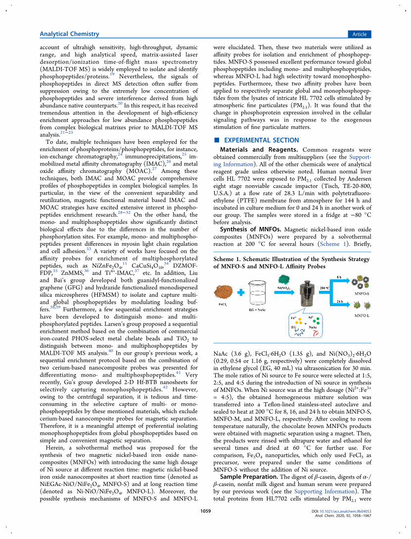
Synthesis of MNFOs. Magnetic nickel-based iron oxidecomposites (MNFOs) were prepared by a solvothermalreaction at 200 °C for several hours (Scheme 1). Briefly,
NaAc (3.6 g), FeCl3·6H2O (1.35 g), and Ni(NO3)2·6H2O(0.29, 0.54 or 1.16 g, respectively) were completely dissolvedin ethylene glycol (EG, 40 mL) via ultrasonication for 30 min.The mole ratios of Ni source to Fe source were selected at 1:5,2:5, and 4:5 during the introduction of Ni source in synthesisof MNFOs. When Ni source was at the high dosage (Ni2+:Fe3+
= 4:5), the obtained homogeneous mixture solution wastransferred into a Teflon-lined stainless-steel autoclave andsealed to heat at 200 °C for 8, 16, and 24 h to obtain MNFO-S,MNFO-M, and MNFO-L, respectively. After cooling to roomtemperature naturally, the chocolate brown MNFOs productswere obtained with magnetic separation using a magnet. Then,the products were rinsed with ultrapure water and ethanol forseveral times and dried at 60 °C for further use. Forcomparison, Fe3O4 nanoparticles, which only used FeCl3 asprecursor, were prepared under the same conditions ofMNFO-S without the addition of Ni source.
Sample Preparation. The digest of β-casein, digests of α-/β-casein, nonfat milk digest and human serum were preparedby our previous work (see the Supporting Information). Thetotal proteins from HL7702 cells stimulated by PM2.1 were
Scheme 1. Schematic Illustration of the Synthesis Strategyof MNFO-S and MNFO-L Affinity Probes
extracted by the protein extraction kit. After total proteincontent was measured by BCA Protein Assay Kit, HL 7702cells lysate digests were obtained by adding cell lysis buffer (4%CHAPS + 65 mM DTT + protease inhibitor (1:100)) togetherwith trypsin (20 μg mL−1) at a ratio of 40:1 (wt/wt) forincubation at 37 °C for 18 h.Selective Enrichment of Global-/Monophosphopep-
tides with Two Magnetic Nickel-Based Iron OxideComposites from Different Samples. The workflow toenrichment of phosphopeptides is shown in Scheme S1.MNFO-S or MNFO-L (5.0 μL, 20 mg mL−1) was added intothe peptide mixture (100 μL) at room temperature, which wasoriginating from β-casein tryptic digests (2.5 μL), the mixtureof α-/β-casein tryptic digests (2.5 μL), nonfat milk trypticdigests (1.0 μL), or human serum (1.0 μL) diluted to 1 mLwith 50% ACN/5.0% TFA solution. The mixture was vortexedfor 15 min at room temperature. Then, the phosphopeptides-loaded probes were collected with a magnet and rinsed with100 μL of the 50% ACN/0.1% TFA solution for three times toremove unspecific absorption and other unbound impurities.Afterward, the trapped phosphopeptides were eluted with 5 μLof NH3·H2O (10%, w/w) by sonication for 10 min. For thetrypsin digests of HL7702 cell lysates, 200 μL of them wereincubated with MNFO-S or MNFO-L (5.0 μL, 20 μg mL−1)for 15 min with vortex. After rinsing, the trappedphosphopeptides were eluted with 5 μL of NH3·H2O (10%,w/w) by sonication for 10 min. Finally, these supernatantswere collected by magnetic separation for further MALDI-TOF MS analysis.Experimental Conditions for MALDI-TOF MS Anal-
ysis. After extraction of phosphopeptides from various samplesby two MNFOs or other affinity probes, 0.5 μL of eluate wasdeposited on a MALDI plate. Then, 0.5 μL of 2,5-dihydroxybenzoic acid (DHB) aqueous solution (20 mgmL−1, 50% ACN and 1% H3PO4) was introduced as a matrixon a MALDI-TOF MS (Bruker Daltonics, Germany) withSmartbeam-II laser technology. The measurements were in apositive reflection mode with the shots number of 500 and theacceleration voltage of 25 kV.
■ RESULTS AND DISCUSSIONSynthesis and Characterization of MNFOs. Briefly,
Fe3O4 and MNFOs were synthesized by a solvothermalprocess with different ratios of Ni2+ to Fe3+. As shown in theTEM image (Figure 1A), the prepared Fe3O4 nanocrystal(Ni2+:Fe3+ = 0:5) had a smooth and uniform sphere at areaction time of 8 h. Upon the addition of nickel source, thesurface of nanoparticles became rough (Ni2+:Fe3+ = 1:5,MNFO-01, Figure 1B), even bringing out semitransparentlayers after introducing more Ni source (Ni2+:Fe3+ = 2:5,MNFO-02, Figure 1C). When the dosage of Ni source furtherenlarged (Ni2+:Fe3+ = 4:5), MNFO-S nanocomposites withsemitransparent flower-like structure were obtained (Figure1D). The flower-shaped layers faded away after prolonging thereaction time to 16 h (MNFO-M, Figure 1E). When thereaction time was further extended to 24 h, MNFO-L wasobtained with a few ribbon layers on the edge of nanoparticles(Figure 1F). The TEM results illustrated that both the dose ofNi source and reaction time could intensively affect theformation and morphology of synthesized MNFOs. Surfacemorphology and composition analysis of the obtained MNFOswere characterized by FESEM for MNFO-S, MNFO-M, andMNFO-L (Figure 2) or SEM for MNFO-01 and MNFO-02
(Figure S1), which were in accordance with the TEM analysisof MNFOs. The EDS spectra were recorded to further identifythe elemental composition of MNFOs, and the results showedthat all of the MNFOs contain the same elementsC, O, Fe,and Niand no contamination elements were detected.As shown in Figure S2, the FT-IR characteristic peak at 570
cm−1 was assigned to the metal−oxygen bonds (Fe−O/Ni−O)in MNFOs. The broad adsorption band centered at 3425 cm−1
was ascribed to surface hydroxyl (−OH). The characteristicpeaks at 2934 and 2856 cm−1 corresponded to −CH2stretching vibration. The peaks at 1650 and 1400 cm−1 wereattributed to COO− and COO−M+ (M: Fe3+, Ni2+), and thepeak at 1102.6 cm−1 was ascribed to C−O. The results impliedthat organic substances (EG and acetate) exist in the affinitymaterials and especially are plentiful in the MNFO-S. Afterintroducing more Ni source, C and H elements in MNFOsgrew in quantity, subsequently (see Table S1 for elementalanalysis). However, under the same dosage of Ni source, the
Figure 1. TEM images of Fe3O4 and MNFOs at the same reactiontime of 8 h from different molar ratios of Ni source to Fe source: (A)Fe3O4 (Ni2+:Fe3+ = 0:5); (B) MNFO-01 (Ni2+:Fe3+ = 1:5); (C)MNFO-02 (Ni2+:Fe3+ = 2:5). TEM images of MNFOs from the samemolar ratio of Ni source to Fe source (Ni2+:Fe3+ = 4:5) at the differentreaction time: (D) MNFO-S (8 h); (E) MNFO-M (16 h); (F)MNFO-L (24 h).
Figure 2. FESEM (A−C) and EDS (D−F) of MNFOs from the fixedmolar ratio of Ni source to Fe source (Ni2+:Fe3+ = 4:5) at differentreaction time. MNFO-S (A, D; 8 h); MNFO-M (B, E; 16 h); MNFO-L (C, F; 24 h).
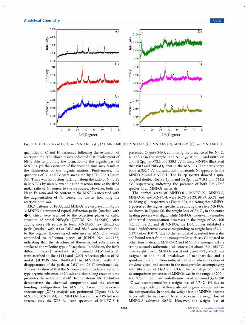
quantities of C and H decreased following the extension ofreaction time. The above results indicated that involvement ofNi is able to promote the formation of the organic part ofMNFOs, yet the extension of the reaction time may result inthe diminution of the organic matters. Furthermore, thequantities of Ni and Fe were measured by ICP-OES (FigureS3). There was no obvious variation about the ratio of Ni to Fein MNFOs by merely extending the reaction time at the fixedmolar ratio of Ni source to the Fe source. However, both theNi to Fe ratio and Ni content in the MNFOs increased withthe augmentation of Ni source, no matter how long thereaction time was.XRD patterns of Fe3O4 and MNFOs are displayed in Figure
3. MNFO-01 presented typical diffraction peaks (marked with◆), which were ascribed to the reflection planes of cubicstructure of spinel NiFe2O4 (JCPDS No. 54-0964). Afteradding more Ni source to form MNFO-S, new diffractionpeaks (marked with Δ) at 7.63° and 26.1° were observed dueto the organic flower-shaped substance in MNFO-S, whichresponded to reflection planes of JCPDS No. 26-1135,indicating that the structure of flower-shaped substances issimilar to the valleriite type of haapalaite. In addition, the freshdiffraction peaks (marked with ★) obtained at 44.5° and 51.8°were ascribed to the (111) and (200) reflection planes of Nimetal (JCPDS No. 04-0850) in MNFO-L, with thedisappearance of the peaks at 7.63° and 26.1° simultaneously.The results showed that the Ni source will introduce a valleriitetype organic substance of Ni salt and that a long reaction timepromotes the reduction of Ni2+ to monatomic Ni. To furtherdemonstrate the chemical composition and the elementbonding configuration for MNFOs, X-ray photoelectronspectroscopy (XPS) analysis was performed (Figure S4). AsMNFO-S, MNFO-M, and MNFO-L have similar XPS full scanspectra, only the XPS full scan spectrum of MNFO-S is
presented (Figure S4A), confirming the presence of Fe, Ni, C,N, and O in the sample. The Ni 2p3/2 at 855.1 and 860.5 eVand Ni 2p1/2 at 872.5 and 880.1 eV in these MNFOs illustratedthat NiO and NiFe2O4 exist in the MNFOs. The new energyband at 852.7 eV indicated that monatomic Ni appeared in theMNFO-M and MNFO-L. The Fe 2p spectra showed a spin-coupled doublet for Fe 2p3/2 and Fe 2p1/2 at 710.5 and 722.5eV, respectively, indicating the presence of both Fe3+/Fe2+
species in all MNFOs materials.The surface areas of MNFO-01, MNFO-02, MNFO-S,
MNFO-M, and MNFO-L were 16.76, 93.28, 98,87, 55.72, and61.38 mg g−1, respectively (Figure S5), indicating that MNFO-S possesses the highest specific area among these five MNFOs.As shown in Figure S6, the weight loss of Fe3O4 in the entireheating process was slight, while MNFOs underwent a numberof thermal decomposition processes in the range of 25−400°C. For Fe3O4 and all MNFOs, the DSC curves exhibited abroad endothermic event corresponding to weight loss of 2.7−5.2% below 100 °C due to the removal of adsorbed free waterand bound water from the nanoparticles surfaces. Compared toother four materials, MNFO-02 and MNFO-S emerged with astrong second exothermic peak centered at about 150−165 °C.The weight loss of MNFOs was about 5.5−18.7%, which wasassigned to the initial breakdown of nanoparticles and aspontaneous combustion induced by the in situ oxidization ofethylene glycol and acetate in the nanoparticles accompanyingwith liberation of H2O and CO2. The last stage of thermaldecomposition processes of MNFOs was in the range of 200−400 °C, and the broad endothermic event at around 330−380°C was accompanied by a weight loss of 7.7−24.1% due tocontinuing oxidation of flower-shaped organic components inthe nanoparticles. In short, the weight loss of MNFOs becamelarger with the increase of Ni source, even the weight loss ofMNFO-S achieved 50.3%. However, the weight loss of
Figure 3. XRD spectra of Fe3O4 and MNFOs: Fe3O4 (A), MNFO-01 (B), MNFO-02 (C), MNFO-S (D), MNFO-M (E), and MNFO-L (F).
MNFO-L was merely 21.7%, which was less than half of theweight loss of MNFO-S. These results were in agreement withthe variation of contents C and H from MNFOs by elementalanalysis (Table S1). As the TG curves showed, there wasalmost no change in weight loss and exothermic orendothermic events in the DSC curves after 400 °C, indicatingthat almost all organic components of MNFOs had beenremoved since then.As shown in Figure S7, the hysteresis loops of Fe3O4 and
MNFOs illustrated that Fe3O4 and MNFOs possessedsuperparamagnetic properties. When the Ni source increasedat the fixed reaction time of 8 h, the saturation magnetization(Ms) values of MNFOs decreased gradually. Ms values weremeasured to 60.6, 41.9, and 15.2 emu g−1, respectively, forMNFO-01, MNFO-02, and MNFO-S. However, Ms values ofthe MNFOs increased to 19.68 emu g−1 (MNFO-M) and 32.6emu g−1 (MNFO-L) under longer reaction time. As a result,both dosage of Ni source and reaction time have a significanteffect on the magnetic properties of MNFOs. Moreover the Nisource induced a weaker magnetic property of MNFOs byvirtue of a more and more nonmagnetic translucent layer inmaterials while the reaction time was fixed at 8 h. In contrast,longer reaction time caused stronger magnetism of materialsdue to the disappearance of nonmagnetic components, as wellas the generation of magnetic Ni atom.Possible Formation Mechanism of MNFOs. According
to the electrode potential (φ(Fe3+/Fe2+) = 0.77 V, φ(Ni2+/Ni)= −0.257 V), metal ions are reduced in the order: Fe3+ > Ni2+.In the EG-NaAC solvent system, Fe3+ and Ni2+ ions formFe(OH)3 and Ni(OH)2 precipitates with the assistance ofNaAc at first, which provides an alkaline environment ofreaction medium (eq 1−3). Then, Fe(OH)3 is partially
reduced by EG to form Fe3O4 monomer and Ni2+ goes intoFe3O4 to form NiFe2O4 monomer (eq 4 and 5). After initialnucleation, the monomers cogrow into nanocrystals. Mean-while, NiO is generated through a dehydration reaction ofNi(OH)2 (eq 6). Compared to the Fe3+ ion, the Ni2+ ion isdifficult to be reduced at the short reaction time; thus, it trendsto coordinate with NaAC-EG to form nickel salt nanoparticle,which is a layered arrangement with a flower-like structurecalled NiEGAc. The above speculation has been testified bythe results of FT-IR, TEM, and XRD. With the extension ofreaction time, a few Ni2+ ions are reduced by EG to obtainelemental Ni (eq 7), and under airtight and high pressureconditions, the Ni atoms go into NiFe2O4 crystal lattices toform MNFO mix-nanocrystals, which have been confirmed byXPS and XRD spectra. As a result, a series of possible chemicalreactions about the formation of MNFOs are summarized asfollows:
+ +− ‐FCH COO H O CH COOH OH3 2 3 (1)
++ − FFe 3OH Fe(OH)33 (2)
++ − FNi 2OH Ni(OH)22 (3)
+ + +F12Fe(OH) H C O 4Fe O H C O 20H O3 6 2 2 3 4 2 2 2 2(4)
+ +
+ + +F
Ni(OH) 14Fe(OH) H C O
NiFe O 4Fe O H C O 24H O2 3 6 2 2
2 4 3 4 2 2 2 2 (5)
+FNi(OH) NiO H O2 2 (6)
Figure 4. Mass spectra of phosphopeptides from tryptic digests of β-casein after treatment with different MNFOs: Fe3O4 (A), MNFO-01 (B),MNFO-02 (C), MNFO-S (D), MNFO-M (E), and MNFO-L (F). “*” indicates phosphopeptides, “#” indicates dephosphorylated residues ofphosphopeptides, “s” indicates monophosphopeptides, and “m” indicates multiphosphopeptides.
+ + +F2Ni(OH) H C O 2Ni H C O 4H O2 6 2 2 2 2 2 2 (7)
Adsorption Capacity of MNFOs toward Phosphopep-tides. p-Nitrophenylphosphate (pNPP) is commonly used as amodel to investigate the intrinsic phosphate adsorptioncapacity.43,44 As shown in Figure S8, 100 μg (5 μL, 20 mgmL−1) of MNFO-S or MNFO-L was used to ensure thecomplete adsorption of pNPP, while 10% NH3·H2O wasoptimized as the eluent during the experiment. According tothe procedure to phosphopeptides enrichment, the adsorptioncapacities of pNPP with MNFO-01, MNFO-02, MNFO-S,MNFO-M, and MNFO-L were measured to be 10.62, 181.5,234.0, 96.77, and 110.8 mg g−1, respectively, which indicatedsatisfactory ability of MNFO-S and MNFO-L towardphosphopeptides.Specific Enrichment of Global- and Monophospho-
peptides by MNFOs. As shown in Figure 4, MNFOsdisplayed different performance of isolation and capture ofmono-/global phosphopeptides form tryptic digest of β-casein(10 pmol). When more Ni source was introduced to formMNFOs at the fixed reaction time of 8 h, the capacity ofMNFOs for multiphosphopeptides gradually declined. How-ever, even at the high doges of Ni source (Ni2+:Fe3+ = 4:5), theobtained MNFO-S can still capture global phosphopeptidesincluding monophosphopeptides and multiphosphopeptideswithout site selectivity. According to the theory of hard andsoft acids and bases (HSAB), Fe3+ ion, as a typical hard acid,
has strong interaction with phosphospeptides of hard baseproperty, and thus, Fe3O4 has strong affinity interaction towardboth mono- and multiphosphopeptides. Ni2+ instead of Fe2+
on the distribution of metal ions at the surface sites of Fe3O4structure, would affect a little affinity interaction of resultingNiFe2O4 toward mono- and multiphosphopeptides due tosofter acidity of Ni2+ ion. Moreover, because of more Ni sourceintroduction, the obtained NiO and Ni salts mixed in theNiFe2O4 further weakened the affinity between MNFOs andphosphopeptides, especially for multiphosphopeptides. Thus,the dosage of doped nickel is an essential factor to affect theselectivity of MNFOs toward mono-/multiphosphopeptides.In addition, when the reaction time was extended at the highdosage of Ni source, the obtained MNFOs will further trend toselectively capture monophosphospeptides, until the MNFO-L(the reaction time of 24 h) only captured monophosphopep-tides. This is mainly because the Ni metal presented in thestructure could further aggravated the MNFO-L selectivity ofmonophosphospeptides, indicating that the reaction time isanother critical factor of selectivity of synthesized MNFOstoward mono/multi-phosphopeptides. It illustrated that theconjugation of divalent cation Ni2+ and metallic Ni seriouslyimpacted on the selectivity of enriching mono/multi-phosphospeptides. Generally, commercial TiO2 is used forcomparison to new affinity materials for phosphopeptides. Inthis work, TiO2 captured global phosphopeptides from β-casein instead of distinguishing mono- and multiphosphopep-
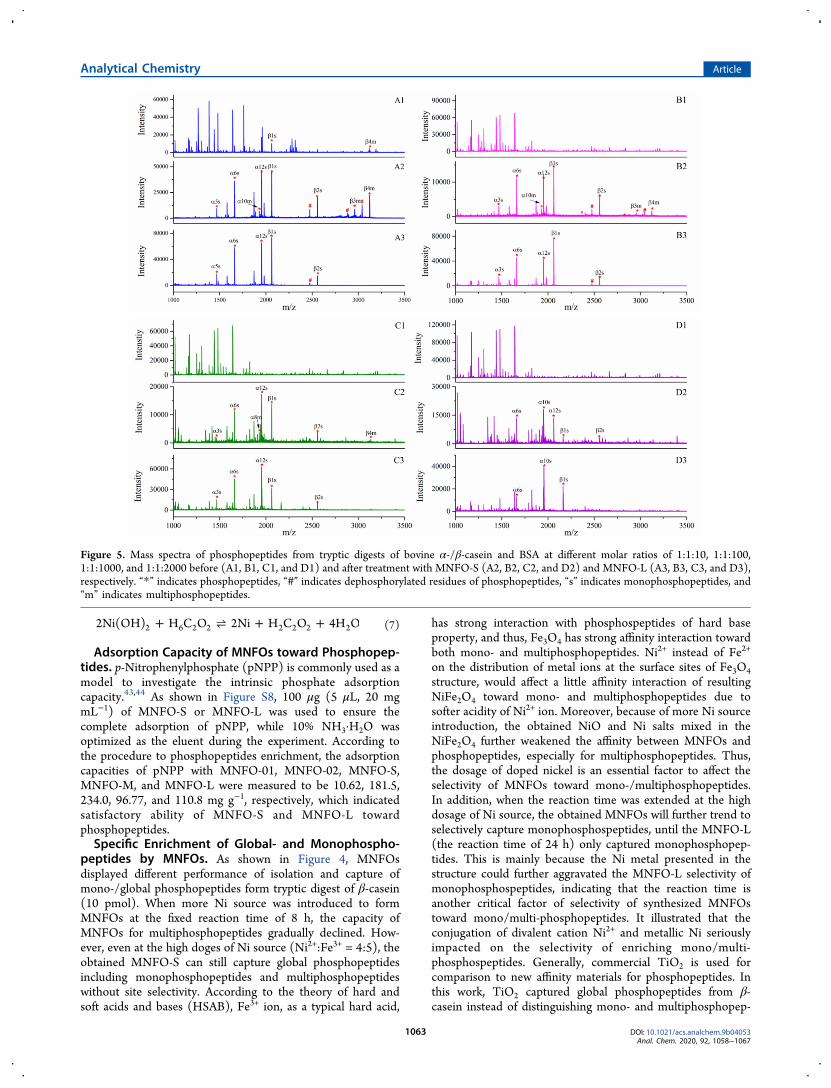
Figure 5. Mass spectra of phosphopeptides from tryptic digests of bovine α-/β-casein and BSA at different molar ratios of 1:1:10, 1:1:100,1:1:1000, and 1:1:2000 before (A1, B1, C1, and D1) and after treatment with MNFO-S (A2, B2, C2, and D2) and MNFO-L (A3, B3, C3, and D3),respectively. “*” indicates phosphopeptides, “#” indicates dephosphorylated residues of phosphopeptides, “s” indicates monophosphopeptides, and“m” indicates multiphosphopeptides.
tides as usual (Figure S9). To further test the difference of
enrichment of phosphopeptides by MNFO-S and MNFO-L,
mixture of bovine α/β-casein tryptic digests (10 pmol, molar
ratio of 1:1) was utilized as another model sample (Figure
S10), which was accordance with above results. Moreover, the
identified mono/multi-phopshopeptides captured by MNFO-S
or MNFO-L in the amino acid sequence pattern are listed inTable S2.To assess the sensitivity of phosphopeptides enrichment by
MNFOs, bovine α/β-casein tryptic digests were mixed indifferent concentrations (Figure S11). Three monophospho-peptides and one multiphosphopeptide were still detected in 5fmol trypsin digests of α-/β-casein after enrichment by
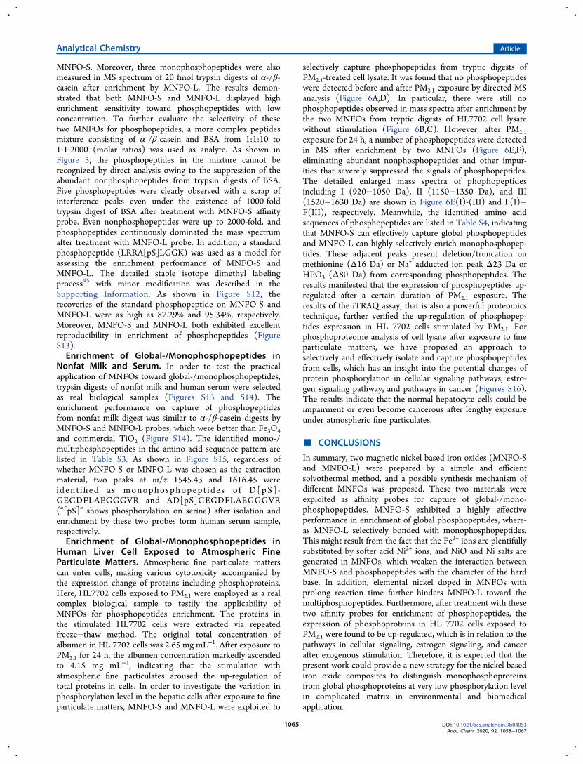
Figure 6.Mass spectra of HL7702 cell lysates digests before and after treatment with MNFOs: before stimulation with PM2.1 by direct analysis (A),before stimulation with PM2.1 by enrichment with MNFO-S (B), before stimulation with PM2.1 by enrichment with MNFO-L (C); afterstimulation with PM2.1 particles for 24 h by direct analysis (D); after stimulation with PM2.1 treated with MNFO-S (E), after stimulation with PM2.1particles for 24 h treated with MNFO-L (F), respectively. E(I)-(III) and F(I)−F(III) present the detailed enlarged mass spectra of phophopeptidesincluding I (920−1050 Da), II (1150−1350 Da), and III (1520−1630 Da). “*” indicates phosphopeptides and “#” indicates dephosphorylation.“Blue line” indicates Δ80 Da, “green line” indicates Δ16 Da, and “red line” indicates Δ23 Da.
MNFO-S. Moreover, three monophosphopeptides were alsomeasured in MS spectrum of 20 fmol trypsin digests of α-/β-casein after enrichment by MNFO-L. The results demon-strated that both MNFO-S and MNFO-L displayed highenrichment sensitivity toward phosphopeptides with lowconcentration. To further evaluate the selectivity of thesetwo MNFOs for phosphopeptides, a more complex peptidesmixture consisting of α-/β-casein and BSA from 1:1:10 to1:1:2000 (molar ratios) was used as analyte. As shown inFigure 5, the phosphopeptides in the mixture cannot berecognized by direct analysis owing to the suppression of theabundant nonphosphopeptides from trypsin digests of BSA.Five phosphopeptides were clearly observed with a scrap ofinterference peaks even under the existence of 1000-foldtrypsin digest of BSA after treatment with MNFO-S affinityprobe. Even nonphosphopeptides were up to 2000-fold, andphosphopeptides continuously dominated the mass spectrumafter treatment with MNFO-L probe. In addition, a standardphosphopeptide (LRRA[pS]LGGK) was used as a model forassessing the enrichment performance of MNFO-S andMNFO-L. The detailed stable isotope dimethyl labelingprocess45 with minor modification was described in theSupporting Information. As shown in Figure S12, therecoveries of the standard phosphopeptide on MNFO-S andMNFO-L were as high as 87.29% and 95.34%, respectively.Moreover, MNFO-S and MNFO-L both exhibited excellentreproducibility in enrichment of phosphopeptides (FigureS13).Enrichment of Global-/Monophosphopeptides in
Nonfat Milk and Serum. In order to test the practicalapplication of MNFOs toward global-/monophosphopeptides,trypsin digests of nonfat milk and human serum were selectedas real biological samples (Figures S13 and S14). Theenrichment performance on capture of phosphopeptidesfrom nonfat milk digest was similar to α-/β-casein digests byMNFO-S and MNFO-L probes, which were better than Fe3O4and commercial TiO2 (Figure S14). The identified mono-/multiphosphopeptides in the amino acid sequence pattern arelisted in Table S3. As shown in Figure S15, regardless ofwhether MNFO-S or MNFO-L was chosen as the extractionmaterial, two peaks at m/z 1545.43 and 1616.45 wereiden t ified a s monophosphopep t ide s o f D[pS] -GEGDFLAEGGGVR and AD[pS]GEGDFLAEGGGVR(“[pS]” shows phosphorylation on serine) after isolation andenrichment by these two probes form human serum sample,respectively.Enrichment of Global-/Monophosphopeptides in
Human Liver Cell Exposed to Atmospheric FineParticulate Matters. Atmospheric fine particulate matterscan enter cells, making various cytotoxicity accompanied bythe expression change of proteins including phosphoproteins.Here, HL7702 cells exposed to PM2.1 were employed as a realcomplex biological sample to testify the applicability ofMNFOs for phosphopeptides enrichment. The proteins inthe stimulated HL7702 cells were extracted via repeatedfreeze−thaw method. The original total concentration ofalbumen in HL 7702 cells was 2.65 mg mL−1. After exposure toPM2.1 for 24 h, the albumen concentration markedly ascendedto 4.15 mg mL−1, indicating that the stimulation withatmospheric fine particulates aroused the up-regulation oftotal proteins in cells. In order to investigate the variation inphosphorylation level in the hepatic cells after exposure to fineparticulate matters, MNFO-S and MNFO-L were exploited to
selectively capture phosphopeptides from tryptic digests ofPM2.1-treated cell lysate. It was found that no phosphopeptideswere detected before and after PM2.1 exposure by directed MSanalysis (Figure 6A,D). In particular, there were still nophosphopeptides observed in mass spectra after enrichment bythe two MNFOs from tryptic digests of HL7702 cell lysatewithout stimulation (Figure 6B,C). However, after PM2.1exposure for 24 h, a number of phosphopeptides were detectedin MS after enrichment by two MNFOs (Figure 6E,F),eliminating abundant nonphosphopeptides and other impur-ities that severely suppressed the signals of phosphopeptides.The detailed enlarged mass spectra of phophopeptidesincluding I (920−1050 Da), II (1150−1350 Da), and III(1520−1630 Da) are shown in Figure 6E(I)-(III) and F(I)−F(III), respectively. Meanwhile, the identified amino acidsequences of phosphopeptides are listed in Table S4, indicatingthat MNFO-S can effectively capture global phosphopeptidesand MNFO-L can highly selectively enrich monophosphopep-tides. These adjacent peaks present deletion/truncation onmethionine (Δ16 Da) or Na+ adducted ion peak Δ23 Da orHPO3 (Δ80 Da) from corresponding phosphopeptides. Theresults manifested that the expression of phosphopeptides up-regulated after a certain duration of PM2.1 exposure. Theresults of the iTRAQ assay, that is also a powerful proteomicstechnique, further verified the up-regulation of phosphopep-tides expression in HL 7702 cells stimulated by PM2.1. Forphosphoproteome analysis of cell lysate after exposure to fineparticulate matters, we have proposed an approach toselectively and effectively isolate and capture phosphopeptidesfrom cells, which has an insight into the potential changes ofprotein phosphorylation in cellular signaling pathways, estro-gen signaling pathway, and pathways in cancer (Figures S16).The results indicate that the normal hepatocyte cells could beimpairment or even become cancerous after lengthy exposureunder atmospheric fine particulates.
■ CONCLUSIONS
In summary, two magnetic nickel based iron oxides (MNFO-Sand MNFO-L) were prepared by a simple and efficientsolvothermal method, and a possible synthesis mechanism ofdifferent MNFOs was proposed. These two materials wereexploited as affinity probes for capture of global-/mono-phosphopeptides. MNFO-S exhibited a highly effectiveperformance in enrichment of global phosphopeptides, where-as MNFO-L selectively bonded with monophosphopeptides.This might result from the fact that the Fe2+ ions are plentifullysubstituted by softer acid Ni2+ ions, and NiO and Ni salts aregenerated in MNFOs, which weaken the interaction betweenMNFO-S and phosphopeptides with the character of the hardbase. In addition, elemental nickel doped in MNFOs withprolong reaction time further hinders MNFO-L toward themultiphosphopeptides. Furthermore, after treatment with thesetwo affinity probes for enrichment of phosphopeptides, theexpression of phosphoproteins in HL 7702 cells exposed toPM2.1 were found to be up-regulated, which is in relation to thepathways in cellular signaling, estrogen signaling, and cancerafter exogenous stimulation. Therefore, it is expected that thepresent work could provide a new strategy for the nickel basediron oxide composites to distinguish monophosphoproteinsfrom global phosphoproteins at very low phosphorylation levelin complicated matrix in environmental and biomedicalapplication.
■ ASSOCIATED CONTENT*S Supporting InformationThe Supporting Information is available free of charge athttps://pubs.acs.org/doi/10.1021/acs.analchem.9b04053.
Observed phosphopeptides from the lysates of HL 7702cells after exposure to PM2.1 for different time by iTRAQexperiment (XLS)
SEM images; EDS spectra; FT-IR spectra; ICP-MSanalysis of Fe and Ni elements in MNFOs; XPS spectra;N2 adsorption−desorption curves of MNFOs; XRDpatterns; hysteresis loops of MNFOs; schematicillustration of the phosphopeptides enrichment frombiological samples using MNFOs materials; adsorptionperformance of pNPP with different MNFOs; perform-ance of MNFOs as well as other affinity probes inphosphopeptides enrichment; tables of elementalanalysis, amino acid sequence of identified phosphopep-tides from the tryptic digests of α-/β-casein, nonfat milk,human serum, and HL 7702 cell lysate treated withMNFOs nanocomposites; detailed comparisons of theMNFO affinity probes with previously reported affinityprobes (PDF)
■ ACKNOWLEDGMENTSThis work was supported by the National Natural ScienceFoundation of China (21874065, 91643105, and 21577057)and the Natural Science Foundation of Jiangsu Province(BK20171335).
■ REFERENCES(1) Chinnasamy, C. N.; Narayanasamy, A.; Ponpandian, N.;Chattopadhyay, K.; Shinoda, K.; Jeyadevan, B.; Tohji, K.;Nakatsuka, K.; Furubayashi, T.; Nakatani, I. Phys. Rev. B: Condens.Matter Mater. Phys. 2001, 63 (18), 184198.(2) Carta, D.; Casula, M. F.; Falqui, A.; Loche, D.; Mountjoy, G.;Sangregorio, C.; Corrias, A. J. Phys. Chem. C 2009, 113 (20), 8606−8615.(3) Liu, M.; Lv, Z. X. J. Nanosci. Nanotechnol. 2018, 18 (6), 4211−4216.(4) Zhang, Y. L.; Zhou, Y.; Li, Z. L.; Chen, G.; Mao, Y.; Guan, H. T.;Dong, C. J. J. Alloys Compd. 2019, 784, 102−110.(5) Ma, Y. H.; Lu, Y.; Gou, H. T.; Zhang, W. X.; Yan, S. H.; Xu, X. L.Ceram. Int. 2018, 44 (2), 2620−2625.(6) Maaz, K.; Karim, S.; Mumtaz, A.; Hasanain, S. K.; Liu, J.; Duan,J. L. J. Magn. Magn. Mater. 2009, 321 (12), 1838−1842.(7) Islam, M.; Ali, G.; Jeong, M. G.; Choi, W.; Chung, K. Y.; Jung,H. G. ACS Appl. Mater. Interfaces 2017, 9 (17), 14833−14843.(8) Shariatdoost, A. S.; Yousefi, M.; Derakhshi, P.; Safekordi, A.;Larijani, K. J. Nanoanal. 2018, 5 (1), 1−6.(9) Sun, W.; Qiao, K.; Liu, J. Y.; Cao, L. M.; Gong, X. Q.; Yang, J.ACS Comb. Sci. 2016, 18 (4), 195−202.(10) Hessien, M. M.; Mostafa, N. Y.; Abd-Elkader, O. H. J. Magn.Magn. Mater. 2016, 398, 109−115.
(11) Zhong, H.; Xiao, X.; Zheng, S.; Zhang, W.; Ding, M.; Jiang, H.;Huang, L.; Kang, J. Nat. Commun. 2013, 4, 1656.(12) Olsen, J. V.; Blagoev, B.; Gnad, F.; Macek, B.; Kumar, C.;Mortensen, P.; Mann, M. Cell 2006, 127 (3), 635−48.(13) Luke Wiseman, R.; Kelly, J. W. Science 2011, 332 (6025), 44−45.(14) Nodeh, H. R.; Ibrahim, W. A. W.; Kamboh, M. A.; Sanagi, M.M. Chemosphere 2017, 166, 21−30.(15) Lee, H. J.; Na, K.; Kwon, M. S.; Kim, H.; Kim, K. S.; Paik, Y. K.Proteomics 2009, 9 (12), 3395−3408.(16) Owen, J. B.; Di Domenico, F.; Sultana, R.; Perluigi, M.; Cini,C.; Pierce, W. M. J. Proteome Res. 2009, 8 (2), 471−482.(17) Hu, Y. W.; Shah, P.; Clark, D. J.; Ao, M. H.; Zhang, H. Anal.Chem. 2018, 90 (13), 8065−8071.(18) Ugras, S.; Daniels, M.; Fazelinia, H.; Gould, N. S.; Yocum, A.K.; Luk, K. C.; Luna, E.; Ding, H.; McKennan, C.; Seeholzer, S.;Martinez, D.; Evans, P.; Brown, D.; Duda, J. E.; Ischiropoulos, H.Ebiomedicine 2018, 31, 307−319.(19) Li, X. S.; Yuan, B. F.; Feng, Y. Q. TrAC, Trends Anal. Chem.2016, 78, 70−83.(20) Eyrich, B.; Sickmann, A.; Zahedi, R. P. Proteomics 2011, 11 (4),554−70.(21) Qi, X.; Chang, C.; Xu, X.; Zhang, Y.; Bai, Y.; Liu, H. J.Chromatogr. A 2016, 1468, 49−54.(22) Long, X. Y.; Song, Q.; Lian, H. Z. J. Mater. Chem. B 2015, 3(48), 9330−9339.(23) Xu, X.; Deng, C.; Gao, M.; Yu, W.; Yang, P.; Zhang, X. Adv.Mater. 2006, 18 (24), 3289−3293.(24) Hennrich, M. L.; van den Toorn, H. W. P.; Groenewold, V.;Heck, A. J. R.; Mohammed, S. Anal. Chem. 2012, 84 (4), 1804−1808.(25) Rush, J.; Moritz, A.; Lee, K. A.; Guo, A.; Goss, V. L.; Spek, E. J.;Zhang, H.; Zha, X. M.; Polakiewicz, R. D.; Comb, M. J. Nat.Biotechnol. 2005, 23 (1), 94−101.(26) Ndassa, Y. M.; Orsi, C.; Marto, J. A.; Chen, S.; Ross, M. M. J.Proteome Res. 2006, 5 (10), 2789−2799.(27) Luo, B.; Yang, M.; Jiang, P.; Lan, F.; Wu, Y. Nanoscale 2018, 10(18), 8391−8396.(28) Ma, W. F.; Zhang, Y.; Li, L. L.; You, L. J.; Zhang, P.; Zhang, Y.T.; Li, J. M.; Yu, M.; Guo, J.; Lu, H. J.; Wang, C. C. ACS Nano 2012,6 (4), 3179−3188.(29) Sun, X. N.; Liu, X. D.; Feng, J. A.; Li, Y.; Deng, C. H.; Duan, G.L. Anal. Chim. Acta 2015, 880, 67−76.(30) Lin, H. Z.; Deng, C. H. Proteomics 2016, 16 (21), 2733−2741.(31) Cheng, G.; Zhang, J. L.; Liu, Y. L.; Sun, D. H.; Ni, J. Z. Chem.Commun. 2011, 47 (20), 5732−5734.(32) Min, Q. H.; Li, S. Y.; Chen, X. Q.; Abdel-Halim, E. S.; Jiang, L.P.; Zhu, J. J. ACS Appl. Mater. Interfaces 2015, 7 (18), 9563−9572.(33) Vicente-Manzanares, M.; Horwitz, A. R. Biochem. Biophys. Res.Commun. 2010, 402, 537−542.(34) Yang, S. S.; Yu, H. X.; Wang, Z. Z.; Liu, H. L.; Zhang, H.; Yu, X.Z.; Shang, W. B.; Chen, G. Q.; Gu, Z. Y. Chem. - Eur. J. 2018, 24 (9),2109−2116.(35) Peng, J. X.; Niu, H.; Zhang, H. Y.; Yao, Y. T.; Zhao, X. Y.;Zhou, X. Y.; Wan, L. H.; Kang, X. H.; Wu, R. A. ACS Appl. Mater.Interfaces 2018, 10 (38), 32613−32621.(36) Bae, S. W.; Kim, J. I.; Cuot, I.; Sung, J.; Hong, J. I.; Yeo, W. S.Anal. Sci. 2017, 33 (12), 1381−1386.(37) Liu, Z.; Wang, F.; Chen, J.; Zhou, Y.; Zou, H. J. Chromatogr. A2016, 1461, 35−41.(38) Xu, L. N.; Li, L. P.; Bai, Y.; Liu, H. W. Chem. Commun. 2014,50, 10963−10966.(39) Xu, L. N.; Ma, W.; Shen, S. S.; Li, L. P.; Bai, Y.; Liu, H. W.Chem. Commun. 2016, 52, 1162−1165.(40) Thingholm, T. E.; Jensen, O. N.; Robinson, P. J.; Larsen, M. R.Mol. Cell. Proteomics 2008, 7 (4), 661−671.(41) Long, X. Y.; Zhang, Z. J.; Li, J. Y.; Sheng, D.; Lian, H. Z. Chem.Commun. 2017, 53 (33), 4620−4623.(42) Xiao, J.; Yang, S. S.; Wu, J. X.; Wang, H.; Yu, X. Z.; Shang, W.B.; Chen, G. Q.; Gu, Z. Y. Anal. Chem. 2019, 91 (14), 9093−9101.