Fluorescence probes have been widely used as ana-lytical tools to monitor the microenvironments incomplex systems [1–6]. The excited-state propertiesof one such fluoroprobe, 8-hydroxy-1,3,6-pyrene tri-sulfonic acid (Pyranine-Py), has been extensively ex-plored for optical sensor applications in measuringpH [7–11], carbon dioxide [12–16], and for investigat-ing the morphology of various systems. The exploi-tation of the photoacidic properties of Py, in its elec-tronically excited state being more acidic than itsground state, results in an excited-state protontransfer (ESPT) [17]. This offers a dual excitation/single emission (ex/em) fluorescence ratio (i.e., ex/em: 403=511nm and ex/em: 454=511nm) that is com-monly used for different applications [18]. Py hasbeen applied as a versatile fluoroprobe in examiningphoto physical and photo chemical processes in com-
plex sample matrices, such as block copolymers, sur-factants (micelles), gels, and in thin films [19–23].Because of the heterogeneous internal environmentin these matrices, a dual excitation and dual emis-sion from Py immobilized in ethyl-cellulose (ex/em:405=440nm and ex/em: 465=510nm) has been ob-served [24]. The band at 440nm has been ascribedto the protonated Py species (PyOH�) and the oneat 510nm is designated to the deprotonated species(PyO−�). The presence of excimers with face-to-faceinteraction of Py molecules in polymer gel [21] andLangmuir–Blodgett (LB) films [25] adds to the com-plexity in handling Py as a probe molecule to studyintricate systems of interest. Rapid temporal fluctua-tions, high local concentrations, and perturbationsduring measurement are factors that affect energytransfer and chemical reactions in microenviron-ments [26]. A detailed characterization of the multi-species system in thin polymer films is hence vital forunderstanding and designing applications in opticalbiosensors.
We explored the use of nanosized materials in oursearch for a real-time spectral resolution techniqueto characterize excited-state fluorescent species ina complex molecular ensemble. Materials at theselength scales have attracted much attention in thepast decade due to their unique properties [27]. Oneinteresting phenomenon in this regard that is asso-ciated with nanometer-thick metal films is the sur-face plasmon coupled emission (SPCE) [28–34]. Thepresence of thin metal film increases the photonicmodes density (PMD) near the fluoroprobe [35,36].The strong coupling of the evanescent fields of sur-face plasmon polartions with the optical emitter un-der study results in a polarized directional emissionat a particular narrow angle [28,31,37,38]. The emis-sion at neighboring wavelengths is strongly attenu-ated. The angularity of the emission is a function ofthe wavelength of the emitting specie on account ofthe spectral dispersive property of SPCE [32,39,40].As a result, the shorter-wavelength light enters thecoupling prism at higher angles [33]. In the absenceof SPCE phenomenon, essentially all detection relieson the fluorescence emission from fluorophores un-der free-space conditions (FS), that is, emission intoa transparent nonabsorbing medium (i.e., water orair) typical of biological samples. As the use of SPCEoffers a significant improvement over the isotropicnature of the fluorescence FS emission, at presentthere is increased interest in finding applications ofSPCE that may be relevant to biomedical field.The SPCE phenomenon offers several advantages
as compared to other fluorescence-based methods[41–45]: (i) a 10–14 fold fluorescence intensity en-hancement is provided by coupling of the probemolecules with surface plasmons; (ii) a significant re-duction of sample volume; (iii) strong backgroundsuppression—only the emission from fluorophoreswithin 200nm from the metal film is coupled, asSPCE is a near-field phenomenon. This presents aunique approach for fluorescence signal detectionin complex sample matrices like whole blood [46,47]and muscle [48] and at the same time facilitatessingle-molecule detection [49,50]. However withgrowing interest in fluoroimmunoassays proceduresfor measuring many different target antigens in asingle step [51], the use of probes with multiple fluor-ophores is sought after. Hence, the current studyfocuses on selective observations of individual fluor-ophores in heterogeneous microenvironments utiliz-ing the intrinsic spectral resolution property ofSPCE.The three discrete excited states of the Py dye-
doped polyvinyl alcohol (PVA) are its protonated, de-protonated, and excimer forms. The use of a PVAchain polymer in this study is on account of its inter-esting structural and thermodynamic properties thatallow for all the manifestations of the Py fluoroprobe.The structural unit of PVA is ½CH2─CHOH�n withthe side chain hydroxyl─OH groups, which are polarand hydrophilic. These ─OH groups can form intra-molecular hydrogen bonds to one another and inter-
molecular hydrogen bonds to other compounds. Thusthe role of H-bonding of PVA with Py in this studywould help stabilize PyOH�. H-bonding lends a proticsolvent behavior to PVA and this would facilitateESPT from Py to PVA and, in turn, stabilize thePyO−� environment to a large extent. The long chaincarbon backbone provides hydrophobic pockets forexcimer π–π stacking, as the competition between hy-drophobic and hydrophilic forces results in the crea-tion of micellar-like structures in some cases [52].The PVA film therefore provides a well-controlledhost environment to study excited-state molecularphenomena. It is generated by spin-coating Py-dopedPVA solution on SPCE substrates.
2. Experimental Methods
The SPCE substrates were fabricated by a low-costsolution-based deposition technique developed inour lab [53]. The wet-chemistry approach was opti-mized to obtain a 47� 3nm uniform thin silver filmon glass substrates by varying the deposition timeand temperature. The substrates were subsequentlyspin coated with a 32� 3nm thick film of Py(10 μM–1mM solution) in 1% PVA. The thicknessand uniformity of the solution-deposited silver filmsand fluoroprobe/polymer layer were determined byAFM measurements. For optical studies, the silver-coated substrates were attached to a BK7 hemicy-lindrical prism with glycerol (n ¼ 1:47) as the index-matching fluid and placed on a 360° rotary platform.The platform’s capability to study these microenvir-onments at any angle relative to the incident anglewas achieved by mounting the PMT and monochro-mator on the rotation stage. This eliminated the needof an optical fiber and simultaneously enhanced thesensitivity of our apparatus. Excitation of the samplewas achieved with a 405nm LED, in reverse Kretsch-mann (RK) configuration [54]. The ability to exciteSPCE with broad band sources like light-emittingdiodes has already been demonstrated by our group[55]. The use of LEDs for sample irradiation enablesbetter matching of the excitation source to the opticalemitter under study. A uniform excitation of thefluorophores across the sample from the air side wasachieved in the RK geometry and the SPCE emissionwas observed on the BK7 prism side of the substrate.The SPCE emission spectra were collected from0–90° and 270–360° with respect to the front ofthe prism as presented in Fig. 1(a). The polarizedemission spectra were obtained through a polarizerplaced ahead of the detector of an ISS K2 (Cham-paign, Illinois) fluorometer. The equations from thesurface plasmon resonance (SPR) theory were ap-plied to the system depicted in Fig. 1(a) to predictthe propogation of surface plasmon polaritons at themetal–dielectric substrate boundary. These equa-tions are the basis of the commercially available soft-ware TFCalc. 3.5 [Software Spectra, Incorporated,Portland, Oregon] used to calculate the angle-dependent reflectivity curves presented in Fig. 1(b).We obtained an excellent agreement between
theoretical and experimental SPCE emission spectrafor the three different excited-state species of Pyin PVA.
3. Results and Discussion
The effect of molecular interactions on the photophy-sics of single-dye systems in constrained environ-ments needs to be understood as temperature andsample heterogeneity are responsible for spectralbroadening. Apart from ground-state molecular ag-gregation, excimer formation due to the proximityamong dye molecules opens new deactivation chan-nels and can further affect discerning a congestedspectrum using conventional fluorescence spectro-scopy. We exploit the use of SPCE to this end to studythe photophysics of Py encapsulated in PVA with in-creased intermolecular interactions. A two-stage pro-cess accounts for the spectral dispersion propertythat involves (i) radiationless conversion of light intosurface plasmon modes on metal films and (ii) sur-face plasmon propagation at the metal–dielectric(substrate) boundary. This offers twin leverage forrealizing an ultranarrowband photon-sorting ofemission from the microenvironments in a multispe-cies system. The effect on spectral dispersion basedon the refractive index of the substrate under consid-eration has previously been explored [37,53]. Earlierstudies [31–33] have also examined the angular de-pendence of emission from fluorophores on couplingwith surface plasmons. In the current paper, anapplication of this effect to selectively disperse wave-lengths of light is achieved using the SPCE phenom-enon, presenting a low-cost analytical tool with the
use of a broadband source for excitation in the RKconfiguration.
The SPR theory predicted plasmon resonance dipsfor the protonated (74:1°), deprotonated (58:0°), andexcimer aggregate (53:4°) of Py match the observedangularity obtained from SPCE at 74°, 58°, and 53°,respectively. The excited-state manifestations of Py:protonated, deprotonated, and the excimer aggre-gate have their emission maximum at specific wave-lengths—441, 496, and 532nm, respectively. Thesespecies couple at particular angles of 74:1°, 58°, and53:4°, respectively, with the surface plasmons on ac-count of the dispersive property of SPCE. The
Fig. 1. (Color online) (a) Schematic representation of sample configuration and angle-dependent SPCE emission of the excited statepopulations of Py in PVA. (b) SPR angle-dependent reflectivity curves calculated for the three-layer system shown in (a), using TFCalc3.0 software at emission wavelengths of 441, 496, and 532nm.
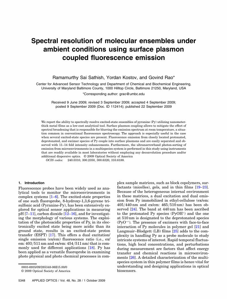
Fig. 2. (Color online) Normalized free-space (FS) and SPCE spec-tra of 1mM Py in 1% PVA. The top portion has photographs takenat the SPCE angles (53°, 58°, and 74°, left to right) of the emissionand at 30° (extreme right) without a filter.
emission at 532nm has been assigned to the excimerspecie, as the band is more pronounced at 1mM andweakens with decreasing concentration of Py(10–100 μM range) in 1% PVA. The normalized spec-tra of 1mM Py obtained on the FS is presented inFig. 2; this resembles the spectra obtained using theconventional fluorescence technique. The crowdedFS spectrum presents the PyO−� having a strongemission at 496nm and shoulders at 441nm and532nm for the PyOH� and excimer, respectively. Acorresponding real-time deconvulated spectra ofthe PyOH�, PyO−� and excimer is obtained byviewing the probe molecule at 74°, 58°, and 53°, re-spectively, on the SPCE side. This finding is the high-light of this paper, presenting a marked difference influorescence spectra recorded on the FS side versusthe SPCE side, in contrast to earlier reports [31–33].The angular spectral resolution capability of theSPCE platform allows for wavelength separation tobe visualized in the horizontal plane with minor shiftin the position of the eye/detector, which in turn al-ters the SPCE emission. The top portion of Fig. 2 dis-plays photographs taken at the SPCE angles of theemission without a filter showing the blue-to-greentransition in the emission from the sample. Thephotograph taken at 30° (extreme right) without a
filter shows only scatter from the 405nm LED.These snap shots were taken in the presence ofother light sources, indicative of the 10–14 fold in-tensity enhancements achieved on the SPCE side.Figures 3(a)–3(d), obtained by wavelength scan at di-verse observation angles (48°–80°) on the SPCE side,clearly show that the PyO−� outweighs the PyOH�and excimeric forms, as the SPCE intensity countsat 496nm is greater than at 441 and 532nm, respec-tively. Similar spectral resolution of three excited-state species was obtained with another fluoroprobe:6,8-dihydroxy-1,3-pyrenedisulfonic acid disodiumsalt (DHDS), a derivative of Py. We examined theemissions from plain PVA film to serve as the back-ground correction, as unintentional scatters are al-ways present in polymeric films.
4. Conclusions
To summarize, we have observed with high resolu-tion different excited-state populations of Py-dopedthin polymeric film in real time using the SPCE phe-nomenon. The spectral lines were resolved by the di-rectional nature of SPCE. Sensor applications can beexploited using the inherent directionality of SPCEfor simultaneous monitoring of different fluoro-phores at the same instant of time and extended
Fig. 3. (Color online) SPCE spectra of 1mM Py in 1% PVA with observation spanning: (a) 48°–53° angles for excimer resolution,(b) 53°–58° angles resolve excimer and PyO−� species, (c) 58°–69° angles projects the dominance of PyO−� species, and (d) 69°–80° anglespresents the emergence of PyOH� band with PyO−� still being the predominant manifestation.
to study complex macromolecule systems and reac-tions. In a larger perspective, this technique can bescaled down to realize low-cost miniaturized spectro-fluorometers with remarkable spectral resolutioncapabilities without the need for additional disper-sive optics.
This work was supported by funding from the Na-tional Science Foundation (NSF), Division of Bioen-gineering and Environmental Systems, grant award:NSF-BES 0517785.
References1. A. Mayer and S. Neuenhofer, “Luminescent labels—more than
just an alternative to radioisotopes?,” Angew. Chem., Int. Ed.33, 1044–1072 (1994).
2. A. Thomas, S. Polarz, and M. Antonietti, “Influence of spatialrestrictions on equilibrium reactions: a case study aboutthe excimer formation of pyrene,” J. Phys. Chem. B 107,5081–5087 (2003).
3. I. Capek, “Fate of excited probes in micellar systems,” Adv.Colloid Interface Sci. 97, 89–147 (2002).
4. H. Morawetz, “On the versatility of fluorescence techniques inpolymer research,” J. Polym. Sci., Part A 37, 1725–1735(1999).
5. D. Kaya, Ö. Pekcan, and Y. Yilmaz, “Direct test of the criticalexponents at the sol-gel transition,” Phys. Rev. E 69,016117 (2004).
6. J. I. Zink and B. S. Dunn, “Photonics materials by the sol-gelprocess: optical materials,” J. Ceram. Soc. Jpn. 99, 878–893(1991).
7. H. R. Kermis, Y. Kostov, and G. Rao, “Rapid method for thepreparation of a robust optical pH sensor,” Analyst 128,1181–1186 (2003).
8. O. S. Wolfbeis, E. Fuerlinger, H. Kroneis, and H. Marsoner,“Fluorimetric analysis. 1. A study on fluorescent indicatorsfor measuring near neutral (“physiological”) pH values,” Fre-senius’ J. Anal. Chem. 314, 119–124 (1983).
9. Z. Zhujun and W. R. Seitz, “A fluorescence sensor for quantify-ing pH in the range from 6.5 to 8.5,” Anal. Chim. Acta 160,47–55 (1984).
10. J. L. Gehrich, D. W. Lubbers, N. Opitz, D. R. Hansmann,W. W. Miller, J. K. Tusa, and M. Yafuso, “Optical fluorescenceand its application to an intravascular blood gas monitoringsystem,” IEEE Trans. Bio-med. Eng. BME-33, 117–132(1986).
11. S. Zhang, S. Tanaka, Y. A. B. D. Wickramasinghe, and P. Rolfe,“Fibre-optical sensor based on fluorescent indicator for mon-itoring physiological pH values,” Med. Bio. Eng. Comput. 33,152–156 (1995).
12. X. Ge, Y. Kostov, and G. Rao, “High-stability non-invasiveautoclavable naked optical CO2 sensor,” Biosens. Bioelectron.18, 857–865 (2003).
13. X. Ge, Y. Kostov, and G. Rao, “Low-cost noninvasive opticalCO2 sensing system for fermentation and cell culture,” Bio-technol. Bioeng. 89, 329–334 (2005).
14. A. Mills and Q. Chang, “Fluorescence plastic thin-film sen-sor for carbon dioxide,” Analyst 118, 839–843(1993).
15. M. Uttamlal and D. R. Walt, “A fiberoptic carbon-dioxidesensor for fermentation monitoring,” Bio/Technology 13,597–601 (1995).
16. R. N. Pattison, J. Swamy, B. Mendenhall, C. Hwang, andB. Frohlich, “Measurement and control of dissolved carbon di-oxide in mammalian cell culture processes using an in situ fi-ber optic chemical sensor,” Biotechnol. Prog. 16, 769–774(2000).
17. M. Rini, B. Z. Magnes, E. Pines, and E. T. J. Nibbering, “Real-time observation of bimodal proton transfer in acid-base pairsin water,” Science 301, 349–352 (2003).
18. R. P. Haugland, The Handbook: A Guide to Fluorescent Probesand Labeling Technologies (Invitrogen Corporation, 2005).
19. Y. Yilmaz, N. Uysal, A. Gelir, O. Guney, D. K. Aktas, S. Goge-bakan, and A. Oner, “Elucidation of multiple-point interac-tions of pyranine fluoroprobe during the gelation,”Spectrochim. Acta. A 72, 332–338 (2009).
20. S. Ghosh, S. Dey, U. Mandal, A. Adhikari, S. K. Mondal, andK. Bhattacharyya, “Ultrafast proton transfer of pyranine in asupramolecular assembly: PEO−PPO−PEO triblock copoly-mer and CTAC,” J. Phys. Chem. B 111, 13504–13510 (2007).
21. R. Barnabas-Rodriguez and J. Estelrich, “Photophysicalchanges of pyranine induced by surfactants: evidence of pre-micellar aggregates,” J. Phys. Chem. B 113, 1972–1982 (2009).
22. S. K. Mondal, K. Sahu, S. Ghosh, P. Sen, and K. Bhattachar-yya, “Excited-state proton transfer from pyranine to acetate inγ-cyclodextrin and hydroxypropyl γ-cyclodextrin,” J. Phys.Chem. A 110, 13646–13652 (2006).
23. R. Gupta and N. K. Chaudhury, “Probing internal environ-ment of sol–gel bulk and thin films using multiple fluorescentprobes,” J. Sol-Gel Sci. Technol. 49, 78–87 (2009).
24. A. Hakonen and S. Hulth, “A high-precision ratiometric fluor-osensor for pH: Implementing time-dependent non-linear ca-libration protocols for drift compensation,” Anal. Chim. Acta606, 63–71 (2008).
25. O. Tsukamoto, M. Villeneuve, A. Sakamoto, and H. Nakahara,“Change in the orientation and packing upon adsorption ofpyranine molecules onto cationic langmuir monolayersand Langmuir–Blodgett films,” Bull. Chem. Soc. Jpn. 80,1723–1730 (2007).
26. R. M. Wightman, P. Runnels, and Kevin Troyer, “Analysis ofchemical dynamics in microenvironments,” Anal. Chim. Acta400, 5–12 (1999).
27. U. Kreibig andM. Vollmer,Optical Properties of Metal Clusters(Springer, 1995).
28. J. R. Lakowicz, “Radiative decay engineering 3. Surfaceplasmon-coupled directional emission,” Anal. Biochem. 324,153–169 (2004).
29. H. M. Hiep, M. Fujii, and S. Hayashi, “Effects of molecular or-ientation on surface-plasmon-coupled emission patterns,”Appl. Phys. Lett. 91, 183110 (2007).
30. D. S. Smith, Y. Kostov, and G. Rao, “Signal enhancement ofsurface plasmon-coupled directional emission by a conicalmirror,” Appl. Opt. 47, 5229–5234 (2008).
31. W. H.Weber and C. F. Eagen, “Energy transfer from an exciteddye molecule to the surface plasmons of an adjacent metal,”Opt. Lett. 4, 236–238 (1979).
32. R. E. Benner, R. Dornhaus, and R. K. Chang, “Angular emis-sion profiles of dye molecules excited by surface plasmonwaves at a metal surface,” Opt. Commun. 30, 145–149(1979).
33. I. Pockrand and A. Brillante, “Nonradiative decay of excitedmolecules near a metal surface,” Chem. Phys. Lett. 69,499–504 (1980).
34. M. Trnavsky, J. Enderlein, T. Ruckstuhl, C. McDonagh, and B.D. Maccraith, “Experimental and theoretical evaluation ofsurface plasmon-coupled emission for sensitive fluorescencedetection,” J. Biomed. Opt. 13, 054021 (2008).
35. T. Liebermann andW. Knoll, “Surface-plasmon field-enhancedfluorescence spectroscopy,” Colloids Surf. A 171, 115–130(2000).
36. E. L. Moal, E. Fort, S. Lévêque-Fort, F. P. Cordelières,M. P. Fontaine-Aupart, and C. Ricolleau, “Enhanced fluores-cence cell imaging with metal-coated slides,” Biophys. J. 92,2150–2161 (2007).
37. E. G. Matveeva, I. Gryczynski, J. Malicka, Z. Gryczynski,E. Goldys, J. Howe, K. W. Berndt, and J. R. Lakowicz, “Plasticversus glass support for an immunoassay on metal coated sur-faces in optically dense samples utilizing directional surfaceplasmon coupled emission,” J. Fluoresc. 15, 865–871 (2005).
38. I. Gryczynski, J. Malicka, Z. Gryczynski, and J. R. Lakowicz,“Radiative decay engineering 4. Experimental studies of sur-face plasmon-coupled directional emission,” Anal. Biochem.324, 170–182 (2004).
39. F. Kaneko, T. Nakano, M. Terakado, K. Shinbo, K. Kato,T. Kawakami, and T. Wakamatsu, “Emission light fromprism/silver/ rhodamine-B LB film and multiple surface plas-mon excitations in the ATR Kretschmann configuration,” Ma-ter. Sci. Eng. C 22, 409–412 (2002).
40. T. Nakano, H. Kobayashi, K. Shinbo, K. Kato, F. Kaneko,T. Kawakami, and T. Wakamatsu, “Emission light propertiesfrom Ag/rhodamine-B LB films due to surface plasmon excita-tions in the Kretschmann and reverse configurations,” Mater.Res. Soc. Symp. Proc. 660, JJ8.35.1–JJ8.35.6 (2001).
41. Y. Kostov, D. S. Smith, L. Tolosa, G. Rao, I. Gryczynski,Z. Gryczynski, J. Malicka, and J. R. Lakowicz, “Directionalsurface plasmon-coupled emission from a 3nm green fluores-cent protein monolayer,” Biotechnol. Prog. 21, 1731–1735(2005).
42. D. S. Smith, Y. Kostov, and G. Rao, “SPCE-based sensors: ul-trafast oxygen sensing using surface-plasmon coupled emis-sion from ruthenium probes,” Sens. Actuator B 127, 432–440(2007).
43. K. Ray, M. H. Chowdhury, and J. R. Lakowicz, “Observation ofsurface plasmon-coupled emission using thin platinum films,”Chem. Phys. Lett. 465, 92–95 (2008).
44. K. Aslan, K. McDonald, M. J. R. Previte, Y. Zhang, andC. D. Geddes, “Angular dependent metal-enhanced fluores-cence from silver island films,” Chem. Phys. Lett. 464,216–219 (2008).
45. K. Ray, H. Szmacinski, J. Enderlein, and J. R. Lakowicz, “Dis-tance dependence of surface plasmon-coupled emission ob-served using Langmuir–Blodgett films,” Appl. Phys. Lett.90, 251116 (2007).
46. E. G. Matveeva, Z. Gryczynski, J. Malicka, J. Lukomska,S. Makowiec, K. W. Berndt, J. R. Lakowicz, and I. Gryczynski,“Directional surface plasmon-coupled emission: Applicationfor an immunoassay in whole blood,” Anal. Biochem. 344,161–167 (2005).
47. K. Aslan, Y. Zhang, and C. D. Geddes, “Surface plasmoncoupled fluorescence in the visible to near-infrared spectralregions using thin nickel films: Application to whole blood as-says,” Anal. Chem. 81, 3801–3808 (2009).
48. J. Borejdo, Z. Gryczynski, N. Calander, P. Muthu, andI. Gryczynski, “Application of surface plasmon coupled emis-sion to study of muscle,” Biophys J. 91, 2626–2635 (2006).
49. F. D. Stefani, K. Vasilev, N. Bocchio, N. Stoyanova, andM. Kreiter, “Surface-plasmon mediated single-molecule fluor-escence through a thin metallic film,” Phys. Rev. Lett. 94,023005 (2005).
50. Z. Gryczynski, I. Gryczynski, E. G. Matveeva, N. Calander,R. Grygorczyk, I. Akopova, S. Bharill, P. Muthu, S. Klidgar,and J. Borejdo, “New surface plasmons approach to single mo-lecule detection (SMD) and fluorescence correlation spectro-scopy (FCS),” Proc. SPIE 8, 64440G.1–64440G.11(2007).
51. A. D. Wellman and M. J. Sepaniak, “Magnetically-assistedtransport evanescent field fluoroimmunoassay,” Anal. Chem.78, 4450–4456 (2006).
52. T. Bryk and M. Holovko, “Hydration structure of a poly(vinylalcohol) chain fragment: Ab initio molecular dynamics study,”J. Mol. Liq. 147, 13–16 (2009).
53. R. Sai Sathish, Y. Kostov, D. S. Smith, and G. Rao, “Solution-deposited thin silver films on plastic surfaces for low-cost ap-plications in plasmon-coupled emission sensors,” Plasmonics4, 127–133 (2009).
54. E. Kreschmann and H. Raether, “Radiative decay of nonradia-tive surface plasmons excited by light,” Z. Naturforsch. A 23,2135–2136 (1968).
55. D. S. Smith, Y. Kostov, G. Rao, I. Gryczynski, J. Malicka,Z. Gryczynski, and J. R. Lakowicz, “First observation of sur-face plasmon-coupled emission due to LED excitation,” J.Fluoresc. 15, 895–900 (2005).