Spontaneous subluxation of the first and second cervical vertebrae, in rheumatoid arthritis, treated with spinal fusion
Report of two cases
S A M U E L R . S A W M I L L E R , M . D . *
A L A N H . W I L D E , M . D .
Department of Orthopaedic Surgery
SP O N T A N E O U S dislocation of the atlas on the axis has been known as an entity for some time, the first case reported is thought to be
that of Bell in 1830.1 Since then there have been numerous published re-ports of at lantoaxial subluxation, most recently in relation to rheumatoid arthritis.2"17
T h e at lantoaxial jo int has two axes of motion; the transverse axis which passes approximately through the center of the annulus osteofibrosus and permits flexion and extension of about 10 degrees; and the vertical axis, which also passes through the center of the annulus osteofibrosus and allows rotation of about 47 degrees. T h e tectorial membrane limits flexion and ex-tension, and the alar ligaments limit rotation. T h e annulus osteofibrosus is composed of the transverse ligament and the anterior arch of the atlas which encircles the odontoid process and prevents anteroposterior motion of it in relation to the atlas.
Pathogenesis a n d incidence
Rheumatoid arthri t is affects the bursae located on either side of the odontoid process and also the joints of Luschka. T h e odontoid process itself can become eroded by the disease and the transverse ligament can then become secondarily loosened.
T h e transverse ligament must be weakened or destroyed in order for the odontoid process to be subluxated in relation to the axis.18 Apparently the only way that the odontoid process is consistently dislocated by t rauma is by gallows hanging, with the knot placed in a submental position.19
Sharp and Purser1 7 in a populat ion study reported an incidence of 64.1 subluxations per thousand patients with clinical symptoms of rheumatoid arthritis in the cervical spine.
* Fellow, Department of Orthopaedic Surgery.
81
82 Sawmiller and Wilde
Diagnosis and clinical features
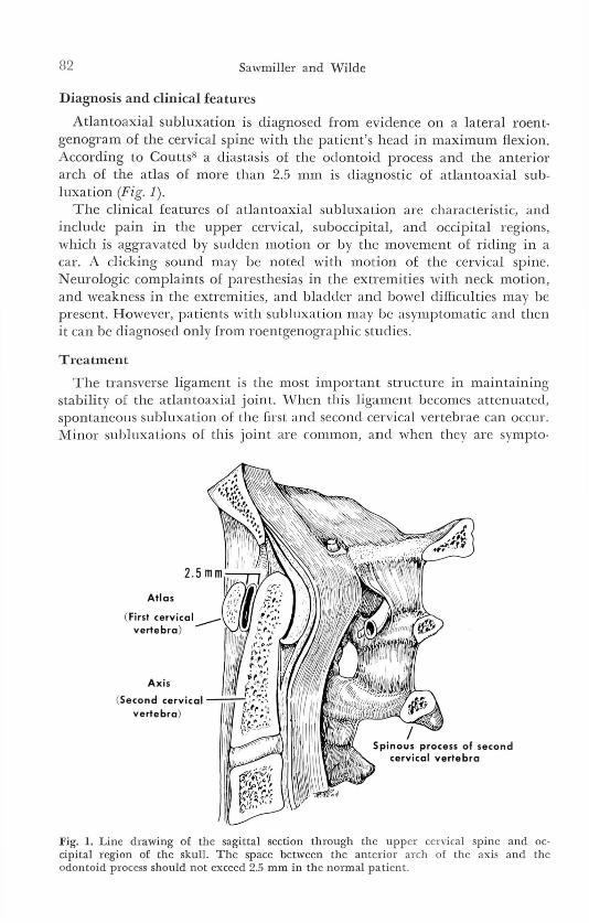
Atlantoaxial subluxation is diagnosed from evidence on a lateral roent-genogram of the cervical spine with the patient's head in maximum flexion. According to Coutts8 a diastasis of the odontoid process and the anterior arch of the atlas of more than 2.5 mm is diagnostic of atlantoaxial sub-luxation (Fig. 1).
T h e clinical features of atlantoaxial subluxation are characteristic, and include pain in the upper cervical, suboccipital, and occipital regions, which is aggravated by sudden motion or by the movement of riding in a car. A clicking sound may be noted with motion of the cervical spine. Neurologic complaints of paresthesias in the extremities with neck motion, and weakness in the extremities, and bladder and bowel difficulties may be present. However, patients with subluxation may be asymptomatic and then it can be diagnosed only from roentgenographic studies.
Trea tment
T h e transverse ligament is the most important structure in maintaining stability of the atlantoaxial joint. When this ligament becomes attenuated, spontaneous subluxation of the first and second cervical vertebrae can occur. Minor subluxations of this joint are common, and when they are sympto-
Fig. 1. Line drawing o£ the sagittal section through the upper cervical spine and oc-cipital region of the skull. The space between the anterior arch of the axis and the odontoid process should not exceed 2.5 mm in the normal patient.
A x i s
( S e c o n d c e r v
v e r t e b r a )
A t l a s
« First ce rv ie
v e r t e b r a )
S p i n o u s p r o c e s s of s e c o n d c e r v i c a l v e r t e b r a
Subluxation of cervical vertebrae in rheumatoid arthritis 83
matic can be managed with a cervical collar. Asymptomatic subluxat ions of the first a n d second cervical ver tebrae requi re only cont inued observat ion a n d per iodic roen tgen examinat ions. M a j o r symptomat ic subluxat ions , which are the subject of this paper , requi re stabilization by spinal fusion, which is desirable no t only to relieve un remi t t i ng pain, b u t to prevent f r igh ten ing episodes of pa in and instabil i ty when the sub luxa t ion is so severe tha t the pa t i en t has a feeling of impend ing doom. T h i s type of lesion is doubtless responsible for previously repor ted incidences of sudden death, and should be t reated with due respect. I t is most likely to be present in pat ients wi th severe polyart icular r heuma to id ar thr i t is in which there is extensive des t ruct ion and deformity in the per iphera l joints.
T w o pat ients wi th rheumato id ar thr i t is and spontaneous sub luxa t ion of the first and second cervical ver tebrae were surgically t reated by us; their cases are next repor ted .
R e p o r t of cases
Case 1. A 58-year-old Caucasian woman who had classical rheumatoid arthritis, stage 3, for 10 years, was first examined at the Cleveland Clinic on April 8, 1968. The onset of pain and stiffness of the neck occurred 14 years previously, after an automobile accident. She also experienced a snapping sensation in the neck, and, with her head in certain positions, "electric" tingling in the feet and legs. She had two episodes of acute pain during which she held her head with her hands to prevent any movement, as her head felt unstable. She had been treated with cervical traction, without relief, and a course of indomethacin which somewhat relieved the discomfort.
Physical examination revealed the typical findings of an inflammatory polyarthritis in the patient's extremities. There was local tenderness over a prominent spinous process of the second cervical vertebra. Rotation of the neck produced a clicking sound that was accompanied by pain. Rotation was limited to 30 degrees bilaterally, and lateral bending was only 20 degrees bilaterally. Extension was possible to 30 degrees; flexion could be accomplished to within 2 cm of the sternum. Neurologic examination revealed no sensory loss in the upper or lower extremities. The deep tendon reflexes in both upper and lower extremities were not increased; there was no ankle clonus, and the Babinski sign was absent bilaterally.
Lateral roentgenograms of the cervical spine demonstrated evidence of a 12-mm space between the odontoid process and the anterior arch of the atlas. In addition, there were erosions of the vertebral end plates of the third, fourth, fifth, and sixth cervical vertebrae typical of rheumatoid arthritis.
Because of the gross instability of the neck, and the neurologic symptoms, the patient was placed in a Halo apparatus, fixed to a shortened Minerva plaster cast. The subluxa-tion was reduced in the Halo apparatus. On April 19, 1968, posterior cervical fusion from the first to the second cervical vertebrae was performed; autogenous iliac bone was used. Postoperatively, there were no complications. Lateral roentgenograms of the cervical spine in flexion and extension 10 weeks postoperatively revealed a stable spinal fusion, and the Halo apparatus was removed. A cervical collar was applied, which the patient wore for the next four weeks.
When last examined on September 8, 1969, 17 months postoperatively, the patient had no pain in her neck. The cervical spine now felt stable and with movement of the neck she had experienced no frightening episodes of instability or feeling of "electricity" in the extremities. Examination revealed that she could flex her chin only to within 6 cm of the sternum, and extension was limited to 30 degrees. Rotation of the neck was possible to 30 degrees bilaterally. Lateral bending was limited to 20 degrees bilaterally. Results of the neurologic examination were again normal. Lateral roentgenograms of the cervical spine in flexion and extension at that time revealed a satisfactory spinal fusion from the occiput to the second cervical vertebra. The fusion had extended spontaneously to the occiput.
84 Sawmiller and Wilde
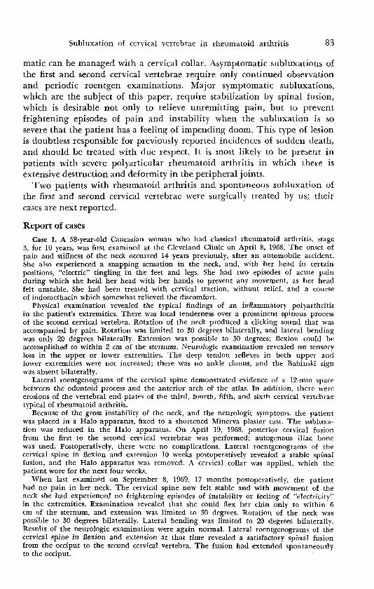
Fig. 2. Case 2. Lateral roentgenogram of the cervical spine, showing an 11-mm anterior subluxation of the atlas on the axis when the head is IL-xcd forward maximally.
Case 2. A 48-year-old Caucasian woman with classical rheumatoid arthritis, stage 4 for 24 years, was first examined at the Cleveland Clinic on May 22, 19(58, because of increasing pain in the ncck and suboccipital region, for about 6 or 7 months. A cervical collar had afforded some relief of the symptoms, but it did not give satisfactory relief of pain, which was increasing in severity. She had no feeling of instability in the neck, nor were there neurologic symptoms of pain in the arm or leg, or paresthesias. Her history was signifi-cant in that four years previously an emergency tracheostomy had been performed for a respiratory obstruction due to cricoarytenoid arthritis.
Physical examination revealed a woman with multiple deformities of her upper and lower extremities typical of rheumatoid arthritis which necessitated her being confined to a wheelchair. The head protruded forward and the spinous process of the second cervical vertebra was prominent. She was able to flex her chin to within 2 cm of her chest, and to extend her ncck 30 degrees. Rotation was 20 degrees bilaterally. There were hyperactive deep tendon reflexes in the upper and lower extremities, but no ankle clonus was present and the ISabinski sign was absent bilaterally. There was no sensory loss in the upper or lower extremities. Lateral roentgenograms of the cervical spine showed an 11-mm diastasis between the first and second cervical vertebrae, with apparent separation of the odontoid process from the body of the second cervical vertebra (Fig- 2). Reduction of the subluxation was attempted with head-halter cervical traction, but without success.
On June 14, 1908, a posterior cervical spinal fusion from the first to the third cervical vertebrae was performed. Further reduction was attempted, at the lime of surgery, bv traction of the fixating wire around the first cervical vertebra, but reduction was incom-plete. A fitted cervical brace was applied postoperatively for fixation. The postoperative course was complicated by laryngeal stridor due to the presence of cricoarytenoid ankylosis which was present previously. This was managed successfully with steam inhalation and intermittent positive-pressure breathing.
There were no further complications referable to the operation. The operation wound healed without infection. She was discharged from the hospital and was wearing a fitted cervical brace. Three months postoperatively she had no ncck pain, and lateral roentgenograms of the cervical spine in flexion and extension demonstrated a satisfactory fusion of the first and second cervical vertebrae posteriorly. The fusion mass between
Subluxation of cervical vertebrae in rheumatoid arthritis 85
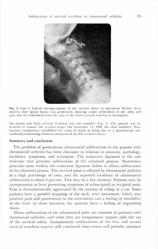
Fig. 3. Case 2. Lateral roentgenogram of the cervical spine in maximum flexion three months after spinal fusion was performed, showing sound arthrodesis of the atlas and axis, but the arthrodesis from the axis to the third cervical vertebra is incomplete.
ihe second and third cervical vertebrae was not complete (Fig. 3). The patient was in-structed to remove the ccrvical brace. On November 11, 1968, she died suddenly. Post-mortem examination established the cause of death as being due to a spontaneous sub-arachnoid hemorrhage from an aneurysm of the left cerebral artery.
Summary a n d conclusion
T h e problem of spontaneous a t lantoaxia l sub luxa t ion in the pat ient wi th rheumato id ar thr i t i s has been discussed in re la t ion to anatomy, pathology, incidence, symptoms, and t rea tment . T h e transverse l igament is the sole s t ructure that prevents subluxa t ion of the odonto id process. Destructive processes must weaken the transverse l igament before it allows subluxat ion of the odontoid process. T h e cervical spine is affected by rheumato id ar thr i t i s in a high percentage of cases, and the repor ted incidence of a t lantoaxia l sub luxa t ion is about 6 percent. T h i s may be a low estimate. Pat ients may be asymptomat ic or have present ing symptoms of suboccipital or occipital pa in . Pa in is characteristically aggravated by the mot ion of r id ing in a car. Some pat ients have a p a i n f u l snapp ing of the neck, with movement , which may produce pa in and paresthesias in the extremities and a feeling of instabil i ty of the head. I n those instances, the pat ients have a feeling of impend ing doom.
Minor subluxat ions of the a t lantoaxia l joint are common in pat ients wi th rheumato id arthri t is , and when they are symptomat ic requi re only the use of the cervical collar. Asymptomat ic subluxat ions of the first and second cervical ver tebrae requ i re only cont inued observation and periodic roentgen
86 Sawmiller and Wilde
examinat ions . Symptomatic subluxat ions of a severe degree, wh ich have been discussed here, requi re stabilization by spinal fusion. A sound sp ina l fus ion n o t only relieves the pat ient ' s p a i n a n d feeling of instabil i ty b u t protects the spinal cord f r o m serious neurologic sequelae.
Refe rences
1. Margulies, M. E.; Katz, I., and Rosenberg, M.: Spontaneous dislocation of the atlanto-axial joint in rheumatoid spondylitis; recovery from quadriplegia following surgical decompression. Neurology 5: 290-294, 1955.
2. Arnesen, A. J. A.: Cervical spondylitis with luxation and medullary compression, treated by operation. Acta Chir. Scand. 70: 104-109, 1932.
3. Ely, L. W.: Subluxation of the atlas; report of two cases. Ann. Surg. 54: 20-29, 1911.
4. Stammers, F. A. R., and Frazer, P.: Spontaneous dislocation of the atlas, with report of a case. Lancet 2: 1203-1205, 1933.
5. Berkheiser, E. J., and Seidler, F.: Nontraumatic dislocations of the atlanto-axial joint. J.A.M.A. 96: 517-523, 1931.
6. de Blecourt, J. J., and Veenstra, S. M.: Transverse lesion in a patient with juvenile rheumatoid arthritis caused by subluxation of some cervical vertebrae. Acta Rheum. Scand. 4: 251-255, 1960.
7. Grogono, B. J. S.: Injuries of the atlas and axis. J. Bone Joint Surg. 36-B: 397-410, 1954.
9. Lourie, H., and Stewart, W. A.: Spontaneous atlantoaxial dislocation. A complication of rheumatoid disease. New Eng. J. Med. 265: 677-681, 1961.
10. Cregan, J. C. F.: Internal fixation of the unstable rheumatoid cervical spine. Ann. Rheum. Dis. 25: 242-252, 1966.
11. Martel, W., and Abell, M. R.: Fatal atlanto-axial luxation in rheumatoid arthritis. Arthritis Rheum. 6: 224-231, 1963.
12. Martel, W., and Page, J. W.: Cervical vertebral erosions and subluxations in rheuma-toid arthritis and ankylosing spondylitis. Arthritis Rheum. 3: 546-556, 1960.
13. Martel, W.: Cervical spondylitis in rheumatoid disease. A comment on neurologic significance and pathogenesis. Amer. J. Med. 44: 441-446, 1968.
14. Martel, W.: T h e occipito-atlanto-axial joints in rheumatoid arthritis and ankylosing spondylitis. Amer. J. Roentgen. 86: 223-240, 1961.
15. Weiss, L. S., and Freehafer, A. A.: Atraumatic subluxation and dislocation of the cervical spine in rheumatoid arthritis. Clin. Orthop. 34: 53-61, 1964.
16. Pratt, T. L. C.: Spontaneous dislocation of the atlanto-axial articulation occurring in ankylosing spondylitis and rheumatoid arthritis. J. Fac. Radiol. 10: 40-43, 1959.
17. Sharp, J., and Purser, D. W.: Spontaneous atlanto-axial dislocation in ankylosing spondylitis and rheumatoid arthritis. Ann. Rheum. Dis. 20: 47-77, 1961.
18. Bland, J. H.: Rheumatoid arthritis of the cervical spine. Bull. Rheum. Dis. 18: 471-476, 1967.
19. Wood-Jones, F.: The ideal lesion produced by judicial hanging. Lancet 1: 53, 1913.