Material Science and Engineering with Advanced Research
Stability of Dental Implants Prototype in Bone: Thread and Design
Daniel V. Pereira1, Oliveira A3,4, Ribeiro CP4, Serafim M. Oliveira1,2*
1IPV – Polytechnic Institute of Viseu (ESTGV), Dep. de Eng. Mecânica e Gestão Industrial, Campus Politécnico, 3504-510 Viseu, Portugal2CI&DETS – Centro de Investigaçãoem Educação, Tecnologia e Saúde, Viseu - Portugal
3UCP – Universidade Católica Portuguesa do Porto43B’s –Research Group (Biomaterials, Biodegradables, Biomimetics), U. do Minho
www.verizonaonlinepublishing.com
*Corresponding author: Serafim M. Oliveira, IPV – Polytechnic Institute of Viseu (ESTGV), Dep. de Eng. Mecânica e Gestão Industrial, Campus Politécnico, 3504-510 Viseu, Portugal; Tel: +351232480500; E mail: [email protected]
Article Type: Research, Submission Date: 12 May 2018, Accepted Date: 18 May 2018, Published Date: 18 July 2018.
Citation: Daniel V. Pereira, Oliveira A, Ribeiro CP, Serafim M. Oliveira (2018) Stability of Dental Implants Prototype in Bone: Thread and Design. Mater. Sci. Eng. Adv. Res 2(4): 1-8.
Dental implants success depends of biological and biomechanics factors which includes the micro and macro design features. In this work is proposed to examine the effect of different designs on the primary stability of commercial and prototypes dental implants. All implants were screwed into posterior regions of fresh pig mandibles and both torque insertion and extraction force were characterized.
In addition to the biomechanical studies, cross-sections of mandibles at the implantation region were analyzed under microscope for bone characterization.
The increase of either diameter or length of implants drives to an increase of extraction force and the primary stability decreases with thread pitch for “V” shape threads and double thread implants presented lower stability than triple thread implants at position P1 and P2 while at positions P3 and P4 no major differences were observed.
A new design implant was created and it presented a good primary stability. Additionally, most of the results obtained shows that the macro design and bone density strongly affects the primary stability of implants corroborating the data found in literature. The new design presented in this work has a great potential to improve the insertion of the implants though it still need new tests to evaluate its performance in other bone structures.
Keywords: Biomechanics, Dental implants, Macro design, Implants thread, Bone microstructure and mechanical assays.
Introduction
Implants are usually produced from titanium, which is a biocompatible material that does not induce the formation of
fibrous tissue at the surface, allowing bone growth and creatinga stablebone-implantinterface [1,2]. However, osteointegration only begins days after implant insertion so an increase in the contact area to promote a good stability is a mandatory factor. On the other hand, the design of implants (shape, thread, diameter, length and thread profile depth) and the bone drilling process for the first fixation of implant are also crucial to promote bone remodeling and osteointegration [3]. High temperature reached during drilling will destroy bone cells therefore a reduced time in drilling process will lead to a better bone remodeling, improving stability.
After the implants in place, they should not be under masticatory forces for three to six months since micromovements affect the osteointegration [2]. In fact, micromovements of the implant can induce the formation of fibrous tissue instead of boneintegration leading to a clinical failure [4,5].
Even though, the dental implants are a useful and viable option for the treatment of edentulous patients. The success of treatment with dental implants is directly related to the various charges and the conditions to which the implant is exposed during their operating life [6]. However, the number of failures is still relevant which creates a continuous research in this area [3].
In fact, the most important factors to the implant success are: surgical technique, design, roughness, topography, chemical composition/surface biocompatibility, the bone region of reception and finally stress conditions since all them affect how to transfer load to the surrounding bone, which influence the integration of bone implants [3,7]. Therefore, engineering implants is an essential art to the study of the relationship between bone integration and mechanical properties of the dental implant.
Citation: Daniel V. Pereira, Oliveira A, Ribeiro CP, Serafim M. Oliveira (2018) Stability of Dental Implants Prototype in Bone: Thread and Design. Mater. Sci. Eng. Adv. Res 2(4): 1-8.
The concept of stability of a dental implantis subdivided in two categories: primary stability and secondary stability [8]. Primary stability is created during implant insertion in the bone at the interface receptor bone and the implant surface. In fact, primary stability is achieved at the level of the cortical bone [9,10].
The secondary stability is the result of both bone healing and bone remodeling processes undergoing on bone-implant interface at the level of the trabecular bone and it is directly dependent of the primary stability [9,11].
In terms of macro design of dental implants, the shape of the thread is an important parameter to improve stability [10]. Some studies show that the larger diameter of the implant increase the stability due to the increase of the bone-implant interface area [2]. On the other hand, larger square threads help the bone condensation, whereas an increase of the number threads stabilize implants especially at the implantation [12].
However most of results were obtained from numerical studies using analysis of the finite element method (FEA). Therefore, there is a lack of information about in vitro studies in this area pushing us into this research to evaluate the impact of geometry, shape, thread pitch, depth, width and profile angle on primary stability in natural bone.
The main purpose of this work was to test various prototypes and commercial dental implants in fresh pig jaws at the areas of molars and pre-molars. Prototype implants depend on threads used and on the design, the first design uses two perpendicular holes (TPH) transverse to the axe of the implant in order to improve the bone anchorage, promoting a better secondary stability and the second design is a self-screwing (SS) implant which easily screws into trabecular bone.
The final goal is to design a new type of implant with optimized characteristics that result in a good biomechanical behavior and successfully perform their duties. With this study we were able to obtain invitro results for force extraction of implants and, at
same time, we demonstrated that the new design is suited to be inserted in the bone using only a small drilled hole on the cortical bone. Moreover, this SS implants presented higher extractions forces than commercial implants when used in low density bone.
Methods
The equipment, materials and procedures used to analyze different types of implants performed in the pre-molars and molars bone area of pig jaws are presented here.
Dental implants
As dental implants, we tested several types of metric screws (M3, M4 and M5) with different ratio diameter/length. The implants tested vary in terms of shape, diameter, length, pitch and number of threads. Besides, in this work were tested square thread implants, commercial implants and it was also designed, manufactured and tested implants (TPH and SS implants) not available at the market, new designs implants, Table 1.
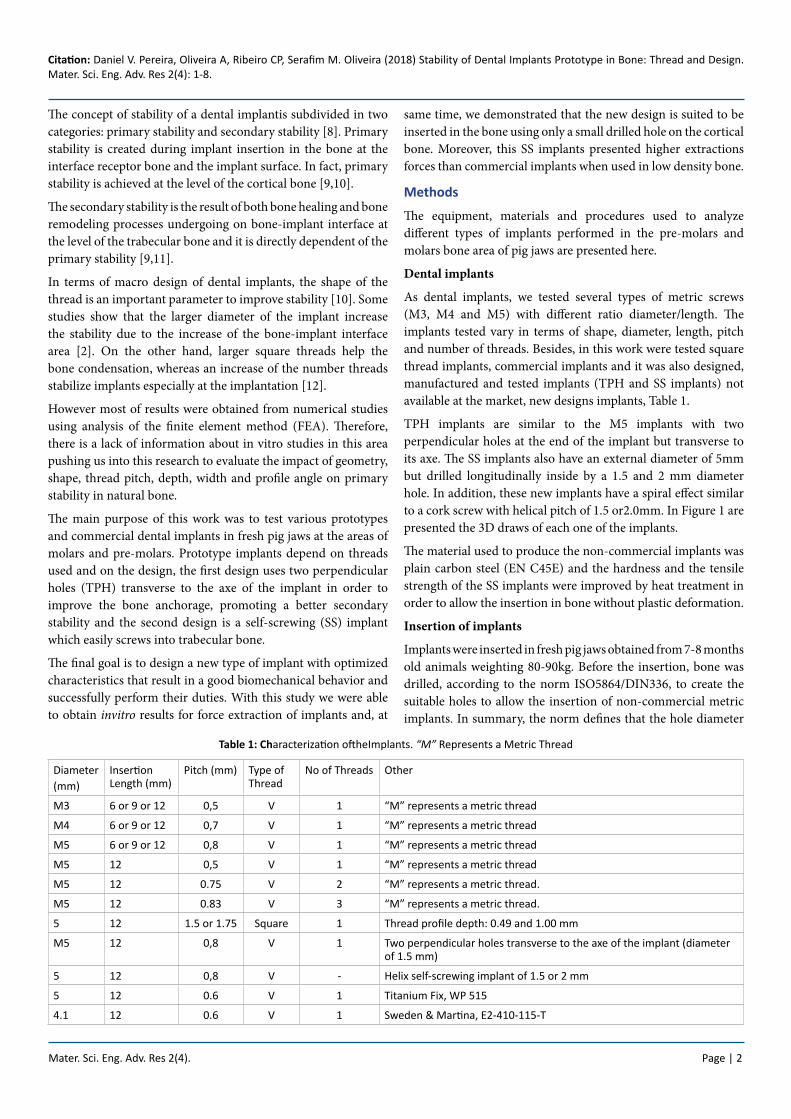
TPH implants are similar to the M5 implants with two perpendicular holes at the end of the implant but transverse to its axe. The SS implants also have an external diameter of 5mm but drilled longitudinally inside by a 1.5 and 2 mm diameter hole. In addition, these new implants have a spiral effect similar to a cork screw with helical pitch of 1.5 or2.0mm. In Figure 1 are presented the 3D draws of each one of the implants.
The material used to produce the non-commercial implants was plain carbon steel (EN C45E) and the hardness and the tensile strength of the SS implants were improved by heat treatment in order to allow the insertion in bone without plastic deformation.
Insertion of implants
Implants were inserted in fresh pig jaws obtained from 7-8 months old animals weighting 80-90kg. Before the insertion, bone was drilled, according to the norm ISO5864/DIN336, to create the suitable holes to allow the insertion of non-commercial metric implants. In summary, the norm defines that the hole diameter
Table 1: Characterization oftheImplants. “M” Represents a Metric Thread
Diameter(mm)
Insertion Length (mm)
Pitch (mm) Type of Thread
No of Threads Other
M3 6 or 9 or 12 0,5 V 1 “M” represents a metric thread
M4 6 or 9 or 12 0,7 V 1 “M” represents a metric thread
M5 6 or 9 or 12 0,8 V 1 “M” represents a metric thread
M5 12 0,5 V 1 “M” represents a metric thread
M5 12 0.75 V 2 “M” represents a metric thread.
M5 12 0.83 V 3 “M” represents a metric thread.
5 12 1.5 or 1.75 Square 1 Thread profile depth: 0.49 and 1.00 mm
M5 12 0,8 V 1 Two perpendicular holes transverse to the axe of the implant (diameter of 1.5 mm)
5 12 0,8 V - Helix self-screwing implant of 1.5 or 2 mm
5 12 0.6 V 1 Titanium Fix, WP 515
4.1 12 0.6 V 1 Sweden & Martina, E2-410-115-T
Mater. Sci. Eng. Adv. Res 2(4). Page | 3
Citation: Daniel V. Pereira, Oliveira A, Ribeiro CP, Serafim M. Oliveira (2018) Stability of Dental Implants Prototype in Bone: Thread and Design. Mater. Sci. Eng. Adv. Res 2(4): 1-8.
corresponds to the diameter of the implantless thepitch. For the SS implants, it was followed the same protocol, however, the hole was only 4 mm deep in the cortical bone. For the square threaded implants, it was used holes of 3.7 and 3 mm in diameter for the thread profile depth of 0.49 and1.00 mm, respectively.
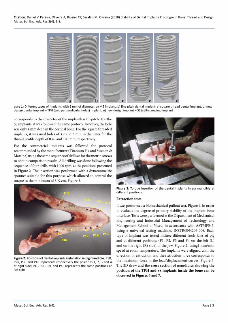
For the commercial implants was followed the protocol recommended by the manufacturer (Titanium Fix and Sweden & Martina) using the same sequence of drills as for the metric screws to obtain comparison results. All drilling was done following the sequence of four drills, with 1000 rpm, at the positions presented in Figure 2. The insertion was performed with a dynamometric spanner suitable for this purpose which allowed to control the torque to the minimum of 5 N.cm, Figure 3.
Figure 2: Positions of dental implants installation in pig mandible. P1R, P2R, P3R and P4R represents respectively the positions 1, 2, 3 and 4 at right side; P1L, P2L, P3L and P4L represents the same positions at left side
Figure 3: Torque insertion of the dental implants in pig mandible at different positions
Extraction tests
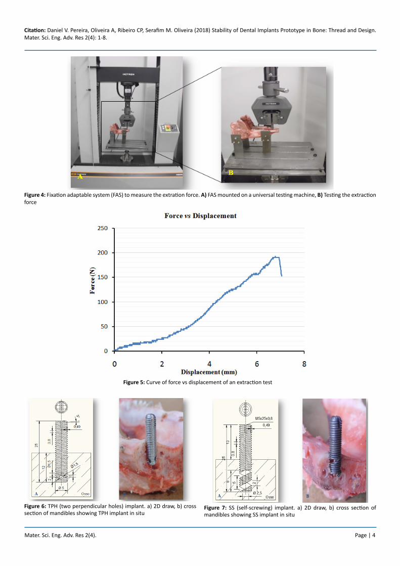
It was performed a biomechanical pullout test, Figure 4, in order to evaluate the degree of primary stability of the implant-bone interface. Tests were performed at the Department of Mechanical Engineering and Industrial Management of Technology and Management School of Viseu, in accordance with ASTMF543, using a universal testing machine, INSTRON4206-006. Each type of implant was tested inthree different fresh jaws of pig and at different positions (P1, P2, P3 and P4 on the left (L) and on the right (R) side) of the jaw, Figure 2, using1 mm/min speed at room temperature. The implants were aligned with the direction of extraction and thee xtraction force corresponds to the maximum force of the load/displacement curves, Figure 5. The 2D draw and the cross section of mandibles showing the position of the TPH and SS implants inside the bone can be observed in Figures 6 and 7.
gure 1: Different types of implants with 5 mm of diameter. a) M5 implant, b) fine pitch dental implant, c) square thread dental implant, d) new design dental implant – TPH (two perpendicular holes) implant, e) new design implant – SS (self-screwing) implant
Mater. Sci. Eng. Adv. Res 2(4). Page | 4
Citation: Daniel V. Pereira, Oliveira A, Ribeiro CP, Serafim M. Oliveira (2018) Stability of Dental Implants Prototype in Bone: Thread and Design. Mater. Sci. Eng. Adv. Res 2(4): 1-8.
Figure 4: Fixation adaptable system (FAS) to measure the extration force. A) FAS mounted on a universal testing machine, B) Testing the extraction force
Figure 5: Curve of force vs displacement of an extraction test
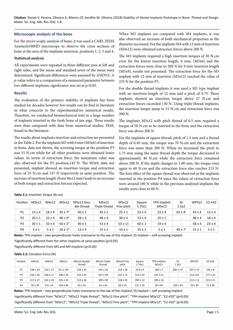
Figure 6: TPH (two perpendicular holes) implant. a) 2D draw, b) cross section of mandibles showing TPH implant in situ
Figure 7: SS (self-screwing) implant. a) 2D draw, b) cross section of mandibles showing SS implant in situ
Mater. Sci. Eng. Adv. Res 2(4). Page | 5
Citation: Daniel V. Pereira, Oliveira A, Ribeiro CP, Serafim M. Oliveira (2018) Stability of Dental Implants Prototype in Bone: Thread and Design. Mater. Sci. Eng. Adv. Res 2(4): 1-8.
Microscopic analysis of the bone
For the micro scopic analysis of bone, it was used a CARL ZEISS Axiotech100HD microscope to observe the cross sections of bone at the area of the implants insertion, positions 1, 2, 3 and 4.
Statistical analysis
All experiments were repeated in three different jaws at left and right sides, and the mean and standard error of the mean were determined. Significant differences were assessed by ANOVA. A p-value refers to a comparison of a measured parameter between two different implants; significance was set at p<0.05.
Results
The evaluation of the primary stability of implants has been studied for decades however few results can be find in literature in what concerns to the experimental/no numerical results. Therefore, we conducted biomechanical tests in a large number of implants inserted in the fresh bone of jaw pigs. These results were than compared with data from numerical studies, FEM, found in the literature.
The results about implants insertion and extraction are presented in the Table 2. For the implants M3 with 6 mm (M3x6) of insertion in bone, data not shown, the screwing torque at the position P3 was 15 N.cm while for all other positions were obtained lower values. In terms of extraction force, the maximum value was also observed for the P3 position,143 N. The M3x9, data not presented, implant showed an insertion torque and extraction force of 25 N.cm and 147 N respectively at same position. The increase of insertion length (from 9to12 mm) leads to an increase of both torque and extraction forceas expected.
When M3 implants are compared with M4 implants, it was also observed an increase of both mechanical properties as the diameter increased. For the implants M4 with 12 mm of insertion (M4x12) were obtained extraction forces above 200 N.
The M5 implants required a high insertion torques of 30 N.cm even for the lowest insertion length, 6 mm, (M5x6) and the extraction forces were close to 200 N for 9 mm insertion length (M5x9), results not presented. The extraction force for the M5 implant with 12 mm of insertion (M5x12) reached the value of 235 N for the position P3.
For the double thread implants it was used a M5 type implant with an insertion length of 12 mm and a pitch of 0.75. These implants showed an insertion torque above 27 N.cm and extraction forces exceeded 130 N. Using triple thread implants, the insertion torque jump to 53 N.cm and extraction force was 200 N.
The implants M5x12 with pitch thread of 0.5 mm required a torque of 50 N.cm to be inserted in the bone and the extraction force was above 200 N.
For the implants of square thread, pitch of 1.5 mm and a thread depth of 0.49 mm, the torque was 70 N.cm and the extraction force was more than 200 N. When we increased the pitch to 1.75 mm using the same thread depth the torque decreased to approximately 40 N.cm while the extraction force remained above 200 N. If the depth changes to 1.00 mm, the torque rises again to 60 N.cm and the extraction force also reaches 215 N. The best effect of the square thread was observed at the implants inserted in the position P4 since the values of extraction force were around 100 N while in the previous analyzed implants the results were close to 60 N.
Notes: TPH implant – two perpendicular holes transverse to the axe of the implant; SS implant – self-screwing implantaSignificantly different from the other implants at same position (p<0.05)bSignificantly different from M3 and M4 implants (p<0.05)
Notes: TPH implant – two perpendicular holes transverse to the axe of the implant; SS implant – self-screwing implantasignificantly different from “M3x12”, “M5x12 Triple thread”, “M5x12 Fine pitch”, “TPH implant M5x12”, “E2-410” (p<0.05)bsignificantly different from “M3x12”, “M5x12 Triple thread”, “M5x12 Fine pitch”, “TPH implant M5x12”, “E2-410” (p<0.05)
Mater. Sci. Eng. Adv. Res 2(4). Page | 6
Citation: Daniel V. Pereira, Oliveira A, Ribeiro CP, Serafim M. Oliveira (2018) Stability of Dental Implants Prototype in Bone: Thread and Design. Mater. Sci. Eng. Adv. Res 2(4): 1-8.
In order to create new implants able to increase the osteointegration and showing good results in terms of primary stability we used theTPH implants, Figure 6, and SS implants, Figure 7. Both types of implants were totally designed and produced for this study and results show that TPH implants led to as light decrease in torque and extraction force when compared with M5x12. On the other side, the SS implants which purpose is also to increase the osteointegration are based on a spiral shape similar to a corkscrew in order to reduce the depth of the drilled hole in the bone required an insertion torque of about 60 N.cm to be inserted at position P1 and about 43 N.cm at position 4 which is explained by the self-screwing effect of these type of implants. As result, the values of extraction forces at position P1 were similar to the values obtained for M5x12, around 200 N, while at position P4 it was observed a slight increase.
The commercial implants, WP515, allowed to obtain mean values greater than 55 N.cm and 200 N of insertion torque and maximum extraction force respectively, while the commercial implants, E2-410-115-T, require insertion torques and extraction forces slightly lower.
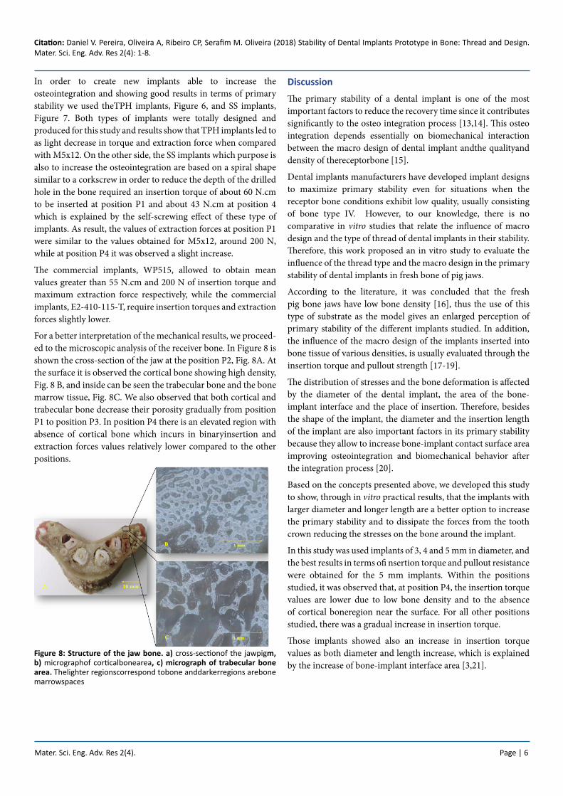
For a better interpretation of the mechanical results, we proceed-ed to the microscopic analysis of the receiver bone. In Figure 8 is shown the cross-section of the jaw at the position P2, Fig. 8A. At the surface it is observed the cortical bone showing high density, Fig. 8 B, and inside can be seen the trabecular bone and the bone marrow tissue, Fig. 8C. We also observed that both cortical and trabecular bone decrease their porosity gradually from position P1 to position P3. In position P4 there is an elevated region with absence of cortical bone which incurs in binaryinsertion and extraction forces values relatively lower compared to the other positions.
Figure 8: Structure of the jaw bone. a) cross-sectionof the jawpigm, b) micrographof corticalbonearea, c) micrograph of trabecular bone area. Thelighter regionscorrespond tobone anddarkerregions arebone marrowspaces
Discussion
The primary stability of a dental implant is one of the most important factors to reduce the recovery time since it contributes significantly to the osteo integration process [13,14]. This osteo integration depends essentially on biomechanical interaction between the macro design of dental implant andthe qualityand density of thereceptorbone [15].
Dental implants manufacturers have developed implant designs to maximize primary stability even for situations when the receptor bone conditions exhibit low quality, usually consisting of bone type IV. However, to our knowledge, there is no comparative in vitro studies that relate the influence of macro design and the type of thread of dental implants in their stability.Therefore, this work proposed an in vitro study to evaluate the influence of the thread type and the macro design in the primary stability of dental implants in fresh bone of pig jaws.
According to the literature, it was concluded that the fresh pig bone jaws have low bone density [16], thus the use of this type of substrate as the model gives an enlarged perception of primary stability of the different implants studied. In addition, the influence of the macro design of the implants inserted into bone tissue of various densities, is usually evaluated through the insertion torque and pullout strength [17-19].
The distribution of stresses and the bone deformation is affected by the diameter of the dental implant, the area of the bone-implant interface and the place of insertion. Therefore, besides the shape of the implant, the diameter and the insertion length of the implant are also important factors in its primary stability because they allow to increase bone-implant contact surface area improving osteointegration and biomechanical behavior after the integration process [20].
Based on the concepts presented above, we developed this study to show, through in vitro practical results, that the implants with larger diameter and longer length are a better option to increase the primary stability and to dissipate the forces from the tooth crown reducing the stresses on the bone around the implant.
In this study was used implants of 3, 4 and 5 mm in diameter, and the best results in terms ofi nsertion torque and pullout resistance were obtained for the 5 mm implants. Within the positions studied, it was observed that, at position P4, the insertion torque values are lower due to low bone density and to the absence of cortical boneregion near the surface. For all other positions studied, there was a gradual increase in insertion torque.
Those implants showed also an increase in insertion torque values as both diameter and length increase, which is explained by the increase of bone-implant interface area [3,21].
Mater. Sci. Eng. Adv. Res 2(4). Page | 7
Citation: Daniel V. Pereira, Oliveira A, Ribeiro CP, Serafim M. Oliveira (2018) Stability of Dental Implants Prototype in Bone: Thread and Design. Mater. Sci. Eng. Adv. Res 2(4): 1-8.
The increase of the insertion length has also increase the insertion torque and pullout force though this phenomenon does not happen for all positions. The shape, depth and width of the dental implant thread are very important factors which affect the primary stability and influences the type offorces that are transferred to the receptor bone. To explore those factors there is in the market different thread types: V, square, trapezoidal, inverted trapezoidal and spiral [3].
We concluded that the increase of the profile depth in implant with square thread allows to decrease the diameter of the hole in the bone increasing the primary stability at all positions tested. Moreover, the increase of the thread width of square thread implants, from 0.3 to 0.5 mm, increases the extraction force (data not shown).
Another important parameter in the macro design of dental implants is the concept of multiple threads. Some manufacturers have introduced multi-threaded implants of two or three entries, allowing to increase the speed of insertion of the implant. Either in terms of insertion torque or extraction force we cannot claim that there is a significant increase or decrease related with the number of entries. Moreover, there is a slight increase in pullout force from position P1 to position P2 and a stagnation at position P3. However, the largest number of entries enables a higher speed of insertion of the implant due to the bigger fillet angle.
The development of new designs is constantly focus on the improvement of the stability of dental implants. It is known that primary stability depends strongly of implants geometric, however, the enhancement of secondary stability in obtained from biological interactions at the bone-implant interface. Therefore, the creation of implants that allow a good primary stability and at the same time, can improve the biological interactions at the bone-implant interface, will lead to a faster correction of tooth structure.
Thus, it was developed the TPH implants to improve the osteointegration using an osteoinductive material inside the holes. As expected, the insertion torque and extration force for these implants dropped slightly due to the decrease of the contact area in about 7 square mm.
In opposition, for the SS implants was observed a slight increase in insertion torque and extration force since the lengthof the initial hole in the bone was decreased, leading there by to an additional force of implant insertion and to a better primary stability. Another advantage of these implantsis the expected improvement of secondary stability since the bone-implant interface is increased.
The results of this study are in agreement with the literature, which indicates that different bone-implant contact are as affect the primary stability, and this is influenced by several macro design factors of dental implants [22,23].
In literature, some studies have been conducted inorder to relate the insertion torque with extration force though some authors observed a direct relation [24,25]others did not [18,19]. In our study it was observed that implants with greater insertion torque also presented higher extraction force values.
Regarding the microscopic analysis of the bone cross-section, it was observed a gradual decrease of both cortical and trabecular bone porosity from position P1 to P3. At position P4, there is only trabecular bone with a medium level of porosity. Those microscopic analyses can explain the low insertion torque and extraction force at position P4.
Conclusions
Different areas of contact between the bone-implant interface, affect the primary stability of dental implants, and this is influenced by the macro design factors studied.
These results allow to predict the primary stability of dental implants but do not allow to establish if implants will succeed in terms of osteointegration/secondary stability. However, TPH and SS implants present new features which increases the possibility of a faster osteointegration, either because there is the possibility to use a osteoinductive material in the holes or because there is an increase of bone-implant interface area and a lower trabecular bone destruction during implantation.
Ohlsson A. Intra-osseous anchorage of dental prostheses. I. Experimental Studies. Scand J Plast Reconstr Surg. 1969; 3(2):81-100. doi:10.3109/02844316909036699.
2. Rokn AR,Rasouli Ghahroudi AAR, Mesgarzadeh A,Miremadi AA, Yaghoobi S. Evaluation of stabilty changes in tapered and parallel wall implants: a human clinical trial. J Dent (Tehran). 2011; 8(4):186–200.
3. Abuhussein H, Pagni G, Rebaudi A, Wang HL. The effect of thread pattern upon implant osseointegration.The effect of thread pattern upon implant osseointegration. Clin Oral Implants Res. 2010; 21(2):129-136. doi:10.1111/j.1600-0501.2009.01800.x
4. Adell R, Lekholm U, Rockler B, Brånemark PI. A 15-year study of osseointegrated implants in the treatment of the edentulous jaw. Int J Oral Surg. 1981; 10(6):387-416. doi:10.1016/S0300-9785(81)80077-4.
5. Skalak R, Zhao Y. Interaction of force-fitting and surface roughness of implants. Clin Implant Dent Relat Res. 2002; 2(4):219-224. doi:10.1016/S0300-9785(81)80077-4.
6. Prado CJ, Neves FD, Soares CJ, Dantas KA, Dantas TS, Naves LZ. Influence of abutment screw design and surface coating on bending flexural strength of implant set. J Oral Implantol.2014; 40(2):123-128. doi:10.1563/AAID-JOI-D-11-00116.
7. Geng JP, Ma QS, Xu W, Tan KB, Liu GR. Finite element analysis of four thread-form configurations in a stepped screw implant. J Oral Rehabil. 2004; 31(3):233-239. doi:10.1046/j.0305-182X.2003.01213.x.
Citation: Daniel V. Pereira, Oliveira A, Ribeiro CP, Serafim M. Oliveira (2018) Stability of Dental Implants Prototype in Bone: Thread and Design. Mater. Sci. Eng. Adv. Res 2(4): 1-8.
8. Cho IH, Lee YI, Kim YM. A comparative study on the accuracy of the devices for measuring the implant stability. J Adv Prosthodont. 2009; 1(3):124-128. doi:10.4047/jap.2009.1.3.124.
9. Atsumi M, Park SH, Wang HL. Methods used to assess implant stability: current status. Int J Oral Maxillofac Implants. 2007; 22(5):743-754. doi:10.4103/0972-4052.176539.
10. Trisi P, Perfetti G, Baldoni E, Berardi D, Colagiovanni M, Scogna G. Implant micromotion is related to peak insertion torque and bone density. Clin Oral Implants Res. 2009; 20(5):467-471. doi: 10.1111/j.1600-0501.2008.01679.x.
12. Koticha T, Fu JH, Chan HL, Wang HL. Influence of Thread Design on Implant Positioning in Immediate Placement. J Periodontol. 2012;83(11):1420-1422. doi:10.1902/jop.2012.110665.
13. Bischof M, Nedir R, Szmukler-Moncler S, Bernard JP, Samson J. Implant stability measurement of delayed and immediately loaded implants during healing. Clin Oral Implants Res. 2004; 15(5):529-539. doi:10.1111/j.1600-0501.
14. Martinez H, Davarpanah M, Missika P, Celletti R, Lazzara R. Optimal implant stabilization in low density bone. Clin Oral Implants Res. 2001; 12(5):423-432.doi:10.1034/j.1600-0501.2001.120501.x.
15. Chong L, Khocht A, Suzuki JB, Gaughan J. Effect of implant design on initial stability of tapered implants. J Oral Implantol. 2009; 35(3):130-135. doi: 10.1563/1548-1336-35.3.130.
16. Dantas C, R. D. Evaluation of primary stability of two types of im-plants installed in standardized porcine bone models, using biome-chanical tests and Micro-CT. An in vitro pilot study. 2012:80-101.
17. Hsu CC, Chao CK, Wang JL, Hou SM, Tsai YT, Lin J. Increase of pullout strength of spinal pedicle screws with conical core: biomechanical tests and finite element analyses. J Orthop Res; 23(4):788-794. doi:10.1016/j.orthres.2004.11.002.
18. Inceoglu S, Ferrara L, McLain RF. Pedicle screw fixation strength: pullout versus insertional torque. Spine J. 2004; 4(5):513-518. doi:10.1016/j.spinee.2004.02.006.
19. Lawson KJ, Brems J. Effect of insertion torque on bone screw pullout strength. Orthopedics. 2001; 24(5):451-454.
20. Cochran DL. A comparison of endosseous dental implant surfaces. J Periodontol. 1999; 70(12):1523-1539.. doi:10.1902/jop.1999.70.12.1523.
21. Baggi L, Cappelloni I, Di Girolamo M, Maceri F, Vairo G. The influence of implant diameter and length on stress distribution of osseointegrated implants related to crestal bone geometry: A three-dimensional finite element analysis. J Prosthet Dent. 2008; 100(6):422-431. doi: 10.1016/S0022-3913(08)60259-0.
22. Kim SJ, Kim MR, Rim JS, Chung SM, Shin SW. Comparison of implant stability after different implant surface treatments in dog bone. J Appl Oral Sci. 2010; 18(4):415-420. doi:10.1590/S1678-77572010000400016.
23. Lan TH, Du JK, Pan CY, Lee HE, Chung WH. Biomechanical analysis of alveolar bone stress around implants with different thread designs and pitches in the mandibular molar area. Clin Oral Investig. 2012; 16(2):363-369. doi: 10.1007/s00784-011-0517-z.
24. Boyle JM 3rd, Frost DE, Foley WL, Grady JJ. (1993a). Comparison between uniaxial pull-out tests and torque measurement of 2.0 mm self-tapping screws. Int J Adult Orthodon Orthognath Surg. 1993; 8(2):129-133.
25. Zdeblick TA, Kunz DN, Cooke ME, McCabe R. Pedicle screw pullout strength. Correlation with insertional torque. Spine (Phila Pa 1976). 1993; 18(12):1673-1676.