Gut, 1964, 5, 309 Steatorrhoea in rats with an intestinal cul-de-sac P. P. HOET1 AND H. EYSSEN From the Rega Institute for Medical Research, University of Louvain, Louvain, Belgium EDITORIAL SUMMARY Steatorrhoea in rats with an intestinal cul-de-sac is mainly due to malabsorption of alimentary fats but faecal lipids of endogenous origin are also increased. Steatorrhoea depends on the site of the blind loop in the small intestine and is mainly caused by bacterial proliferation in the lumen of the gut. The aetiological role of Gram-positive anaerobic microbes, especially Clostridium welchii, is suggested. Malabsorption of fat is a prominent feature in patients with anatomical lesions of the small intestine (Badenoch, 1960), as in strictures, multiple diverticulosis (Cooke, Cox, Fone, Meynell, and Gaddie, 1963), entero-enteric fistulas, and surgically produced blind loops (Goldstein, Wirts, and Kramer, 1961). Malabsorption of vitamin B12 occurs inde- pendently or in association with steatorrhoea and can lead to a megaloblastic anaemia (Gellman, 1956). The structural abnormalities of the intestine in the blind loop syndrome cause stasis and microbial proliferation in the lumen of the gut. In patients suffering from blind loop syndrome, high bacterial counts were found in aspirated material from the afferent loop in contrast to the transient flora of a normal small intestine (Wirts and Goldstein, 1963). Further evidence of the bacterial aetiology is provided bythe therapeutic effect of broad-spectrum antibiotics or surgical correction of the structural defect. Surgically formed small intestinal cul-de-sacs in rats and in dogs provided an experimental approach to the malabsorption of vitamin B12 and showed the role of bacterial proliferation in this condition (Cameron, Watson, and Witts, 1949). The increased faecal fat excretion in these animals was recognized (Aitken, Badenoch, and Spray, 1950). The purpose of this investigation was to study the fat absorption and excretion in rats with surgically produced blind loops. The effect of different antibiotics on fat excretion was correlated with bacteriological findings in order to identify the organism or organisms responsible for this experi- mental steatorrhoea. MATERIALS AND METHODS EXPERIMENTAL ANIMALS AND DIETS Male rats, each weighing 150-200 g., from a stock colony were used. 'Aspirant aan het Nationaal Fonds voor Wetenschappelijk Onderzoek (N.F.W.O.). A cul-de-sac was produced surgically on the small intestine under ether anaesthesia, according to the method of Cameron, Watson, and Witts (1950). This self-filling loop, 7 to 8 cm. long, became dilated by the action of peristalsis on the intestinal contents. Most of the animals supported the operation well and lived for more than 15 months. Operated and control animals were kept in individual cages with a wire screen bottom in order to reduce coprophagy and to facilitate collection of faeces. Adaptation to the diet was allowed for at least five days before faeces were collected. The basal com- position of the diets is given in Table I. TABLE I BASAL COMPOSITION OF DIETS Sucrose Casein Vitamins and choline Mineral salts Chromium oxide 705 25-0 1-3 3.0 0-2 To obtain a fat-containing diet, 9 % com-oil (w/w) was added to the basic diet, at the expense of sucrose. In some experiments, animal feed doses of antibiotics (200 parts per million) were added to the 9 % corn-oil diet. Chromium oxide was mixed with all diets and the amount determined in the faeces served as a measure of the amount of food ingested per gram faeces produced. Daily food intake of controls and of rats with a proximal blind loop was also determined, and the total faecal output per 24 hours was calculated. ANALYTICAL METHODS Faeces were dried in vacuo over phosphorus pentoxide. Free fatty acids and total fatty acids were measured in faeces according to the method of van de Kamer, ten Bokkel Huinink, and Weyers (1949). The petroleum ether extract was titrated with 0-1 N NaOH, an alcohol-water solution of thymol-blue being used as indicator, and the two phases mixed by bubbling N, (Dole and Meinertz, 1960). Free and esterified fatty acids were analysed by liquid gas chromatography after separate extraction (Evrard, 309 on 13 September 2018 by guest. Protected by copyright. http://gut.bmj.com/ Gut: first published as 10.1136/gut.5.4.309 on 1 August 1964. Downloaded from
Transcript
Gut, 1964, 5, 309
Steatorrhoea in rats with an intestinal cul-de-sacP. P. HOET1 AND H. EYSSEN
From the Rega Institute for Medical Research, University ofLouvain, Louvain, Belgium
EDITORIAL SUMMARY Steatorrhoea in rats with an intestinal cul-de-sac is mainly due to malabsorptionof alimentary fats but faecal lipids of endogenous origin are also increased. Steatorrhoea depends on
the site of the blind loop in the small intestine and is mainly caused by bacterial proliferation inthe lumen of the gut. The aetiological role of Gram-positive anaerobic microbes, especiallyClostridium welchii, is suggested.
Malabsorption of fat is a prominent feature inpatients with anatomical lesions of the smallintestine (Badenoch, 1960), as in strictures, multiplediverticulosis (Cooke, Cox, Fone, Meynell, andGaddie, 1963), entero-enteric fistulas, and surgicallyproduced blind loops (Goldstein, Wirts, and Kramer,1961). Malabsorption of vitamin B12 occurs inde-pendently or in association with steatorrhoea andcan lead to a megaloblastic anaemia (Gellman, 1956).The structural abnormalities of the intestine in the
blind loop syndrome cause stasis and microbialproliferation in the lumen of the gut. In patientssuffering from blind loop syndrome, high bacterialcounts were found in aspirated material from theafferent loop in contrast to the transient flora of anormal small intestine (Wirts and Goldstein, 1963).Further evidence ofthe bacterial aetiology is providedbythe therapeutic effect of broad-spectrum antibioticsor surgical correction of the structural defect.
Surgically formed small intestinal cul-de-sacs inrats and in dogs provided an experimental approachto the malabsorption of vitamin B12 and showed therole of bacterial proliferation in this condition(Cameron, Watson, and Witts, 1949). The increasedfaecal fat excretion in these animals was recognized(Aitken, Badenoch, and Spray, 1950).The purpose of this investigation was to study
the fat absorption and excretion in rats withsurgically produced blind loops. The effect ofdifferent antibiotics on fat excretion was correlatedwith bacteriological findings in order to identify theorganism or organisms responsible for this experi-mental steatorrhoea.
MATERIALS AND METHODS
EXPERIMENTAL ANIMALS AND DIETS Male rats, eachweighing 150-200 g., from a stock colony were used.
'Aspirant aan het Nationaal Fonds voor Wetenschappelijk Onderzoek(N.F.W.O.).
A cul-de-sac was produced surgically on the smallintestine under ether anaesthesia, according to themethod of Cameron, Watson, and Witts (1950). Thisself-filling loop, 7 to 8 cm. long, became dilated by theaction of peristalsis on the intestinal contents. Most ofthe animals supported the operation well and lived formore than 15 months. Operated and control animalswere kept in individual cages with a wire screen bottomin order to reduce coprophagy and to facilitate collectionof faeces. Adaptation to the diet was allowed for at leastfive days before faeces were collected. The basal com-position of the diets is given in Table I.
TABLE IBASAL COMPOSITION OF DIETS
SucroseCaseinVitamins and cholineMineral saltsChromium oxide
70525-01-33.00-2
To obtain a fat-containing diet, 9% com-oil (w/w) wasadded to the basic diet, at the expense of sucrose. Insome experiments, animal feed doses of antibiotics(200 parts per million) were added to the 9% corn-oil diet.Chromium oxide was mixed with all diets and the amountdetermined in the faeces served as a measure of theamount of food ingested per gram faeces produced.Daily food intake of controls and of rats with a proximalblind loop was also determined, and the total faecaloutput per 24 hours was calculated.
ANALYTICAL METHODS Faeces were dried in vacuo overphosphorus pentoxide. Free fatty acids and total fattyacids were measured in faeces according to the methodof van de Kamer, ten Bokkel Huinink, and Weyers (1949).The petroleum ether extract was titrated with 0-1 NNaOH, an alcohol-water solution of thymol-blue beingused as indicator, and the two phases mixed by bubblingN, (Dole and Meinertz, 1960).
Free and esterified fatty acids were analysed by liquidgas chromatography after separate extraction (Evrard,
309
on 13 Septem
ber 2018 by guest. Protected by copyright.
http://gut.bmj.com
/G
ut: first published as 10.1136/gut.5.4.309 on 1 August 1964. D
Hoet, Eyssen, Charlier, and Sacquet, 1964). Chromiumoxide was measured in faeces according to the method ofEdwards and Gillis (1959).
BACTERIOLOGICAL METHODS The abdomen was openedunder ether anaesthesia, and the blind loop was removed.The wall of the loop was incised, and the liquid contentwas collected. After application of Gram's stain to thismaterial, successive dilutions were made with sterilesaline.
Different media were inoculated with the final dilutions:pour plates of 10 ml. brain heart infusion agar (B.B.L.)with 0.5% yeast extract and 10% bovine blood; S.F.medium (B.B.L.), for the detection of faecal streptococci(enterococci); Tergitol 7 agar (Difco) for members ofthe coliform groups; Rogosa SL broth (Difco), aselective medium for the cultivation of lactobacilli;reinforced clostridial agar (Oxoid) for anaerobes,especially of the Clostridium species; brain heart infusionagar (B.B.L.) with 0.5% yeast extract and 0.5% Na-thioglycolate for total anaerobic counts. After 24 to 27hours' growth on these media, colonies were enumerated,examined after being stained by Gram's method, andeventually isolated.
HISTOLOGICAL METHODS Fragments of the blind loop,stomach, jejunum, and ileum were fixed either in formol-saline or in Bouin. After paraffin embedding the followingstaining methods were routinely applied: haematoxylin,trichrome Masson, and Gram's stain. Several sectionswere stained by the silver impregnation method ofTibor-Pap and by the P.A.S. stain.
RESULTS
THE EXPERIMENTAL STEATORRHOEA In three groupsof rats an intestinal cul-de-sac was producedrespectively on the proximal, middle, or distal partof the small intestine. The rats were fed a 9% corn-oildiet, and faecal fat was determined one and fiveweeks after operation. As shown in Table II, thesteatorrhoea was maximal when the cul-de-sac wason the proximal part of the intestine, whereas thefat excretion of animals with a distal cul-de-sac wassimilar to that in normal rats.
In further experiments, only rats with proximalblind loops were used. In these animals the steator-rhoea developed soon after operation, as shown by thefigures for faecal fat on the seventh post-operativeday and by the increase in faecal fat between thefirst and fifth week after operation. The differencebetween control and operated animals was evenmore pronounced when results were expressed asdaily faecal fat excretion rather than as fat contentper gram faeces.To compare the absorption of alimentary fat in
control and blind loop rats, the animals were fed afat-free or a 9% corn-oil diet, and free fatty acids
TABLE IIFAECAL OUTPUT IN RATS WITH A BLIND LOOP AT THE
PROXIMAL, MIDDLE, OR DISTAL PART OF THE SMALLINTESTINE
Animals Fat (mg.lg. faeces)± S.D. (N)
Control 87+22 (8) 76+ 12
Blind loop One Week Five WeeksPost- Post-operatively operatively
'Values determined between one and five weeks after operation.
and total fatty acids were determined in faeces. Theresults are given in Figure 1.
Fat excretion in control animals was relativelyindependent of fat intake, whereas in blind looprats it increased proportionally to alimentary fat,indicating a malabsorption. The faecal lipids of non-alimentary origin were also increased in blind looprats, since even on a fat-free diet they excreted 30%more fat than did control animals.On a corn-oil diet, the proportion of faecal free
and esterified fatty acids was the same in control andblind loop rats. On a fat-free diet, control and blindloop rats excrete the same amount of free fatty acid,the difference in total fat being made up of esterfiedfatty acids. The faecal free fatty acids, as shown bygas liquid chromatography, contained the odd-numbered, branched fatty acids, in both normal andblind loop rats; these acids are derived from bacterialbodies, as was found in faeces of normal rats(Evrard, et al., 1964).
THERAPEUTIC EFFECT OF ANTIBIOTICS ON EXPERIMENTALSTEATORRHOEA The therapeutic effect of antibiotics
200
0
i 100-
fat-free diet
O
9% Corn-oil diet
C. B.L. C. B.L.
FIG. 1. Faecal fat in control and blind loop rats on afat-free and a 9°% corn-oil diet. C = control rats; B.L. =blind loop rats; black area indicates free fatty acids.
310
on 13 Septem
ber 2018 by guest. Protected by copyright.
http://gut.bmj.com
/G
ut: first published as 10.1136/gut.5.4.309 on 1 August 1964. D
Steatorrhoea in rats with an intestinal cul-de-sac
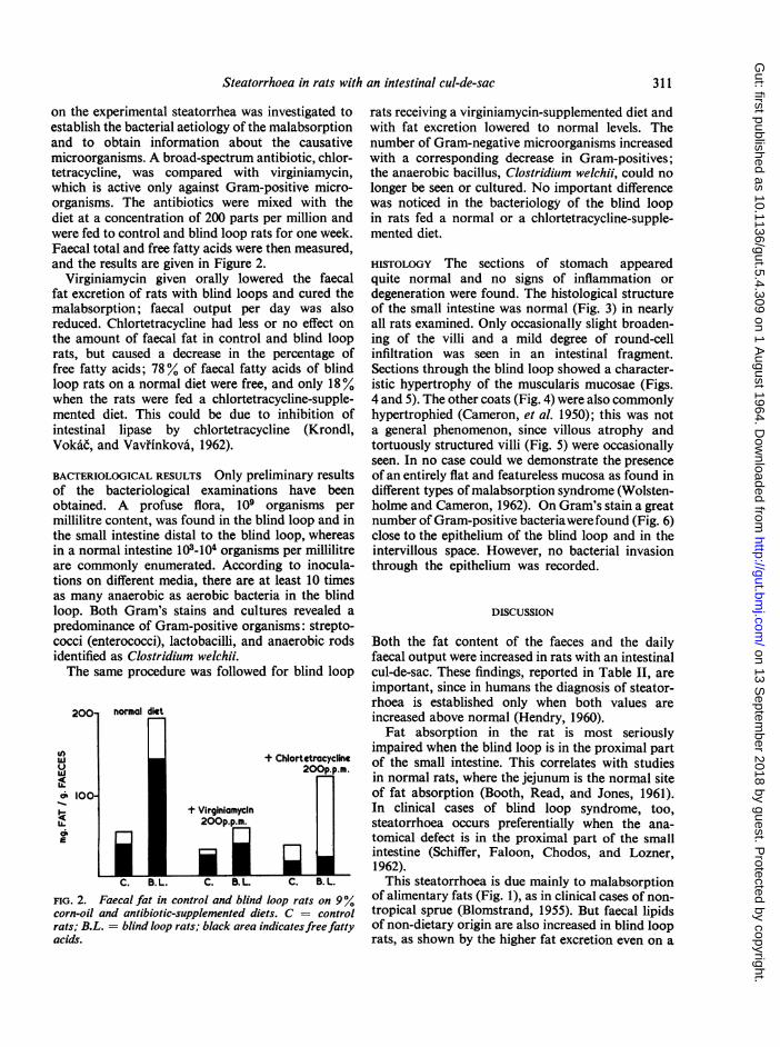
on the experimental steatorrhea was investigated toestablish the bacterial aetiology of the malabsorptionand to obtain information about the causativemicroorganisms. A broad-spectrum antibiotic, chlor-tetracycline, was compared with virginiamycin,which is active only against Gram-positive micro-organisms. The antibiotics were mixed with thediet at a concentration of 200 parts per million andwere fed to control and blind loop rats for one week.Faecal total and free fatty acids were then measured,and the results are given in Figure 2.
Virginiamycin given orally lowered the faecalfat excretion of rats with blind loops and cured themalabsorption; faecal output per day was alsoreduced. Chlortetracycline had less or no effect onthe amount of faecal fat in control and blind looprats, but caused a decrease in the percentage offree fatty acids; 78% of faecal fatty acids of blindloop rats on a normal diet were free, and only 18%when the rats were fed a chlortetracycline-supple-mented diet. This could be due to inhibition ofintestinal lipase by chlortetracycline (Krondl,Vokac, and Vavfi'nkovai, 1962).
BACTERIOLOGICAL RESULTS Only preliminary resultsof the bacteriological examinations have beenobtained. A profuse flora, 109 organisms permillilitre content, was found in the blind loop and inthe small intestine distal to the blind loop, whereasin a normal intestine 103-104 organisms per millilitreare commonly enumerated. According to inocula-tions on different media, there are at least 10 timesas many anaerobic as aerobic bacteria in the blindloop. Both Gram's stains and cultures revealed apredominance of Gram-positive organisms: strepto-cocci (enterococci), lactobacilli, and anaerobic rodsidentified as Clostridium welchii.The same procedure was followed for blind loop
200' normal diet
Wii + ChlortetracyclineU - 200p.p.m.
M 100-+ Virginiomycin
aEu. 200p.p.m.
iI;1FIG. 2. Faecal fat in control and blind loop rats on 9%corn-oil and antibiotic-supplemented diets. C = controlrats; B.L. = blind loop rats; black area indicates freefattyacids.
rats receiving a virginiamycin-supplemented diet andwith fat excretion lowered to normal levels. Thenumber of Gram-negative microorganisms increasedwith a corresponding decrease in Gram-positives;the anaerobic bacillus, Clostridium welchii, could nolonger be seen or cultured. No important differencewas noticed in the bacteriology of the blind loopin rats fed a normal or a chlortetracycline-supple-mented diet.
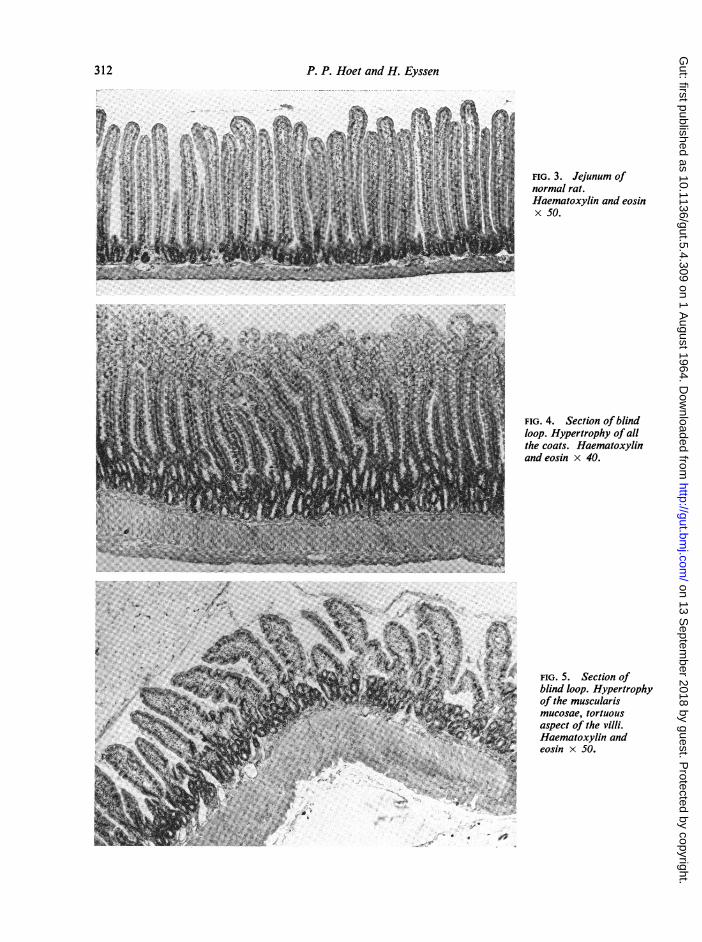
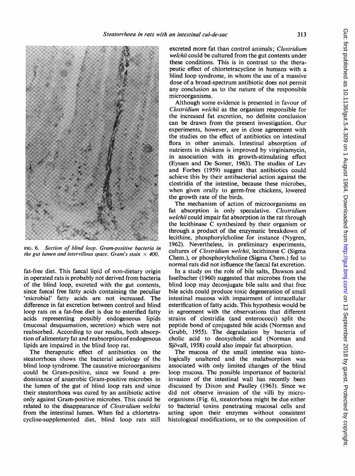
HISTOLOGY The sections of stomach appearedquite normal and no signs of inflammation ordegeneration were found. The histological structureof the small intestine was normal (Fig. 3) in nearlyall rats examined. Only occasionally slight broaden-ing of the villi and a mild degree of round-cellinfiltration was seen in an intestinal fragment.Sections through the blind loop showed a character-istic hypertrophy of the muscularis mucosae (Figs.4 and 5). The other coats (Fig. 4) were also commonlyhypertrophied (Cameron, et al. 1950); this was nota general phenomenon, since villous atrophy andtortuously structured villi (Fig. 5) were occasionallyseen. In no case could we demonstrate the presenceof an entirely flat and featureless mucosa as found indifferent types of malabsorption syndrome (Wolsten-holme and Cameron, 1962). On Gram's stain a greatnumber of Gram-positive bacteriawerefound (Fig. 6)close to the epithelium of the blind loop and in theintervillous space. However, no bacterial invasionthrough the epithelium was recorded.
DISCUSSION
Both the fat content of the faeces and the dailyfaecal output were increased in rats with an intestinalcul-de-sac. These findings, reported in Table II, areimportant, since in humans the diagnosis of steator-rhoea is established only when both values areincreased above normal (Hendry, 1960).
Fat absorption in the rat is most seriouslyimpaired when the blind loop is in the proximal partof the small intestine. This correlates with studiesin normal rats, where the jejunum is the normal siteof fat absorption (Booth, Read, and Jones, 1961).In clinical cases of blind loop syndrome, too,steatorrhoea occurs preferentially when the ana-tomical defect is in the proximal part of the smallintestine (Schiffer, Faloon, Chodos, and Lozner,1962).This steatorrhoea is due mainly to malabsorption
of alimentary fats (Fig. 1), as in clinical cases of non-tropical sprue (Blomstrand, 1955). But faecal lipidsof non-dietary origin are also increased in blind looprats, as shown by the higher fat excretion even on a
311
on 13 Septem
ber 2018 by guest. Protected by copyright.
http://gut.bmj.com
/G
ut: first published as 10.1136/gut.5.4.309 on 1 August 1964. D
Steatorrhoea in rats with an intestinal cul-de-sac
FIG. 6. Section of blind loop. Gram-positive bacteria inthe gut lumen and intervillous space. Gram's stain x 400.
fat-free diet. This faecal lipid of non-dietary originin operated rats is probably not derived from bacteriaof the blind loop, excreted with the gut contents,since faecal free fatty acids containing the peculiar'microbial' fatty acids are not increased. Thedifference in fat excretion between control and blindloop rats on a fat-free diet is due to esterified fattyacids representing possibly endogeneous lipids(mucosal desquamation, secretion) which were notreabsorbed. According to our results, both absorp-tion of alimentary fat and reabsorption ofendogenouslipids are impaired in the blind loop rat.The therapeutic effect of antibiotics on the
steatorrhoea shows the bacterial aetiology of theblind loop syndrome. The causative microorganismscould be Gram-positive, since we found a pre-dominance of anaerobic Gram-positive microbes inthe lumen of the gut of blind loop rats and sincetheir steatorrhoea was cured by an antibiotic activeonly against Gram-positive microbes. This could berelated to the disappearance of Clostridium welchiifrom the intestinal lumen. When fed a chlortetra-cycline-supplemented diet, blind loop rats still
excreted more fat than control animals; Clostridiumwelchii could be cultured from the gut contents underthese conditions. This is in contrast to the thera-peutic effect of chlortetracycline in humans with ablind loop syndrome, in whom the use of a massivedose of a broad-spectrum antibiotic does not permitany conclusion as to the nature of the responsiblemicroorganisms.Although some evidence is presented in favour of
Clostridium welchii as the organism responsible forthe increased fat excretion, no definite conclusioncan be drawn from the present investigation. Ourexperiments, however, are in close agreement withthe studies on the effect of antibiotics on intestinalflora in other animals. Intestinal absorption ofnutrients in chickens is improved by virginiamycin,in association with its growth-stimulating effect(Eyssen and De Somer, 1963). The studies of Levand Forbes (1959) suggest that antibiotics couldachieve this by their antibacterial action against theclostridia of the intestine, because these microbes,when given orally to germ-free chickens, loweredthe growth rate of the birds.The mechanism of action of microorganisms on
fat absorption is only speculative. Clostridiumwelchii could impair fat absorption in the rat throughthe lecithinase C synthesized by their organism orthrough a product of the enzymatic breakdown oflecithine, phosphorylcholine for instance (Nygren,1962). Nevertheless, in preliminary experiments,cultures of Clostridium welchii, lecithinase C (SigmaChem.), or phosphorylcholine (Sigma Chem.) fed tonormal rats did not influence the faecal fat excretion.
In a study on the role of bile salts, Dawson andIsselbacher (1960) suggested that microbes from theblind loop may deconjugate bile salts and that freebile acids could produce toxic degeneration of smallintestinal mucosa with impairment of intracellularesterification of fatty acids. This hypothesis would bein agreement with the observations that differentstrains of clostridia (and enterococci) split thepeptide bond of conjugated bile acids (Norman andGrubb, 1955). The degradation by bacteria ofcholic acid to deoxycholic acid (Norman andSjovall, 1958) could also impair fat absorption.The mucosa of the small intestine was histo-
logically unaltered and the malabsorption wasassociated with only limited changes of the blindloop mucosa. The possible importance of bacterialinvasion of the intestinal wall has recently beendiscussed by Dixon and Paulley (1963). Since wedid not observe invasion of the villi by micro-organisms (Fig. 6), steatorrhoea might be due eitherto bacterial toxins penetrating mucosal cells andacting upon their enzymes without consistenthistological modifications, or to the composition of
313
on 13 Septem
ber 2018 by guest. Protected by copyright.
http://gut.bmj.com
/G
ut: first published as 10.1136/gut.5.4.309 on 1 August 1964. D
the gut contents modified by the microorganisms.Work is in progress to establish the exact roleof clostridia and other bacterial strains in themalabsorption associated with the blind loopsyndrome.
The histological study was done by Dr. M. Vandeputteat the Rega Institute for Medical Research. We aregrateful to him for his contribution and his interest inthis work.
REFERENCES
Aitken, M. A., Badenoch, J., and Spray, G. H. (1950). Fat excretionin rats with intestinal culs-de-sac. Brit. J. exp. Pathol., 31,355-357.
Badenoch, J. (1960). Steatorrhoea in the adult. Brit. med. J., 2,879-887; 963-974.
Blomstrand, R. (1955). A study on the intestinal absorption of fat innormal adults and in non-tropical sprue with carbon-labelledoleic acid and palmitic acid. Acta med. scand., 152, 129-138.
Booth, C. C., Read, A. E., and Jones, E. (1961). Studies on the site offat absorption. Gut, 2, 23-31.
Cameron, D. G., Watson, G. M., and Witts, L. J. (1949). The experi-mental production of macrocytic anaemia by operations on theintestinal tract. Blood, 4, 803-815.
-, -, - (1950). The alimentary tract of rats with intestinalculs-de-sac. Brit. J. exp. Pathol., 31, 349-354.
Cooke, W. T., Cox, E. V., Fone, D. J., Meynell, M. J., and Gaddie, R.(1963). The clinical and metabolic significance of jejunaldiverticula. Gut, 4, 115-131.
Dawson, A. M., and Isselbacher, K. J. (1960). Studies on lipidmetabolism in the small intestine with observations on therole of bile salts. J. clin. Invest., 39, 730-740.
Dixon, J. M. S., and Paulley, J. W. (1963). Bacteriological and histo-logical studies of the small intestine of rats treated withmecamylamine. Gut, 4, 169-173.
Dole, V. P., and Meinertz, H. (1960). Microdetermination of long-chain fatty acids in plasma and tissues. J. biol. Chem., 235,2595-2599.
Edwards, H. M., Jr., and Gillis, M. B. (1959). A chromic oxidebalance method for determining phosphate availability.Poultry Sci., 38, 569-574.
Evrard, E., Hoet, P. P., Eyssen, H., Charlier, H., and Sacquet, E.(1964). Faecal lipids in germ-free and conventional rats. Tobe published.
Eyssen, H., and De Somer, P. (1963). The mode of action of anti-biotics in stimulating growth of chicks. J. exp. Med., 117,127-138.
Gellman, D. D. (1956). Diverticulosis of the small intestine withsteatorrhoea and megaloblastic anaemia. Lancet, 2, 873-874.
Goldstein, F., Wirts, C. W., and Kramer, S. (1961). The relationshipof afferent limb stasis and bacterial flora to the production ofpost-gastrectomy steatorrhoea. Gastroenterology, 40, 47-55.
Hendry, E. B. (1960). The chemical diagnosis of steatorrhoea. Brit.med. J., 2, 975-979.
Kamer, J. H., van de, ten Bokkel Huinink, H., and Weyers, H. A.(1949). Rapid method for the determination of fat in faeces.J. biol. Chem., 177, 347-355.
Krondl, A., Vokac, V., and Vavfinkovai, H. (1962). Influence ofchlortetracycline and neomycin on digestion and absorptionof fat in rats. Amer. J. Physiol., 202, 437-439.
Lev, M., and Forbes, M. (1959). Growth response to dietary penicillinof germ-free chicks and of chicks with a defined intestinal flora.Brit. J. Nutr., 13, 78-84.
Norman, A., and Grubb, R. (1955). Hydrolysis of conjugated bileacids by Clostridia and enterococci. Acta pathol. microbiol.scand., 36, 537-547.
, and Sjovall, J. (1958). On the transformation and enterohepaticcirculation of cholic acid in the rat. J. biol. Chem., 233, 872-884.
Nygren, B. (1962). Phospholipase C-producing bacteria and foodpoisoning. Acta pathol. microbiol. scand., suppl., 160.
Schiffer, L. M., Faloon, W. W., Chodos, R. B., and Lozner, E. L.(1962). Malabsorption syndrome associated with intestinaldiverticulosis. Gastroenterology, 42, 63-68.
Wirts, C. W., and Goldstein, F. (1963). Studies on the mechanism ofpost-gastrectomy steatorrhoea. Ann. intern. Med., 58, 25-26.
Wolstenholme, G. E. W., and Cameron, M. P. (1962). IntestinalBiopsy. (Ciba Foundation Study Group, No. 14.) Churchill,London.
on 13 Septem
ber 2018 by guest. Protected by copyright.
http://gut.bmj.com
/G
ut: first published as 10.1136/gut.5.4.309 on 1 August 1964. D