Streptococcosis is an economically important diseasethat has been reported in over 30 farmed fresh andsaltwater fish species including catfish (Shewmaker etal. 2007), tilapia (Eldar et al. 1995), trout (Hoshina etal. 1958, Eldar et al. 1995), salmon (Romalde et al.2008), red drum (Eldar et al. 1999), flounder (Baeck etal. 2006), yellowtail (Kusuda et al. 1991), hybrid stripedbass (Evans et al. 2000) and turbot (Toranzo et al. 1994).The occurrence of streptococcosis in fish appears to bestress related with low grade chronic mortalities seen insome fish species and acute mortalities of 30 to 50% oc-
curring in others as a result of an encephalitis and/orsystemic infection affecting multiple organs (Eldar etal. 1995, Ringo & Gattesoup 1998, Camus et al. 2008).This group of bacteria appear to have no host speci-ficity, are capable of infecting both fish and mammalianspecies and include Streptococcus iniae, S. agalactiae(S. dificilis), S. phocae and S. parauberis with S. ictaluribeing the most recently described member of thisgenus to infect an aquaculture fish species (Ringo &Gattesoup 1998, Shewmaker et al. 2007).
Lethal infections due to Gram-positive cocci in crus-taceans are rare and have only been reported in thefreshwater prawn Macrobrachium rosenbergii (Cheng
Streptococcosis in farmed Litopenaeus vannamei: anew emerging bacterial disease of penaeid shrimp
Ken W. Hasson*, Ernesto Matheu Wyld, Yaping Fan, Sonia W. Lingsweiller,Stephanie J. Weaver, Jinling Cheng, Patricia W. Varner
Texas Veterinary Medical Diagnostic Lab, 1 Sippel Rd, College Station, Texas 77843, USA
ABSTRACT: Presumptive systemic streptococcal infections were detected histologically in farmedLitopenaeus vannamei juveniles submitted from a Latin American country and the bacteria isolated.Characterization work demonstrated that the Gram-positive cocci form chains, grow aerobically andanaerobically, are oxidase- and catalase-negative, non-hemolytic, non-motile, Lancefield Group Bpositive and PCR positive when amplified with a universal streptococcal primer set. Differing Strep-tococcus identifications were obtained using API 20 Strep and Biolog systems, the former identifyingthe isolate as S. uberis and the latter as S. parauberis. Injection of specific pathogen-free (SPF) L. van-namei with the bacteria resulted in 100% mortality by 3 d post-injection with successful recovery ofthe agent from moribund test shrimp hemolymph samples. The recovered isolate was used in per osand waterborne exposure studies of SPF L. vannamei with mortalities ranging from 40 to 100% and80 to 100%, respectively. Histologic analysis of 5 to 8 moribund shrimp from each exposure methoddemonstrated that all contained a severe bacteremia characterized by numerous free cocci within thehemolymph and aggregates of vacuolated hemocytes with notable intravacuolar cocci. This uniquelesion type was most pronounced within the lymphoid organ and considered pathodiagnostic for thisdisease. Experimentally induced lesions were identical to those in naturally infected farmed shrimpand the Streptococcus sp. responsible was re-isolated, fulfilling Koch’s postulates. Five freeze/thawcycles of 10 experimentally infected shrimp were performed over a 2 mo period and the bacteria suc-cessfully cultured from all shrimp at each interval. These collective findings describe the firstreported case of streptococcosis in marine penaeid shrimp in the Western Hemisphere and indicatethat the agent may be disseminated via live or frozen infected shrimp.
Resale or republication not permitted without written consent of the publisher
OPENPEN ACCESSCCESS
Dis Aquat Org 86: 93–106, 2009
& Chen 1998). The disease, ‘white muscle disease’,causes significant losses in M. rosenbergii populationsand has been attributed to 2 different species of Lacto-coccus, L. lactis subsp. lactis and L. garvieae, whichare included in the family Streptococcaceae (Vendrellet al. 2006, Wang et al. 2008). Bacterial diseases offarmed marine shrimp are very common, can result inhigh production losses and are principally caused byopportunistic Gram-negative bacilli corresponding tothe Vibrionaceae (Sindermann & Lightner 1988, Brock& Main 1994). To date, no reports of Streptococcusinfections in penaeid shrimp have been published.
During September 2008, Litopenaeus vannamei, pre-served in Davidson’s AFA (alcohol, formalin, acid) fixa-tive and originating from 2 separate intensive shrimpculture farms located in a Latin American country, weresubmitted to the Texas Veterinary Medical DiagnosticLaboratory (TVMDL) for diagnostic evaluation. Onefarm utilized full strength sea water (29 to 32 ppt) whilethe other operated with estuarine water (12 to 18 ppt).Both farms contained a mixture of lined and unlinedponds with stocking densities of 135 shrimp m–2 and85 shrimp m–2, respectively. All ponds contained aera-tors with water quality parameters reported as withinnormal limits and nighttime dissolved oxygen lows of2.5 to 3.5 ppm when the disease problem began.Chronic mortality was reported in lined and unlinedponds of both farms and was first observed among juve-nile shrimp weighing 6 to 7 g. Losses ranging from 20 to40% occurred in ponds during a 3 wk period.
No obvious clinical signs of disease or externallesions were observed, with dead shrimp typicallyfound on pond bottoms. Many of these dead shrimpdisplayed intact empty exoskeletons with large por-tions of internal tissue and tail muscle absent as ifeaten or degraded from the inside outwards. Histologicand Gram-stain analyses of representative preservedshrimp from each farm demonstrated the presence of apreviously unreported systemic bacterial infectioncharacterized by numerous Gram-positive cocci freewithin the hemolymph of the vasculature and hemalsinuses as well as within cytoplasmic vacuoles of indi-vidual hemocytes or small aggregates of these cells.Variable numbers of extracellular cocci, bacteria-ladenhemocytes and small hemocytic nodules were detectedwithin the lymphoid organ, heart, gills, skeletal mus-cle, foregut, midgut, hindgut, nerve cord, connectivetissues of both ceca and the hemal sinuses of the anten-nal gland and hepatopancreas.
Since no published reports of a bacterial septicemiadue to a Gram-positive coccus in penaeid shrimp couldbe found in the literature and this appeared to be anewly emerging disease, the current study was initi-ated upon the submission of fresh shrimp and hem-olymph samples from the affected farms. The objec-
tives of the current study were 4-fold: (1) to isolate andperform initial characterization work on the bacterialagent, (2) to fulfill Koch’s postulates by reproducingthe disease and recovering the isolate from specificpathogen-free (SPF) Litopenaeus vannamei test shrimpusing intramuscular injection, waterborne, and per osexposure methods, (3) to describe the histologicchanges induced by this disease, and (4) to determinethe effect of repeated freezing and thawing cycles onthe organism’s viability.
MATERIALS AND METHODS
Bacterial isolation and identification. Chilled shrimp(n = 6 per pond) and corresponding pooled hemolymphsamples, originating from a single pond on 2 separateintensive Litopenaeus vannamei farms (hereafterreferred to as Farms A and B), were submitted from theaffected region (exact location withheld at farm’srequest). The cephalothorax of each of the 12 shrimpwas swabbed with 70% ethanol, the exoskeleton re-moved from one side using sterile scissors, and thelymphoid organ exposed for inoculation onto bloodagar (BA) plates (tryptic soy agar with 5% sheep’sblood, BBL) using a sterile disposable plastic loopfollowing the methods of Cheng & Chen (1998). Simi-larly, each of the 2 pooled hemolymph samples werestreaked on BA, and the cultures incubated aerobicallyand anaerobically overnight at 28°C to determine thetrue hemolytic pattern of the isolates.
Biochemical tests: Pure Streptococcus-like colonies,characterized by the development of numerous smallcircular white colonies on BA, were isolated from boththe shrimp and hemolymph samples originating fromthe 2 farms after 18 to 24 h incubation, and thenstreaked for purity on BA plates. One purified isolateper farm was selected and subjected to oxidase andcatalase tests, Gram-staining and biochemical identifi-cation using the API 20 Strep strip kit (Biomerieux) fol-lowing the manufacturer’s instructions. The isolateswere also inoculated into tryptose broth (Remel) andincubated aerobically at 28°C for 18 h with subsequentwet mount analysis to observe cell morphology. Theisolate selected for use in all 3 bioassays was later ana-lyzed with a PathoDx kit (Remel) to determine itsLancefield group designation (Lancefield 1933) andidentified using the Omnilog ID system (Biolog) to-gether with an ATCC S. porcinus isolate to verify thesystems’ ability to identify a known Streptococcus sp.These 2 latter tests were performed according to themanufacturer’s instructions.
PCR analysis: Bacterial genomic DNA was extractedfrom 4 separate bacterial samples (Streptococcus-likebacteria from Farm A, Streptococcus-like bacteria from
94
Hasson et al.: Streptococcosis in farmed Litopenaeus vannamei
Farm B, Streptococcus-like bacteriarecovered from Bioassay 2 treatmentshrimp, and ATCC S. porcinus isolate)following the protocol of Berridge etal. (1998) with modifications. Briefly,bacteria were aerobically cultured in3 ml of tryptose broth overnight (28°C)and then equally divided into two1.5 ml Eppendorf tubes. After centrifu-gation at 11 000 × g (15 min), the 2resulting pellets were suspended andcombined in 300 µl of strep lysis buffer(50 mM Tris, 100 mM NaCl, 20 mMEDTA, 0.5% sodium dodecyl sulfate,pH 6.0) together with 10 µl muta-nolysin (10 U µl–1, Sigma). The solu-tion was briefly mixed by vortexing,incubated at 37°C for 45 min and thenplaced in boiling water for 10 min toinactivate the mutanolysin. Followingcentrifugation at 16 000 × g (3 min),100 µl of the DNA lysate was trans-ferred to a chromaspin column (Clonetech) accordingto the manufacturer’s instructions, and centrifuged at700 × g (15 min). The resulting filtrate was stored at4°C until analyzed by PCR. The PCR reaction mixtures(25 µl) and thermocycler program used were accordingto Meiri-Bendek et al. (2002) and utilized their univer-sal 16S rDNA streptococcus primer set (C1: 5’-GCGTGC CTA ATA CAT GCA A-3’; C2: 5’-TAC AAC GCAGGT CCA TCT-3’), which was designed to produce a207 bp amplicon. The resulting PCR products were runon a 2% agarose gel (80 V, 40 min), which was thenstained by submersion in 0.001% ethidium bromide(20 min). The gel was then destained under runningwater for 3 min, the banding pattern visualized by UVtransillumination and photographed using a PolaroidPhoto-Documentation camera (Fisher Scientific).
Test shrimp. Juvenile SPF Fast Growth line Litope-naeus vannamei (mean weight = 8 g; provided byShrimp Improvement Systems) were used to conduct 3separate bioassays. Static 10 gallon (37.9 l) glassaquaria were utilized in each bioassay, filled with 10 lof artificially prepared 30 ppt saltwater (Crystal SeaBioassay Salt, Marine Enterprises International) andeach stocked with 5 or 6 shrimp (4.0 to 4.8 g l–1 bio-mass). Two aquariums were used for the first injectionexposure study (Bioassay 1), 6 for the waterborne/peros exposure study (Bioassay 2) and 2 for the secondinjection study (Bioassay 3) as outlined in Table 1. Theaquariums were continuously aerated with a single air-stone, covered to prevent animal escape and a 90%water exchange performed each morning. Bacteria-exposed aquariums were maintained in a room sepa-rate from the negative control aquariums so as to min-
imize the possibility of cross-contamination. A meanwater temperature of 28°C was maintained in bothbioassay rooms during the 3 studies through place-ment of continuously operating electric area heaters.All shrimp were fed a pelleted ration (Ziegler No. 4pellet) ad libitum once daily during the studies. Theonly exceptions were the per os-exposed treatmentshrimp and a corresponding per os negative controlgroup (described below).
Bioassay 1. Injection exposure to the Streptococ-cus-like isolate. The bacterial isolate used in thisbioassay was cultured from the lymphoid organ of anaturally infected Farm A Litopenaeus vannamei andstored at –80°C in Brucella broth with glycerol(CryosaverTM tube). The bacteria was later thawed,streaked and aerobically cultured on BA for 18 h at28°C and a colony then transferred to 50 ml of tryp-tose broth. Following 18 h incubation with agitation(28 to 30°C), the bacterial suspension was pelleted bycentrifugation (2080 × g, 30 min, 4°C), the supernatantdiscarded and the pellet suspended in sterile 2%saline to achieve a turbidity equivalent to a McFar-land No. 2 standard (a concentration of ~600 × 106
colony forming units [CFU] ml–1). A 10 µl aliquot wasthen transferred to a 15 ml tube and diluted 1:1000with 2% sterile saline to obtain a concentration of~6000 CFU µl–1. Six shrimp were then intramuscularlyinjected (IM) in the third abdominal segment with ~50to 80 µl of the bacterial dilution using a 1 ml tuber-culin syringe to achieve 3 to 5 × 10⁵ CFU shrimp–1,similar to dosages used in past Vibrio sp. infectivitystudies (Saulnier et al. 2000). To serve as negativecontrols, 6 shrimp were similarly injected with sterile
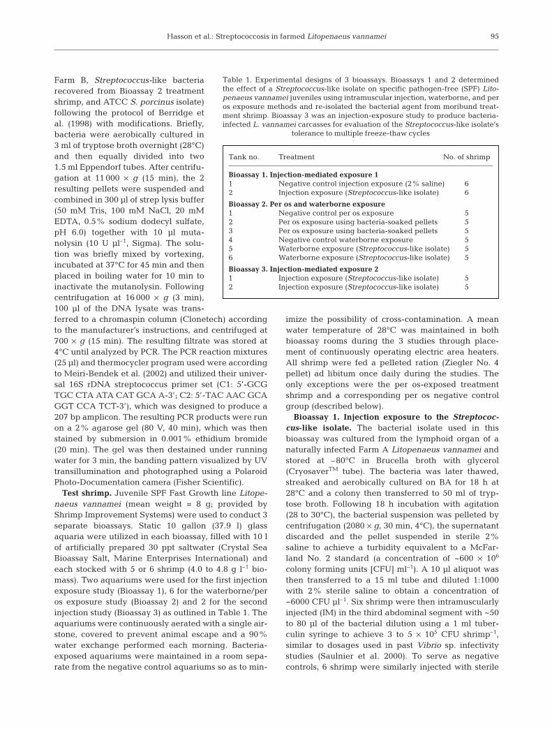
Bioassay 2. Per os and waterborne exposure1 Negative control per os exposure 52 Per os exposure using bacteria-soaked pellets 53 Per os exposure using bacteria-soaked pellets 54 Negative control waterborne exposure 55 Waterborne exposure (Streptococcus-like isolate) 56 Waterborne exposure (Streptococcus-like isolate) 5
Table 1. Experimental designs of 3 bioassays. Bioassays 1 and 2 determinedthe effect of a Streptococcus-like isolate on specific pathogen-free (SPF) Lito-penaeus vannamei juveniles using intramuscular injection, waterborne, and peros exposure methods and re-isolated the bacterial agent from moribund treat-ment shrimp. Bioassay 3 was an injection-exposure study to produce bacteria-infected L. vannamei carcasses for evaluation of the Streptococcus-like isolate’s
tolerance to multiple freeze-thaw cycles
Dis Aquat Org 86: 93–106, 2009
2% saline. Moribund and freshly dead (i.e. no muscleopacity or difference in color from live shrimpobserved) shrimp were collected, the needle of a 1 mltuberculin syringe inserted into the base of the fourthperiopod and ~50 to 100 µl of hemolymph extractedfrom the ventral sinus. Hemolymph samples (1 or 2drops sample–1) were immediately streaked on BAplates and observed for growth after 18 to 24 h aero-bic incubation (28°C). One representative colony perplate was Gram-stained and observed by lightmicroscopy to confirm the presence of Gram-positivecocci. Finally, 3 representative colonies from a singleplate were randomly selected, streaked onto 3 BAslants, and incubated for 24 h (28°C). The slants werethen filled with sterile mineral oil and stored at roomtemperature until used in Bioassay 2. The bioassaywas terminated on Day 3 when the last bacteria-injected shrimp was collected in a moribund state.
Bioassay 2. Waterborne and per os exposures to theStreptococcus-like isolate. Six 10 gal aquariums con-taining 5 SPF Litopenaeus vannamei juveniles eachwere utilized (Table 1). Two aquaria were designatedas replicate per os treatment groups and 2 aquariumsas replicate waterborne treatment groups. The remain-ing 2 aquariums were placed in a separate room to pre-vent the possibility of bacterial contamination andserved as per os and waterborne negative controltanks. Waterborne and per os exposures to the Strepto-coccus-like isolate were conducted each morning dur-ing the first 5 d of this 12 d study following the dailywater exchange. All moribund and freshly deadshrimp were bled for bacterial culture as described inBioassay 1 and preserved for histologic analysis (see‘Materials and methods: Histopathology’). Streptococ-cus-like bacteria recovered from the hemolymph of 2moribund per os-exposed shrimp and 2 waterborne-exposed shrimp were analyzed using the API 20 Strepkit to confirm that the isolates were the same andequivalent to the original wild type isolate used inBioassay 1.
Bacterial culture and shrimp exposure methods: A1 l sterile flask containing 400 ml of tryptose broth wasinoculated each evening with the bacterial isolaterecovered during Bioassay 1 and the culture aerobi-cally incubated with continuous agitation at 28 to 30°Covernight (18 to 20 h). The culture was then equallydivided between eight 50 ml centrifuge tubes, the bac-teria pelleted by centrifugation (2060 × g, 30 min, 4°C)and the supernatant removed using a disposablepipette. The bacterial pellets from 6 tubes were indi-vidually re-suspended in 50 ml of artificial seawater bymixing and 150 ml of bacterial suspension (3 tubes)added to each of the 2 waterborne treatment tanks.The bacterial pellets in the remaining 2 tubes wereeach re-suspended in 15 ml of artificial seawater, the
suspensions transferred into 2 medium size plasticweighing dishes and feed pellets (Ziegler No. 4 pellet)equivalent to 5% of the biomass (2 g) aquarium–1
added. After 30 s the hydrated pellets were fed to the 2per os treatment groups together with the excess bac-terial suspension. Similarly, 2 g of pelleted feed washydrated with artificial seawater and fed to the per osnegative control group.
Bacterial counts: Water samples (50 ml aquarium–1)were collected from the 2 waterborne treatment tanksand corresponding negative control tank ~30 minpost-inoculation each day during the first 4 d of this12 d study. Three serial tenfold dilutions were pre-pared from each water sample (1:1000 to 1:100 000)and 100 µl streaked on BA plates using a bacterialcell spreader. An undiluted 100 µl water samplefrom the negative control tank was similarly plated.Following 18 to 24 h aerobic incubation (28°C), Strep-tococcus-like colonies were counted and CFUs ml–1
determined.Bioassay 3. Freeze-thaw effects on the Streptococ-
cus-like isolate. A total of 10 SPF Litopenaeus van-namei juvenile shrimp were intramuscularly injectedwith the same Streptococcus-like isolate used in Bioas-say 2. Injections were performed following the protocoldescribed in Bioassay 1 and the shrimp then equallydivided between two 10 gal aquariums as previouslydescribed. Resulting moribund and dead shrimp (n =10) were collected, equally divided into 2 groups andindividually frozen in Whirl-pakTM bags (Nasco) ateither –80°C (n = 5) or –20°C (n = 5). Four days post-freezing, all shrimp were thawed at room temperaturefor 60 min, the head and first tail segment swabbedwith 70% ethanol, and the abdomen aseptically sev-ered from the head, cutting ventral to dorsal using anew sterile scalpel blade for each shrimp. A sterile dis-posable loop was inserted into the ventral sinus area ofthe first tail segment and aseptically extracted fluidthen plated on BA. The shrimp were then returned totheir respective freezers and the BA plates incubatedovernight at 28°C. Each plate was examined the nextmorning for the presence of Streptococcus-likecolonies, which were Gram-stained and examinedmicroscopically for the presence of Gram-positivecocci. Thawing and plating of the shrimp samples wasrepeated once per day over the next 2 d and then againat 30 and 60 d post-collection for a total of 5 freeze-thaw cycles.
Histopathology. Upon receipt of the juvenile SPFLitopenaeus vannamei population used in the 3 bio-assays, 5 shrimp were preserved with Davidson’s AFAand paraffin-embedded tissues were prepared forroutine H&E histological analysis according to themethods of Bell & Lightner (1988) in order to confirmtheir disease-free status. During Bioassays 1 and 2 all
96
Hasson et al.: Streptococcosis in farmed Litopenaeus vannamei
moribund and fresh dead shrimp from the negativecontrol and treatment groups were bled for bacterialculture as previously described and then preservedwith Davidson’s AFA for histological analysis follow-ing standard methods. Upon terminating Bioassay 2,all surviving negative control and bacteria-exposedshrimp were similarly preserved. Streptococcus lesionseverity was graded on a scale from 0 to 4 similar tothat for scoring Taura syndrome virus (TSV)-inducedlesions (Hasson et al. 1995). Streptococcus-inducedlesions were defined as foci containing single or mul-tiple, variably sized aggregates of vacuolated pre-sumptive hemocytes with intravacuolar and extracel-lular cocci, and varying degrees of melanization.Complete absence of such lesions was graded as 0,lesions in <25% of an infected tissue was graded as 1,lesions in 25 to 50% of a tissue graded as 2, lesions in50 to 75% of a tissue graded as 3 and lesions in >75%of a tissue graded as 4. As these lesions were typicallypresent in multiple tissues, a lesion severity grade wasassigned to a given sample based on the single mostseverely affected tissue or organ. To better visualizethe bacteria in the affected tissue, select sampleblocks were re-cut and the sections Gram-stainedaccording to Carson (1997) together with positive con-trol tissue samples containing Gram-positive and neg-ative organisms.
RESULTS
Bacterial identification
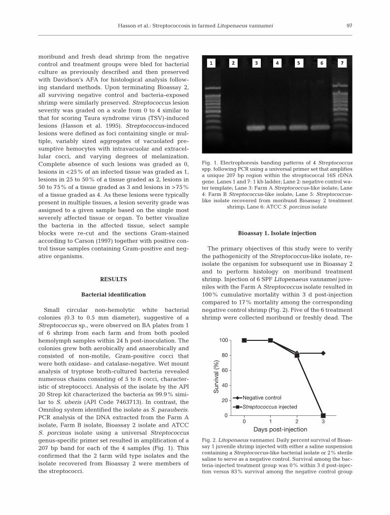
Small circular non-hemolytic white bacterialcolonies (0.3 to 0.5 mm diameter), suggestive of aStreptococcus sp., were observed on BA plates from 1of 6 shrimp from each farm and from both pooledhemolymph samples within 24 h post-inoculation. Thecolonies grew both aerobically and anaerobically andconsisted of non-motile, Gram-positive cocci thatwere both oxidase- and catalase-negative. Wet mountanalysis of tryptose broth-cultured bacteria revealednumerous chains consisting of 5 to 8 cocci, character-istic of streptococci. Analysis of the isolate by the API20 Strep kit characterized the bacteria as 99.9% simi-lar to S. uberis (API Code 7463713). In contrast, theOmnilog system identified the isolate as S. parauberis.PCR analysis of the DNA extracted from the Farm Aisolate, Farm B isolate, Bioassay 2 isolate and ATCCS. porcinus isolate using a universal Streptococcusgenus-specific primer set resulted in amplification of a207 bp band for each of the 4 samples (Fig. 1). Thisconfirmed that the 2 farm wild type isolates and theisolate recovered from Bioassay 2 were members ofthe streptococci.
Bioassay 1. Isolate injection
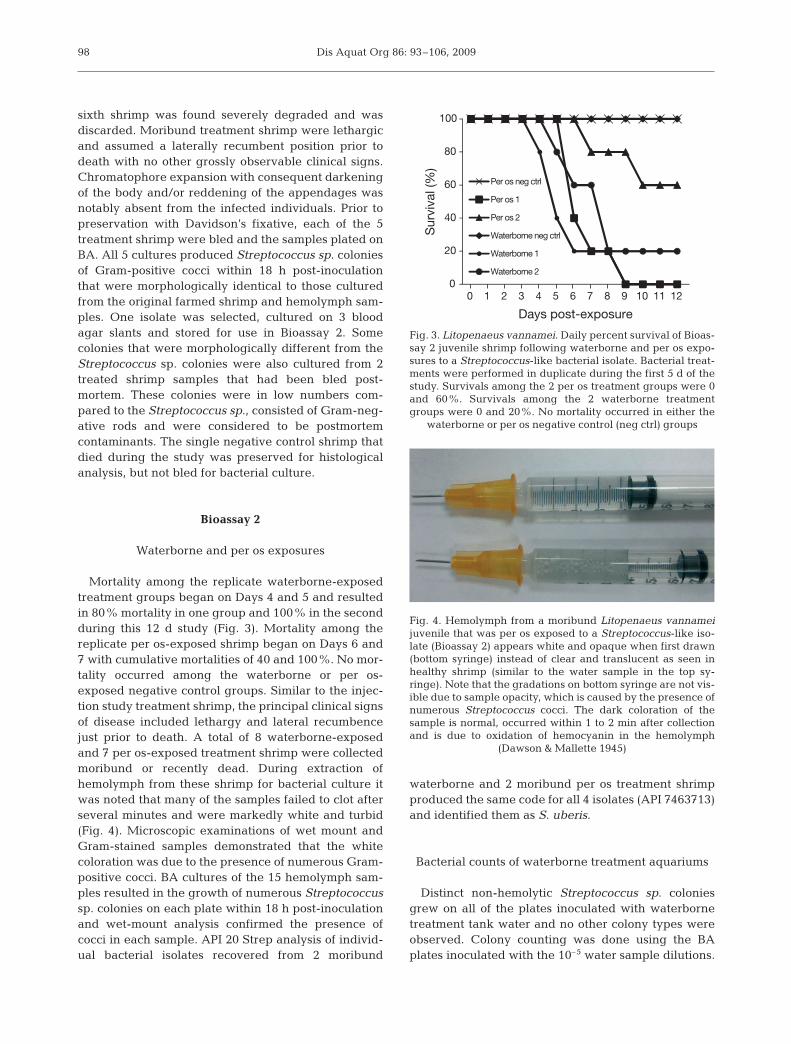
The primary objectives of this study were to verifythe pathogenicity of the Streptococcus-like isolate, re-isolate the organism for subsequent use in Bioassay 2and to perform histology on moribund treatmentshrimp. Injection of 6 SPF Litopenaeus vannamei juve-niles with the Farm A Streptococcus isolate resulted in100% cumulative mortality within 3 d post-injectioncompared to 17% mortality among the correspondingnegative control shrimp (Fig. 2). Five of the 6 treatmentshrimp were collected moribund or freshly dead. The
97
Fig. 1. Electrophoresis banding patterns of 4 Streptococcusspp. following PCR using a universal primer set that amplifiesa unique 207 bp region within the streptococcal 16S rDNAgene. Lanes 1 and 7: 1 kb ladder; Lane 2: negative control wa-ter template; Lane 3: Farm A Streptococcus-like isolate; Lane4: Farm B Streptococcus-like isolate; Lane 5: Streptococcus-like isolate recovered from moribund Bioassay 2 treatment
shrimp; Lane 6: ATCC S. porcinus isolate
0
20
40
60
80
100
0 1 2 3
Sur
viva
l (%
)
Days post-injection
Negative control
Streptococcus injected
Fig. 2. Litopenaeus vannamei. Daily percent survival of Bioas-say 1 juvenile shrimp injected with either a saline suspensioncontaining a Streptococcus-like bacterial isolate or 2% sterilesaline to serve as a negative control. Survival among the bac-teria-injected treatment group was 0% within 3 d post-injec-tion versus 83% survival among the negative control group
Dis Aquat Org 86: 93–106, 2009
sixth shrimp was found severely degraded and wasdiscarded. Moribund treatment shrimp were lethargicand assumed a laterally recumbent position prior todeath with no other grossly observable clinical signs.Chromatophore expansion with consequent darkeningof the body and/or reddening of the appendages wasnotably absent from the infected individuals. Prior topreservation with Davidson’s fixative, each of the 5treatment shrimp were bled and the samples plated onBA. All 5 cultures produced Streptococcus sp. coloniesof Gram-positive cocci within 18 h post-inoculationthat were morphologically identical to those culturedfrom the original farmed shrimp and hemolymph sam-ples. One isolate was selected, cultured on 3 bloodagar slants and stored for use in Bioassay 2. Somecolonies that were morphologically different from theStreptococcus sp. colonies were also cultured from 2treated shrimp samples that had been bled post-mortem. These colonies were in low numbers com-pared to the Streptococcus sp., consisted of Gram-neg-ative rods and were considered to be postmortemcontaminants. The single negative control shrimp thatdied during the study was preserved for histologicalanalysis, but not bled for bacterial culture.
Bioassay 2
Waterborne and per os exposures
Mortality among the replicate waterborne-exposedtreatment groups began on Days 4 and 5 and resultedin 80% mortality in one group and 100% in the secondduring this 12 d study (Fig. 3). Mortality among thereplicate per os-exposed shrimp began on Days 6 and7 with cumulative mortalities of 40 and 100%. No mor-tality occurred among the waterborne or per os-exposed negative control groups. Similar to the injec-tion study treatment shrimp, the principal clinical signsof disease included lethargy and lateral recumbencejust prior to death. A total of 8 waterborne-exposedand 7 per os-exposed treatment shrimp were collectedmoribund or recently dead. During extraction ofhemolymph from these shrimp for bacterial culture itwas noted that many of the samples failed to clot afterseveral minutes and were markedly white and turbid(Fig. 4). Microscopic examinations of wet mount andGram-stained samples demonstrated that the whitecoloration was due to the presence of numerous Gram-positive cocci. BA cultures of the 15 hemolymph sam-ples resulted in the growth of numerous Streptococcussp. colonies on each plate within 18 h post-inoculationand wet-mount analysis confirmed the presence ofcocci in each sample. API 20 Strep analysis of individ-ual bacterial isolates recovered from 2 moribund
waterborne and 2 moribund per os treatment shrimpproduced the same code for all 4 isolates (API 7463713)and identified them as S. uberis.
Bacterial counts of waterborne treatment aquariums
Distinct non-hemolytic Streptococcus sp. coloniesgrew on all of the plates inoculated with waterbornetreatment tank water and no other colony types wereobserved. Colony counting was done using the BAplates inoculated with the 10–5 water sample dilutions.
98
0
20
40
60
80
100
0 1 2 3 4 5 6 7 8 9 10 11 12
Sur
viva
l (%
)
Days post-exposure
Per os neg ctrl
Per os 1
Per os 2
Waterborne neg ctrl
Waterborne 1
Waterborne 2
Fig. 3. Litopenaeus vannamei. Daily percent survival of Bioas-say 2 juvenile shrimp following waterborne and per os expo-sures to a Streptococcus-like bacterial isolate. Bacterial treat-ments were performed in duplicate during the first 5 d of thestudy. Survivals among the 2 per os treatment groups were 0and 60%. Survivals among the 2 waterborne treatmentgroups were 0 and 20%. No mortality occurred in either the
waterborne or per os negative control (neg ctrl) groups
Fig. 4. Hemolymph from a moribund Litopenaeus vannameijuvenile that was per os exposed to a Streptococcus-like iso-late (Bioassay 2) appears white and opaque when first drawn(bottom syringe) instead of clear and translucent as seen inhealthy shrimp (similar to the water sample in the top sy-ringe). Note that the gradations on bottom syringe are not vis-ible due to sample opacity, which is caused by the presence ofnumerous Streptococcus cocci. The dark coloration of thesample is normal, occurred within 1 to 2 min after collectionand is due to oxidation of hemocyanin in the hemolymph
(Dawson & Mallette 1945)
Hasson et al.: Streptococcosis in farmed Litopenaeus vannamei
Streptococcus sp. colony counts of the 2 waterborne-exposed tanks ranged from 0.2 × 10⁶ to 14 × 106 CFUml–1 with a mean range of ~5.5 to 6 × 10⁶ CFU ml–1
(Table 2). Bacteria counts were not performed on Day5 when the last waterborne exposure was performed.The counts on Days 0 and 1 were considerably lowerthan the subsequent counts as marine broth was erro-neously used to produce the first 2 cultures. All subse-quent cultures were conducted using tryptose broth,which resulted in 10-fold higher bacterial counts. NoStreptococcus sp. colonies were cultured from the cor-responding negative control group water samples.
Histopathology
No viral or bacterial infections were detected by rou-tine histology in the 5 SPF Litopenaeus vannamei test
shrimp that were preserved upon arrival at TVMDL orin the single dead negative control shrimp collectedduring Bioassay 1 (Table 3). The cause of death of thiscontrol shrimp collected on Day 2 of that study couldnot be determined by histology, but was possibly dueto molt-induced stress. Upon terminating Bioassay 2 onDay 12, 4 surviving per os-negative control shrimp, 3surviving per os treatment shrimp and 1 waterbornetreatment shrimp were collected and analyzed by rou-tine histology. One of the 3 per os-exposed sampleswas found to have low numbers (Severity Grade 1) ofcocci within the lymphoid organ (LO), whereas noinfections or lesions were detected in the remaining 7samples.
Fresh dead or moribund shrimp from the injectiontreatment group of Bioassay 1 (n = 5), per os treatmentgroup of Bioassay 2 (n = 7) and waterborne treatmentgroup of Bioassay 2 (n = 8) were analyzed by routine
99
Tank no. Day 0 Day 1 Day 2 Day 3 Day 4 Day 5 Mean CFU ml–1
Table 2. Daily bacterial counts (colony-forming units [CFU] ml–1) of tank water from Bioassay 2 treatment tanks (Tanks 5 and 6)subjected to waterborne Streptococcus sp. exposure. No Streptococcus sp. colonies were cultured from the negative control
group water samples. NA: not analyzed
Tank Treatment Histology Streptococcus-positive Cumulativeno. Day No. Lesion Lesion Haemolymph API mortality
sampled examined prevalence severity (%)
Bioassay 11 Injection negative ctrl Arrival day 5 0 of 5 ND NA NA 1/6 (17)
Day 2 1 0 of 1 ND NA NA2 Injection Streptococcus Day 2 1 1 of 1 G4 1 of 1 NA 6/6 (100)
Day 3 4 4 of 4 G3–4 4 of 4 NABioassay 21 Per os negative ctrl – 0 NA NA NA NA 0/5 (0)2 Per os Streptococcus Day 6 3 3 of 3 G3–4 3 of 3 2 of 2 5/5 (100)
Day 7 1 1 of 1 G4 1 of 1 NADay 9 1 1 of 1 G4 1 of 1 1 of 1
3 Per os Streptococcus Day 7 1 1 of 1 G4 1 of 1 NA 2/5 (40)Day 10 1 1 of 1 G4 1 of 1 NADay 12 3 1 of 3 G1 0 of 3 NA
4 Waterborne negative ctrl Day 12 4 0 of 4 ND 0 of 4 NA 0/5 (0)5 Waterborne Streptococcus Day 4 1 1 of 1 G3 1 of 1 NA 5/5 (100)
Day 5 2 2 of 2 G4 2 of 2 2 of 2Day 6 1 1 of 1 G4 1 of 1 NA
6 Waterborne Streptococcus Day 5 1 1 of 1 G4 1 of 1 NA 4/5 (80)Day 6 1 1 of 1 G4 1 of 1 NADay 8 2 2 of 2 G3–4 2 of 2 NADay 12 1 0 of 1 ND 0 of 1 NA
Table 3. Litopenaeus vannamei. Histologic findings of Bioassays 1 and 2 summarizing the number of shrimp per treatmentanalyzed by histology, day of collection, the number with Streptococcus infections, overall lesion severity grade and the cumula-tive percent mortality upon termination on Day 3 (Bioassay 1) or Day 12 (Bioassay 2). Test shrimp were exposed to a Strepto-coccus sp. isolate via injection in Bioassay 1 and by waterborne and per os routes in Bioassay 2. Lesion severity grade (G) is basedon a scale from 1 to 4: 1 = mild focal to multifocal lesions in <25% of the affected tissue, 4 = severe multifocal to diffuse lesions
in >75% of an affected tissue. NA: not analyzed; ND: not detected
Dis Aquat Org 86: 93–106, 2009
histology. Severe Grade 3 to 4 systemic bacterial infec-tions were detected in all 20 samples (Table 3) with theLO being the most severely affected tissue. Both lesionmorphology and distribution were found to be identi-cal to those observed in naturally infected farmed
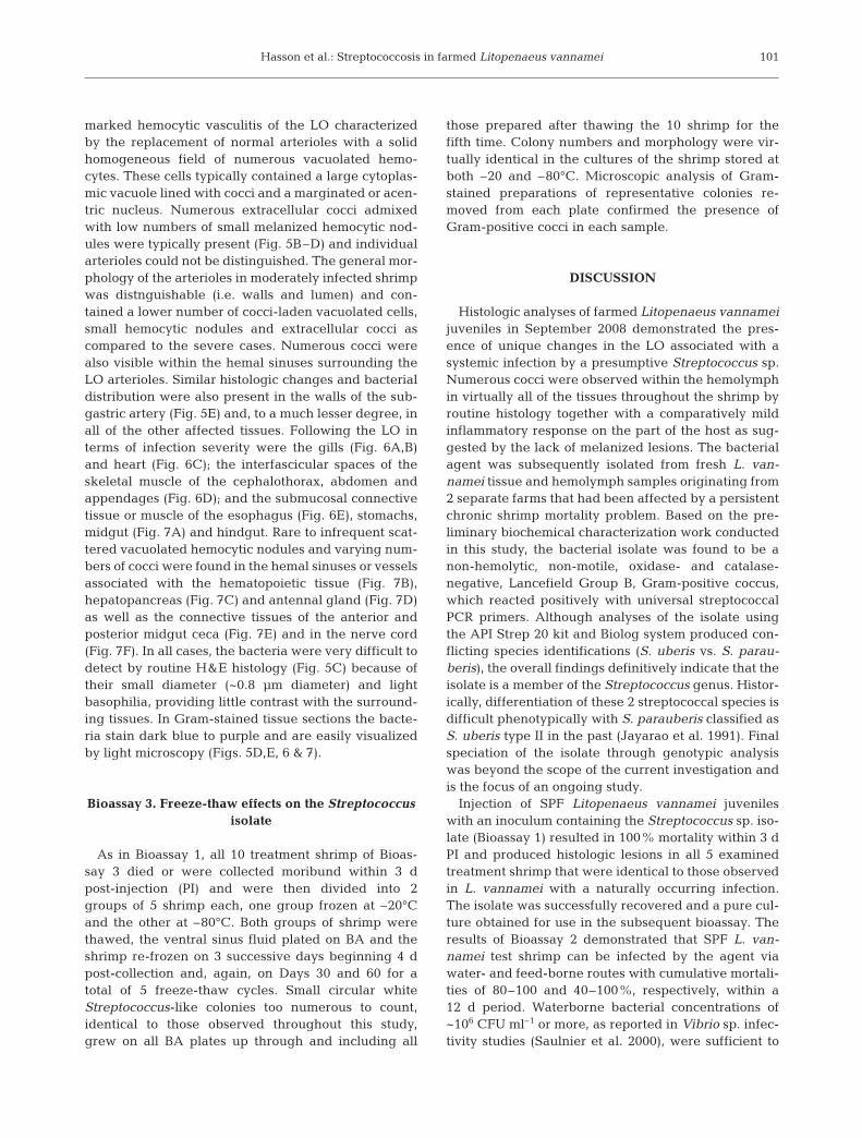
Litopenaeus vannamei. Normal LO sections containnumerous arterioles characterized by a thick wall sur-rounding an endothelial-lined lumen when viewed incross-section (Fig. 5A). In contrast, the severelyinfected treatment shrimp of this study displayed a
100
Fig. 5. Litopenaeus vannamei. Histological sections of naturally occurring and experimentally induced Streptococcus sp. infec-tions within the lymphoid organ and subgastric artery. (A) Normal L. vannamei lymphoid organ showing individual arterioles incross-section (arrows). H&E stain. (B) Lymphoid organ of a L. vannamei with a naturally occurring Streptococcus sp. infection.Note the homogeneous and highly vacuolated appearance of the organ and loss of arteriole morphology. A few small melanizedhemocytic nodules are present (arrows). H&E stain. (C) High magnification view of lymphoid organ cells in (B); all of the cells arehighly vacuolated. The fine stippling visible around the cells and lining the inner perimeter of the vacuoles are cocci. H&E stain.(D) High magnification view of a section of lymphoid organ in a shrimp with an experimentally induced infection. Numerousminute cocci (blue spheres) are evident within the interstitium and the numerous cytoplasmic vacuoles that are present. Normalarteriole architecture is absent and a few melanized nodules are present (arrows). Gram stain. (E) Longitudinal section throughthe subgastric artery of a shrimp with an experimentally induced infection (Bioassay 2). Note the Gram-positive cocci within
the arteriolar wall, vacuoles and lumen (L). Gram stain
Hasson et al.: Streptococcosis in farmed Litopenaeus vannamei
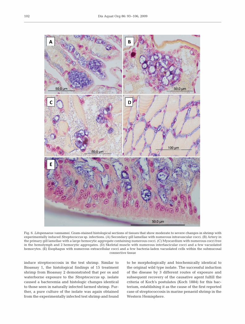
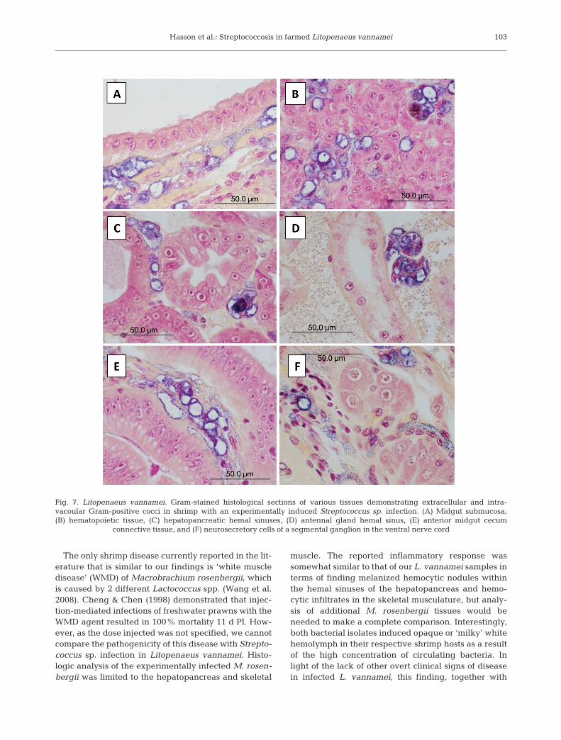
marked hemocytic vasculitis of the LO characterizedby the replacement of normal arterioles with a solidhomogeneous field of numerous vacuolated hemo-cytes. These cells typically contained a large cytoplas-mic vacuole lined with cocci and a marginated or acen-tric nucleus. Numerous extracellular cocci admixedwith low numbers of small melanized hemocytic nod-ules were typically present (Fig. 5B–D) and individualarterioles could not be distinguished. The general mor-phology of the arterioles in moderately infected shrimpwas distnguishable (i.e. walls and lumen) and con-tained a lower number of cocci-laden vacuolated cells,small hemocytic nodules and extracellular cocci ascompared to the severe cases. Numerous cocci werealso visible within the hemal sinuses surrounding theLO arterioles. Similar histologic changes and bacterialdistribution were also present in the walls of the sub-gastric artery (Fig. 5E) and, to a much lesser degree, inall of the other affected tissues. Following the LO interms of infection severity were the gills (Fig. 6A,B)and heart (Fig. 6C); the interfascicular spaces of theskeletal muscle of the cephalothorax, abdomen andappendages (Fig. 6D); and the submucosal connectivetissue or muscle of the esophagus (Fig. 6E), stomachs,midgut (Fig. 7A) and hindgut. Rare to infrequent scat-tered vacuolated hemocytic nodules and varying num-bers of cocci were found in the hemal sinuses or vesselsassociated with the hematopoietic tissue (Fig. 7B),hepatopancreas (Fig. 7C) and antennal gland (Fig. 7D)as well as the connective tissues of the anterior andposterior midgut ceca (Fig. 7E) and in the nerve cord(Fig. 7F). In all cases, the bacteria were very difficult todetect by routine H&E histology (Fig. 5C) because oftheir small diameter (~0.8 µm diameter) and lightbasophilia, providing little contrast with the surround-ing tissues. In Gram-stained tissue sections the bacte-ria stain dark blue to purple and are easily visualizedby light microscopy (Figs. 5D,E, 6 & 7).
Bioassay 3. Freeze-thaw effects on the Streptococcusisolate
As in Bioassay 1, all 10 treatment shrimp of Bioas-say 3 died or were collected moribund within 3 dpost-injection (PI) and were then divided into 2groups of 5 shrimp each, one group frozen at –20°Cand the other at –80°C. Both groups of shrimp werethawed, the ventral sinus fluid plated on BA and theshrimp re-frozen on 3 successive days beginning 4 dpost-collection and, again, on Days 30 and 60 for atotal of 5 freeze-thaw cycles. Small circular whiteStreptococcus-like colonies too numerous to count,identical to those observed throughout this study,grew on all BA plates up through and including all
those prepared after thawing the 10 shrimp for thefifth time. Colony numbers and morphology were vir-tually identical in the cultures of the shrimp stored atboth –20 and –80°C. Microscopic analysis of Gram-stained preparations of representative colonies re-moved from each plate confirmed the presence ofGram-positive cocci in each sample.
DISCUSSION
Histologic analyses of farmed Litopenaeus vannameijuveniles in September 2008 demonstrated the pres-ence of unique changes in the LO associated with asystemic infection by a presumptive Streptococcus sp.Numerous cocci were observed within the hemolymphin virtually all of the tissues throughout the shrimp byroutine histology together with a comparatively mildinflammatory response on the part of the host as sug-gested by the lack of melanized lesions. The bacterialagent was subsequently isolated from fresh L. van-namei tissue and hemolymph samples originating from2 separate farms that had been affected by a persistentchronic shrimp mortality problem. Based on the pre-liminary biochemical characterization work conductedin this study, the bacterial isolate was found to be anon-hemolytic, non-motile, oxidase- and catalase-negative, Lancefield Group B, Gram-positive coccus,which reacted positively with universal streptococcalPCR primers. Although analyses of the isolate usingthe API Strep 20 kit and Biolog system produced con-flicting species identifications (S. uberis vs. S. parau-beris), the overall findings definitively indicate that theisolate is a member of the Streptococcus genus. Histor-ically, differentiation of these 2 streptococcal species isdifficult phenotypically with S. parauberis classified asS. uberis type II in the past (Jayarao et al. 1991). Finalspeciation of the isolate through genotypic analysiswas beyond the scope of the current investigation andis the focus of an ongoing study.
Injection of SPF Litopenaeus vannamei juvenileswith an inoculum containing the Streptococcus sp. iso-late (Bioassay 1) resulted in 100% mortality within 3 dPI and produced histologic lesions in all 5 examinedtreatment shrimp that were identical to those observedin L. vannamei with a naturally occurring infection.The isolate was successfully recovered and a pure cul-ture obtained for use in the subsequent bioassay. Theresults of Bioassay 2 demonstrated that SPF L. van-namei test shrimp can be infected by the agent viawater- and feed-borne routes with cumulative mortali-ties of 80–100 and 40–100%, respectively, within a12 d period. Waterborne bacterial concentrations of~106 CFU ml–1 or more, as reported in Vibrio sp. infec-tivity studies (Saulnier et al. 2000), were sufficient to
101
Dis Aquat Org 86: 93–106, 2009
induce streptococcosis in the test shrimp. Similar toBioassay 1, the histological findings of 15 treatmentshrimp from Bioassay 2 demonstrated that per os andwaterborne exposure to the Streptococcus sp. isolatecaused a bacteremia and histologic changes identicalto those seen in naturally infected farmed shrimp. Fur-ther, a pure culture of the isolate was again obtainedfrom the experimentally infected test shrimp and found
to be morphologically and biochemically identical tothe original wild type isolate. The successful inductionof the disease by 3 different routes of exposure andsubsequent recovery of the causative agent fulfill thecriteria of Koch’s postulates (Koch 1884) for this bac-terium, establishing it as the cause of the first reportedcase of streptococcosis in marine penaeid shrimp in theWestern Hemisphere.
102
Fig. 6. Litopenaeus vannamei. Gram-stained histological sections of tissues that show moderate to severe changes in shrimp withexperimentally induced Streptococcus sp. infections. (A) Secondary gill lamellae with numerous intravascular cocci. (B) Artery inthe primary gill lamellae with a large hemocytic aggregate containing numerous cocci. (C) Myocardium with numerous cocci freein the hemolymph and 2 hemocytic aggregates. (D) Skeletal muscle with numerous interfascicular cocci and a few vacuolatedhemocytes. (E) Esophagus with numerous extracellular cocci and a few bacteria-laden vacuolated cells within the submucosal
connective tissue
Hasson et al.: Streptococcosis in farmed Litopenaeus vannamei
The only shrimp disease currently reported in the lit-erature that is similar to our findings is ‘white muscledisease’ (WMD) of Macrobrachium rosenbergii, whichis caused by 2 different Lactococcus spp. (Wang et al.2008). Cheng & Chen (1998) demonstrated that injec-tion-mediated infections of freshwater prawns with theWMD agent resulted in 100% mortality 11 d PI. How-ever, as the dose injected was not specified, we cannotcompare the pathogenicity of this disease with Strepto-coccus sp. infection in Litopenaeus vannamei. Histo-logic analysis of the experimentally infected M. rosen-bergii was limited to the hepatopancreas and skeletal
muscle. The reported inflammatory response wassomewhat similar to that of our L. vannamei samples interms of finding melanized hemocytic nodules withinthe hemal sinuses of the hepatopancreas and hemo-cytic infiltrates in the skeletal musculature, but analy-sis of additional M. rosenbergii tissues would beneeded to make a complete comparison. Interestingly,both bacterial isolates induced opaque or ‘milky’ whitehemolymph in their respective shrimp hosts as a resultof the high concentration of circulating bacteria. Inlight of the lack of other overt clinical signs of diseasein infected L. vannamei, this finding, together with
103
Fig. 7. Litopenaeus vannamei. Gram-stained histological sections of various tissues demonstrating extracellular and intra-vacoular Gram-positive cocci in shrimp with an experimentally induced Streptococcus sp. infection. (A) Midgut submucosa,(B) hematopoietic tissue, (C) hepatopancreatic hemal sinuses, (D) antennal gland hemal sinus, (E) anterior midgut cecum
connective tissue, and (F) neurosecretory cells of a segmental ganglion in the ventral nerve cord
Dis Aquat Org 86: 93–106, 2009
microscopic verification of numerous Gram-positivecocci within a given hemolymph sample, may serve asvaluable diagnostic tools for making a rapid presump-tive diagnosis of streptococcosis in the field.
The histologic changes caused by this disease in theLO, together with the widespread distribution of coccithroughout the tissues, are unique in penaeids, notpreviously reported and can be used to make a defini-tive diagnosis of streptococcosis in Gram-stained sec-tions. Unlike the formation of LO spheroids followingthe sequestration of various shrimp viruses and othersmall antigenic agents by hemocytes (Hasson et al.1999) or the hemocytic encapsulation of bacteria form-ing melanized nodules within the LO and other vascu-larized tissues as seen with a systemic vibriosis (Light-ner 1996), streptococcosis in L. vannamei induces adistinctive vasculitis in the LO. The disease is charac-terized by the presence of cocci-containing cytoplas-mic vacuoles (phagosomes) in numerous presumptivehemocytes together with low numbers of small intersti-tial melanized hemocytic nodules. In severe infections,normal LO arteriole morphology is lost and replacedby vacuolated, bacteria-laden cells (Fig. 5). These cellsappear to progressively increase in number within thewalls of the arterioles, spreading into the arteriolelumens and surrounding hemal sinuses until individualarterioles are no longer discernable by light micro-scopy and replaced by a solid field of vacuolated cells(Fig. 5B,C). These changes may be considered the his-tologic hallmark of this disease and suggest that thebacteria are either successfully phagocytized andsequestered by these presumed hemocytes or thehemocytes are actively invaded by the bacteria as hasbeen demonstrated to occur in some mammalianepithelial cell lines following experimental exposure toGroup B Streptococci (Rubens et al. 1992, Almeida &Oliver 1995). Once in the phagosome the cocci maycontinue replicating, leading to eventual cell lysis andthe release of bacterial progeny into surrounding tis-sues. Another possibility is that the bacteria simply usethe phagosome as a safe haven where they are pro-tected from host immune responses and exposure toantimicrobials without replicating. Zlotkin et al. (2003)reported a similar phenomenon in salmonid phago-cytes exposed to S. iniae. Dubbed the ‘Trojan horseeffect’, these investigators demonstrated that phagocy-tized S. iniae not only survive in fish macrophages, butinduce apoptosis of these cells, effectively minimizingthe host immune response while being transported anddelivered to typically inaccessible regions such as thecentral nervous system. Ultrastructural analysis ofinfected LO cells in time course-sampled shrimp,together with an assessment of their apoptotic index,are needed to determine if the mode of entry andoccurrences reported in Streptococcus-infected fish
phagocytes or mammalian epithelial cells are compa-rable to those in Streptococcus-infected shrimp hemo-cytes.
Prevention and treatment of this new bacterial dis-ease has already proved difficult for the farmers con-fronting this problem. The intracellular location of theagent will influence the development of managementstrategies for treating this disease. Antibiogram resultsindicated the isolate to be susceptible to a variety ofantibiotics including oxytetracycline (OTC) (P. W.Varner & K. W. Hasson unpubl. data). However, addi-tion of OTC-medicated feed to affected ponds did notend the mortalities. We speculate that the intracellularlocation of the organism may shield it against antimi-crobials like OTC, making treatment of alreadyinfected shrimp ineffectual. This is similar to the prob-lem of treating necrotizing hepatopancreatitis (NHP),an intracellular rickettsial-like disease of Litopenaeusvannamei (Frelier et al. 1992). Successful prevention ofNHP has been achieved through the rapid applicationof OTC-medicated feed at the first signs of an outbreakwhile the shrimp are still feeding (Brock & Main 1994)and may be a strategy worth testing in Streptococcus-affected regions. However, the threat of inducingOTC-resistant Streptococcus sp. is a concern, particu-larly in regions already using this antibiotic to combatNHP. Another strategy employed by farmers is toreduce bacterial disease susceptibility in their stocksby minimizing stressful environmental culture condi-tions based on the premise that the Streptococcus sp.responsible is an opportunistic pathogen and out-breaks are stress-related. This was attempted byreducing stocking densities, increasing water ex-changes and through early harvests with mixed resultsreported. Low dissolved oxygen concentration hasbeen reported to increase the virulence and distribu-tion of Enterococcus seriolicida disease in yellowtailSeriola quinqueradiata (Vendrell et al. 2006). Night-time dissolved oxygen (DO) lows of 2.5 to 3.5 ppmwere reported in the affected shrimp farms and contin-uous aeration at night should be tested to determine ifavoidance of suboptimal DO concentrations mayimprove survivals in Streptococcus-infected shrimpponds. Other efforts have been focused on finding andeliminating the source of the disease. However, elimi-nation of this bacteria through farm dry outs may provedifficult as it has been demonstrated experimentallythat S. parauberis isolates from cattle and turbot canendure for 1 mo in saltwater and up to 6 mo in marinesediment by entering into a dormant state in order topersist for longer periods in the marine environment.These viable yet non-culturable bacteria can be pre-sent in high numbers in the environment, but are notdetectable via normal direct plate count methods (Cur-rás et al. 2002). This trait, combined with the fact that
104
Hasson et al.: Streptococcosis in farmed Litopenaeus vannamei
many streptococcal species occur naturally in the envi-ronment and can become endemic to a farm (Yanong &Francis-Floyd 2006), suggests that their detection andelimination may prove problematic.
In Bioassay 3 a total of 10 shrimp were experimen-tally infected by injection with the Streptococcus sp.isolate and died within 3 d PI as in Bioassay 1. Multiplefreezing and thawing trials of 5 shrimp held at –20°Cand 5 held at –80°C were then conducted. After a totalof 5 freeze-thaw cycles over a 2 mo period, the Strepto-coccus sp. organism was successfully cultured from theventral sinus of all 10 shrimp on each occasion. Theseresults demonstrate that the isolate used in this seriesof studies is highly resistant to long-term freezing andmultiple freeze/thaw cycles and differences in freezingtemperatures had no apparent effect on the organism’sviability.
The collective findings of this study establish theexistence of a new streptococcal disease of penaeidshrimp that can potentially spread to other countriesthrough either live or frozen infected shrimp. Researchemphasis needs to focus on developing PCR primersfor the detection of this Streptococcus sp., identifyingthe source(s) of the bacteria and developing a manage-ment plan for the prevention, containment and treat-ment of future outbreaks.
Acknowledgements. Our appreciation and thanks to Dr. P.Frelier for his editorial comments concerning this manuscript.The authors also express their gratitude to Shrimp Improve-ment Systems and Dr. G. Jaramillo for supplying the SPF testshrimp used in this study, to Ziegler Bros. and Ms. C. Shew forsupplying the feed used in this study and to Dr. T. Samochafor his continued support and contributions to our bioassaysystem.
LITERATURE CITED
Almeida RA, Oliver SP (1995) Invasion of bovine mammaryepithelial cells by Streptococcus dysgalactiae. J Dairy Sci78:1310–1317
Baeck GW, Kim JH, Gomez DK, Park SC (2006) Isolation andcharacterization of Streptococcus sp. from diseased flounder(Paralichthys olivaceus) in Jeju island. J Vet Sci 7:53–58
Bell T, Lightner DV (1988) A handbook of normal penaeidshrimp histology. World Aquaculture Society, BatonRouge, LA
Berridge BR, Fuller JD, Azavedo J, Low DE, Bercovier H, Fre-lier PR (1998) Development of specific nested oligonu-cleotide PCR primers for the Streptococcus iniae 16S-23Sribosomal DNA intergenic spacer. J Clin Microbiol 36:2778–2781
Brock JA, Main KL (1994) A guide to the common problemsand diseases of cultured Penaeus vannamei. World Aqua-culture Society, Baton Rouge, LA
Camus AC, Shewmaker PL, Mauel MJ, Wise DJ (2008) Strep-tococcus ictaluri arthritis, osteolysis, myositis, and spinalmeningitis in channel catfish broodstock. J Aquat AnimHealth 20:54–62
Carson FL (1997) Histotechnology — a self instructional text,2nd edn. American Society of Clinical Pathologists,Chicago, IL
Cheng W, Chen JC (1998) Isolation and characterization of anEnterococcus-like bacterium causing muscle necrosis andmortality in Macrobrachium rosenbergii in Taiwan. DisAquat Org 34:93–101
Currás M, Magariños B, Toranzo AE, Romalde JL (2002) Dor-mancy as a survival strategy of the fish pathogen Strepto-coccus parauberis in the marine environment. Dis AquatOrg 52:129–136
Dawson CR, Mallette MF (1945) The copper proteins. In:Anson ML, Edsall JT (eds) Advances in protein chemistry.Elsevier, Amsterdam, p 179–248
Eldar A, Bejerano Y, Livoff A, Hurvitz A, Bercovier H (1995)Experimental streptococcal meningo-encephalitis in cul-tured fish. Vet Microbiol 43:33–40
Eldar A, Perl S, Frelier PF, Bercovier H (1999) Red drum Sci-aenops ocellatus mortalities associated with Streptococcusiniae infection. Dis Aquat Org 36:121–127
Evans JJ, Shoemaker CA, Klesius PJ (2000) ExperimentalStreptococcus iniae infection of hybrid striped bass(Morone chrysops × Morone saxatilis) and tilapia (Ore-ochromis niloticus) by nares inoculation. Aquaculture189:197–210
Frelier PF, Sis RF, Bell TA, Lewis DH (1992) Microscopic andultrastructural studies of necrotizing hepatopancreatitis inPacific white shrimp (Penaeus vannamei) cultured inTexas. Vet Pathol 29:269–277
Hasson KW, Lightner DV, Poulos BT, Redman RM, White BL,Brock JA, Bonami JR (1995) Taura syndrome in Penaeusvannamei: demonstration of a viral etiology. Dis AquatOrg 23:115–126
Hasson KW, Lightner DV, Mohney LL, Redman RM, WhiteBM (1999) Role of lymphoid organ spheroids in chronicTaura syndrome virus (TSV) infections in Penaeus van-namei. Dis Aquat Org 38:93–105
Hoshina T, Sano T, Morimoto Y (1958) A streptococcus patho-genic to fish (1933). J Tokyo Univ Fish 44:57–68
Koch R (1884) Die Aetiologie der Tuberkulose. Mitt KaiserlGesundheitsamte 2:1–88
Kusuda R, Kawai K, Salati F, Banner CR, Freyer JL (1991)Enterococcus seriolicida sp. nov., a fish pathogen. Int JSyst Bacteriol 41:406–409
Lancefield RC (1933) A serological differentiation of humanand other groups of hemolytic streptococci. J Exp Med57:571–595
Lightner DV (1996) A handbook of shrimp pathology anddiagnostic procedures for diseases of cultured penaeidshrimp. World Aquaculture Society, Baton Rouge, LA
Meiri-Bendek I, Lipkin E, Friedmann A, Leitner G, Saran A,Friedman S, Kashi Y (2002) A PCR-based method for thedetection of Streptococcus agalactiae in milk. J Dairy Sci85:1717–1723
Ringo E, Gatesoupe FJ (1998) Lactic acid bacteria: a review.Aquaculture 160:177–203
Romalde JL, Ravelo C, Valdés I, Magariños B and others(2008) Streptococcus phocae, an emerging pathogen forsalmonid culture. Vet Microbiol 130:198–207
Rubens CE, Smith S, Hulse M, Chi EY, van Belle G (1992)Respiratory epithelial cell invasion by Group B strepto-cocci. Infect Immun 60:5157–5163
105
Dis Aquat Org 86: 93–106, 2009
Saulnier D, Haffner P, Goarant C, Levy P, Ansquer D (2000)Experimental infection models for shrimp vibriosis studies:a review. Aquaculture 191:133–144
Shewmaker PL, Camus AC, Bailiff T, Steigerwalt AG, MoreyRE, Carvalho MS (2007) Streptococcus ictaluri sp. nov.,isolated from channel catfish Ictalurus punctatus brood-stock. Int J System Evol Microbiol 57:1603–1606
Sindermann CJ, Lightner DV (1988) Disease diagnosis andcontrol in North American marine aquaculture. ElsevierScience Publishing, New York
Toranzo AE, Devesa S, Heinen P, Riaza A, Nunez S, Barja JL(1994) Streptococcosis in cultured turbot caused by anEnterococcus-like bacterium. Bull Eur Assoc Fish Pathol14:19–23
Vendrell D, Balcazar JL, Ruiz-Zarzuela I, de Blas I, Girones O,Muzquiz JL (2006) Lactococcus garvieae in fish: a review.Comp Immunol Microbiol Infect Dis 29:177–198
Wang PC, Lin YD, Liaw LL, Chern RS, Chen SC (2008) Lacto-coccus lactis subspecies lactis also causes white muscledisease in farmed giant freshwater prawns Marcro-brachium rosenbergii. Dis Aquat Org 79:9–17
Yanong R, Francis-Floyd R (2006) Streptococcal infections offish. Circular FA057. Institute of Food and AgriculturalSciences, University of Florida, Miami, FL, p 1–6
Zlotkin A, Chilmonczyk S, Eyngor M, Hurvitz A, Ghittino C,Eldar A (2003) Trojan horse effect: phagocyte-mediatedStreptococcus iniae infection of fish. Infect Immun71:2318–2325
106
Editorial responsibility: Grant Stentiford,Weymouth, UK
Submitted: March 19, 2009; Accepted: August 20, 2009Proofs received from author(s): September 14, 2009