Structural diversity of bacterial flagellar motors Songye Chen 1,6 , Morgan Beeby 1,2,6 , Gavin E Murphy 1,7 , Jared R Leadbetter 3 , David R Hendrixson 4 , Ariane Briegel 1,2 , Zhuo Li 1,2,8 , Jian Shi 1,2 , Elitza I Tocheva 1 , Axel Mu ¨ ller 5 , Megan J Dobro 1 and Grant J Jensen 1,2, * 1 Division of Biology, California Institute of Technology, Pasadena, CA, USA, 2 Howard Hughes Medical Institute, California Institute of Technology, Pasadena, CA, USA, 3 Division of Environmental Science and Engineering, California Institute of Technology, Pasadena, CA, USA, 4 Department of Microbiology, University of Texas Southwestern Medical Center, Dallas, TX, USA and 5 Division of Chemistry, California Institute of Technology, Pasadena, CA, USA The bacterial flagellum is one of nature’s most amazing and well-studied nanomachines. Its cell-wall-anchored motor uses chemical energy to rotate a microns-long filament and propel the bacterium towards nutrients and away from toxins. While much is known about flagellar motors from certain model organisms, their diversity across the bacterial kingdom is less well characterized, allowing the occasional misrepresentation of the motor as an invariant, ideal machine. Here, we present an electron cryotomographical survey of flagellar motor architectures throughout the Bacteria. While a conserved structural core was observed in all 11 bacteria imaged, surprisingly novel and divergent structures as well as different symme- tries were observed surrounding the core. Correlating the motor structures with the presence and absence of parti- cular motor genes in each organism suggested the loca- tions of five proteins involved in the export apparatus including FliI, whose position below the C-ring was con- firmed by imaging a deletion strain. The combination of conserved and specially-adapted structures seen here sheds light on how this complex protein nanomachine has evolved to meet the needs of different species. The EMBO Journal (2011) 30, 2972–2981. doi:10.1038/ emboj.2011.186; Published online 14 June 2011 Subject Categories: microbiology & pathogens; structural biology Keywords: bacterial flagellar motor; electron cryotomography; motility; phylogenetic profiling; subtomogram average Introduction The bacterial flagellum is a paradigm in modern molecular biology. Its structural complexity, multi-phasic and strictly regulated self-assembly, mechanical capabilities, functional interdependence with the chemosensory system, and both pathogenic and ecophysiological importance have made it central to studies and discussions in both the scientific and popular literature (Berg, 2003; McCarter, 2006; Pallen and Matzke, 2006; Chevance and Hughes, 2008; Minamino et al, 2008; Sowa and Berry, 2008; Snyder et al, 2009). The flagel- lum consists of a motor also known as the basal body, a flexible linker termed the hook, and a filament that behaves as a helical propeller and is typically many times the length of the bacterium itself. The motor converts ion flux across the cytoplasmic membrane into a torque that rotates the flagel- lum. In some organisms such as Escherichia coli, counter- clockwise rotation generates thrust that propels the cell forward. Signals from chemoreceptor arrays (Briegel et al, 2009) modulate the probability of the flagellar motor rever- sing direction (to spin clockwise) (Hazelbauer et al, 2008), which causes the bacterium to randomly re-orient. Thus, if a bacterium detects that it is swimming towards nutrients, the chemosensory system can prolong its movement in a specific direction. Although all bacterial flagella share similarities in struc- ture and mechanism, extensive variation in number, place- ment and usage exist between species. While cells such as Vibrio cholerae and Caulobacter crescentus, for example, exhibit a single polar flagellum, others, including Salmonella enterica and E. coli distribute several or many flagella around their periphery. Under different conditions, some bacteria alternate between polar and lateral flagellar systems (McCarter, 2004). The flagella of spirochaetes do not typi- cally pierce the outer membrane, but instead remain in the periplasm where they rotate and/or contort the cell (Murphy et al, 2006; Liu et al, 2009; Kudryashev et al, 2010). Flagellar rotation is powered either by a proton- motive force (e.g., E. coli) or by a sodium-motive force (e.g., V. cholerae). The ‘run and tumble’ swimming mode that switches between clockwise and counter-clockwise rota- tions is best known, but other bacteria function differently, for example by varying the speed of a unidirectional motor. The molecular architectures of the helical flagellar filaments are also diverse (Galkin et al, 2008). The flagellar motor is constructed from 420 proteins, and its basic morphology consists of an extended axial rod with coaxial rings termed the L-, P-, S-, M- and C-rings based on their locations relative to the cell. Previous analyses of flagellar motor genes have shown that they are widely dis- tributed throughout and conserved across many bacterial lines of descent, but it has not been clear how observed sequence variations relate to the final structure of the macro- molecular complex (Pallen et al, 2005; Liu and Ochman, 2007; Snyder et al, 2009). Structural studies of the motor to date have focussed on a small number of key organisms and have been hampered by the motor’s size, complexity, Received: 20 December 2010; accepted: 17 May 2011; published online: 14 June 2011 *Corresponding author. Division of Biology, California Institute of Technology, 1200 E. California Blvd, MC114-96, Pasadena, CA 91125, USA. Tel.: þ 1 626 395 8827; Fax: þ 1 626 395 5730; E-mail: [email protected]6 These authors contributed equally to this work 7 Present address: Laboratory of Cell Biology, Center for Cancer Research, National Cancer Institute, National Institutes of Health, Bethesda, MD 20892, USA 8 Present address: Molecular and Cellular Biology Department, City of Hope Beckman Research Institute, Electron Microscopy Facility, Duarte, CA 91010, USA The EMBO Journal (2011) 30, 2972–2981 | & 2011 European Molecular Biology Organization | All Rights Reserved 0261-4189/11 www.embojournal.org The EMBO Journal VOL 30 | NO 14 | 2011 & 2011 European Molecular Biology Organization EMBO THE EMBO JOURNAL THE EMBO JOURNAL 2972
Transcript
Structural diversity of bacterial flagellar motors
Songye Chen1,6, Morgan Beeby1,2,6,Gavin E Murphy1,7, Jared R Leadbetter3,David R Hendrixson4, Ariane Briegel1,2,Zhuo Li1,2,8, Jian Shi1,2, Elitza I Tocheva1,Axel Muller5, Megan J Dobro1
and Grant J Jensen1,2,*1Division of Biology, California Institute of Technology, Pasadena,CA, USA, 2Howard Hughes Medical Institute, California Institute ofTechnology, Pasadena, CA, USA, 3Division of Environmental Scienceand Engineering, California Institute of Technology, Pasadena, CA, USA,4Department of Microbiology, University of Texas Southwestern MedicalCenter, Dallas, TX, USA and 5Division of Chemistry, California Instituteof Technology, Pasadena, CA, USA
The bacterial flagellum is one of nature’s most amazing
and well-studied nanomachines. Its cell-wall-anchored
motor uses chemical energy to rotate a microns-long
filament and propel the bacterium towards nutrients and
away from toxins. While much is known about flagellar
motors from certain model organisms, their diversity
across the bacterial kingdom is less well characterized,
allowing the occasional misrepresentation of the motor as
an invariant, ideal machine. Here, we present an electron
cryotomographical survey of flagellar motor architectures
throughout the Bacteria. While a conserved structural
core was observed in all 11 bacteria imaged, surprisingly
novel and divergent structures as well as different symme-
tries were observed surrounding the core. Correlating the
motor structures with the presence and absence of parti-
cular motor genes in each organism suggested the loca-
tions of five proteins involved in the export apparatus
including FliI, whose position below the C-ring was con-
firmed by imaging a deletion strain. The combination of
conserved and specially-adapted structures seen here
sheds light on how this complex protein nanomachine
has evolved to meet the needs of different species.
The EMBO Journal (2011) 30, 2972–2981. doi:10.1038/
interdependence with the chemosensory system, and both
pathogenic and ecophysiological importance have made it
central to studies and discussions in both the scientific and
popular literature (Berg, 2003; McCarter, 2006; Pallen and
Matzke, 2006; Chevance and Hughes, 2008; Minamino et al,
2008; Sowa and Berry, 2008; Snyder et al, 2009). The flagel-
lum consists of a motor also known as the basal body, a
flexible linker termed the hook, and a filament that behaves
as a helical propeller and is typically many times the length of
the bacterium itself. The motor converts ion flux across the
cytoplasmic membrane into a torque that rotates the flagel-
lum. In some organisms such as Escherichia coli, counter-
clockwise rotation generates thrust that propels the cell
forward. Signals from chemoreceptor arrays (Briegel et al,
2009) modulate the probability of the flagellar motor rever-
sing direction (to spin clockwise) (Hazelbauer et al, 2008),
which causes the bacterium to randomly re-orient. Thus, if a
bacterium detects that it is swimming towards nutrients, the
chemosensory system can prolong its movement in a specific
direction.
Although all bacterial flagella share similarities in struc-
ture and mechanism, extensive variation in number, place-
ment and usage exist between species. While cells such
as Vibrio cholerae and Caulobacter crescentus, for example,
exhibit a single polar flagellum, others, including Salmonella
enterica and E. coli distribute several or many flagella around
their periphery. Under different conditions, some bacteria
alternate between polar and lateral flagellar systems
(McCarter, 2004). The flagella of spirochaetes do not typi-
cally pierce the outer membrane, but instead remain in
the periplasm where they rotate and/or contort the cell
(Murphy et al, 2006; Liu et al, 2009; Kudryashev et al,
2010). Flagellar rotation is powered either by a proton-
motive force (e.g., E. coli) or by a sodium-motive force
(e.g., V. cholerae). The ‘run and tumble’ swimming mode
that switches between clockwise and counter-clockwise rota-
tions is best known, but other bacteria function differently,
for example by varying the speed of a unidirectional motor.
The molecular architectures of the helical flagellar filaments
are also diverse (Galkin et al, 2008).
The flagellar motor is constructed from 420 proteins, and
its basic morphology consists of an extended axial rod with
coaxial rings termed the L-, P-, S-, M- and C-rings based on
their locations relative to the cell. Previous analyses of
flagellar motor genes have shown that they are widely dis-
tributed throughout and conserved across many bacterial
lines of descent, but it has not been clear how observed
sequence variations relate to the final structure of the macro-
molecular complex (Pallen et al, 2005; Liu and Ochman,
2007; Snyder et al, 2009). Structural studies of the motor
to date have focussed on a small number of key organisms
and have been hampered by the motor’s size, complexity,Received: 20 December 2010; accepted: 17 May 2011; publishedonline: 14 June 2011
*Corresponding author. Division of Biology, California Institute ofTechnology, 1200 E. California Blvd, MC114-96, Pasadena, CA 91125,USA. Tel.: þ 1 626 395 8827; Fax: þ 1 626 395 5730;E-mail: [email protected] authors contributed equally to this work7Present address: Laboratory of Cell Biology, Center for CancerResearch, National Cancer Institute, National Institutes of Health,Bethesda, MD 20892, USA8Present address: Molecular and Cellular Biology Department, City ofHope Beckman Research Institute, Electron Microscopy Facility, Duarte,CA 91010, USA
The EMBO Journal (2011) 30, 2972–2981 | & 2011 European Molecular Biology Organization | All Rights Reserved 0261-4189/11
www.embojournal.org
The EMBO Journal VOL 30 | NO 14 | 2011 &2011 European Molecular Biology Organization
membrane localization and attachment to the cell envelope.
These factors make motors difficult if not impossible to purify
intact. X-ray crystallography has nevertheless revealed
the structures of some of the isolated motor proteins, and
complementary biochemical and genetic analyses have lo-
cated many of them within the intact motor. Single-particle
cryoEM studies have produced nanometre-resolution struc-
tures of the purified S. enterica motor, albeit without stators
or the export apparatus (Thomas et al, 2006). In just the past
few years, the development of electron cryotomography
(ECT) has made it possible to image the structures of
complete motors within intact cells in 3D to ‘macromolecu-
lar’ (several nanometres) resolution (Li and Jensen, 2009;
Milne and Subramaniam, 2009). The first in situ structures
have been from the thin spirochaetes Treponema primitia
(Murphy et al, 2006) and Borrelia burgdorferi (Liu et al, 2009;
Kudryashev et al, 2010). Those species demonstrated large
peripheral structures not seen in the S. enterica single-particle
reconstruction, hinting that the diversity of motor structures
might be great.
Results and discussion
Data collected and overall appearance of the motors
We sought to sample the structural diversity of bacterial
flagellar motors. To this end, we imaged the flagellar motors
from 11 phylogenetically diverse bacteria chosen for their
general interest as model organisms; involvement in animal
host associations or free-living lifestyles, and suitability
for ECT. The selected bacteria include those with polar
(C. crescentus and Campylobacter jejuni), peritrichous
(S. enterica and E. coli) and periplasmic (the spirochaetes
T. primitia and B. burgdorferi) flagella. Bacteria employing
Na-driven motors (V. cholerae) or ‘sheathed’ flagella
(V. cholerae and Helicobacter hepaticus) were also included
(see Supplementary Table S1 for comprehensive details on
the phylogenetics and characteristics of each motor). Using
high-throughput imaging methods (Suloway et al, 2009), for
each species hundreds of cryotomograms of whole cells were
collected. Subtomograms containing motors were computa-
tionally extracted from the data, mutually aligned, averaged
and cylindrically symmetrized to obtain resolutions of a few
nanometres (see Materials and methods).
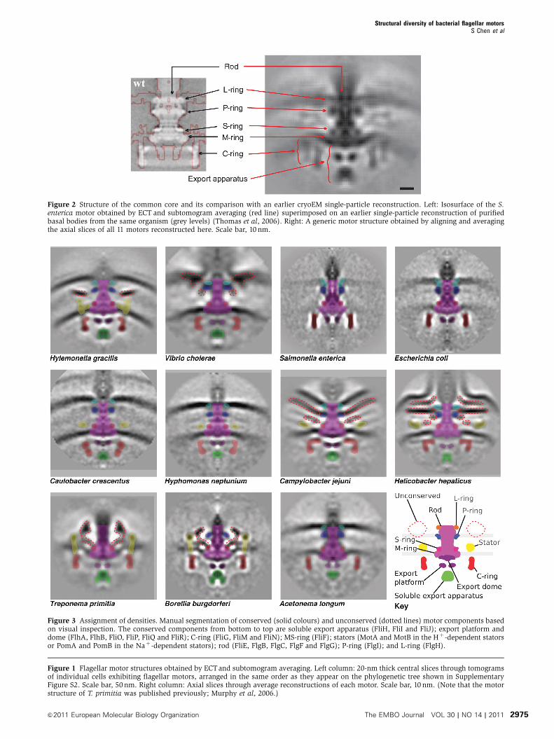
While all the motors had clear similarities, their overall
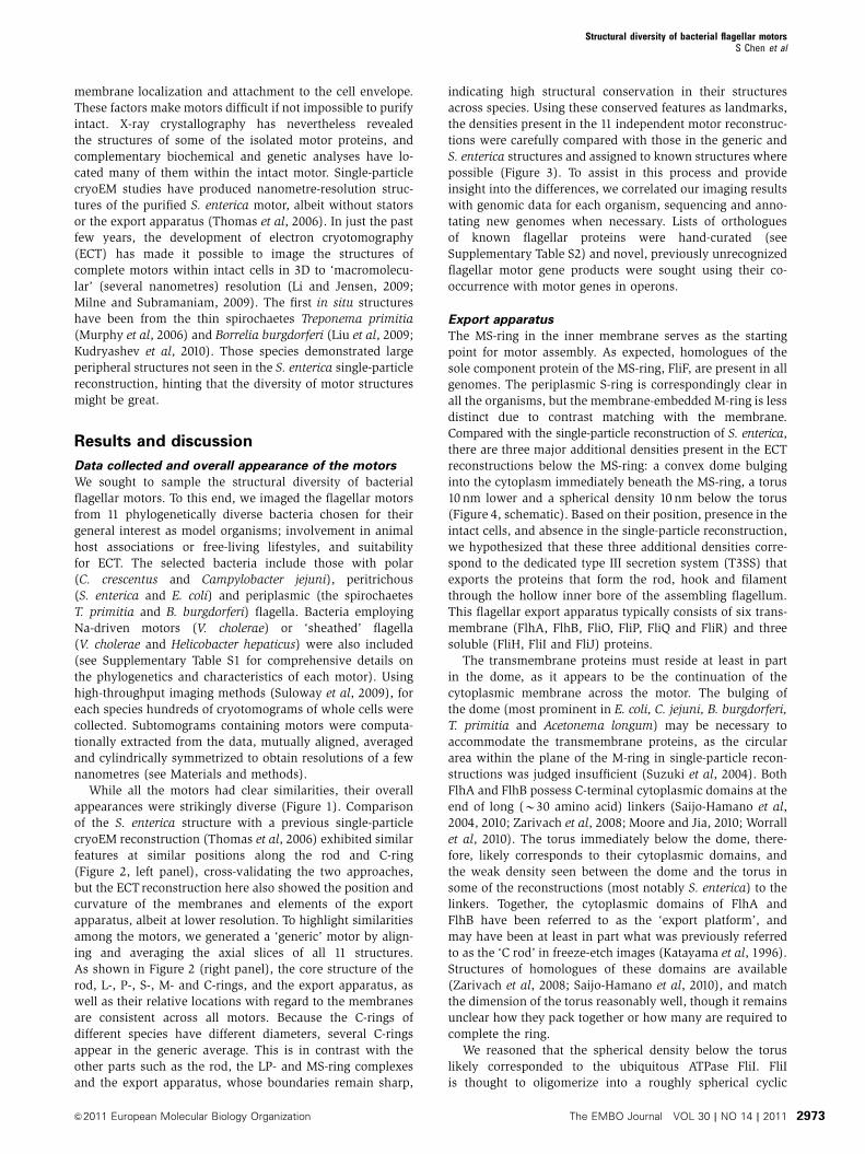
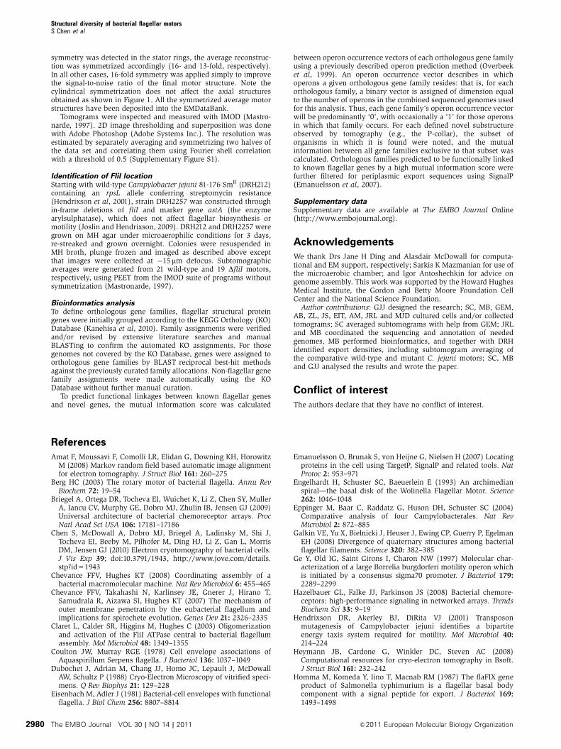
appearances were strikingly diverse (Figure 1). Comparison
of the S. enterica structure with a previous single-particle
cryoEM reconstruction (Thomas et al, 2006) exhibited similar
features at similar positions along the rod and C-ring
(Figure 2, left panel), cross-validating the two approaches,
but the ECT reconstruction here also showed the position and
curvature of the membranes and elements of the export
apparatus, albeit at lower resolution. To highlight similarities
among the motors, we generated a ‘generic’ motor by align-
ing and averaging the axial slices of all 11 structures.
As shown in Figure 2 (right panel), the core structure of the
rod, L-, P-, S-, M- and C-rings, and the export apparatus, as
well as their relative locations with regard to the membranes
are consistent across all motors. Because the C-rings of
different species have different diameters, several C-rings
appear in the generic average. This is in contrast with the
other parts such as the rod, the LP- and MS-ring complexes
and the export apparatus, whose boundaries remain sharp,
indicating high structural conservation in their structures
across species. Using these conserved features as landmarks,
the densities present in the 11 independent motor reconstruc-
tions were carefully compared with those in the generic and
S. enterica structures and assigned to known structures where
possible (Figure 3). To assist in this process and provide
insight into the differences, we correlated our imaging results
with genomic data for each organism, sequencing and anno-
tating new genomes when necessary. Lists of orthologues
of known flagellar proteins were hand-curated (see
Supplementary Table S2) and novel, previously unrecognized
flagellar motor gene products were sought using their co-
occurrence with motor genes in operons.
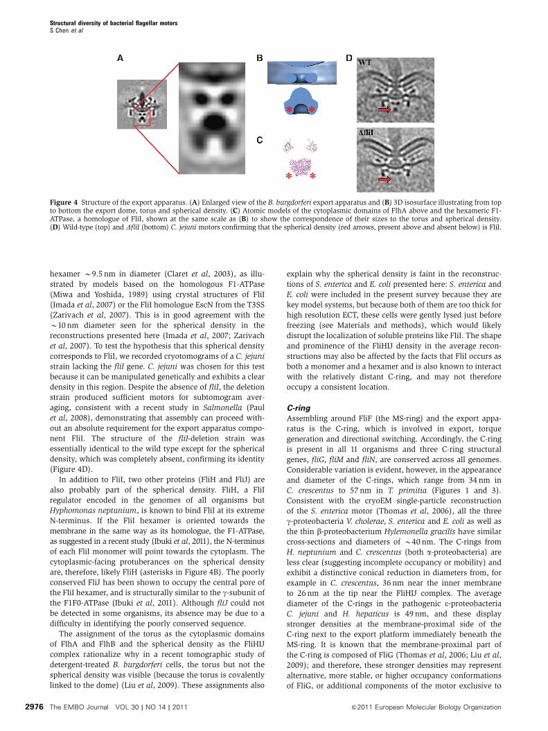
Export apparatus
The MS-ring in the inner membrane serves as the starting
point for motor assembly. As expected, homologues of the
sole component protein of the MS-ring, FliF, are present in all
genomes. The periplasmic S-ring is correspondingly clear in
all the organisms, but the membrane-embedded M-ring is less
distinct due to contrast matching with the membrane.
Compared with the single-particle reconstruction of S. enterica,
there are three major additional densities present in the ECT
reconstructions below the MS-ring: a convex dome bulging
into the cytoplasm immediately beneath the MS-ring, a torus
10 nm lower and a spherical density 10 nm below the torus
(Figure 4, schematic). Based on their position, presence in the
intact cells, and absence in the single-particle reconstruction,
we hypothesized that these three additional densities corre-
spond to the dedicated type III secretion system (T3SS) that
exports the proteins that form the rod, hook and filament
through the hollow inner bore of the assembling flagellum.
This flagellar export apparatus typically consists of six trans-
membrane (FlhA, FlhB, FliO, FliP, FliQ and FliR) and three
soluble (FliH, FliI and FliJ) proteins.
The transmembrane proteins must reside at least in part
in the dome, as it appears to be the continuation of the
cytoplasmic membrane across the motor. The bulging of
the dome (most prominent in E. coli, C. jejuni, B. burgdorferi,
T. primitia and Acetonema longum) may be necessary to
accommodate the transmembrane proteins, as the circular
area within the plane of the M-ring in single-particle recon-
structions was judged insufficient (Suzuki et al, 2004). Both
FlhA and FlhB possess C-terminal cytoplasmic domains at the
end of long (B30 amino acid) linkers (Saijo-Hamano et al,
2004, 2010; Zarivach et al, 2008; Moore and Jia, 2010; Worrall
et al, 2010). The torus immediately below the dome, there-
fore, likely corresponds to their cytoplasmic domains, and
the weak density seen between the dome and the torus in
some of the reconstructions (most notably S. enterica) to the
linkers. Together, the cytoplasmic domains of FlhA and
FlhB have been referred to as the ‘export platform’, and
may have been at least in part what was previously referred
to as the ‘C rod’ in freeze-etch images (Katayama et al, 1996).
Structures of homologues of these domains are available
(Zarivach et al, 2008; Saijo-Hamano et al, 2010), and match
the dimension of the torus reasonably well, though it remains
unclear how they pack together or how many are required to
complete the ring.
We reasoned that the spherical density below the torus
likely corresponded to the ubiquitous ATPase FliI. FliI
is thought to oligomerize into a roughly spherical cyclic
Structural diversity of bacterial flagellar motorsS Chen et al
&2011 European Molecular Biology Organization The EMBO Journal VOL 30 | NO 14 | 2011 2973
Structural diversity of bacterial flagellar motorsS Chen et al
The EMBO Journal VOL 30 | NO 14 | 2011 &2011 European Molecular Biology Organization2974
Figure 2 Structure of the common core and its comparison with an earlier cryoEM single-particle reconstruction. Left: Isosurface of the S.enterica motor obtained by ECT and subtomogram averaging (red line) superimposed on an earlier single-particle reconstruction of purifiedbasal bodies from the same organism (grey levels) (Thomas et al, 2006). Right: A generic motor structure obtained by aligning and averagingthe axial slices of all 11 motors reconstructed here. Scale bar, 10 nm.
Figure 3 Assignment of densities. Manual segmentation of conserved (solid colours) and unconserved (dotted lines) motor components basedon visual inspection. The conserved components from bottom to top are soluble export apparatus (FliH, FliI and FliJ); export platform anddome (FlhA, FlhB, FliO, FliP, FliQ and FliR); C-ring (FliG, FliM and FliN); MS-ring (FliF); stators (MotA and MotB in the Hþ -dependent statorsor PomA and PomB in the Naþ -dependent stators); rod (FliE, FlgB, FlgC, FlgF and FlgG); P-ring (FlgI); and L-ring (FlgH).
Figure 1 Flagellar motor structures obtained by ECTand subtomogram averaging. Left column: 20-nm thick central slices through tomogramsof individual cells exhibiting flagellar motors, arranged in the same order as they appear on the phylogenetic tree shown in SupplementaryFigure S2. Scale bar, 50 nm. Right column: Axial slices through average reconstructions of each motor. Scale bar, 10 nm. (Note that the motorstructure of T. primitia was published previously; Murphy et al, 2006.)
Structural diversity of bacterial flagellar motorsS Chen et al
&2011 European Molecular Biology Organization The EMBO Journal VOL 30 | NO 14 | 2011 2975
hexamer B9.5 nm in diameter (Claret et al, 2003), as illu-
strated by models based on the homologous F1-ATPase
(Miwa and Yoshida, 1989) using crystal structures of FliI
(Imada et al, 2007) or the FliI homologue EscN from the T3SS
(Zarivach et al, 2007). This is in good agreement with the
B10 nm diameter seen for the spherical density in the
reconstructions presented here (Imada et al, 2007; Zarivach
et al, 2007). To test the hypothesis that this spherical density
corresponds to FliI, we recorded cryotomograms of a C. jejuni
strain lacking the fliI gene. C. jejuni was chosen for this test
because it can be manipulated genetically and exhibits a clear
density in this region. Despite the absence of fliI, the deletion
strain produced sufficient motors for subtomogram aver-
aging, consistent with a recent study in Salmonella (Paul
et al, 2008), demonstrating that assembly can proceed with-
out an absolute requirement for the export apparatus compo-
nent FliI. The structure of the fliI-deletion strain was
essentially identical to the wild type except for the spherical
density, which was completely absent, confirming its identity
(Figure 4D).
In addition to FliI, two other proteins (FliH and FliJ) are
also probably part of the spherical density. FliH, a FliI
regulator encoded in the genomes of all organisms but
Hyphomonas neptunium, is known to bind FliI at its extreme
N-terminus. If the FliI hexamer is oriented towards the
membrane in the same way as its homologue, the F1-ATPase,
as suggested in a recent study (Ibuki et al, 2011), the N-terminus
of each FliI monomer will point towards the cytoplasm. The
cytoplasmic-facing protuberances on the spherical density
are, therefore, likely FliH (asterisks in Figure 4B). The poorly
conserved FliJ has been shown to occupy the central pore of
the FliI hexamer, and is structurally similar to the g-subunit of
the F1F0-ATPase (Ibuki et al, 2011). Although fliJ could not
be detected in some organisms, its absence may be due to a
difficulty in identifying the poorly conserved sequence.
The assignment of the torus as the cytoplasmic domains
of FlhA and FlhB and the spherical density as the FliHIJ
complex rationalize why in a recent tomographic study of
detergent-treated B. burgdorferi cells, the torus but not the
spherical density was visible (because the torus is covalently
linked to the dome) (Liu et al, 2009). These assignments also
explain why the spherical density is faint in the reconstruc-
tions of S. enterica and E. coli presented here: S. enterica and
E. coli were included in the present survey because they are
key model systems, but because both of them are too thick for
high resolution ECT, these cells were gently lysed just before
freezing (see Materials and methods), which would likely
disrupt the localization of soluble proteins like FliI. The shape
and prominence of the FliHIJ density in the average recon-
structions may also be affected by the facts that FliI occurs as
both a monomer and a hexamer and is also known to interact
with the relatively distant C-ring, and may not therefore
occupy a consistent location.
C-ring
Assembling around FliF (the MS-ring) and the export appa-
ratus is the C-ring, which is involved in export, torque
generation and directional switching. Accordingly, the C-ring
is present in all 11 organisms and three C-ring structural
genes, fliG, fliM and fliN, are conserved across all genomes.
Considerable variation is evident, however, in the appearance
and diameter of the C-rings, which range from 34 nm in
C. crescentus to 57 nm in T. primitia (Figures 1 and 3).
Consistent with the cryoEM single-particle reconstruction
of the S. enterica motor (Thomas et al, 2006), all the three
g-proteobacteria V. cholerae, S. enterica and E. coli as well as
the thin b-proteobacterium Hylemonella gracilis have similar
cross-sections and diameters of B40 nm. The C-rings from
H. neptunium and C. crescentus (both a-proteobacteria) are
less clear (suggesting incomplete occupancy or mobility) and
exhibit a distinctive conical reduction in diameters from, for
example in C. crescentus, 36 nm near the inner membrane
to 26 nm at the tip near the FliHIJ complex. The average
diameter of the C-rings in the pathogenic e-proteobacteria
C. jejuni and H. hepaticus is 49 nm, and these display
stronger densities at the membrane-proximal side of the
C-ring next to the export platform immediately beneath the
MS-ring. It is known that the membrane-proximal part of
the C-ring is composed of FliG (Thomas et al, 2006; Liu et al,
2009); and therefore, these stronger densities may represent
alternative, more stable, or higher occupancy conformations
of FliG, or additional components of the motor exclusive to
Figure 4 Structure of the export apparatus. (A) Enlarged view of the B. burgdorferi export apparatus and (B) 3D isosurface illustrating from topto bottom the export dome, torus and spherical density. (C) Atomic models of the cytoplasmic domains of FlhA above and the hexameric F1-ATPase, a homologue of FliI, shown at the same scale as (B) to show the correspondence of their sizes to the torus and spherical density.(D) Wild-type (top) and DfliI (bottom) C. jejuni motors confirming that the spherical density (red arrows, present above and absent below) is FliI.
Structural diversity of bacterial flagellar motorsS Chen et al
The EMBO Journal VOL 30 | NO 14 | 2011 &2011 European Molecular Biology Organization2976
the e-proteobacteria. The spirochaetes B. burgdorferi and
T. primitia and the diderm firmicute A. longum possess
C-rings with an even larger average diameter of 54 nm,
but maintain the same cross-section as S. enterica. No corre-
lation could be made between tomographic density and the
presence of FliY, a FliN paralogue with an additional domain
found in A. longum, T. primitia, H. hepaticus, C. jejuni and
S. enterica.
Rod and L/P-rings
Assembling atop the S-ring is the rod, which acts as the
central driveshaft through the cell envelope, transmitting
torque applied to the MS-ring to the hook and filament. The
rod is comprised of ubiquitous paralogous proteins that are
known to form a proximal (FliE, FlgB, FlgC and FlgF) and a
distal (FlgG) rod, but no new details about the arrangement
of these proteins could be gleaned from the tomograms.
The length of the rod was well conserved, as judged by the
distinct densities seen in the generic average for the S- and
P-rings and inner and outer membranes. The distance be-
tween the distal end of the S-ring and the centre of the outer
membrane was 22 nm (measured in the generic average),
confirming earlier measurements of the rod length on isolated
flagellar hook-basal bodies (HBBs) from wild-type S. enterica
(Takahashi et al, 2009).
The periplasmic P- and L-rings around the rod are thought
to function as bushings through the peptidoglycan layer and
lipopolysaccharide of the outer membrane, respectively.
Because the rings coat the outside of the rod, both proteins
(FlgH and FlgI) are exported into the periplasm via the Sec
pathway instead of through the dedicated flagellar T3SS
(Homma et al, 1987; Jones and Macnab, 1990). Unlike the
rod proteins, however, it has previously been noted that the
genes encoding the L/P-ring components are not ubiquitous,
and the variation seen here calls into question the presumed
roles of these proteins (Pallen et al, 2005). The reconstruc-
tions of S. enterica, E. coli, H. neptunium, C. crescentus and
A. longum represent the ‘standard’ cases, where both the
L- and P-ring genes are present, the P-ring is visible in the
tomograms, and the L-ring is not, likely because it is em-
bedded in the outer membrane. In H. gracilis, both genes are
present, and two rings are visible underneath the outer
membrane, suggesting that the L-ring may not be embedded
in the membrane in this species, although it is not clear why
this alternative arrangement is seen. In the sheathed flagella
of H. hepaticus and V. cholerae, the outer membrane con-
tinues as a sheath around the flagellar hook and filament. The
L-ring proteins in these two organisms presumably still form a
complex with the P-ring, but the diverse structures in that
region make assignments unclear. It is noteworthy that the
H. hepaticus L-ring gene does not encode an otherwise
ubiquitous cysteine residue in the vicinity of the amino
terminus (Schoenhals and Macnab, 1996). This residue has
been shown to be part of a signal peptide II cleavage con-
sensus motif that is subsequently lipoylated. The situation is
further complicated by the fact that this cysteine residue is
nevertheless retained in the other organism with a sheathed
flagellum, V. cholerae. In the spirochaetes B. burgdorferi and
T. primitia, the L-ring protein FlgH is absent, as is thought due
to the fact that the flagellar filaments in these organisms never
cross the outer membrane (Pallen et al, 2005), but remain
within the periplasm. The P-ring protein FlgI is absent only in
the genome of T. primitia. This was first observed by tomo-
graphy in a recent study of a B. burgforferi FlgI mutant, in
which a density around the rod was absent (Liu et al, 2009).
Here, we observe a similar absence of ring density on the rod
of T. primitia, corresponding to FlgI. It is noteworthy that FlgI
in the spirochaetes is distant from the peptidoglycan layer, at
odds with its supposed function. FlgI is also found in the
Leptospiraceae, another family within the spirochaete class,
so it may have a special alternative function in the spiro-
chaetes (Chevance et al, 2007).
Stator complex
Above the C-ring and surrounding the MS-ring in the inner
membrane are stator complexes that are thought to extend
towards and bind to the peptidoglycan layer. Through inter-
actions with the C-ring proteins, the stators transform the
flow of Hþ or Naþ ions across the membrane into torque to
drive the motor. Each stator complex is composed of two
proteins, MotA and MotB in the Hþ -dependent stators (e.g.,
E. coli), or PomA and PomB in the Naþ -dependent stators
(e.g., V. cholerae), with an apparent 4:2 stoichiometry. In our
averaged structures, we see distinctive rod-shaped stator ring
structures above the inner membrane with connections
on the other side of the membrane to the C-ring in the
b-proteobacterium H. gracilis and the spirochaetes B. burg-
dorferi and T. primitia. As previously shown elsewhere
(Murphy et al, 2006; Liu et al, 2009; Kudryashev et al,
2010) and reproduced in Figure 5, 16 copies of the stator
complexes are clearly seen in the averaged tomograms of the
spirochaetes B. burgdorferi and T. primitia even before rota-
tional averaging. In contrast, the symmetry observed in the H.
gracilis stator ring is 13 (Figure 5). In the a-proteobacteria (H.
neptunium and C. crescentus), stator densities are visible, but
they are less prominent and their symmetries could not be
discerned. In the e-proteobacteria C. jejuni and H. hepaticus,
stator densities are clear, but they have a different shape in
cross-section, lie nearly parallel to the cytoplasmic mem-
brane, and extend further into the periplasm. The reason
that the symmetry of these motors could not be discerned
may be that the stator system is highly dynamic with rapid
turnover of subunits (Leake et al, 2006) and perhaps variable
conformations, causing low occupancy and heterogeneity.
These cells also have single, polar flagella, and so because
the rod-shaped cells freeze lying flat on the grid, no direct
‘top’ views of the motor were obtained, which makes detect-
ing rotational symmetries more difficult. The rest of the
species may not exhibit clear stator density because the
periplasmic domains of the stator complex (MotA/B or
PomA/B) are thin, as shown in cryoEM images of purified
PomA/B in liposomes (Yonekura et al, 2006).
Unconserved densities
In addition to this somewhat conserved structural core, many
of the organisms exhibited additional, presumably proteinac-
eous densities that are likely to be unrecognized, novel
components of the motor. The most striking is the spiro-
chaetes’ large ‘P-collar’ that forms a bowl-like structure around
the rod within the stator ring, and which has been postulated
to stabilize the motor (Murphy et al, 2006). To generate
hypotheses about which proteins form the P-collar, we
exploited the fact that genes encoding proteins involved in
stable complexes are frequently encoded in genomic proximity
Structural diversity of bacterial flagellar motorsS Chen et al
&2011 European Molecular Biology Organization The EMBO Journal VOL 30 | NO 14 | 2011 2977
to one-another (Huynen et al, 2000). Furthermore, as the
protein is not a component of the cylindrical core of the rod,
hook or filament, we assumed its secretion would be via the
Sec pathway as opposed to the dedicated flagellar T3SS. Using
these criteria (see Materials and methods), we narrowed our
search to a candidate gene for involvement in the P-collar,
flbB, first identified in a comprehensive study of B. burgdorferi
flagellar genetics (Ge et al, 1997). This gene is consistently
near other flagellar motor genes in spirochaetes and includes
the appropriate periplasmic export signal.
While the L/P-ring complex simply surrounds the rod as it
passes through the lipopolysaccharide membrane and pepti-
doglycan layer in the well-studied flagellar motors, in
V. cholerae it appears to serve as a foundation for two
additional structures. V. cholerae possesses two proteins,
MotX and MotY, which form a so-called T-ring that is thought
to assist in the formation and binding of the MotA/MotB
stators (Terashima et al, 2006). Here, this T-ring can be seen
lower and to the outside of the P-ring, to which it is
connected. There is also a substantial increase in density
near the presumed position of the L-ring. Hosogi et al (2011)
recently described this entire density in the closely related
Vibrio alginolyticus HBB as the L-ring. Based on the fact that
the L-ring-encoding flgH gene is B360 amino acids long in
both S. enterica and V. cholerae, we propose that the unu-
sually large and oddly shaped ring structure must also
include an additional, as-yet unidentified protein.
The L/P-ring complex also appears to serve as a structural
anchor in the e-proteobacteria C. jejuni and H. hepaticus.
Here, the L/P-ring complex is attached to large periplasmic
basal disks. In C. jejuni, an extensive disk-like density is
found in the periplasm immediately below and parallel to the
outer membrane around the rod and above the stators. This
disk connects to the P-ring and has an outer radius of
48±9 nm, calculated from the edge of the rod along the
disk. A second disk with a radius of 32±7 nm sits beneath
the first and seems to be faintly connected to the M/S-ring.
In H. hepaticus, a similarly extensive (32±2 nm in radius)
but flat disk is found between what are probably enhanced
L- and P-rings. A similarly large disk (85 nm average radius)
connected to the L/P-ring complex was observed previously
by EM of negatively stained flagellar motors purified
from another e-proteobacterium, Wolinella succinogenes,
which is closely related to C. jejuni and H. hepaticus
(Engelhardt et al, 1993). While all three of these large basal
disks are likely composed of homologous proteins, bioinfor-
matic analyses yielded no candidate proteins for this struc-
ture, likely unaided by the well-discussed genomic
fragmentation and lack of synteny within the e-proteobacteria
(Eppinger et al, 2004).
It is interesting to note that the b-proteobacterium H.
gracilis also exhibited a periplasmic disk above the stators,
but the relationship of this disk to the larger e-proteobacterial
basal disks is unclear. The H. gracilis disk is at the same height
as the P-ring but does not connect, exhibiting an inner and
outer radius of 18±2 and 31±2 nm, respectively. Previously,
similar disks have been observed in another b-proteobacter-
ium, Aquaspirillum serpens (Coulton and Murray, 1978). The
function of these disks remains unclear, as their occurrence
does not seem to correlate with any obvious phenotype (such
as sheathed flagella) or habitat (such as viscous environ-
ment), besides that they are so far exclusive to (but not
Figure 5 Symmetries in the stator region. Upper row: Radial slices through the stator regions of three average motors before rotationalaveraging showing 16-, 16- and 13-fold symmetry, respectively. Lower row: Axial slices through the averaged and symmetrized motors witharrows showing the height at which the radial slices were taken. Scale bar, 10 nm.
Structural diversity of bacterial flagellar motorsS Chen et al
The EMBO Journal VOL 30 | NO 14 | 2011 &2011 European Molecular Biology Organization2978
required for) polar flagella. Based on their position, they may
stabilize the stators to sustain larger torques.
Above the basal disk and immediately below the highly
curved bend in the outer membrane of the sheathed
H. hepaticus is yet another disk-like density. As mentioned
above, in the other sheathed bacterium, V. cholerae, the L-ring
is unusually large and odd-shaped. These structures may
stabilize the outer membrane and the sheath against the
rotation of the flagellum. Finally, two previous studies of
negatively stained, purified C. crescentus motors gave con-
flicting reports about whether or not a so-called E-ring existed
between the M/S- and L/P-ring complexes (Stallmeyer et al,
1989; Kanbe et al, 2005). It remains unclear whether there are
additional rings here in the C. crescentus and H. neptunium
reconstructions.
Conclusion
In summary, three-dimensional structures of 11 phylogeneti-
cally diverse bacterial flagellar motors were obtained in situ
at a few nanometre resolutions via ECT and subtomogram
averaging. The positions of key proteins of the export appa-
ratus were determined. While the motors all exhibited a
common core built from the products of conserved flagellar
genes, their overall appearances were strikingly different due
to variations in the structure of that core and the presence of
unique peripheral densities, showing that each is a related
but specially adapted nanomachine.
Materials and methods
Culture conditionsC. crescentus CB15N, E. coli RP437 and MG1655, V. choleraeTRH7000, H. hepaticus (ATCC 51449), C. jejuni (ATCC 29428),B. burgdorferi B31 (ATCC 35210), A. longum APO-1 DSM 6540 andT. primitia strain ZAS-2 cells were grown as described previously(Murphy et al, 2006; Briegel et al, 2009).
H. neptunium (ATCC 15444) was grown in 2216 marine broth(Difco) at 301C for 24–48 h. The cell culture was incubated on iceduring plunge freezing.
S. enterica subsp. enterica serovar Typhimurium str. LT2 wasgrown in LB plus 0.3 M sucrose and up to 10 mM MgSO4. Penicillin(466 IU/ml) was added 15 and 60 min before plunge freezing.
H. gracilis was cultivated from a rotten lily taken from a Caltechpond by placing a drop of liquid upon a 0.22-mm filter resting atopagar containing 10 mM MOPS pH 7.0, 0.5 g tryptone and 0.5 g yeastextract per litre. Colonies appeared several weeks later. The 16SrRNA was sequenced from a liquid culture and found to be identicalto the type strain of H. gracilis. Cultures were grown in the abovemedia without MOPS for 2 days and only reached an OD of 0.05.The cells were checked for motility with a light microscope. Thecells were then centrifuged and concentrated 10-fold in the samemedia for plunge freezing onto the EM grids (Murphy, 2007).
Genome sequencingH. gracilis (ATCC 19624) cultures were obtained from the ATCC andcultured in liquid and agar media containing 5 g peptone, 0.5 g yeastextract, 0.02 g Tween-80 and 0.1 g K2HPO4 made up to 1 l withnanopure water with optional addition of 15 g agar. Colonies wereinitially grown on agar plates and subsequently used to inoculateliquid cultures. The identity of the cultures was confirmed by PCRamplification and sequencing of 16S rRNA. DNA was purified from2 l cultures by lysing cells with lysozyme followed by consecutiveRnaseA, proteinase K, SDS, sodium acetate and chloroform/phenolmixture additions. DNA was washed with ethanol and dried.A paired-end genomic DNA library was prepared from this sampleand 76 bp reads generated using Illumina sequencing at Caltech’sMillard & Muriel Jacobs Genetics & Genomics Laboratory (http://mmjggl.caltech.edu/sequencing/). Reads were truncated to 67 bpand assembled using Velvet (Zerbino and Birney, 2008). Using a
kmer size of 49, 3.6 Mb of sequence data was generated anddistributed over 152 contigs with an n50 of 75 kb and a maximumcontig length of 192 kb. This partially assembled genome wasannotated for ORFs using the NCBI PGAAP service and flagellargenes semi-automatically annotated using reciprocal best-hitmethodology and manual curation. This Whole Genome Shotgunproject has been deposited at DDBJ/EMBL/GenBank under theaccession AEGR00000000. The version described in this paper is thefirst version, AEGR01000000.
The genome sequencing of A. longum APO-1 was performedat Stanford in the laboratory facility of Stephen Quake and willbe described elsewhere. This Whole Genome Shotgun project hasbeen deposited at DDBJ/EMBL/GenBank under the accessionAFGF00000000. The version described in this paper is the firstversion, AFGF01000000. Genome sequencing results for T. primitiaZAS-2 will be described elsewhere; the closed sequence for thisstrain has been deposited into GenBank as CP001843.
EM sample preparation, data collection and tomogramreconstructionEM R2/2 copper/rhodium Quantifoil or lacy carbon grids were glowdischarged and coated with a 3� -concentrated solution of 10 nmcolloidal gold particles (Ted Pella). A 5� -concentrated solution of10 nm colloidal gold was also added to the cells immediately beforeplunge freezing. A 4-ml droplet of the sample solution was appliedto the EM grid, then blotted and plunge frozen into liquid ethane(Dubochet et al, 1988) or into a liquid ethane–propane mixture(Tivol et al, 2008) using a Vitrobot (FEI Company) (Iancu et al,2006) or in-house plunger. To flatten the thickest cell types, E. coliand S. enterica cells were incubated with 466 IU/ml penicillin G forup to 60 min at 301C (Eisenbach and Adler, 1981), which causedsome cells to rupture when blotted just before plunge freezing. Thegrids were stored under liquid nitrogen until data collection.
EM images were collected using a Polara 300-kV FEG transmis-sion electron microscope (FEI Company) equipped with an energyfilter (slit width 20 eV; Gatan) on a 2k� 2k Ultrascan CCD cameraor, later, a lens-coupled 4k� 4k UltraCam (Gatan). Typically, tiltseries were recorded from�601 to 601 with an increment of 11 semi-automatically around 1 or 2 axes (Iancu et al, 2005) at 8–12mmunder-focus using the predictive UCSF-Tomo package (Zheng et al,2007) or Leginon (Suloway et al, 2009). Cumulative doses of up to200 e�/A2 were used.
Image tilt series were generally binned by two (in X and Y) and3D tomograms were calculated automatically by Raptor (Amat et al,2008) using the Peach distributed computing system (Leong et al,2005). In cases where Raptor was not available or failed, the semi-automatic IMOD software package (Mastronarde, 1997) was used.No digital filters were used to reduce noise. Image tilt series andfinal reconstructions were deposited into an in-house web-baseddatabase. For a more detailed protocol, see Chen et al (2010).
Subtomogram extraction, alignment, averaging andsymmetrizationBsoft (Heymann et al, 2008) and the Peach (Leong et al, 2005)distributed computing system were used for iterative subtomo-gram extraction, alignment, averaging and symmetrization. ForV. cholerae, H. neptunium and A. longum, tomograms of differentmagnifications were used, and the tomograms of higher magnifica-tion were re-sampled to match the pixel size of the tomograms oflowest magnification. The positions of flagellar motors in thetomograms were marked by eye at the entry point of the flagellarfilament into the outer membrane. For alignment, subtomogramscentred on the motors were extracted, bandpass filtered between200 nm and the first CTF zero and masked in reciprocal spacewithin the missing wedge. Rotational alignments were done with31 step sizes, as no further improvement was seen with finer steps.Subtomograms containing flagellar motors were first extracted byhand and aligned to a reference arbitrarily chosen from the datasets. The aligned motors were then averaged, and the averagewas rotated so that the rod axis coincided with the z-axis. Thisresult was cylindrically symmetrized around the z-axis. Subtomo-grams were computationally re-extracted based on the previousalignment result, realigned to the symmetrized average motor,re-averaged and symmetrized. The whole process was iterated untilthe coordinates of motors stabilized. Average motors were checkedcomputationally for symmetry about the rod before applying anysymmetry. In the cases of the spirochaetes and H. gracilis, where
Structural diversity of bacterial flagellar motorsS Chen et al
&2011 European Molecular Biology Organization The EMBO Journal VOL 30 | NO 14 | 2011 2979
symmetry was detected in the stator rings, the average reconstruc-tion was symmetrized accordingly (16- and 13-fold, respectively).In all other cases, 16-fold symmetry was applied simply to improvethe signal-to-noise ratio of the final motor structure. Note thecylindrical symmetrization does not affect the axial structuresobtained as shown in Figure 1. All the symmetrized average motorstructures have been deposited into the EMDataBank.
Tomograms were inspected and measured with IMOD (Mastro-narde, 1997). 2D image thresholding and superposition was donewith Adobe Photoshop (Adobe Systems Inc.). The resolution wasestimated by separately averaging and symmetrizing two halves ofthe data set and correlating them using Fourier shell correlationwith a threshold of 0.5 (Supplementary Figure S1).
Identification of FliI locationStarting with wild-type Campylobacter jejuni 81-176 SmR (DRH212)containing an rpsL allele conferring streptomycin resistance(Hendrixson et al, 2001), strain DRH2257 was constructed throughin-frame deletions of fliI and marker gene astA (the enzymearylsulphatase), which does not affect flagellar biosynthesis ormotility (Joslin and Hendrixson, 2009). DRH212 and DRH2257 weregrown on MH agar under microaerophilic conditions for 3 days,re-streaked and grown overnight. Colonies were resuspended inMH broth, plunge frozen and imaged as described above exceptthat images were collected at �15mm defocus. Subtomographicaverages were generated from 21 wild-type and 19 DfliI motors,respectively, using PEET from the IMOD suite of programs withoutsymmetrization (Mastronarde, 1997).
Bioinformatics analysisTo define orthologous gene families, flagellar structural proteingenes were initially grouped according to the KEGG Orthology (KO)Database (Kanehisa et al, 2010). Family assignments were verifiedand/or revised by extensive literature searches and manualBLASTing to confirm the automated KO assignments. For thosegenomes not covered by the KO Database, genes were assigned toorthologous gene families by BLAST reciprocal best-hit methodsagainst the previously curated family allocations. Non-flagellar genefamily assignments were made automatically using the KODatabase without further manual curation.
To predict functional linkages between known flagellar genesand novel genes, the mutual information score was calculated
between operon occurrence vectors of each orthologous gene familyusing a previously described operon prediction method (Overbeeket al, 1999). An operon occurrence vector describes in whichoperons a given orthologous gene family resides: that is, for eachorthologous family, a binary vector is assigned of dimension equalto the number of operons in the combined sequenced genomes usedfor this analysis. Thus, each gene family’s operon occurrence vectorwill be predominantly ‘0’, with occasionally a ‘1’ for those operonsin which that family occurs. For each defined novel substructureobserved by tomography (e.g., the P-collar), the subset oforganisms in which it is found were noted, and the mutualinformation between all gene families exclusive to that subset wascalculated. Orthologous families predicted to be functionally linkedto known flagellar genes by a high mutual information score werefurther filtered for periplasmic export sequences using SignalP(Emanuelsson et al, 2007).
Supplementary dataSupplementary data are available at The EMBO Journal Online(http://www.embojournal.org).
Acknowledgements
We thank Drs Jane H Ding and Alasdair McDowall for computa-tional and EM support, respectively; Sarkis K Mazmanian for use ofthe microaerobic chamber; and Igor Antoshechkin for advice ongenome assembly. This work was supported by the Howard HughesMedical Institute, the Gordon and Betty Moore Foundation CellCenter and the National Science Foundation.
Author contributions: GJJ designed the research; SC, MB, GEM,AB, ZL, JS, EIT, AM, JRL and MJD cultured cells and/or collectedtomograms; SC averaged subtomograms with help from GEM; JRLand MB coordinated the sequencing and annotation of neededgenomes, MB performed bioinformatics, and together with DRHidentified export densities, including subtomogram averaging ofthe comparative wild-type and mutant C. jejuni motors; SC, MBand GJJ analysed the results and wrote the paper.
Conflict of interest
The authors declare that they have no conflict of interest.
References
Amat F, Moussavi F, Comolli LR, Elidan G, Downing KH, HorowitzM (2008) Markov random field based automatic image alignmentfor electron tomography. J Struct Biol 161: 260–275
Berg HC (2003) The rotary motor of bacterial flagella. Annu RevBiochem 72: 19–54
Briegel A, Ortega DR, Tocheva EI, Wuichet K, Li Z, Chen SY, MullerA, Iancu CV, Murphy GE, Dobro MJ, Zhulin IB, Jensen GJ (2009)Universal architecture of bacterial chemoreceptor arrays. ProcNatl Acad Sci USA 106: 17181–17186
Chen S, McDowall A, Dobro MJ, Briegel A, Ladinsky M, Shi J,Tocheva EI, Beeby M, Pilhofer M, Ding HJ, Li Z, Gan L, MorrisDM, Jensen GJ (2010) Electron cryotomography of bacterial cells.J Vis Exp 39; doi:10.3791/1943, http://www.jove.com/details.stp?id=1943
Chevance FFV, Takahashi N, Karlinsey JE, Gnerer J, Hirano T,Samudrala R, Aizawa SI, Hughes KT (2007) The mechanism ofouter membrane penetration by the eubacterial flagellum andimplications for spirochete evolution. Genes Dev 21: 2326–2335
Claret L, Calder SR, Higgins M, Hughes C (2003) Oligomerizationand activation of the FliI ATPase central to bacterial flagellumassembly. Mol Microbiol 48: 1349–1355
Dubochet J, Adrian M, Chang JJ, Homo JC, Lepault J, McDowallAW, Schultz P (1988) Cryo-Electron Microscopy of vitrified speci-mens. Q Rev Biophys 21: 129–228
Eisenbach M, Adler J (1981) Bacterial-cell envelopes with functionalflagella. J Biol Chem 256: 8807–8814
Emanuelsson O, Brunak S, von Heijne G, Nielsen H (2007) Locatingproteins in the cell using TargetP, SignalP and related tools. NatProtoc 2: 953–971
Engelhardt H, Schuster SC, Baeuerlein E (1993) An archimedianspiral—the basal disk of the Wolinella Flagellar Motor. Science262: 1046–1048
Eppinger M, Baar C, Raddatz G, Huson DH, Schuster SC (2004)Comparative analysis of four Campylobacterales. Nat RevMicrobiol 2: 872–885
Galkin VE, Yu X, Bielnicki J, Heuser J, Ewing CP, Guerry P, EgelmanEH (2008) Divergence of quaternary structures among bacterialflagellar filaments. Science 320: 382–385
Ge Y, Old IG, Saint Girons I, Charon NW (1997) Molecular char-acterization of a large Borrelia burgdorferi motility operon whichis initiated by a consensus sigma70 promoter. J Bacteriol 179:2289–2299
Hendrixson DR, Akerley BJ, DiRita VJ (2001) Transposonmutagenesis of Campylobacter jejuni identifies a bipartiteenergy taxis system required for motility. Mol Microbiol 40:214–224
Heymann JB, Cardone G, Winkler DC, Steven AC (2008)Computational resources for cryo-electron tomography in Bsoft.J Struct Biol 161: 232–242
Homma M, Komeda Y, Iino T, Macnab RM (1987) The flaFIX geneproduct of Salmonella typhimurium is a flagellar basal bodycomponent with a signal peptide for export. J Bacteriol 169:1493–1498
Structural diversity of bacterial flagellar motorsS Chen et al
The EMBO Journal VOL 30 | NO 14 | 2011 &2011 European Molecular Biology Organization2980
Hosogi N, Shigematsu H, Terashima H, Homma M, Nagayama K(2011) Zernike phase contrast cryo-electron tomography of so-dium-driven flagellar hook-basal bodies from Vibrio alginolyti-cus. J Struct Biol 173: 67–76
Huynen M, Snel B, Lathe W, Bork P (2000) Predicting proteinfunction by genomic context: quantitative evaluation and quali-tative inferences. Genome Res 10: 1204–1210
Iancu CV, Tivol WF, Schooler JB, Dias DP, Henderson GP, MurphyGE, Wright ER, Li Z, Yu Z, Briegel A, Gan L, He Y, Jensen GJ(2006) Electron cryotomography sample preparation using theVitrobot. Nat Protoc 1: 2813–2819
Iancu CV, Wright ER, Benjamin J, Tivol WF, Dias DP, Murphy GE,Morrison RC, Heymann JB, Jensen GJ (2005) A ‘flip-flop’ rotationstage for routine dual-axis electron cryotomography. J Struct Biol151: 288–297
Ibuki T, Imada K, Minamino T, Kato T, Miyata T, Namba K (2011)Common architecture of the flagellar type III protein export appa-ratus and F- and V-type ATPases. Nat Struct Mol Biol 18: 277–282
Imada K, Minamino T, Tahara A, Namba K (2007) Structuralsimilarity between the flagellar type III ATPase Flil and F-1-ATPase subunits. Proc Natl Acad Sci USA 104: 485–490
Jones CJ, Macnab RM (1990) Flagellar assembly in Salmonellatyphimurium: analysis with temperature-sensitive mutants.J Bacteriol 172: 1327–1339
Joslin SN, Hendrixson DR (2009) Activation of the Campylobacterjejuni FlgSR two-component system is linked to the flagellarexport apparatus. J Bacteriol 191: 2656–2667
Kanbe M, Shibata S, Umino Y, Jenal U, Aizawa SI (2005) Proteasesusceptibility of the Caulobacter crescentus flagellar hook-basalbody: a possible mechanism of flagellar ejection during celldifferentiation. Microbiology 151: 433–438
Kanehisa M, Goto S, Furumichi M, Tanabe M, Hirakawa M (2010)KEGG for representation and analysis of molecular networksinvolving diseases and drugs. Nucleic Acids Res 38: D355–D360
Katayama E, Shiraishi T, Oosawa K, Baba N, Aizawa S (1996)Geometry of the flagellar motor in the cytoplasmic membraneof Salmonella typhimurium as determined by stereo-photogram-metry of quick-freeze deep-etch replica images. J Mol Biol 255:458–475
Kudryashev M, Cyrklaff M, Wallich R, Baumeister W, FrischknechtF (2010) Distinct in situ structures of the Borrelia flagellar motor.J Struct Biol 169: 54–61
Leake MC, Chandler JH, Wadhams GH, Bai F, Berry RM, ArmitageJP (2006) Stoichiometry and turnover in single, functioningmembrane protein complexes. Nature 443: 355–358
Leong PA, Heymann JB, Jensen GJ (2005) Peach: a simple Perl-based system for distributed computation and its application tocryo-EM data processing—Ways & means. Structure 13: 505–511
Li Z, Jensen GJ (2009) Electron cryotomography: a new view intomicrobial ultrastructure. Curr Opin Microbiol 12: 333–340
Liu J, Lin T, Botkin DJ, McCrum E, Winkler H, Norris SJ (2009)Intact flagellar motor of Borrelia burgdorferi revealed by cryo-electron tomography: evidence for stator ring curvature androtor/C-ring assembly flexion. J Bacteriol 191: 5026–5036
Liu RY, Ochman H (2007) Stepwise formation of the bacterialflagellar system. Proc Natl Acad Sci USA 104: 7116–7121
Mastronarde DN (1997) Dual-axis tomography: an approach withalignment methods that preserve resolution. J Struct Biol 120:343–352
McCarter LL (2006) Regulation of flagella. Curr Opin Microbiol 9:180–186
Milne JLS, Subramaniam S (2009) Cryo-electron tomography ofbacteria: progress, challenges and future prospects. Nat RevMicrobiol 7: 666–675
Minamino T, Imada K, Namba K (2008) Molecular motors of thebacterial flagella. Curr Opin Struct Biol 18: 693–701
Miwa K, Yoshida M (1989) The alpha 3 beta 3 complex, the catalyticcore of F1-ATPase. Proc Natl Acad Sci USA 86: 6484–6487
Moore SA, Jia YH (2010) Structure of the cytoplasmic domain of theflagellar secretion apparatus component FlhA from Helicobacterpylori. J Biol Chem 285: 21060–21069
Murphy GE (2007) Cryoelectron Tomography of Bacteria and TheirMacromolecular Machines. Pasadena, USA: California Institute ofTechnology
Overbeek R, Fonstein M, D’Souza M, Pusch GD, Maltsev N (1999)The use of gene clusters to infer functional coupling. Proc NatlAcad Sci USA 96: 2896–2901
Pallen MJ, Matzke NJ (2006) From the origin of species to the originof bacterial flagella. Nat Rev Microbiol 4: 784–790
Pallen MJ, Penn CW, Chaudhuri RR (2005) Bacterial flagellardiversity in the post-genomic era. Trends Microbiol 13:143–149
Paul K, Erhardt M, Hirano T, Blair DF, Hughes KT (2008) Energysource of flagellar type III secretion. Nature 451: 489–492
Saijo-Hamano Y, Imada K, Minamino T, Kihara M, Shimada M,Kitao A, Namba K (2010) Structure of the cytoplasmic domain ofFlhA and implication for flagellar type III protein export. MolMicrobiol 76: 260–268
Saijo-Hamano Y, Minamino T, Macnab RM, Namba K (2004)Structural and functional analysis of the C-terminal cytoplasmicdomain of FIhA, an integral membrane component of the type IIIflagellar protein export apparatus in Salmonella. J Mol Biol 343:457–466
Schoenhals GJ, Macnab RM (1996) Physiological and biochemicalanalyses of FlgH, a lipoprotein forming the outer membrane Lring of the flagellar basal body of Salmonella typhimurium.J Bacteriol 178: 4200–4207
Stallmeyer MJB, Hahnenberger KM, Sosinsky GE, Shapiro L,Derosier DJ (1989) Image reconstruction of the flagellar basalbody of Caulobacter crescentus. J Mol Biol 205: 511–518
Suloway C, Shi J, Cheng A, Pulokas J, Carragher B, Potter CS, ZhengSQ, Agard DA, Jensen GJ (2009) Fully automated, sequential tilt-series acquisition with Leginon. J Struct Biol 167: 11–18
Suzuki H, Yonekura K, Namba K (2004) Structure of the rotor of thebacterial flagellar motor revealed by electron cryomicroscopy andsingle-particle image analysis. J Mol Biol 337: 105–113
Takahashi N, Mizuno S, Hirano T, Chevance FFV, Hughes KT,Aizawa SI (2009) Autonomous and FliK-dependent lengthcontrol of the flagellar rod in Salmonella enterica. J Bacteriol191: 6469–6472
Terashima H, Fukuoka H, Yakushi T, Kojima S, Homma M (2006)The Vibrio motor proteins, MotX and MotY, are associated withthe basal body of Na+-driven flagella and required for statorformation. Mol Microbiol 62: 1170–1180
Thomas DR, Francis NR, Xu C, DeRosier DJ (2006) The three-dimensional structure of the flagellar rotor from a clockwise-locked mutant of Salmonella enterica serovar Typhimurium.J Bacteriol 188: 7039–7048
Tivol WF, Briegel A, Jensen GJ (2008) An improved cryogen forplunge freezing. Microsc Microanal 14: 375–379
Worrall LJ, Vuckovic M, Strynadka NCJ (2010) Crystal structure ofthe C-terminal domain of the Salmonella type III secretion systemexport apparatus protein InvA. Protein Sci 19: 1091–1096
Yonekura K, Yakushi T, Atsumi T, Maki-Yonekura S, Homma M,Namba K (2006) Electron cryomicroscopic visualization ofPomA/B stator units of the sodium-driven flagellar motor inliposomes. J Mol Biol 357: 73–81
Zarivach R, Deng WY, Vuckovic M, Felise HB, Nguyen HV, Miller SI,Finlay BB, Strynadka NCJ (2008) Structural analysis of theessential self-cleaving type III secretion proteins EscU and SpaS.Nature 453: 124–127
Zarivach R, Vuckovic M, Deng WY, Finlay BB, Strynadka NCJ(2007) Structural analysis of a prototypical ATPase from thetype III secretion system. Nat Struct Mol Biol 14: 131–137
Zerbino DR, Birney E (2008) Velvet: algorithms for de novoshort read assembly using de Bruijn graphs. Genome Res 18:821–829
Zheng SQ, Keszthelyi B, Branlund E, Lyle JM, Braunfeld MB,Sedat JW, Agard DA (2007) UCSF tomography: an integratedsoftware suite for real-time electron microscopic tomographicdata collection, alignment, and reconstruction. J Struct Biol 157:138–147
Structural diversity of bacterial flagellar motorsS Chen et al
&2011 European Molecular Biology Organization The EMBO Journal VOL 30 | NO 14 | 2011 2981