ARTICLE Received 30 Dec 2015 | Accepted 31 May 2016 | Published 15 Jul 2016 Structural investigation of heteroyohimbine alkaloid synthesis reveals active site elements that control stereoselectivity Anna Stavrinides 1, *, Evangelos C. Tatsis 1, *, Lorenzo Caputi 1 , Emilien Foureau 2 , Clare E.M Stevenson 1 , David M. Lawson 1 , Vincent Courdavault 2 & Sarah E. O’Connor 1 Plants produce an enormous array of biologically active metabolites, often with stereo- chemical variations on the same molecular scaffold. These changes in stereochemistry dramatically impact biological activity. Notably, the stereoisomers of the heteroyohimbine alkaloids show diverse pharmacological activities. We reported a medium chain dehydrogenase/reductase (MDR) from Catharanthus roseus that catalyses formation of a heteroyohimbine isomer. Here we report the discovery of additional heteroyohimbine synthases (HYSs), one of which produces a mixture of diastereomers. The crystal structures for three HYSs have been solved, providing insight into the mechanism of reactivity and stereoselectivity, with mutation of one loop transforming product specificity. Localization and gene silencing experiments provide a basis for understanding the function of these enzymes in vivo. This work sets the stage to explore how MDRs evolved to generate structural and biological diversity in specialized plant metabolism and opens the possibility for metabolic engineering of new compounds based on this scaffold. DOI: 10.1038/ncomms12116 OPEN 1 The John Innes Centre, Department of Biological Chemistry, Norwich NR4 7UH, UK. 2 Universite ´ Franc ¸ois-Rabelais de Tours, EA2106 ‘Biomole ´cules et Biotechnologies Ve ´ge ´tales’, Tours 37200, France. *These authors contributed equally to this work. Correspondence and requests for materials should be addressed to V.C. (email: [email protected]) or to S.E.O’C. (email: [email protected]). NATURE COMMUNICATIONS | 7:12116 | DOI: 10.1038/ncomms12116 | www.nature.com/naturecommunications 1
Transcript
ARTICLE
Received 30 Dec 2015 | Accepted 31 May 2016 | Published 15 Jul 2016
Structural investigation of heteroyohimbine alkaloidsynthesis reveals active site elements that controlstereoselectivityAnna Stavrinides1,*, Evangelos C. Tatsis1,*, Lorenzo Caputi1, Emilien Foureau2, Clare E.M Stevenson1,
David M. Lawson1, Vincent Courdavault2 & Sarah E. O’Connor1
Plants produce an enormous array of biologically active metabolites, often with stereo-
chemical variations on the same molecular scaffold. These changes in stereochemistry
dramatically impact biological activity. Notably, the stereoisomers of the heteroyohimbine
alkaloids show diverse pharmacological activities. We reported a medium chain
dehydrogenase/reductase (MDR) from Catharanthus roseus that catalyses formation of a
heteroyohimbine isomer. Here we report the discovery of additional heteroyohimbine
synthases (HYSs), one of which produces a mixture of diastereomers. The crystal structures
for three HYSs have been solved, providing insight into the mechanism of reactivity and
stereoselectivity, with mutation of one loop transforming product specificity. Localization and
gene silencing experiments provide a basis for understanding the function of these enzymes
in vivo. This work sets the stage to explore how MDRs evolved to generate structural and
biological diversity in specialized plant metabolism and opens the possibility for metabolic
engineering of new compounds based on this scaffold.
DOI: 10.1038/ncomms12116 OPEN
1 The John Innes Centre, Department of Biological Chemistry, Norwich NR4 7UH, UK. 2 Universite Francois-Rabelais de Tours, EA2106 ‘Biomolecules etBiotechnologies Vegetales’, Tours 37200, France. * These authors contributed equally to this work. Correspondence and requests for materials should beaddressed to V.C. (email: [email protected]) or to S.E.O’C. (email: [email protected]).
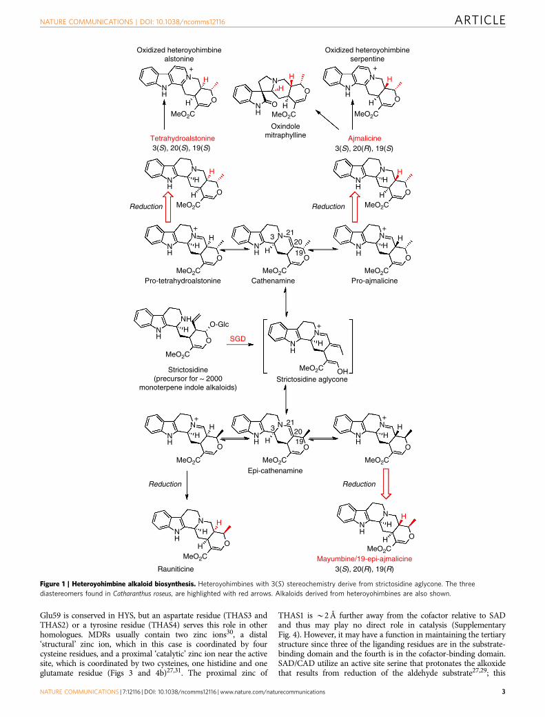
Heteroyohimbines are a prevalent subclass of the mono-terpene indole alkaloids (Corynanthe type skeleton),having been isolated from many plant species, primarily
from the Apocynaceae and Rubiaceae families1. Thesealkaloids exhibit numerous biological activities: ajmalicine is ana1-adrenergic receptor antagonist2–5, and mayumbine (19-epi-ajmalicine) is a ligand for the benzodiazepine receptor (Fig. 1)6.Oxidized beta-carboline heteroyohimbines also exhibit potentpharmacological activity: serpentine has shown topoisomeraseinhibition activity7 and alstonine has been shown to interact with5-HT2A/C receptors and shows promise as an anti-psychoticagent8–13. In addition, heteroyohimbines are biosyntheticprecursors of many oxindole alkaloids, which also display awide range of biological activities14. Although a total of 16heteroyohimbine stereoisomers are possible, only 8 are reportedto be found in nature, at stereocentres C3, C19, C20 (Fig. 1)14–20.How and why the stereoselectivity is controlled in thebiosynthesis of these alkaloids remains unclear.
The medicinal plant Catharanthus roseus produces three ofthese isomers, ajmalicine (raubasine), tetrahydroalstonine and 19-epi-ajmalicine (mayumbine) (Fig. 1)21. These heteroyohimbines,along with the majority of monoterpene indole alkaloids,are derived from deglycosylated strictosidine (strictosidineaglycone)22. The removal of a glucose unit from strictosidine bystrictosidine glucosidase (SGD) forms a reactive dialdehydeintermediate that can rearrange to form numerous isomers23.The stabilization of these isomers by enzyme-catalyzed reductionis hypothesized to be the stepping stone for the extensivechemical diversity observed in the monoterpene indole alkaloids(Fig. 1)21,22. We recently reported the first cloning of abiosynthetic gene encoding an enzyme that acts on strictosidineaglycone. This zinc-dependent medium chain dehydrogenase/reductase (MDR), named tetrahydroalstonine synthase (THAS),produces the heteroyohimbine tetrahydroalstonine (Fig. 1)24.Although these studies demonstrated that THAS is a key enzymein heteroyohimbine biosynthesis, the mechanism by which thisenzyme controls the stereoselectivity of the reduction remainedunknown. Moreover, it is important to note that strictosidineaglycone serves as the precursor for many alkaloid scaffolds, andtherefore represents a central branch point in the monoterpeneindole alkaloid biosynthetic pathway. Therefore, we set out toidentify additional heteroyohimbine synthases (HYSs) withdifferent stereochemical product profiles that would moreclearly define how structural diversity, in this case theformation of different stereoisomers, is controlled in this system.
In this study, we assayed 14 MDR homologues identified fromthe C. roseus transcriptome25,26 that have homology to THAS(Cr_024553). This screen revealed three additional enzymes withTHAS activity (Cr_010119, Cr_021691, Cr_032583a), and,importantly, an enzyme that produced a mixture of hetero-yohimbine diastereomers (Cr_032583b). Crystal structures ofTHAS (here referred to as THAS1), a second representativeTHAS (Cr_021691, THAS2) and the structure of the promiscuoushomologue (Cr_032583b, HYS) were solved and mutants revealedkey residues that control the stereochemistry of the productprofiles. Notably, analysis of the subcellular localization of some ofthese HYSs indicates an unusual nuclear localization pattern and aninteraction with the previous enzyme, SGD. These discoveriesprovide insight into the mechanism and evolution of a crucialbranch point in a specialized metabolic pathway with bothpharmacological and evolutionary importance.
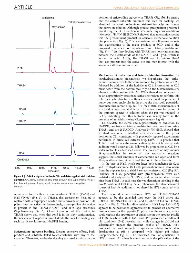
ResultsDiscovery of HYSs. Guided by our initial discovery of THAS1(ref. 24) we identified candidates from the MDR protein family in
the C. roseus transcriptome25,26 based on amino acid similarity tothis enzyme (Supplementary Table 1). Each of these candidateswas cloned from C. roseus cDNA and expressed in Escherichiacoli, with the exception of Cr_017994, which could not beexpressed and was not considered further. The remainingcandidates were assayed with the substrate strictosidineaglycone, and product formation was monitored by liquidchromatography mass spectrometry (LC-MS). Of these, four(Cr_010119, Cr_021691, Cr_032583a, Cr_032583b) reducedstrictosidine aglycone to a product corresponding to one of theheteroyohimbines (Fig. 2, Supplementary Fig. 1). The products ofthe enzymatic reactions were identified based on LC-MS data andcomparison to authentic standards (Supplementary Fig. 2).Enzymes that failed to produce a heteroyohimbine productwere not studied further (Supplementary Fig. 1). Three of theenzymes (Cr_021691, THAS2; Cr_010119, THAS3; Cr_032583a,THAS4) produced tetrahydroalstonine in B85% yield, withsmall amounts of 19-epi-ajmalicine (mayumbine) (o15%) alsoobserved in these reactions, similar to the previously reportedTHAS1. Notably, one enzyme (Cr_032583b, HYS) produced adramatically different product profile consisting of a mixture ofajmalicine/tetrahydroalstonine/mayumbine (55:27:15, at pH 6)(Fig. 2). The discovery of this enzyme, HYS, now provides amolecular basis to understand the generation of stereochemicaldiversity in this alkaloid family.
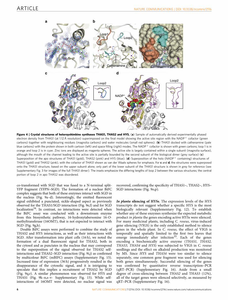
Crystallography of three HYSs. To understand the mechanismof stereochemical control at this crucial biosynthetic branchpoint, we crystallized three HYSs. THAS1 and THAS2, whichproduce predominantly tetrahydroalstonine, were both crystal-lized, since their amino acid sequence identity is relatively low(55%) and the predicted active sites have numerous differences(Fig. 3). HYS, which has a distinctly different product profile, wasalso crystallized to explore the structural basis behind this distinctstereochemical outcome. Structures (Supplementary Table 3)were obtained for THAS1 and THAS2 with NADPþ bound(THAS1, 1.05 Å resolution (Fig. 4a,b; Supplementary Figs 3–5);THAS2, 2.10 Å resolution (Fig. 4d, Supplementary Fig. 4)) and inapo form (THAS1, 2.25 Å resolution (Fig. 4c, SupplementaryFig. 5); THAS2, 2.05 Å resolution (Fig. 4c)), while HYS could onlybe crystallized in the apo form (2.25 Å resolution, Fig. 4c).
Structural features of HYS active sites. The five HYS structuresdescribed here are similar to sinapyl alcohol dehydrogenase(SAD; PDB accession codes 1YQX and 1YQD) and the SADhomologue cinnamyl alcohol dehydrogenase (CAD; PDB acces-sion codes 2CF5 and 2CF6)27–29, which reduce the aldehydemoiety of lignin precursors. Indeed, pairwise superpositions ofsubunits from these structures gave RMSD values of o2 Å(Supplementary Table 4). The biological unit is an elongatedhomodimer, with each subunit divided into a substrate andcofactor-binding domain; the latter also being responsible forforming the dimer interface (Supplementary Fig. 3). The overallstructure of THAS1, with active site, cofactor and loopshighlighted, is shown in Supplementary Fig. 3.
The active site cavities of the HYSs are framed by helix a2, thecatalytic zinc coordination sphere, and loops 1 and 2, with theNADP(H) co-substrate binding at the base of the active site (Figs 3and 4a,b,d, , Supplementary Fig. 4). Loop 2, which is positionedabove the active site, is highly variable in length and sequence(Figs 3 and 4d). In both THAS1 and THAS2, a network of aminoacids holds NADPþ in place. Most notably, Glu59 of THAS1anchors NADP(H) through a bidentate interaction with bothribose hydroxyls, with His59 playing a comparable role in SAD,although here the interaction is with the 30 OH only (Fig. 4b).
Glu59 is conserved in HYS, but an aspartate residue (THAS3 andTHAS2) or a tyrosine residue (THAS4) serves this role in otherhomologues. MDRs usually contain two zinc ions30, a distal‘structural’ zinc ion, which in this case is coordinated by fourcysteine residues, and a proximal ‘catalytic’ zinc ion near the activesite, which is coordinated by two cysteines, one histidine and oneglutamate residue (Figs 3 and 4b)27,31. The proximal zinc of
THAS1 is B2 Å further away from the cofactor relative to SADand thus may play no direct role in catalysis (SupplementaryFig. 4). However, it may have a function in maintaining the tertiarystructure since three of the liganding residues are in the substrate-binding domain and the fourth is in the cofactor-binding domain.SAD/CAD utilize an active site serine that protonates the alkoxidethat results from reduction of the aldehyde substrate27,29; this
serine is replaced with a tyrosine residue in THAS1 (Tyr56) andHYS (Tyr53) (Fig. 3). In THAS2, this tyrosine on helix a2 isreplaced with a tryptophan residue, but a tyrosine at position 120points into the active site. Interestingly, a non-proline cis-peptideis present in the THAS1-NADPþ and HYS apo structures(Supplementary Fig. 5). Closer inspection of this region inTHAS1 shows that when this bond is in the trans conformation,the side chain of Asp340 is projected into the cofactor-binding sitesuch that it would prevent NADPH binding.
Strictosidine aglycone binding. Despite extensive efforts, bothproduct and substrate failed to co-crystallize with any of theenzymes. Therefore, molecular docking was used to visualize the
position of strictosidine aglycone in THAS1 (Fig. 4b). To ensurethat the correct substrate tautomer was used for docking, weidentified the most predominant strictosidine aglycone isomerthat forms in solution. Although product precipitation preventedmonitoring the SGD reaction in situ under aqueous conditions(Methods), 1H,15N-HMBC NMR showed that an enamine specieswas the predominant product in aqueous methanolic solution(Supplementary Fig. 6). This is consistent with literature reportsthat cathenamine is the major product of SGD, and is theproposed precursor of ajmalicine and tetrahydroalstonine(Fig. 1)21,23. In silico docking with THAS1 positions cathenaminebetween the nicotinamide of the NADPþ and Tyr56, which islocated on helix a2 (Fig. 4b). THAS1 loop 1 contains Phe65that also projects into the active site and may interact with thearomatic cathenamine substrate.
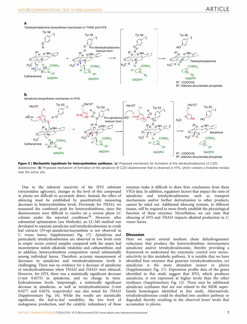
Mechanism of reduction and heteroyohimbine formation. Intetrahydroalstonine biosynthesis, we hypothesize that cathe-namine tautomerizes to the iminium form by protonation at C20,followed by addition of the hydride at C21. Protonation at C20must occur from the bottom face to yield the S stereochemistryobserved at this position (Fig. 5a). While there does not appear tobe an appropriately positioned active site residue to perform thisrole, the crystal structures of these enzymes reveal the presence ofnumerous water molecules in the active site that could potentiallyprotonate this carbon (Fig. 4a). 1H,15N-HMBC measurements ofstrictosidine aglycone at different pH values show formation ofthe iminium species in solution when the pH was reduced toB3.5, indicating that this tautomer can readily form in thepresence of an acidic moiety (Supplementary Fig. 6).
To elucidate the stereo and regioselectivity of reduction byNADPH, we isolated tetrahydroalstonine from reactions usingTHAS1 and pro-R-NADPD. Analysis by 1H-NMR showed thattetrahydroalstonine is labelled with deuterium in the pro-Rposition at C21, consistent with previously reported experimentsperformed in crude cell extracts (Fig. 6a)32. It is possible thatTHAS1 could reduce the enamine directly, in which case hydrideaddition would occur at C21, followed by protonation at C20 by awater molecule as described above. The presence of mayumbine/19-epi-ajmalicine in some of the enzymatic reactionssuggests that small amounts of cathenamine can open and form19-epi-cathenamine, either in solution or in the active site.
In the case of HYS, which produces both ajmalicine (R C20)and tetrahydroalstonine (S C20), protonation must also occurfrom the opposite face to yield R stereochemistry at C20 (Fig. 5b).Products of HYS generated with pro-R-NADPD were alsoisolated and analysed by 1H-NMR, and, as for tetrahydroalsto-nine from THAS1 in each case showed deuterium labelling in thepro-R position at C21 (Fig. 6a–c). Therefore, the stereochemicalcourse of hydride addition is not altered in HYS compared withTHAS1.
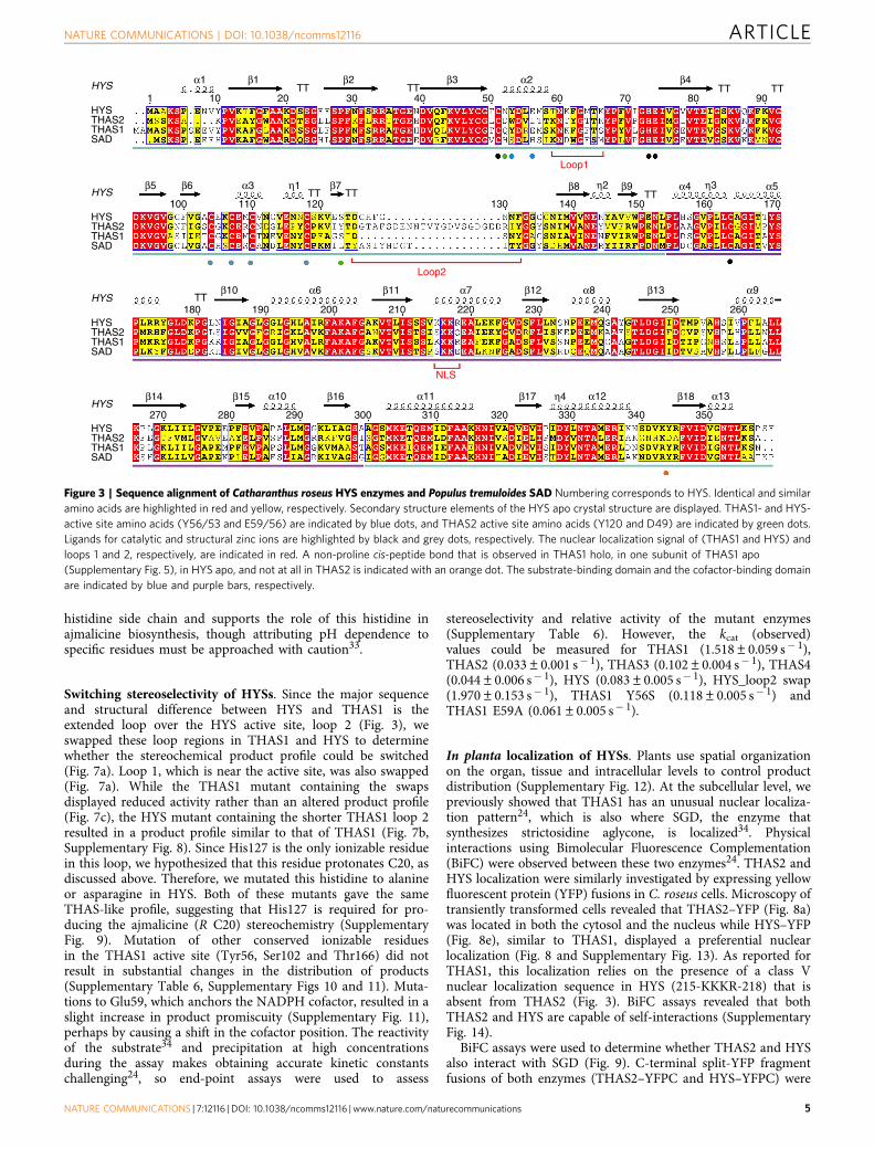
The major difference between HYS and THAS1/THAS2appears to be the extended loop over the HYS active site(D125-GHFGNN-F132 in HYS and D128-SN-Y131 in THAS1,loop 2 in Fig. 3). The histidine residue in HYS loop 2 (His127)appears to be positioned appropriately to provide an alternativeproton source for the opposite (‘top’) face of the substrate, whichcould explain the appearance of ajmalicine in the product profileof HYS. Reactions with THAS1 and HYS performed at differentpH conditions (5–8) revealed that while changes in pH did notsubstantially impact the product profile of THAS1, HYSproduced increased amounts of ajmalicine relative to tetrahy-droalstonine at pH 6 compared with higher pH values(Supplementary Fig. 7). The increased level of ajmalicine inHYS at lower pH values is consistent with the pKa value of the
Ajmalicine
Tetrahydroalstonine
Mayumbine
HYS
THAS4
THAS3
THAS2
THAS1
Cr033062
8.00 9.00 10.00 11.00 12.00 13.00 14.00Time (min)
Figure 2 | LC-MS analysis of active MDR candidates against strictosidine
aglycone. Cr033062 exhibited only trace activity. See Supplementary Fig. 1
for chromatograms of assays with inactive enzymes and negative
histidine side chain and supports the role of this histidine inajmalicine biosynthesis, though attributing pH dependence tospecific residues must be approached with caution33.
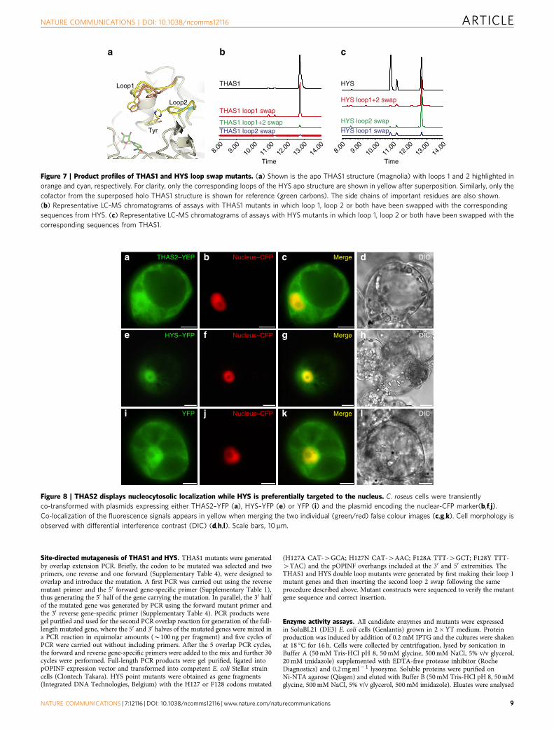
Switching stereoselectivity of HYSs. Since the major sequenceand structural difference between HYS and THAS1 is theextended loop over the HYS active site, loop 2 (Fig. 3), weswapped these loop regions in THAS1 and HYS to determinewhether the stereochemical product profile could be switched(Fig. 7a). Loop 1, which is near the active site, was also swapped(Fig. 7a). While the THAS1 mutant containing the swapsdisplayed reduced activity rather than an altered product profile(Fig. 7c), the HYS mutant containing the shorter THAS1 loop 2resulted in a product profile similar to that of THAS1 (Fig. 7b,Supplementary Fig. 8). Since His127 is the only ionizable residuein this loop, we hypothesized that this residue protonates C20, asdiscussed above. Therefore, we mutated this histidine to alanineor asparagine in HYS. Both of these mutants gave the sameTHAS-like profile, suggesting that His127 is required for pro-ducing the ajmalicine (R C20) stereochemistry (SupplementaryFig. 9). Mutation of other conserved ionizable residuesin the THAS1 active site (Tyr56, Ser102 and Thr166) did notresult in substantial changes in the distribution of products(Supplementary Table 6, Supplementary Figs 10 and 11). Muta-tions to Glu59, which anchors the NADPH cofactor, resulted in aslight increase in product promiscuity (Supplementary Fig. 11),perhaps by causing a shift in the cofactor position. The reactivityof the substrate34 and precipitation at high concentrationsduring the assay makes obtaining accurate kinetic constantschallenging24, so end-point assays were used to assess
stereoselectivity and relative activity of the mutant enzymes(Supplementary Table 6). However, the kcat (observed)values could be measured for THAS1 (1.518±0.059 s� 1),THAS2 (0.033±0.001 s� 1), THAS3 (0.102±0.004 s� 1), THAS4(0.044±0.006 s� 1), HYS (0.083±0.005 s� 1), HYS_loop2 swap(1.970±0.153 s� 1), THAS1 Y56S (0.118±0.005 s� 1) andTHAS1 E59A (0.061±0.005 s� 1).
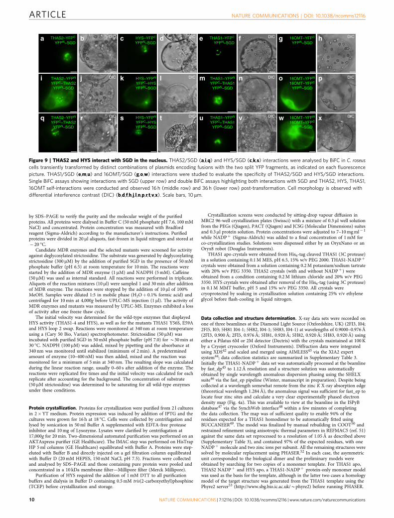
In planta localization of HYSs. Plants use spatial organizationon the organ, tissue and intracellular levels to control productdistribution (Supplementary Fig. 12). At the subcellular level, wepreviously showed that THAS1 has an unusual nuclear localiza-tion pattern24, which is also where SGD, the enzyme thatsynthesizes strictosidine aglycone, is localized34. Physicalinteractions using Bimolecular Fluorescence Complementation(BiFC) were observed between these two enzymes24. THAS2 andHYS localization were similarly investigated by expressing yellowfluorescent protein (YFP) fusions in C. roseus cells. Microscopy oftransiently transformed cells revealed that THAS2–YFP (Fig. 8a)was located in both the cytosol and the nucleus while HYS–YFP(Fig. 8e), similar to THAS1, displayed a preferential nuclearlocalization (Fig. 8 and Supplementary Fig. 13). As reported forTHAS1, this localization relies on the presence of a class Vnuclear localization sequence in HYS (215-KKKR-218) that isabsent from THAS2 (Fig. 3). BiFC assays revealed that bothTHAS2 and HYS are capable of self-interactions (SupplementaryFig. 14).
BiFC assays were used to determine whether THAS2 and HYSalso interact with SGD (Fig. 9). C-terminal split-YFP fragmentfusions of both enzymes (THAS2–YFPC and HYS–YFPC) were
1 10 20TT TT
30 40 50 60 70 80TT TT
90
α1 β1
β5 β6
β10
β14 β15 β16 β17 β18
α6
α10 α11
β11 α7 β12 α8
α12 α13
β13 α9
β7α3 η1
β2 β3 α2 β4
HYS
HYSTHAS2THAS1SAD
HYS
HYSTHAS2THAS1SAD
HYS
HYSTHAS2THAS1SAD
HYS
HYSTHAS2THAS1SAD
100
180
270 280 290 300 310 320 330 340 350
110
190 200 210 220 230 240 250 260
120 130 140 150 160 170TT
TT
TTβ8 β9
Loop1
Loop2
NLS
α4 α5η2 η3TT
η4
Figure 3 | Sequence alignment of Catharanthus roseus HYS enzymes and Populus tremuloides SAD Numbering corresponds to HYS. Identical and similar
amino acids are highlighted in red and yellow, respectively. Secondary structure elements of the HYS apo crystal structure are displayed. THAS1- and HYS-
active site amino acids (Y56/53 and E59/56) are indicated by blue dots, and THAS2 active site amino acids (Y120 and D49) are indicated by green dots.
Ligands for catalytic and structural zinc ions are highlighted by black and grey dots, respectively. The nuclear localization signal of (THAS1 and HYS) and
loops 1 and 2, respectively, are indicated in red. A non-proline cis-peptide bond that is observed in THAS1 holo, in one subunit of THAS1 apo
(Supplementary Fig. 5), in HYS apo, and not at all in THAS2 is indicated with an orange dot. The substrate-binding domain and the cofactor-binding domain
are indicated by blue and purple bars, respectively.
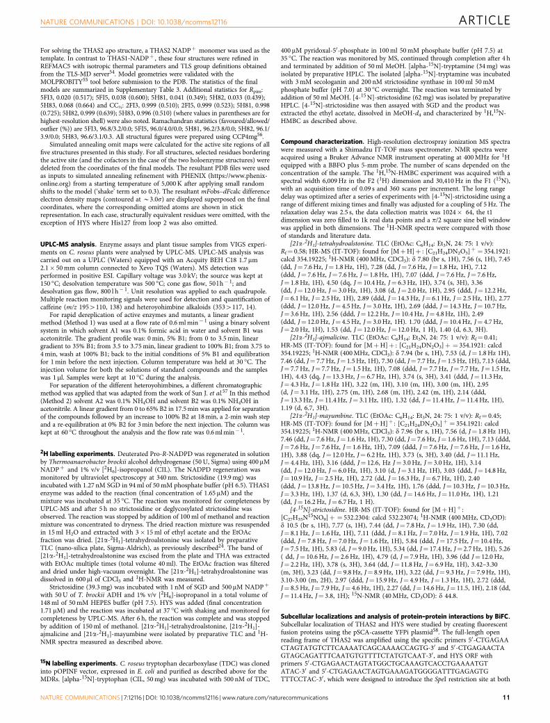
co-transformed with SGD that was fused to a N-terminal split-YFP fragment (YFPN–SGD). The formation of a nuclear BiFCcomplex suggests that both of these enzymes interact with SGD inthe nucleus (Fig. 9a–d). Interestingly, the emitted fluorescentsignal exhibited a punctated, sickle-shaped aspect as previouslyobserved for the THAS1/SGD interaction (Fig. 9e,f) and for SGDlocalization34. In contrast, no interactions were detected whenthe BiFC assay was conducted with a downstream enzymefrom this biosynthetic pathway, 16-hydroxytabersonine 16-O-methyltransferase (16OMT), that is not expected to interact withSGD (Fig. 9g,h).
Double BiFC assays were performed to combine the study ofTHAS2 and HYS interactions, as well as their interactions withSGD. After transformation into plant cells (16 h), we noted theformation of a dual fluorescent signal for THAS2, both inthe cytosol and as punctates in the nucleus that may correspondto the superposition of the signal observed for THAS2 self-interactions and THAS2–SGD interaction (Fig. 9i,j) as confirmedby multicolour BiFC (mBIFC) assays (Supplementary Fig. 15).Increased time of expression (36 h) progressively resulted in thedisappearance of the cytosolic signal, and it is intriguing tospeculate that this implies a recruitment of THAS2 by SGD(Fig. 9q,r). A similar phenomenon was observed for HYS andTHAS1 (Fig. 9k–n,s–v- Supplementary Fig. 15). While self-interactions of 16OMT were detected, no nuclear signal was
recovered, confirming the specificity of THAS1–, THAS2–, HYS–SGD interactions (Fig. 9o,p).
In planta silencing of HYSs. The expression levels of the HYStranscripts do not suggest whether a specific HYS is the mostbiologically relevant (Supplementary Fig. 12). To establishwhether any of these enzymes synthesize the expected metabolicproduct in planta the genes encoding active HYSs were silenced.For many medicinal plants, including C. roseus, virus-inducedgene silencing (VIGS) is the only established method to silencegenes in the whole plant. In C. roseus, the effect of VIGS istemporally and spatially limited to the first two leaves thatemerge immediately after infection35. Each of the genesencoding a biochemically active enzyme (THAS1, THAS2,THAS3, THAS4 and HYS) was subjected to VIGS in C. roseusseedlings and the effect on alkaloid production was monitoredby MS. Since HYS and THAS4 were too similar to silenceseparately, one common gene fragment was used for silencingboth genes simultaneously. Successful silencing of the geneswas confirmed by quantitative reverse transcription–PCR(qRT–PCR) (Supplementary Fig. 16). Aside from a smalldegree of cross-silencing between THAS2 and THAS3 (12%),all of the target genes were silenced selectively, as measured byqRT–PCR (Supplementary Fig. 16).
LooLooLooooLoL oL p111
b
c d
a
LooLooLooLooLooLoLoLoLoLLLoLo p1p1p1p11111p1
CCatCatC tCatCatCatCatCatCatC tCatCCatatatattthenhenhenhenhenhenhenhenhenhennhenhehenamiamiamiamiamiamiamiamiamamaa neneeeeeeeenennCatCatatCatCatCatCatCatCC hhenenhenhhehenhennhhh amamamamiaaa ne
Figure 4 | Crystal structures of heteroyohimbine synthases THAS1, THAS2 and HYS. (a) Sample of automatically derived experimentally phased
electron density from THAS1 (at 1.12 Å resolution) superimposed on the final model showing the active site region with the NADPþ cofactor (green
carbons) together with neighbouring residues (magnolia carbons) and water molecules (small red spheres). (b) THAS1 docked with cathenamine (pale
blue carbons) with the protein shown in both cartoon (left) and space filling (right) modes. The NADPþ cofactor is shown with green carbons; loop 1 is in
orange and loop 2 is in cyan. Zinc ions are displayed as magenta spheres. The active site is largely contained within a single subunit (magnolia surface),
although the mouth of the channel leading to the active site is partially bounded by the second subunit of the biological dimer (grey surface) (c)
Superposition of the apo structures of THAS1 (gold), THAS2 (pink) and HYS (blue). (d) Superposition of the holo (NADPþ containing) structures of
THAS1 (gold) and THAS2 (pink), with the cofactor of THAS1 shown as van der Waals spheres for emphasis. For c and d, the structures were superposed
onto the THAS1 structure, based on the upper subunit alone; only part of the lower subunit of the THAS1 structure is shown in grey for reference (see
Supplementary Fig. 3 for images of the full THAS1 dimer). The insets emphasize the differing lengths of loop 2 between the various structures; the central
Due to the inherent reactivity of the HYS substrate(strictosidine aglycone), changes in the level of this compoundin planta are difficult to accurately detect. Instead, the effect ofsilencing must be established by quantitatively measuringdecreases in heteroyohimbine levels. Previously for THAS1, wemeasured the combined peak for heteroyohimbines, since thediastereomers were difficult to resolve on a reverse phase LCcolumn under the reported conditions24. However, aftersubstantial optimization (see Methods), an LC-MS method wasdeveloped to separate ajmalicine and tetrahydroalstonine in crudeleaf extracts (19-epi-ajamlicine/mayumbine is not observed inC. roseus leaves, Supplementary Fig. 17). Ajmalicine andparticularly tetrahydroalstonine are observed in low levels evenin empty vector control samples compared with the major leafmonoterpene indole alkaloids vindoline and catharanthine, andin addition, heteroyohimbine composition varied substantiallyamong individual leaves. Therefore, accurate measurement ofdecreases in ajmalicine and tetrahydroalstonine levels ischallenging. There was no evidence for a decrease of ajmalicineor tetrahydroalstonine when THAS2 and THAS3 were silenced.However, for HYS, there was a statistically significant decrease(t-test 0.0275) in ajmalicine, and no change in tetra-hydroalstonine levels. Surprisingly, a statistically significantdecrease in ajmalicine, as well as tetrahydroalstonine (t-test0.0277 and 0.0276, respectively) was also noted for THAS1(Supplementary Fig. 16). While the results are statisticallysignificant, the leaf-to-leaf variability, the low level ofendogenous production, and the catalytic redundancy of these
enzymes make it difficult to draw firm conclusions from theseVIGS data. In addition, regulatory factors that impact the ratio ofajmalicine and tetrahydroalstonine, such as transportmechanisms and/or further derivatization to other products,cannot be ruled out. Additional silencing systems, in differenttissues, will be required to more firmly establish the physiologicalfunction of these enzymes. Nevertheless, we can state thatsilencing of HYS and THAS1 impacts alkaloid production in C.roseus leaves.
DiscussionHere we report several medium chain dehydrogenases/reductases that produce the heteroyohimbine stereoisomersajmalicine and/or tetrahydroalstonine, thereby providing aframework to understand the enzymatic control over stereo-selectivity in this metabolic pathway. It is notable that we haveidentified four enzymes that generate tetrahydroalstonine, yetajmalicine is the more abundant isomer in planta(Supplementary Fig. 17). Expression profile data of the genesidentified in this study suggest that HYS, which producesajmalicine, is not expressed at higher levels than the othersynthases (Supplementary Fig. 12). There may be additionalajmalicine synthases that are not related to the MDR super-family homologues identified in this study. Alternatively,tetrahydroalstonine could be shuttled into another pathway ordegraded, thereby resulting in the observed lower levels thataccumulate in planta.
Tetrahydroalstonine biosynthesis mechanism in THAS and HYSa
b
Pro-tetrahydroalstonineiminium form
Tyr 56
O
O ON N+
Cathenamine
Ajmalicine biosynthesis mechanism in HYS
His127Tyr 53
Tyr 53 Tyr 53 Tyr 53
Pro-ajmalicineiminium from
HO-H H O
His127 His127 His127
H3C H3C
SR
H H CONH2
N R2
NADPH
H
HCONH2
NR2
R2
R2
NADPH
NADPH NADPH NADPH NADPH
H
H
CONH2 CONH2
N N+R2 R2
R
NADPH NADPH
HO
SH
H OH
HR1
H NH
OO
NN+
Cathenamine
H3CH3C H3C
H2O
S
R
R
H
H
HNN
H
H
H
HO
H
HH NHH
HO
O O
O
HO
H HR1R1 R1
H NHHR1
HNHNH
-HO
HR1
H NH
O ON N
H3C H3CH H H
Oδ–
δ–
δ+
δ+
HH H HR1 R1
H NH
Tetrahydroalstonine
Ajmalicine
R1: -COOCH3R2: Adenine dinucleotide phosphate
R1: -COOCH3R2: Adenine dinucleotide phosphate
NH
HO
Tyr 56 Tyr 56 Tyr 56
H H H3C
OH
H NN H N
NHN
N
CONH2
N
H
HCONH2
NR2
H
H
CONH2
NR2
CONH2
N+
RH
H
Figure 5 | Mechanistic hypothesis for heteroyohimbine synthases. (a) Proposed mechanism for formation of the tetrahydroalstonine (S C20)
diastereomer. (b) Proposed mechanism of formation of the ajmalicine (R C20) diastereomer that is observed in HYS, which contains a histidine residue
Importantly, the pharmacological activity of heteroyohimbinesis impacted by the stereochemistry. Ajmalicine has recently beenused in combination with almitrine in post-stroke treatments,
though the side effects caused by almitrine resulted in widespreadwithdrawal of the drug in 2013 (ref. 4). While tetrahydroalstoninehas no reported pharmacological function, its oxidized product,alstonine (Fig. 1), has recently been shown to act by a uniquemechanism for modulating dopamine uptake and shows potentialas an anti-psychotic drug13. The heteroyohimbines have excellentpromise as a scaffold for pharmacological activity. The discoveryof the HYSs, along with recently developed heterologousproduction platforms for monoterpene indole alkaloids36, nowallows the possibility of generating these alkaloids and unnaturalderivatives through metabolic engineering/synthetic biologystrategies.
The crystal structures of three HYSs reveal the potential ofbiosynthetic machinery to generate stereochemical variation.Flexible loop regions can be the key to unlocking chemicaldiversity: as we have demonstrated here, mutating the extendedloop over the HYS active site (loop 2 in Fig. 3) impactsstereochemical outcome. Notably, the MDRs that we haveidentified demonstrate high variability at this region (Fig. 3).Phylogenetic analysis (Supplementary Fig. 18) suggests that theseHYSs, which appear to have originated from a common ancestor,may have undergone neo-functionalization through mutation inthis loop region. This loop could potentially be harnessed inprotein engineering efforts to generate novel catalytic activity.
While the in planta function of heteroyohimbines is unknown,deglycosylated strictosidine is toxic and may act as a defensecompound34, similar to the defense roles of the aglycones of theiridoids from which stictosidine is derived37,38. SGD is expressedin most tissues (Supplementary Fig. 12), suggesting that the plantmust have evolved mechanisms to control the levels of the toxicstrictosidine aglycone. In directed overflow metabolism, excessreactive intermediates are converted into non-reactivebyproducts39. It is intriguing to speculate that monoterpeneindole alkaloid biosynthesis may have initially arisen as amechanism for handling overflow of strictosidine aglycone. TheHYSs perform a single, chemically straightforward reductionreaction that immediately neutralizes the reactivity ofstrictosidine aglycone/cathenamine. The co-localization andinteraction of THAS1, THAS2 and HYS with SGD reinforcesthe hypothesis of an evolutionary mechanism deployed bystrictosidine-accumulating plants to manage the reactivity ofthe strictosidine aglycone. It also raises the question of a possiblecompetition between HYSs for recruitment by SGD when distinctenzymes are co-expressed in the same tissue/cells. Whether theheteroyohimbines serve an active biological function in the plant,or whether they are simply the end product of directed overflowmetabolism, or both, remains to be investigated. Regardless, it isclear that MDRs play an important role in the generation of awide variety of chemical structures. Duplication of theevolutionary dehydrogenase ancestor may have given rise tomultiple HYSs, along with MDRs with other biosyntheticactivities, such as tabersonine-3-reductase that is involved inthe biosynthesis of the anti-cancer alkaloid vinblastine(Supplementary Fig. 18)40.
MethodsSelection and cloning of candidate MDRs. The nucleotide and the proteinsequences of THAS1 were subjected to a BLAST search against the C. roseusSunstorm Apricot V1.0 Transcript sequences (http://medicinalplantgen-omics.msu.edu) and the MDR sequences with the highest identity to THAS1 at theactive site and which showed non-negligible expression levels in young and matureleaves were selected as candidates for cloning and expression. The protein sequenceof SAD was blasted against the same database and MDRs were also selected basedon their active site similarity to that of SAD. The genes coding the candidate MDRswere amplified from C. roseus leaf cDNA and cloned into the E. coli expressionvector pOPINF using the In-Fusion cloning kit (Clontech Takara)41 by usingprimers designed based on the transcript sequences (Supplementary Table 1).
Site-directed mutagenesis of THAS1 and HYS. THAS1 mutants were generatedby overlap extension PCR. Briefly, the codon to be mutated was selected and twoprimers, one reverse and one forward (Supplementary Table 4), were designed tooverlap and introduce the mutation. A first PCR was carried out using the reversemutant primer and the 50 forward gene-specific primer (Supplementary Table 1),thus generating the 50 half of the gene carrying the mutation. In parallel, the 30 halfof the mutated gene was generated by PCR using the forward mutant primer andthe 30 reverse gene-specific primer (Supplementary Table 4). PCR products weregel purified and used for the second PCR overlap reaction for generation of the full-length mutated gene, where the 50 and 30 halves of the mutated genes were mixed ina PCR reaction in equimolar amounts (B100 ng per fragment) and five cycles ofPCR were carried out without including primers. After the 5 overlap PCR cycles,the forward and reverse gene-specific primers were added to the mix and further 30cycles were performed. Full-length PCR products were gel purified, ligated intopOPINF expression vector and transformed into competent E. coli Stellar straincells (Clontech Takara). HYS point mutants were obtained as gene fragments(Integrated DNA Technologies, Belgium) with the H127 or F128 codons mutated
(H127A CAT-4GCA; H127N CAT-4AAC; F128A TTT-4GCT; F128Y TTT-4TAC) and the pOPINF overhangs included at the 30 and 50 extremities. TheTHAS1 and HYS double loop mutants were generated by first making their loop 1mutant genes and then inserting the second loop 2 swap following the sameprocedure described above. Mutant constructs were sequenced to verify the mutantgene sequence and correct insertion.
Enzyme activity assays. All candidate enzymes and mutants were expressedin SoluBL21 (DE3) E. coli cells (Genlantis) grown in 2�YT medium. Proteinproduction was induced by addition of 0.2 mM IPTG and the cultures were shakenat 18 �C for 16 h. Cells were collected by centrifugation, lysed by sonication inBuffer A (50 mM Tris-HCl pH 8, 50 mM glycine, 500 mM NaCl, 5% v/v glycerol,20 mM imidazole) supplemented with EDTA-free protease inhibitor (RocheDiagnostics) and 0.2 mg ml� 1 lysozyme. Soluble proteins were purified onNi-NTA agarose (Qiagen) and eluted with Buffer B (50 mM Tris-HCl pH 8, 50 mMglycine, 500 mM NaCl, 5% v/v glycerol, 500 mM imidazole). Eluates were analysed
THAS1 loop2 swapTHAS1 loop1+2 swap
THAS1 loop1 swap
THAS1
HYS loop2 swap
HYS loop1+2 swap
HYS loop1 swap
HYSLoop1
a b c
Loop2
Tyr
8.00
9.00
10.0
011
.00
12.0
013
.00
14.0
0
Time
8.00
9.00
10.0
011
.00
12.0
013
.00
14.0
0
Time
Figure 7 | Product profiles of THAS1 and HYS loop swap mutants. (a) Shown is the apo THAS1 structure (magnolia) with loops 1 and 2 highlighted in
orange and cyan, respectively. For clarity, only the corresponding loops of the HYS apo structure are shown in yellow after superposition. Similarly, only the
cofactor from the superposed holo THAS1 structure is shown for reference (green carbons). The side chains of important residues are also shown.
(b) Representative LC-MS chromatograms of assays with THAS1 mutants in which loop 1, loop 2 or both have been swapped with the corresponding
sequences from HYS. (c) Representative LC-MS chromatograms of assays with HYS mutants in which loop 1, loop 2 or both have been swapped with the
corresponding sequences from THAS1.
THAS2–YEPa b c d
e f g h
i j k l
HYS–YFP
YFP Nucleus–CFP
Nucleus–CFP
Nucleus–CFP Merge
Merge
Merge DIC
DIC
DIC
Figure 8 | THAS2 displays nucleocytosolic localization while HYS is preferentially targeted to the nucleus. C. roseus cells were transiently
co-transformed with plasmids expressing either THAS2–YFP (a), HYS–YFP (e) or YFP (i) and the plasmid encoding the nuclear-CFP marker(b,f,j).
Co-localization of the fluorescence signals appears in yellow when merging the two individual (green/red) false colour images (c,g,k). Cell morphology is
observed with differential interference contrast (DIC) (d,h,l). Scale bars, 10mm.
by SDS–PAGE to verify the purity and the molecular weight of the purifiedproteins. All proteins were dialysed in Buffer C (50 mM phosphate pH 7.6, 100 mMNaCl) and concentrated. Protein concentration was measured with Bradfordreagent (Sigma-Aldrich) according to the manufacturer’s instructions. Purifiedproteins were divided in 20 ml aliquots, fast-frozen in liquid nitrogen and stored at� 20 �C.
Candidate MDR enzymes and the selected mutants were screened for activityagainst deglycosylated strictosidine. The substrate was generated by deglycosylatingstrictosidine (300 mM) by the addition of purified SGD in the presence of 50 mMphosphate buffer (pH 6.5) at room temperature for 10 min. The reactions werestarted by the addition of MDR enzyme (1mM) and NADPH (5 mM). Caffeine(50 mM) was used as internal standard. All reactions were performed in triplicate.Aliquots of the reaction mixtures (10ml) were sampled 1 and 30 min after additionof MDR enzyme. The reactions were stopped by the addition of 10 ml of 100%MeOH. Samples were diluted 1:5 in mobile phase (H2Oþ 0.1% formic acid) andcentrifuged for 10 min at 4,000g before UPLC-MS injection (1ml). The activity ofMDR enzymes and mutants was measured by UPLC-MS. Enzymes exhibited a lossof activity after one freeze thaw cycle.
The initial velocity was determined for the wild-type enzymes that displayedHYS activity (THAS1-4 and HYS), as well as for the mutants THAS1 Y56S, E59Aand HYS loop 2 swap. Reactions were monitored at 340 nm at room temperatureusing a (Cary 50 Bio, Varian) spectrophotometer. Strictosidine (50mM) wasincubated with purified SGD in 50 mM phosphate buffer (pH 7.0) for B30 min at30 �C. NADPH (100 mM) was added, mixed by pipetting and the absorbance at340 nm was monitored until stabilized (minimum of 2 min). A predeterminedamount of enzyme (10–400 nM) was then added, mixed and the reaction wasmonitored for a minimum of 5 min at 340 nm. The resulting slope was calculatedduring the linear reaction range, usually 0–60 s after addition of the enzyme. Thereactions were replicated five times and the initial velocity was calculated for eachreplicate after accounting for the background. The concentration of substrate(50 mM strictosidine) was determined to be saturating for all wild-type enzymesunder these conditions.
Protein crystallization. Proteins for crystallization were purified from 2 l culturesin 2�YT medium. Protein expression was induced by addition of IPTG and thecultures were grown for 16 h at 18 �C. Cells were collected by centrifugation andlysed by sonication in 50 ml Buffer A supplemented with EDTA-free proteaseinhibitor and 10 mg of Lysozyme. Lysates were clarified by centrifugation at17,000g for 20 min. Two-dimensional automated purification was performed on anAKTAxpress purifier (GE Healthcare). The IMAC step was performed on HisTrapHP 5 ml columns (GE Healthcare) equilibrated with Buffer A. Proteins were step-eluted with Buffer B and directly injected on a gel filtration column equilibratedwith Buffer D (20 mM HEPES, 150 mM NaCl, pH 7.5). Fractions were collectedand analysed by SDS–PAGE and those containing pure protein were pooled andconcentrated in a 10 kDa membrane filter—Millipore filter (Merck Millipore).
Purification of HYS required the addition of 1 mM DTT to all purificationbuffers and dialysis in Buffer D containing 0.5 mM tris(2-carboxyethyl)phosphine(TCEP) before crystallization and storage.
Crystallization screens were conducted by sitting-drop vapour diffusion inMRC2 96-well crystallization plates (Swissci) with a mixture of 0.3 ml well solutionfrom the PEGs (Qiagen), PACT (Qiagen) and JCSG (Molecular Dimensions) suitesand 0.3 ml protein solution. Protein concentrations were adjusted to 7–10 mg ml� 1
while NADPþ (Sigma-Aldrich) was added to a final concentration of 1 mM forco-crystallization studies. Solutions were dispensed either by an OryxNano or anOryx8 robot (Douglas Instruments).
THAS1 apo crystals were obtained from His6-tag cleaved THAS1 (3C protease)in a solution containing 0.1 M MES, pH 6.5, 15% w/v PEG 2000. THAS1-NADPþ
crystals were obtained from a solution containing 0.2 M potassium/sodium tartratewith 20% w/v PEG 3350. THAS2 crystals (with and without NADPþ ) wereobtained from a condition containing 0.2 M lithium chloride and 20% w/v PEG3350. HYS crystals were obtained after removal of the His6-tag (using 3C protease)in 0.1 M MMT buffer, pH 5 and 15% w/v PEG 3350. All crystals werecryoprotected by soaking in crystallization solution containing 25% v/v ethyleneglycol before flash-cooling in liquid nitrogen.
Data collection and structure determination. X-ray data sets were recorded onone of three beamlines at the Diamond Light Source (Oxfordshire, UK) (2FI3, I04;2FI5, I03; 5H81 I04-1; 5H82, I04-1; 5H83, I04-1) at wavelengths of 0.9000–0.976 Å(2FI3, 0.900 Å; 2FI5, 0.976 Å; 5H81, 0.920 Å; 5H82, 0.920 Å; 5H83, 0.920 Å) usingeither a Pilatus 6M or 2M detector (Dectris) with the crystals maintained at 100 Kby a Cryojet cryocooler (Oxford Instruments). Diffraction data were integratedusing XDS42 and scaled and merged using AIMLESS43 via the XIA2 expertsystem44; data collection statistics are summarized in Supplementary Table 3.Initially the THAS1-NADPþ data set was automatically processed at the beamlineby fast_dp45 to 1.12 Å resolution and a structure solution was automaticallyobtained by single wavelength anomalous dispersion phasing using the SHELXsuite46 via the fast_ep pipeline (Winter, manuscript in preparation). Despite beingcollected at a wavelength somewhat remote from the zinc K X-ray absorption edge(theoretical wavelength 1.284 Å), the anomalous signal was sufficient for fast_ep tolocate four zinc sites and calculate a very clear experimentally phased electrondensity map (Fig. 4a). This was available to view at the beamline in the ISPyBdatabase47 via the SynchWeb interface48 within a few minutes of completingthe data collection. The map was of sufficient quality to enable 94% of theresidues expected for a THAS1 homodimer to be automatically fitted usingBUCCANEER49. The model was finalized by manual rebuilding in COOT50 andrestrained refinement using anisotropic thermal parameters in REFMAC5 (ref. 51)against the same data set reprocessed to a resolution of 1.05 Å as described above(Supplementary Table 3), and contained 97% of the expected residues, with oneNADPþ molecule and two zinc ions per subunit. All the remaining structures weresolved by molecular replacement using PHASER.52 In each case, the asymmetricunit corresponded to the biological dimer and the preliminary models wereobtained by searching for two copies of a monomer template. For THAS1 apo,THAS2 NADPþ and HYS apo, a THAS1-NADPþ protein-only monomer modelwas used as the basis for the template, although in the latter two cases a homologymodel of the target structure was generated from the THAS1 template using thePhyre2 server53 (http://www.sbg.bio.ic.ac.uk/Bphyre2) before running PHASER.
THAS2–YFPC
YFPN–SGDHYS–YFPC
YFPN–SGD
THAS2–YFPN
YFPC–THAS2YFPN–SGD
HYS–YFPN
YFPC–HYSYFPN–SGD
THAS2–YFPN
YFPC–THAS2YFPN–SGD
THAS1–YFPC
YFPN–SGD
THAS1–YFPN
YFPC–THAS1YFPN–SGD
16OMT–YFPN
16OMT–YFPC
YFPN–SGD
16OMT–YFPN
16OMT–YFPC
YFPN–SGD
16OMT–YFPC
YFPN–SGD
THAS1–YFPN
YFPC–THAS1YFPN–SGD
HYS–YFPN
YFPC–HYSYFPN–SGD
DIC DIC DIC
DIC
DIC DIC
DIC
DIC
DIC
DIC
DIC
DICa b c d e f g h
i k l m n o p
q r s t u v w x
j
Figure 9 | THAS2 and HYS interact with SGD in the nucleus. THAS2/SGD (a,i,q) and HYS/SGD (c,k,s) interactions were analysed by BiFC in C. roseus
cells transiently transformed by distinct combinations of plasmids encoding fusions with the two split YFP fragments, as indicated on each fluorescence
picture. THAS1/SGD (e,m,u) and 16OMT/SGD (g,o,w) interactions were studied to evaluate the specificity of THAS2/SGD and HYS/SGD interactions.
Single BiFC assays showing interactions with SGD (upper row) and double BiFC assays highlighting both interactions with SGD and THAS2, HYS, THAS1,
16OMT self-interactions were conducted and observed 16 h (middle row) and 36 h (lower row) post-transformation. Cell morphology is observed with
For solving the THAS2 apo structure, a THAS2 NADPþ monomer was used as thetemplate. In contrast to THAS1-NADPþ , these four structures were refined inREFMAC5 with isotropic thermal parameters and TLS group definitions obtainedfrom the TLS-MD server54. Model geometries were validated with theMOLPROBITY55 tool before submission to the PDB. The statistics of the finalmodels are summarized in Supplementary Table 3. Additional statistics for Rpim:5FI3, 0.020 (0.517); 5FI5, 0.038 (0.600); 5H81, 0.041 (0.349); 5H82, 0.033 (0.439);5H83, 0.068 (0.664) and CC½: 2FI3, 0.999 (0.510); 2FI5, 0.999 (0.523); 5H81, 0.998(0.725); 5H82, 0.999 (0.639); 5H83, 0.996 (0.510) (where values in parentheses are forhighest-resolution shell) were also noted. Ramachandran statistics (favoured/allowed/outlier (%)) are 5FI3, 96.8/3.2/0.0; 5FI5, 96.0/4.0/0.0; 5H81, 96.2/3.8/0.0; 5H82, 96.1/3.9/0.0; 5H83, 96.6/3.1/0.3. All structural figures were prepared using CCP4mg56.
Simulated annealing omit maps were calculated for the active site regions of allfive structures presented in this study. For all structures, selected residues borderingthe active site (and the cofactors in the case of the two holoenzyme structures) weredeleted from the coordinates of the final models. The resultant PDB files were usedas inputs to simulated annealing refinement with PHENIX (https://www.phenix-online.org) from a starting temperature of 5,000 K after applying small randomshifts to the model (‘shake’ term set to 0.3). The resultant mFobs–dFcalc differenceelectron density maps (contoured at B3.0s) are displayed superposed on the finalcoordinates, where the corresponding omitted atoms are shown in stickrepresentation. In each case, structurally equivalent residues were omitted, with theexception of HYS where His127 from loop 2 was also omitted.
UPLC-MS analysis. Enzyme assays and plant tissue samples from VIGS experi-ments on C. roseus plants were analysed by UPLC-MS. UPLC-MS analysis wascarried out on a UPLC (Waters) equipped with an Acquity BEH C18 1.7 mm2.1� 50 mm column connected to Xevo TQS (Waters). MS detection wasperformed in positive ESI. Capillary voltage was 3.0 kV; the source was kept at150 �C; desolvation temperature was 500 �C; cone gas flow, 50 l h� 1; anddesolvation gas flow, 800 l h� 1. Unit resolution was applied to each quadrupole.Multiple reaction monitoring signals were used for detection and quantification ofcaffeine (m/z 1954110, 138) and heteroyohimbine alkaloids (3534117, 14).
For rapid dereplication of active enzymes and mutants, a linear gradientmethod (Method 1) was used at a flow rate of 0.6 ml min� 1 using a binary solventsystem in which solvent A1 was 0.1% formic acid in water and solvent B1 wasacetonitrile. The gradient profile was: 0 min, 5% B1; from 0 to 3.5 min, lineargradient to 35% B1; from 3.5 to 3.75 min, linear gradient to 100% B1; from 3.75 to4 min, wash at 100% B1; back to the initial conditions of 5% B1 and equilibrationfor 1 min before the next injection. Column temperature was held at 30 �C. Theinjection volume for both the solutions of standard compounds and the sampleswas 1 ml. Samples were kept at 10 �C during the analysis.
For separation of the different heteroyohimbines, a different chromatographicmethod was applied that was adapted from the work of Sun J. et al.57 In this method(Method 2) solvent A2 was 0.1% NH4OH and solvent B2 was 0.1% NH4OH inacetonitrile. A linear gradient from 0 to 65% B2 in 17.5 min was applied for separationof the compounds followed by an increase to 100% B2 at 18 min, a 2-min wash stepand a re-equilibration at 0% B2 for 3 min before the next injection. The column waskept at 60 �C throughout the analysis and the flow rate was 0.6 ml min� 1.
2H labelling experiments. Deuterated Pro-R-NADPD was regenerated in solutionby Thermoanaerobacter brockii alcohol dehydrogenase (50 U, Sigma) using 400 mMNADPþ and 1% v/v [2H6]-isopropanol (CIL). The NADPD regeneration wasmonitored by ultraviolet spectroscopy at 340 nm. Strictosidine (19.9 mg) wasincubated with 1.27 nM SGD in 94 ml of 50 mM phosphate buffer (pH 6.5). THAS1enzyme was added to the reaction (final concentration of 1.65 mM) and themixture was incubated at 35 �C. The reaction was monitored for completeness byUPLC-MS and after 5 h no strictosidine or deglycosylated strictosidine wasobserved. The reaction was stopped by addition of 100 ml of methanol and reactionmixture was concentrated to dryness. The dried reaction mixture was resuspendedin 15 ml H2O and extracted with 3� 15 ml of ethyl acetate and the EtOAcfraction was dried. [21a-2H1]-tetrahydroalstonine was isolated by preparativeTLC (nano-silica plate, Sigma-Aldrich), as previously described24. The band of[21a-2H1]-tetrahydroalstonine was excised from the plate and THA was extractedwith EtOAc multiple times (total volume 40 ml). The EtOAc fraction was filteredand dried under high-vacuum overnight. The [21a-2H1]-tetrahydroalstonine wasdissolved in 600ml of CDCl3 and 1H-NMR was measured.
Strictosidine (39.3 mg) was incubated with 1 nM of SGD and 500mM NADPþ
with 50 U of T. brockii ADH and 1% v/v [2H6]-isopropanol in a total volume of148 ml of 50 mM HEPES buffer (pH 7.5). HYS was added (final concentration1.71 mM) and the reaction was incubated at 37 �C with shaking and monitored forcompleteness by UPLC-MS. After 6 h, the reaction was complete and was stoppedby addition of 150 ml of methanol. [21a-2H1]-tetrahydroalstonine, [21a-2H1]-ajmalicine and [21a-2H1]-mayumbine were isolated by preparative TLC and 1H-NMR spectra measured as described above.
15N labelling experiments. C. roseus tryptophan decarboxylase (TDC) was clonedinto pOPINF vector, expressed in E. coli and purified as described above for theMDRs. [alpha-15N]-tryptophan (CIL, 50 mg) was incubated with 500 nM of TDC,
400 mM pyridoxal-50-phosphate in 100 ml 50 mM phosphate buffer (pH 7.5) at35 �C. The reaction was monitored by MS, continued through completion after 4 hand terminated by addition of 50 ml MeOH. [alpha-15N]-tryptamine (34 mg) wasisolated by preparative HPLC. The isolated [alpha-15N]-tryptamine was incubatedwith 3 mM secologanin and 200 nM strictosidine synthase in 100 ml 50 mMphosphate buffer (pH 7.0) at 30 �C overnight. The reaction was terminated byaddition of 50 ml MeOH. [4-15 N]-strictosidine (62 mg) was isolated by preparativeHPLC. [4-15N]-strictosidine was then assayed with SGD and the product wasextracted the ethyl acetate, dissolved in MeOH-d4 and characterized by 1H,15N-HMBC as described above.
Compound characterization. High-resolution electrospray ionization MS spectrawere measured with a Shimadzu IT-TOF mass spectrometer. NMR spectra wereacquired using a Bruker Advance NMR instrument operating at 400 MHz for 1Hequipped with a BBFO plus 5-mm probe. The number of scans depended on theconcentration of the sample. The 1H,15N-HMBC experiment was acquired with aspectral width 6,009 Hz in the F2 (1H) dimension and 30,410 Hz in the F1 (15N),with an acquisition time of 0.09 s and 360 scans per increment. The long rangedelay was optimized after a series of experiments with [4-15N]-strictosidine using arange of different mixing times and finally was adjusted for a coupling of 5 Hz. Therelaxation delay was 2.5 s, the data collection matrix was 1024� 64, the t1dimension was zero filled to 1k real data points and a p/2 square sine bell windowwas applied in both dimensions. The 1H-NMR spectra were compared with thoseof standards and literature data.
Subcellular localizations and analysis of protein–protein interactions by BiFC.Subcellular localization of THAS2 and HYS were studied by creating fluorescentfusion proteins using the pSCA-cassette YFPi plasmid58. The full-length openreading frame of THAS2 was amplified using the specific primers 50-CTGAGAACTAGTATGTCTTCAAAATCAGCAAAACCAGTG-30 and 50-CTGAGAACTAGTAGCAGATTTCAATGTGTTTTCTATGTCAAT-30 , and HYS ORF withprimers 50-CTGAGAACTAGTATGGCTGCAAAGTCACCTGAAAATGTATAC-30 and 50-CTGAGAACTAGTGAAAGATGGGGATTTGAGAGTGTTTCCTAC-30 , which were designed to introduce the SpeI restriction site at both
cDNA extremities. PCR products were sequenced and cloned at the 50 end of theYFP-coding sequence to generate the THAS2–YFP, HYS–YFP fusion proteins or atthe 30 end to express the YFP–THAS2 and YFP–HYS fusions.
The interaction of THAS2 and HYS with SGD were characterized by BiFCassays using the previously amplified THAS2 and HYS PCR products cloned viaSpeI into the pSPYCE (M) vector34, which allows expression of THAS2 and HYSfused to the amino-terminal extremity of the split-YFPC fragment (THAS2–YFPC,HYS–YFPC, respectively), and into the pSPYNE(R)173-SGD plasmid34 expressingSGD fused to the carboxy-terminal extremity of the split YFPN fragment(YFPN–SGD). Plasmids encoding THAS1–YFPN, THAS1–YFPC, YFPC–THAS1and plasmids expressing 16OMT–YFPN and 16OMT–YFPC were used as controlsand were constructed previously24,59.
THAS2 and HYS self-interactions were analysed via additional cloning of theTHAS2 and HYS PCR products into the pSCA-SPYNE173, pSPYNE(R)173 andpSCA-SPYCE (MR) plasmids34,60, to express THAS2–YFPN, HYS–YFPC andYFPC–THAS2, YFPC–HYS, respectively.
The capacities of THAS2 and HYS to interact with SGD were also characterizedby double BiFC and mBIFC. The previously amplified THAS2 and HYS PCRproducts were fused to the coding sequences of the amino-terminal or carboxy-terminal of the split YFP fragments into the pSCA-SPYNE173, pSCA-SPYCE (M)and pSCA-SPYCE (MR) plasmids34,60, allowing expression of THAS2–YFPN,YFPC–THAS2 HYS–YFPN and HYS–YFPC respectively. SGD was subsequentlyfused to the carboxy-terminal extremity of the split YFPN fragment (YFPN–SGD)and the CFPN fragment (CFPN-SGD).
Transient transformation of C. roseus cells by particle bombardment andfluorescence imaging were performed following the procedures previouslydescribed58. Briefly, C. roseus-plated cells were bombarded with DNA-coated goldparticles (1mm) and 1,100 psi rupture disc at a stopping-screen-to-target distance of6 cm, using the Bio-Rad PDS1000/He system. Cells were cultivated for 16–38 h beforebeing harvested and observed. The subcellular localization was determined using anOlympus BX-51 epifluorescence microscope equipped with an Olympus DP-71digital camera and a combination of YFP and CFP filters. The pattern of localizationpresented in this work is representative of circa 50 observed cells. The nuclearlocalizations of the different fusion proteins were confirmed by co-transformationexperiments using a nuclear-CFP marker34. Such plasmid transformations wereperformed using 400 ng of each plasmid or 100 ng for BiFC assays.
Agrobacterium VIGS and qPCR. The THAS1, THAS2, THAS3 and THAS4-HYSsilencing fragments were amplified with primers (Supplementary Table 6) and theresulting fragments were cloned into the pTRV2u vector as described61. SinceTHAS4 and HYS are B91% identical it was not possible to design silencingfragments to avoid cross-silencing. Therefore, a common silencing fragment for bothof the two genes was designed. The resulting pTRV2u constructs were used to silencethe different THASs and HYS in C. roseus seedlings essentially as described before35.Leaves from the first two pairs to emerge following inoculation were harvested fromeight plants transformed with the empty pTRV2u and pTRV2u carrying thesilencing fragment. The collected leaves were frozen in liquid nitrogen, powderedusing a pre-chilled mortar and pestle, and subjected to LC-MS and qRT–PCRanalysis. The heteroyohimbine content of silenced leaves was determined by LC-MS.Leaf powder was weighed (10–20 mg), extracted with methanol (2 ml) and vortexedfor 1 min. After a 10-min centrifugation step at 17,000g, an aliquot of thesupernatant (20ml) was diluted to 200ml with methanol, filtered through 0.2-mmPTFE filters and analysed on Waters Xevo TQ-MS. The chromatographic separationand MS measurements were carried out as described above (method 2). To morecomprehensively assess the global effect of silencing the HYS genes by VIGS on C.roseus metabolism, an untargeted metabolomics analysis by LC-MS was performedas previously reported.24 However, aside from the changes in the heteroyohimbinesreported in Supplementary Fig. 16, no substantial differences in metabolic profileswere noted using this approach.
Gene silencing was confirmed by qRT–PCR. qRT–PCR was also used to checkthe expression of the other HYS genes to ensure that no cross-silencing occurred.RNA extraction was performed using the RNeasy Plant Mini Kit (Qiagen). RNA(1mg) was used to synthesize cDNA in 20-ml reactions using the iScript cDNASynthesis Kit (Bio-Rad). The cDNA served as template for quantitative PCRperformed using the CFX96 Real Time PCR Detection System (Bio-Rad) using theSSO Advanced SYBR Green Supermix (Bio-Rad). Each reaction was performed in atotal reaction volume of 20ml containing an equal amount of cDNA, 0.25 mMforward and reverse primers and 1� Sso Advanced SYBR Green Supermix (Bio-Rad). The reaction was initiated by a denaturation step at 95 �C for 10 min followedby 41 cycles at 95 �C for 15 s and 60 �C for 1 min. Melting curves were used todetermine the specificity of the amplifications. Relative quantification of geneexpression was calculated according to thedelta–delta cycle threshold method using the 40 S ribosomal protein S9 (RPS9). Allprimer pair (Supplementary Table 7) efficiencies were between 98 and 108%, and theindividual efficiency values were considered in the calculation of normalized relativeexpression, which was performed using the Gene Study feature of CFX ManagerSoftware. All biological samples were measured in technical duplicates.
pH effect on product profile. Strictosidine was deglycosylated using purified SGDfor 25 min at room temperature using assay conditions as described above.
Strictosidine aglycone was then incubated at a final concentration of 300 mM at pH5, 6, 7 and 8 in a buffer mix to avoid buffer ingredient effect on activity ((50 mMphosphate buffer, 50 mM citric acid, 50 mM HEPES). Caffeine (50 mM) was used asan internal standard.
At time 0 the enzyme, either THAS1 or HYS (1mM final concentration),premixed with NADPH (500 mM) was added to the substrate solution. In parallel, achemical reducing agent, NaBH4 (3 mM final concentration), was added todeglycosylated strictosidine as a control reaction. All reactions were carried out intriplicate. An end-point sample (10 ml) was taken for each assay and prepared forUPLC-MS by addition of 10 ml of 100% MeOH to stop the reaction, and thendiluted 1:5 with H2O, and centrifuged for 10 min at 4,000 r.p.m. UPLC-MS anddata collection were performed as described above for heteroyohimbine separationand quantification.
CD spectra and analysis. Far ultraviolet CD spectra of the wild-type enzymesTHAS1 and HYS, as well as the loop mutants of THAS1 and HYS were recordedon a Chirascan Plus spectropolarimeter (Applied Photophysics) at 20 �C in 10 mMpotassium phosphate buffer pH 7.0. Samples were analysed from 180 to 260 nmusing a 0.5-nm step at a speed of 1 s per step. Four replicate measurements wereperformed on each sample and baseline correction was applied to all data. Spectraare presented as the CD absorption coefficient calculated on a mean residueellipticity basis.
Melting curves of HYS and the HYS loop 2 swap mutant were also acquired byCD. The samples were subjected to temperature ramping at the rate of 1 �C min� 1
from 20 to 90 �C. Data collection was done from 260 to 201 nm using a 1-nm stepand 0.75 s time per point. Data were analysed using the Global 3 software. HYSmelting point was measured as 61.0±0.1 �C; enthalpy 351.5±3.6 KJ mol� 1. HYSloop 2 swap melting point was measured at 62.0±0.1 �C; enthalpy535.8±4.5 KJ mol� 1.
Protein sequence alignments and phylogenetic tree. Protein sequence align-ment was generated using ClustalW algorithm with Geneious v.8 (http://www.geneious.com)62,63. The alignment was edited manually using SeaviewV4 (ref. 64) and secondary structure depiction was added using ESPript V3(http://espript.ibcp.fr)65. Phylogenetic analysis was performed using the neighbour-joining66 algorithm and Bootstrap analysis with 1,000 replicates.
Docking of cathenamine in THAS1-NADPþ structure. Cathenamine wasdocked into the THAS1-NADPþ crystal structure using Autodock 4.2 (ref. 67).The ligand (cathenamine) was prepared with two torsions at the C16, the rest of themolecule being rigid and the receptor consisted of the desolvated high-resolutioncrystal structure. The search space was defined by a 40� 40� 40 Å box with a0.375-Å grid spacing, centred between the nicotinamide ring and the side chain ofTyr56, and encompassed the entire active site cavity. Searches were performedusing the Lamarckian Genetic Algorithm, consisting of 100 runs with a populationsize of 150 and 2,500,000 energy evaluations. A total of 27,000 generations wereanalysed and clustered with an RMS tolerance of 2 Å per cluster. This resulted injust two distinct clusters, which constituted 98% and 2% of the resultant poses,respectively. The latter cluster placed the indole moiety such that the nitrogenatom was closest to the cofactor. Thus, this cluster was eliminated since it wasinconsistent with the results of the deuterium labelling experiments (Fig. 6). Theposes contained within the major cluster were all deemed to be ‘productive’ sincethey placed the indole moiety of cathenamine towards the entrance of the activesite and the C20 and C21 3.3 Å above the nicotinamide C4 atom. The top rankedpose (with an estimated free energy of binding¼ � 8.76 kcal mol� 1), as selected bythe software, is used in the structures illustrated here (Fig. 4b).
Data availability. The atomic coordinates and structure factors of the five X-raystructures described in this manuscript have been deposited in the Protein DataBank (http://www.pdb.org/), with accession codes 5FI3, 5FI5, 5H81, 5H82 and5H83. Accession numbers: THAS1 (AKF02528.1); THAS2 (KU865323); THAS3(KU865322); THAS4 (KU865324); HYS (KU865325); Cro_017994 (KU865326);Cro_011702 (KU865327); Cro_030442 (KU865328); Cro_006840 (KU865329);Cro_022770 (KU865330); Cro_033537 (KU865331); Cro_027234 (AHK60846);Cro_033062 (KU865332); Tabersonine-3-reductase (AKM12281). Data supportingthe findings of this study are available within the article and its SupplementaryInformation files and from the corresponding author upon reasonable request.
References1. Shamma, M. & Richey, J. M. The stereochemistry of the heteroyohimbine
alkaloids. J. Am. Chem. Soc. 85, 2507–2512 (1963).2. Allain, H. & Bentue-Ferrer, D. Clinical efficacy of almitrine-raubasine. An
overview. Eur. Neurol. 39, 39–44 (1998).3. Benzi, G. Pharmacological features of an almitrine-raubasine combination.
Activity at cerebral levels. Eur. Neurol. 39, 31–38 (1998).4. Li, S. et al. Assessment of the therapeutic activity of a combination of almitrine
and raubasine on functional rehabilitation following ischaemic stroke. Curr.Med. Res. Opin. 20, 409–415 (2004).
5. Roquebert, J. & Demichel, P. Inhibition of the a1- and a2-adrenoceptor-mediated pressor response in pithed rats by raubasine, tetrahydroalstonine andakuammigine. Eur. J. Pharmacol. 106, 203–205 (1984).
6. Ai, J., Dekermendjian, K., Nielsen, M. & Witt, M. R. The heteroyohimbinemayumbine binds with high affinity to rat brain benzodiazepine receptorsin vitro. Nat. Prod. Lett. 11, 73–76 (1997).
7. Dassonneville, L. et al. Stimulation of topoisomerase II-mediated DNA cleavageby three DNA-intercalating plant alkaloids: cryptolepine, matadine, andserpentine. Biochemistry 38, 7719–7726 (1999).
8. Costa-Campos, L., Iwu, M. & Elisabetsky, E. Lack of pro-convulsant activityof the antipsychotic alkaloid alstonine. J. Ethnopharmacol. 93, 3017–3310(2004).
9. Elisabetsky, E. & Costa-Campos, L. The alkaloid alstonine: A review of Itspharmacological properties. eCAM 3, 39–48 (2006).
10. Herrmann, A. P. et al. Effects of the putative antipsychotic alstonine on glutamateuptake in acute hippocampal slices. Neurochem. Int. 61, 1144–1150 (2012).
11. Linck, V. M. et al. Alstonine as an antipsychotic: effects on brain amines andmetabolic changes. eCAM, 418597 (2011).
12. Linck, V. M. et al. 5-HT2A/C receptors mediate the antipsychotic-like effects ofalstonine. Prog. Neuropsychopharmacol. Biol. Psychiatry 36, 29–33 (2012).
13. Linck, V. M. et al. Original mechanisms of antipsychotic action by the indolealkaloid alstonine (Picralima nitida). Phytomedicine 22, 52–55 (2015).
14. Saxton, J. E. The Chemistry of Heterocyclic Compounds, Indoles: theMonoterpenoid Indole Alkaloids (Wiley, 2009).
15. Amer, M. A. & Court, W. E. P. Alkaloids of Rauwolfia nitida root bark.Phytochemistry 20, 2569–2573 (1981).
16. Hochstein, F. A. Alkaloids of Rauwolfia sellowii. J. Am. Chem. Soc. 77,5744–5745 (1955).
17. Melchio, J., Bouquet, A., Pais, M. & Goutarel, R. Alcaloides indoliques CVI (1)Identite de la mayumbine et de l’epi-19 ajmalicine. L’iso-3 rauniticine, unnouvel alcaloıde extrait du Corynanthe mayumbensis (R. Good) N. Halle.Tetrahedron Lett. 18, 315–316 (1977).
18. Phillipson, J. D. & Supavita, N. Alkaloids of uncaria elliptica. Phytochemistry22, 1809–1813 (1983).
19. Ponglux, D., Supavita, T., Verpoorte, R. & Phillipson, D. P. Alkaloids ofUncaria attenuata from Thailand. Phytochemistry 19, 2013–2016 (1980).
20. Robinson, R. & Thomas, A. F. The alkaloids of picralima nitida, Stapf, Th. and H.Durand. Part I. The structure of alkuammigine. J. Chem. Soc. 3479–3482 (1954).
21. Stoeckigt, J., Husson, H. P., Kan-Fan, C. & Zenk, M. H. Cathenamine, a centralintermediate in the cell free biosynthesis of ajmalicine and related indolealkaloids. J. Chem. Soc. Chem. Commun. 164–166 (1977).
22. O’Connor, S. E. & Maresh, J. J. Chemistry and biology of monoterpene indolealkaloid biosynthesis. Nat. Prod. Rep. 23, 532–547 (2006).
23. Gerasimenko, I., Sheludko, Y., Ma, X. & Stockigt, J. Heterologous expression ofa Rauvolfia cDNA encoding strictosidine glucosidase, a biosynthetic key to over2000 monoterpenoid indole alkaloids. Eur. J. Biochem. 269, 2204–2213 (2002).
24. Stavrinides, A. et al. Unlocking the diversity of alkaloids in Catharanthusroseus: nuclear localization suggests metabolic channeling in secondarymetabolism. Chem. Biol. 22, 336–341 (2015).
25. Gongora-Castillo, E. et al. Development of transcriptomic resources forinterrogating the biosynthesis of monoterpene indole alkaloids in medicinalplant species. PloS ONE 7, e52506 (2012).
26. Kellner, F. et al. Genome-guided investigation of plant natural productbiosynthesis. Plant J. 82, 680–692 (2015).
27. Bomati, E. K. & Noel, J. P. Structural and kinetic basis for substrate selectivity inPopulus tremuloides sinapyl alcohol dehydrogenase. Plant Cell 17, 1698–1611(2005).
28. Pan, H. et al. Structural studies of cinnamoyl-CoA reductase and cinnamyl-alcohol dehydrogenase, key enzymes of monolignol biosynthesis. Plant Cell 26,3709–3727 (2014).
29. Youn, B. et al. Crystal structures and catalytic mechanism of the Arabidopsiscinnamyl alcohol dehydrogenases AtCAD5 and AtCAD4. Org. Biomol. Chem.4, 687–1697 (2006).
30. Auld, D. S. & Bergman, T. Medium- and short-chain dehydrogenase/reductasegene and protein families: The role of zinc for alcohol dehydrogenase structureand function. Cell. Mol. Life Sci. 65, 3961–3970 (2008).
31. Eklund, H. & Ramaswamy, S. Medium- and short-chain dehydrogenase/reductase gene and protein families: Three-dimensional structures of MDRalcohol dehydrogenases. Cell. Mol. Life Sci. 65, 3907–3917 (2008).
32. Stoeckigt, J., Hemscheidt, T., Hoefle, G., Heinstein, P. & Formacek, V.Steric course of hydrogen transfer during enzymatic formation of3a-heteroyohimbine alkaloids. Biochemistry 22, 3448–3452 (1983).
33. Knowles, J. R. & Jencks, W. P. The intrinsic pka-values of functional groups inenzymes: Improper deductions from the ph-dependence of steady-stateparameter. Crit. Rev. Biochem. 4, 165–173 (1976).
34. Guirimand, G. et al. Strictosidine activation in Apocynaceae: towards a "nucleartime bomb"? BMC Plant Biol. 10, 182 (2010).
35. Liscombe, D. K. & O’Connor, S. E. A virus-induced gene silencing approach tounderstanding alkaloid metabolism in Catharanthus roseus. Phytochemistry 72,1969–1977 (2011).
36. Brown, S., Clastre, M., Courdavault, V. & O’Connor, S. E. De novo productionof the plant-derived alkaloid strictosidine in yeast. Proc. Natl Acad. Sci. USA112, 3205–3210 (2015).
37. Konno, K., Hirayama, C., Yasui, H. & Nakamura, M. Enzymatic activation ofoleuropein: A protein crosslinker used as a chemical defense in the privet tree.Proc. Natl Acad. Sci. USA 96, 9159–9164 (1999).
38. Pankoke, H., Buschmann, T. & Muller, C. Role of plant beta-glucosidasesin the dual defense system of iridoid glycosides and their hydrolyzingenzymes in Plantago lanceolata and Plantago major. Phytochemistry 94, 99–107(2013).
39. Frelin, O. et al. A directed-overflow and damage-control N-glycosidase inriboflavin biosynthesis. Biochem. J. 466, 137–145 (2015).
40. Qu, Y. et al. Completion of the seven-step pathway from tabersonine to theanticancer drug precursor vindoline and its assembly in yeast. Proc. Natl Acad.Sci. USA 112, 6224–6229 (2015).
41. Berrow, N. S. et al. A versatile ligation-independent cloning method suitable forhigh-throughput expression screening applications. Nucleic Acids Res. 35, e45(2007).
42. Kabsch, W. XDS. Acta Crystallogr. D Biol. Crystallogr. 66, 125–132 (2010).43. Evans, P. R. & Murshudov, G. N. Scaling and assessment of data quality. Acta
Crystallogr. D Biol. Crystallogr. 69, 1204–1214 (2013).44. Winter, G. Xia2: an expert system for macromolecular crystallography data
reduction. J. Appl. Crystallogr. 43, 186–190 (2010).45. Winter, G. & McAuley, K. E. Automated data collection for macromolecular
crystallography. Methods 55, 81–93 (2011).46. Sheldrick, G. M. A short history of SHELX. Acta Crystallogr. Sect. A 64,
112–122 (2008).47. Delageniere, S. et al. ISPyB: an information management system for synchrotron
macromolecular crystallography. Bioinformatics 27, 3186–3192 (2011).48. Fisher, S. J., Levik, K. E., Williams, M. A., Ashton, A. W. & McAuley, K. E.
SynchWeb: a modern interface for ISPyB. J. Appl. Crystallogr. 48, 927–932(2015).
49. Cowtan, K. The Buccaneer software for automated model building. 1. Tracingprotein chains. Acta Crystallogr. Sect. D 62, 1002–1011 (2006).
50. Emsley, P., Lohkamp, B., Scott, W. G. & Cowtan, K. Features and developmentof Coot. Acta Crystallogr. D Biol. Crystallogr. 66, 486–501 (2010).
51. Winn, M. D., Murshudov, G. N. & Papiz, M. Z. Macromolecular TLSrefinement in REFMAC at moderate resolutions. Methods Enzymol. 374,300–321 (2003).
52. McCoy, A. J. et al. Phaser crystallographic software. J. Appl. Crystallogr. 40,658–674 (2007).
53. Kelley, L. A., Mezulis, S., Yates, C. M., Wass, M. N. & Sternberg, M. J. ThePhyre2 web portal for protein modeling, prediction and analysis. Nat. Protoc.10, 845–858 (2015).
54. Painter, J. & Merritt, E. A. TLSMD web server for the generation of multi-groupTLS models. J. Appl. Crystallogr 39, 109–111 (2006).
55. Chen, V. B. et al. MolProbity: all-atom structure validation for macromolecularcrystallography. Acta Crystallogr. D Biol. Crystallogr. 66, 12–21 (2010).
56. McNicholas, S., Potterton, E., Wilson, K. S. & Noble, M. E. Presenting yourstructures: the CCP4mg molecular-graphics software. Acta Crystallogr. D Biol.Crystallogr. 67, 386–394 (2011).
57. Sun, J., Baker, A. & Chen, P. Profiling the indole alkaloids in yohimbe bark withultra-performance liquid chromatography coupled with ion mobilityquadrupole time-of-flight mass spectrometry. Rapid Commun. Mass Spectrom.25, 2591–2602 (2011).
58. Guirimand, G. et al. Optimization of the transient transformation ofCatharanthus roseus cells by particle bombardment and its application to thesubcellular localization of hydroxymethylbutenyl 4-diphosphate synthase andgeraniol 10-hydroxylase. Plant Cell Rep. 28, 1215–1234 (2009).
59. Guirimand, G. et al. Spatial organization of the vindoline biosynthetic pathwayin Catharanthus roseus. J. Plant Physiol. 168, 549–557 (2011).
60. Waadt, R. et al. Multicolor bimolecular fluorescence complementation revealssimultaneous formation of alternative CBL/CIPK complexes in planta. Plant J.56, 505–516 (2008).
61. Geu-Flores, F. et al. An alternative route to cyclic terpenes by reductivecyclization. Nature 492, 138–142 (2012).
62. Kearse, M. et al. Geneious Basic: An integrated and extendable desktop softwareplatform for the organization and analysis of sequence data. Bioinformatics 28,1647–1649 (2012).
63. Larkin, M. A. et al. Clustal W and Clustal X version 2.0. Bioinformatics 23,2947–2948 (2007).
64. Gouy, M., Guindon, S. & Gascuel, O. Seaview version 4: A multiplatformgraphical user interface for sequence alignment and phylogenetic tree building.Mol. Biol. Evol. 27, 221–224 (2010).
65. Gouet, P., Robert, X. & Courcelle, E. ESPript/ENDscript: extracting andrendering sequence and 3D information from atomic structures of proteins.Nucleic Acids Res. 31, 3320–3323 (2003).
66. Saitou, N. & Nei, M. The neighbor-joining method: a new method forreconstructing phylogenetic trees. Mol. Biol. Evol. 4, 406–425 (1987).
67. Morris, G. M. et al. Autodock4 and autodocktools4: automated docking withselective receptor flexibility. J. Comput. Chem. 30, 2785–2791 (2009).
AcknowledgementsWe gratefully acknowledge support from the ERC (311363) and a BBSRC Institute StrategicProgramme grant (MET; BB/J004561/1) to S.E.O’C. and from the Region Centre (France,ABISAL grant) to V.C. A.S. is supported by a BBSRC DTP studentship. The DiamondLight Source provided access to beamlines I03, I04 and I04-1 (proposal MX9475).
Author contributionsA.S. and E.C.T. and S.E.O’C. designed the project; A.S., E.C.T. and L.C. performedmolecular cloning/enzyme assays; L.C., A.S., C.E.M.S. and D.M.L. assisted with crystal-lization, X-ray data acquisition and structure refinement; E.C.T. and L.C.performed VIGS; E.C.T. performed NMR structural characterization; E.F. and V.C.performed all localization experiments; S.E.O’C. supervised the work; A.S. and E.C.T. andS.E.O’C. wrote the manuscript with input from all authors.
Additional informationSupplementary Information accompanies this paper at http://www.nature.com/naturecommunications
Competing financial interests: The authors declare no competing financial interests.
Reprints and permission information is available online at http://npg.nature.com/reprintsandpermissions.
How to cite this article: Stavrinides, A. et al. Structural investigation of heteroyohimbinealkaloid synthesis reveals active site elements that control stereoselectivity. Nat.Commun. 7:12116 doi: 10.1038/ncomms12116 (2016).
This work is licensed under a Creative Commons Attribution 4.0International License. The images or other third party material in this
article are included in the article’s Creative Commons license, unless indicated otherwisein the credit line; if the material is not included under the Creative Commons license,users will need to obtain permission from the license holder to reproduce the material.To view a copy of this license, visit http://creativecommons.org/licenses/by/4.0/