Structural investigations of poly(methyl methacrylate) by two-dimensional NMR A.S. Brar* , Gurmeet Singh, Ravi Shankar Chemistry Department, Indian Institute of Technology, Delhi, Hauz Khas, New Delhi 110016, India Received 24 April 2004; accepted 11 May 2004 Abstract In the present paper a comprehensive microstructure analysis of PMMA based on the combination of one-dimensional and two- dimensional (TOCSY, HSQC, and HMBC) NMR experiments is reported. To resolve complex resonance assignments, empirical chemical shift modeling supported by the 2D NMR spectroscopy has been done. Rigorous assignments of resonance signals have been done by investigating the one bond couplings between carbon/proton nuclei by the HSQC (heteronuclear single quantum coherence) and two bond proton/proton couplings by TOCSY (total correlation spectroscopy) experiments. Investigations of the two and three bond order carbon/proton couplings from HMBC (heteronuclear multi bond correlation) spectrum are reported. Keywords: NMR; PMMA; Microstructure; Modeling 1. Introduction NMR spectroscopy has created an important niche in polymer science as it is being applied to a large spectrum of polymer science fields. In recent years, analysis of long range carbon-proton couplings from HMBC spectral analysis has proven to be highly informative in the microstructure analysis of polymers [1-3]. PMMA is a commercial polymer and finds large range of applications, so it has been in centre stage of polymer microstructure analysis. PMMA has been analyzed by many investigators [4-11]. Berger et al. reported partial configuration assignments of PMMA on the basis of HMBC spectrum analysis [8]. Kawamura et al. reported the assignments of a-methyl, quaternary carbon and carbonyl carbon by the comparison of intensities of the resonance signals of isotactic PMMA and syndiotactic PMMA with the calculated intensities following first-order Markov model [9]. Matlengiewicz et al. [10] reported the methylene carbon resonances of PMMA based on the analysis done by Berger et al. [8] which were different from the assignments done by Kawamura et al. In this report rigorous configuration assignments of PMMA from investigating two and three bond order 1 H/ 13 C couplings from HMBC spectrum, in conjugation with HSQC and TOCSY spectra are reported for the unambiguous microstructure analysis. Pentads mmrr and rmrm, having equal intensities for the free radically synthesized PMMA were difficult to be distinguished. To resolve these resonances, empirical chemical shift modeling based on the models proposed by Cheng et al. [12-15] and Matlengiewicz et al. [10,16] was done. The chemical shift additive parameters obtained from the empirical chemical shift modeling of carbonyl carbon resonances were optimized by Genetic Algorithm. Numerical optimization techniques based on biological processes such as Artificial Neural Networking and Genetic Algorithm are finding ever increasing applications. Genetic Algorithm based on Darwin's theory of survival of the fittest is highly suited for the large-scale optimization problems finding applications in a wide range of fields such as robotics, protein folding, engineering design problems etc [17-19]. Chemists have also started to get benefit from it indicated by the increase in the number of publications involving genetic algorithm, like for automated wavelength selection, reactivity ratios optimization and automated structure elucidation [20-23].
Transcript
Structural investigations of poly(methyl methacrylate)by two-dimensional NMR
A.S. Brar* , Gurmeet Singh, Ravi ShankarChemistry Department, Indian Institute of Technology, Delhi, Hauz Khas, New Delhi 110016, India
Received 24 April 2004; accepted 11 May 2004
Abstract
In the present paper a comprehensive microstructure analysis of PMMA based on the combination of one-dimensional and two-dimensional (TOCSY, HSQC, and HMBC) NMR experiments is reported. To resolve complex resonance assignments, empirical chemicalshift modeling supported by the 2D NMR spectroscopy has been done. Rigorous assignments of resonance signals have been done byinvestigating the one bond couplings between carbon/proton nuclei by the HSQC (heteronuclear single quantum coherence) and two bondproton/proton couplings by TOCSY (total correlation spectroscopy) experiments. Investigations of the two and three bond ordercarbon/proton couplings from HMBC (heteronuclear multi bond correlation) spectrum are reported.
Keywords: NMR; PMMA; Microstructure; Modeling
1. Introduction
NMR spectroscopy has created an important niche inpolymer science as it is being applied to a large spectrum ofpolymer science fields. In recent years, analysis of longrange carbon-proton couplings from HMBC spectralanalysis has proven to be highly informative in themicrostructure analysis of polymers [1-3]. PMMA is acommercial polymer and finds large range of applications,so it has been in centre stage of polymer microstructureanalysis.
PMMA has been analyzed by many investigators [4-11].Berger et al. reported partial configuration assignments ofPMMA on the basis of HMBC spectrum analysis [8].Kawamura et al. reported the assignments of a-methyl,quaternary carbon and carbonyl carbon by the comparisonof intensities of the resonance signals of isotactic PMMAand syndiotactic PMMA with the calculated intensitiesfollowing first-order Markov model [9]. Matlengiewicz et al.[10] reported the methylene carbon resonances of PMMAbased on the analysis done by Berger et al. [8] which weredifferent from the assignments done by Kawamura et al.
In this report rigorous configuration assignments ofPMMA from investigating two and three bond order1H/13C couplings from HMBC spectrum, in conjugationwith HSQC and TOCSY spectra are reported for theunambiguous microstructure analysis.
Pentads mmrr and rmrm, having equal intensities for thefree radically synthesized PMMA were difficult to bedistinguished. To resolve these resonances, empiricalchemical shift modeling based on the models proposed byCheng et al. [12-15] and Matlengiewicz et al. [10,16] wasdone. The chemical shift additive parameters obtained fromthe empirical chemical shift modeling of carbonyl carbonresonances were optimized by Genetic Algorithm.
Numerical optimization techniques based on biologicalprocesses such as Artificial Neural Networking and GeneticAlgorithm are finding ever increasing applications. GeneticAlgorithm based on Darwin's theory of survival of the fittestis highly suited for the large-scale optimization problemsfinding applications in a wide range of fields such asrobotics, protein folding, engineering design problems etc[17-19]. Chemists have also started to get benefit from itindicated by the increase in the number of publicationsinvolving genetic algorithm, like for automated wavelengthselection, reactivity ratios optimization and automatedstructure elucidation [20-23].
70 A.S. Brar et al. /Journal of Molecular Structure 703 (2004) 69-81
2. Experimental
2.1. Polymer synthesis
Poly(methyl methacrylate) was synthesized in bulk at508C using AIBN (0.05 mol%) as initiator. The polymeriz-ation was stopped by precipitating the reaction mixtures inlarge excess of methanol. Further purification was doneusing chloroform/methanol mixture as solvent/precipitantsystem.
2.2. NMR studies
The 1D and 2D NMR spectra were recorded on BrukerDPX-300 spectrometer in CDCl3.
1H and 13C measurementswere made at frequencies of 300.13 and 75.5 MHz,respectively, and calibrated with respect to the solvent signal.Gradient HSQC and HMBC experiments were recorded usingthe pulse sequence invigpt and inv4gplplrnd of the Brukersoftware, respectively. The spectra were acquired with 512increments in the F1 dimension and 2048 data points in the F2dimension. Total correlation spectroscopy (TOCSY) experi-ment was performed using standard pulse sequence. 32 scanswere accumulated for 512 experiments with 2 s delay time.
2.3. Genetic algorithm
In the real coded genetic algorithm (RCGA), apopulation of individuals or random strings of chemicalshift additive parameters are generated. The strings areevaluated to find the fitness value and then operated by threemain operators: selection, crossover and mutation to create anew set of population. Over generations, the individualsmove towards increasingly optimal solutions, makingRCGA highly probable for finding optimal solutions to
a mathematical problem for which there may not be onecorrect answer. This characteristic feature and its capabilityto narrow down to good fitness after a small number offunction evaluations prompted application of RCGA forchemical shift additive parameters optimization. To the bestof our knowledge, this is the first report reporting theapplication of Genetic Algorithm for the optimization of 13CNMR empirical additive parameters.
3. Results and discussion
Carbonyl carbon resonances of PMMA as shown inFig. 1 constituted three envelopes of resonances from176.2-176.6, 177.0-177.4 and 177.8-178.6 ppm assignedto mm, mr/rm and rr triads, respectively. Areas under theseregions were used to calculate Bovey's s parameter (srepresents the probability of isotactic placement of adjacentmonomer units). Value of s for the PMMA synthesized wasdetermined to be 0.24, indicating that the PMMA waspredominantly syndiotactic.
The mm triads were further split at pentad level ofconfigurational sensitivity. The rmmr, rmmm/mmmr andmmmm pentads, based on their intensities were assignedto peaks at 176.34, 176.45 and 176.57 ppm, respectively(Fig. 1). Similarly, rr centered rrrr, mrrr/rrrm and mrrmpentads were assigned to peaks at 177.98, 178.27 and178.56 ppm, respectively.
The mr centered pentads constituted two resonancesignals at 177.14 and 177.31 ppm. Based on the differencein the intensities of rmrr and mmrm pentads, they wereassigned to the peaks at 177.14 and 177.31, respectively.PMMA synthesized by free radical polymerization followsBernoullian statistics and thus mmrr and rmrm pentadswill have equal concentration, making their identification
—I—178 176PPm 179
l177
Fig. 1. Expanded 13C NMR spectrum of carbonyl carbon region of PMMA.
A.S. Brar et al. /Journal of Molecular Structure 703 (2004) 69-81
Pm(r) Pr(m)
CH
71
(a)
(b)
CH CH3 .••
(m) (m) (r) (r)
Scheme 1. Chemical shift modeling of (a) mrmr and (b) mmrr pentads for carbonyl carbon.
a difficult problem. To resolve this anomaly, empiricalchemical shift modeling based on Cheng's model wasapplied.
The chemical shift modeling of carbonyl carbon atpentad level of compositional sensitivity is shown inScheme 1. The carbonyl carbon will have a base value,denoted by d8, and the adjacent neighboring groups willimpart additive effects on it. The carbonyl carbon will havean additive chemical shift effect a due to the nearestneighboring groups (marked by solid circles). It can beeither am or ar depending on the placement of theneighboring groups. The next nearest neighboring groups(marked by dotted circles) were modeled to be sensitive tothe placement of the nearest groups as well. Thus they willimpart chemical shift additive parameters: brðrÞ; where ðrÞ
Table 1Chemical shift modeling of the pentads
represents the tacticity of the nearest neighboring group andr represents tacticity of the next nearest neighboring group,brðmÞ; bmðrÞ a nd bmðmÞ: This modeling enabled to differentiatebetween mmrr and rmrm pentads. The pentads weremodeled as shown in Table 1.
The pentads rrrr, mrrr, mrrm, rmmr, rmmm and mmmmwere well resolved and the chemical shifts of these six
Table 2Optimized empirical additive parameters
Parameter
Optimized value (ppm)
so
173.36 1.67
(X
1.27
ACT)
0.64
Aw
0.22
An(m)
0.30
An(r>
0.90
Table 3
Calculated and experimental chemical shift values
Pentad Chemical shift modeling
Pentad Calculated chemical
shift (ppm)
Experimental chemical
shift (ppm)
rrrr
mrrr
mrrm
rmrr
mmrr
rmrm
mmrm
rmmr
rmmm
mmmm
s -s°-s° -
s°-s°-s°-s° -
s°-s°-s°-
h 2*arþh 2*arþh 2*ar þþ a m þN n +I" «m +h am þþ2*am
pentads were then used for the modeling. These six pentadsgave six equations and seven parameters to be optimized.Genetic algorithm was used to optimize the additiveparameters. The optimized parameters are listed in Table 2and the calculated and experimental chemical shift valuesare listed in Table 3. From the calculated chemical shiftvalues it was concluded that mmrm and rmrm pentads andmmrr and rmrr pentads having chemical shifts in vicinitywould form two sets of resonance signals. The grouping ofmmrm/rmrm and mmrr/rmrr pentads was similar to thatreported by Kawamura et al.
13C{1H} NMR spectrum of a-methyl region was splitinto three regions corresponding to rr, mr/rm and mm triadsfrom low to high chemical shift, shown in Fig. 2. Triad rrconstituted a single peak at 16.65 ppm, mr triad constitutedtwo peaks at 18.89 and 19.14 ppm. For mm triad tworesonance signals at 21.21 and 21.76 ppm were observedand assigned to rmmr and rmmm pentads, respectively.Quaternary carbon resonances, shown in Fig. 3 wereresolved at triad level of configurational sensitivity.Peaks at 44.50, 44.71 and 45.05 ppm were assigned to therr, mr/rm and mm triads, respectively.
1 1 1
46
Fig. 3. Expanded 13C
ppm 45 44
NMR spectrum of quaternary carbon of PMMA.
A.S. Brar et al. /Journal of Molecular Structure 703 (2004) 69-81 73
OCH,
ppm 56 55 54 53 52 51
Fig. 4. 13C {1H} NMR spectrum of methylene and -OCH3 region of PMMA.
50
CH,
(m) (r) (r) (m)
Scheme 2. Depiction of couplings in rr centered pentads.
74 A.S. Brar et al. /Journal of Molecular Structure 703 (2004) 69-81
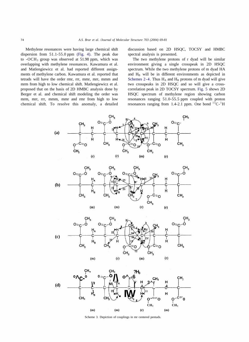
Methylene resonances were having large chemical shiftdispersion from 51.1-55.0 ppm (Fig. 4). The peak dueto -OCH3 group was observed at 51.98 ppm, which wasoverlapping with methylene resonances. Kawamura et al.and Matlengiewicz et al. had reported different assign-ments of methylene carbon. Kawamura et al. reported thattetrads will have the order rmr, rrr, mmr, mrr, mmm andmrm from high to low chemical shift. Matlengiewicz et al.proposed that on the basis of 2D HMBC analysis done byBerger et al. and chemical shift modeling the order wasmrm, mrr, rrr, mmm, mmr and rmr from high to lowchemical shift. To resolve this anomaly, a detailed
discussion based on 2D HSQC, TOCSY and HMBCspectral analysis is presented.
The two methylene protons of r dyad will be similarenvironment giving a single crosspeak in 2D HSQCspectrum. While the two methylene protons of m dyad HAand HB will be in different environments as depicted inSchemes 2-4. Thus HA and HB protons of m dyad will givetwo crosspeaks in 2D HSQC and so will give a cross-correlation peak in 2D TOCSY spectrum. Fig. 5 shows 2DHSQC spectrum of methylene region showing carbonresonances ranging 51.0-55.5 ppm coupled with protonresonances ranging from 1.4-2.1 ppm. One bond 13C-1H
(a)
CH
CH CH,(m)
H
(r)
CH3
CH,
(b)
(r)
CH,
(c)
(d)
CH.,i 3
°^c-°
CH3
0
V*1
f
CH
CH3
°/\Mi |
?H3o^c-6
f\.
r. IW
H1C^
CH33^r
/ • ^
III
O"CcrO
H1
H
CH,
; c r oCH3 CH3
(m) (m) (r) (m)
Scheme 3. Depiction of couplings in mr centered pentads.
A.S. Brar et al. /Journal of Molecular Structure 703 (2004) 69-81 75
(a)0*c-0 CH.
H
CH,
(b)
(r)
IH
CH,
(C) CH, CH,H B 3
YHA
CH3
Y
HB
YHA
CH3
c
I I I I ICH3 CH3 CH3 CH3 CH3
(m) (m) (m) (m)
Scheme 4. Depiction of couplings in mm centered pentads.
—\—1.8
—I—1.6ppn 2.0
Fig. 5. Methylene region in 2D HSQC spectrum of PMMA.
—I—1.4
-50
-52
-54
-56
ppm
76 A.S. Brar et al. /Journal of Molecular Structure 703 (2004) 69-81
-1.4
- l .B
- 1 .
-2 .0
ppm
ppm 2 .0 l .B 1.6 1.4
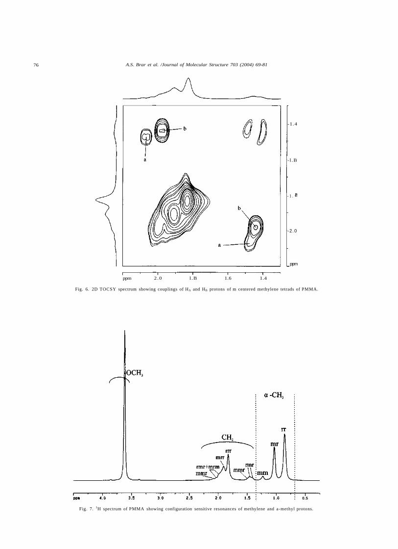
Fig. 6. 2D TOCSY spectrum showing couplings of HA and HB protons of m centered methylene tetrads of PMMA.
0.5
Fig. 7. 1H spectrum of PMMA showing configuration sensitive resonances of methylene and a-methyl protons.
A.S. Brar et al. /Journal of Molecular Structure 703 (2004) 69-81 11
couplings between methylene carbon and proton are labeledfrom I to IX in Schemes 2-4. For clarity the couplings andthe crosspeaks arising due to them have been labeled by thesame symbol.
Couplings I (Scheme 2 (a) and (b)), II (Scheme 2(b) and(c) and 3 (a) and (b)) and III (Scheme 3(c) and (d))corresponding to r centered rrr, mrr, mrm tetrads, respect-ively, will have single crosspeaks in 2D HSQC spectrum.These couplings are attributed to crosspeaks I, II and III,respectively, as shown in Fig. 5. Couplings IV and V(Scheme 3(c)) and VI and VII (Scheme 3(a)) correspond tormr tetrad which were observed to be higher order sensitive.Owing to m centered methylene group, couplings IV/VI andV/VII were assigned to HA and HB protons, respectively.The nonequivalent protons showed a cross-relation peak in2D TOCSY spectrum labeled b in Fig. 6. The couplings VIIIand IX were attributed to HA and HB of mmr tetrad(Scheme 3(b) and (d) and Scheme 4(a) and (b)). Thesecouplings gave rise to crosspeaks VIII and IX in 2DHSQC (Fig. 5) and the nonequivalent protons gavecross correlation peak a in 2D TOCSY spectrum (Fig. 6).
Thus using HSQC in conjugation with TOCSY enabled tostrengthen the assignments. Using these assignments 1Hspectrum was assigned as shown in Fig. 7 was similar to the1H spectrum discussed by Bovey [24] except that mrm andrmr (HA) proton resonances were assigned by Bovey at lowand high chemical shift, respectively, while in this studythey were observed to be overlapped.
The HMBC spectrum enabled to study the three bondcouplings between a-methyl carbon and methylene protons.As shown in Fig. 8, a-methyl carbon showed couplingswith methylene protons. Coupling 1 (Scheme 2(a)) betweena-methyl carbon of rr triad with methylene protons of rrrtetrad gave crosspeak 1 in Fig. 8. Coupling 2 of rr a-methylcarbon with mrr methylene (Scheme 2(b) and (c)) gavecrosspeak 2 in HMBC spectrum. a-Methyl carbon of mrtriad can be of rrmr, mmrr, mrmr or mmrm pentads.
Triad mr showed two resonance signals as shown inFig. 2. Comparing intensities of the signals enabled toassign rrmr and mmrm pentads to signals at low and highchemical shifts, respectively. Pentads mmrr and mrmrcould not be assigned. The difference between these
-12.5
-15.0
-17.5
r20.0
-22.5
ppm
ppm 2.0 1.8 1.6 1.4
Fig. 8. 2D HMBC spectrum showing three bond couplings between a-methyl carbon and methylene protons.
78 A.S. Brar et al. /Journal of Molecular Structure 703 (2004) 69-81
pentads is that mmrr pentad can have methylene in eithermmr or mrr tetrads, thus a-methyl carbon can couple eitherto mmr or mrr methylene. Similarly for mrmr pentad thea-methyl carbon can couple to methylene protons of mrmor rmr tetrads.
On analyzing the HMBC spectrum it was observed thatpeaks labeled 3 and 4 corresponded to the low chemicalshift signal of mr triad, while crosspeaks 5 and 6 to the highchemical shift signal. The couplings 3 and 4 are depicted inScheme 3(a) and couplings 5 and 6 in Scheme 3(c). Oncorrelating with the results of HSQC and TOCSY spectrum,the crosspeaks 3-6 were assigned to methylene of rmrtetrads. HA and HB protons resulted in the formation of twocrosspeaks. If mmrr pentad would have been at highchemical shift then there should not have been crosspeakscorresponding to rmr tetrads at high chemical shift. Thus itwas ascertained that mmrr pentad can be assigned to thesignal at low chemical shift and pentad rmrm to signal athigh chemical shift. The rmr tetrad which is part of the rrmr
pentad came at low chemical shift and rmr which is thepart of mrmr came at high chemical shift on carbon axis.The assignments are shown in Fig. 2. The coupling 7(Scheme 3(a) and (b)) was attributed to crosspeak 7 assignedto mrr methylene proton coupled with mr a-methyl carbon.The coupling 8 (Scheme 3(c) and (d)) attributed tocrosspeak 8 in Fig. 8 to mrm methylene.
Carbonyl carbon showed three bond order coupling witha-methyl protons and with adjacent methylene protons,shown in HMBC spectrum in Fig. 9. Crosspeak A0
corresponding to coupling A0 (Scheme 2) was assigned tocoupling of a-methyl proton of rr triad with rr carbonylcarbon. The crosspeaks B0 and C0 corresponding tocouplings B0 (Scheme 3) and C0 (Scheme 4) were assignedto couplings of mr and mm triads, respectively.
Crosspeaks 10-19 in Fig. 9 were assigned to the threebond order couplings between carbonyl carbon andmethylene protons. Crosspeaks 10 and 11 were assignedto rmr tetrad of rmrr pentad and crosspeaks 12 and 13
-174
-176
-179
rl80
:_ppm
ppm 2.0 1.5 1.0
Fig. 9. 2D HMBC spectrum showing three bond couplings between methylene protons and carbonyl carbon and a-methyl protons with carbonyl carbon.
A.S. Brar et al. /Journal of Molecular Structure 703 (2004) 69-81 79
assigned to rmr tetrad of rmrm pentad on the basis ofargument given for the a-methyl carbon analysis. Thecouplings 10 and 11 are depicted in Scheme 3(a) and 12 and13 are depicted in Scheme 3(c). The assignments of rmrmand mmrr pentads made from 2D NMR spectroscopy weresame as made from the empirical chemical shift modeling,strengthening the methodology used.
Crosspeak 16 was assigned to coupling 16 (Scheme 3(a)and (b)) between carbonyl carbon of rrmr or mmrr pentadand methylene protons of mrr tetrad. Crosspeaks 14 and 15were assigned to couplings between HA and HB protons ofmmr tetrad with mm carbonyl carbon as depicted inScheme 4(a) and (b). Crosspeak 17 was assigned to thecoupling 17 as shown in Scheme 2(a).
The couplings of HA with a-methyl carbon gave strongcrosspeaks 3 and 5 as shown in Fig. 8 in comparison tocouplings between HB with a-methyl carbon shown bycrosspeaks 4 and 6. While in the case of carbonyl carbon asshown in Fig. 9, HA protons showed weaker couplings(crosspeaks 10 and 12) than the HB protons (crosspeaks 11and 13). This indicated that while a-methyl carbon wascoupled more strongly to HA proton the carbonyl carbonshowed strong coupling with HB proton.
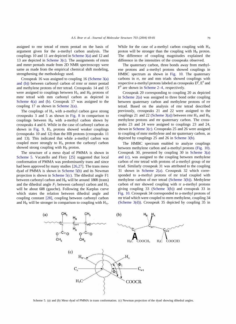
The structure of a meso dyad of PMMA is shown inScheme 5. Vacatello and Flory [25] suggested that localconformation of PMMA was predominantly trans and sincehad been approved by many studies [26,27]. The trans mesodyad of PMMA is shown in Scheme 5(b) and its Newmanprojection is shown in Scheme 5(c). The dihedral angle F1between carbonyl carbon and HB will be around 1808 (trans)and the dihedral angle F2 between carbonyl carbon and HA
will be about 608 (gauche). Following the Karplus curvewhich states the relation between dihedral angle andcoupling constant [28], coupling between carbonyl carbonand HB will be stronger in comparison to coupling with HA.
While for the case of a-methyl carbon coupling with HA
proton will be stronger than the coupling with HB proton.The difference of coupling magnitudes explained thedifference in the intensities of the crosspeaks observed.
The quaternary carbon, three bonds away from methyl-ene protons and a-methyl protons showed couplings inHMBC spectrum as shown in Fig. 10. The quaternarycarbons in rr, mr and mm triads showed couplings withrespective a-methyl protons labeled as crosspeaks D0, E0 andF0 are shown in Scheme 2-4, respectively.
Crosspeak 20 corresponding to coupling 20 as depictedin Scheme 2(a) was assigned to three bond order couplingbetween quaternary carbon and methylene protons of rrrtetrad. Based on the analysis of rmr tetrad describedpreviously, crosspeaks 21 and 22 were assigned to thecouplings 21 and 22 (Scheme 3(a)) between rmr HA and HB
methylene protons and mr quaternary carbon. The cross-peaks 23 and 24 were assigned to couplings 23 and 24,shown in Scheme 3(c). Crosspeaks 25 and 26 were assignedto coupling of mmr methylene and mr quaternary carbon, asdepicted by couplings 25 and 26 in Scheme 3(b).
The HMBC spectrum enabled to analyze couplingsbetween methylene carbon and a-methyl protons (Fig. 10).Crosspeak 30, presented by coupling 30 in Scheme 3(a)and (c), was assigned to the coupling between methylenecarbon of rmr tetrad with protons of a-methyl group of mrtriad. Similarly crosspeak 31 was attributed to the coupling31 shown in Scheme 2(a). Crosspeak 32 which corre-sponded to a-methyl protons of mr triad coupled withmethylene carbon of mrr tetrad (Scheme 3(b)). Methylenecarbon of mrr showed coupling with rr a-methyl protonsgiving coupling 33 (Scheme 3(b)) and crosspeak 33 inFig. 10. Crosspeak 34 corresponded to a-methyl protons ofmr triad which were coupled to mrm methylene, coupling 34(Scheme 3(d)). Crosspeak 35 depicted by coupling 35 in
AH3C COOCH3 H3C COOCH3
H3C COOCR
Scheme 5. (a) and (b) Meso dyad of PMMA in trans conformation. (c) Newman projection of the dyad showing dihedral angles.
80 A.S. Brar et al. /Journal of Molecular Structure 703 (2004) 69-81
-45
-50
-55
_ppm
ppnt 2.0 1.5 1.0
Fig. 10. 2D HMBC spectrum showing three bond couplings of quaternary carbon with methylene protons and a-methyl protons and methylene carbon witha-methyl protons and adjacent methylene carbon.
Scheme 4(a) and (b) was assigned to coupling of a-methylprotons of mm triad with methylene of mmr tetrad.Crosspeak 36 was assigned to the coupling of methylenecarbon of mmm tetrad with the a-methyl protons of mmtriad as shown by coupling 36 in Scheme 4(b) and (c).
Three bond order couplings between methylene carbonand methylene protons of two adjacent methylene unitswere analyzed from HMBC spectrum. For clarity thesecouplings aren't marked in mr centered pentads. Thecrosspeak 40 corresponded to carbon of either rmr or rrrtetrad with proton of mrm tetrad. This crosspeak wasassigned to the coupling between rmr centered carbon andmrm centered proton constituting rmrm unit, as rmrmpentad contains both rmr and mrm tetrads. Using thisanalogy contribution of rrr tetrad to this crosspeak was ruledout. On similar argument crosspeak 41 was assigned tocoupling of carbon of rrr tetrad with methylene of rrr tetrad,
combination of these tetrads are part of the rrrr pentad asshown in Scheme 2(a). Crosspeaks 42 and 43 wereattributed the couplings of methylene carbon of mrr tetradwith HA and HB protons of rmr tetrad (which was part ofm0rmr0 pentad) pointing to the mrmrr hexad. Similarlycrosspeaks 44 and 45 were assigned to the couplings ofmethylene carbon of mrr tetrad with HA and HB protons ofrmr tetrad (which was part of r0rmr0 pentad) pointing to therrmrr hexad. The crosspeak 46 was assigned to the coupling46 (Scheme 2(b)) of mrr methylene carbon with rrr proton inthe mrrr pentad.
4. Conclusions
Comprehensive microstructure study of PMMA hasbeen done using 2D HSQC, TOCSY and HMBC NMR
A.S. Brar et al. /Journal of Molecular Structure 703 (2004) 69-81 81
spectroscopy. Empirical chemical shift modeling has beenapplied for the differentiation of rmrm and mmrr carbonresonances of carbonyl carbon and the results obtained weresimilar to the experimental observations. Complete con-figuration assignments of methylene carbon and protonresonances have been made at tetrad level. From the HMBCspectral analysis it was ascertained that the PMMA waspredominantly in trans conformation.
Acknowledgements
Authors thank the Council of Scientific and IndustrialResearch (CSIR), India for the financial support.
[3] M. Monwar, S.J. Oh, P.L. Rinaldi, E.F. McCord, R.A. Hutchinson,M.M. Buback, H. Latz, Anal. Bioanal. Chem. 378 (2004) 1414.
[4] Y. Inoue, A. Nishioka, R. Chujo, Polym. J. 4 (1971) 535.[5] I.R. Peat, W.F. Reynolds, Tetrahedron Lett. 14 (1972) 1359.[6] G. Moad, E. Rizzardo, D.H. Solomon, S.R. Johns, R.I. Wiling,