Structure of M pro from COVID-19 virus and discovery of its inhibitors 1 Zhenming Jin 1,2,10 , Xiaoyu Du 2,10 , Yechun Xu 3,10 , Yongqiang Deng 4,10 , Meiqin Liu 5,10 , Yao 2 Zhao 1 , Bing Zhang 1 , Xiaofeng Li 4 , Leike Zhang 5 , Chao Peng 6 , Yinkai Duan 1 , Jing Yu 1 , Lin 3 Wang 1 , Kailin Yang 7 , Fengjiang Liu 1 , Rendi Jiang 5 , Xinglou Yang 5 , Tian You 1 , Xiaoce Liu 1 , 4 Xiuna Yang 1 , Fang Bai 1 , Hong Liu 3 , Xiang Liu 8 , Luke W. Guddat 9 , Wenqing Xu 1,6 , Gengfu 5 Xiao 5 , Chengfeng Qin 4 , Zhengli Shi 5 , Hualiang Jiang 1,3* , Zihe Rao 1,2,8* & Haitao Yang 1* 6 1 Shanghai Institute for Advanced Immunochemical Studies and School of Life Science and 7 Technology, ShanghaiTech University, Shanghai, China. 8 2 Laboratory of Structural Biology, School of Life Sciences and School of Medicine, 9 Tsinghua University, Beijing, China. 10 3 Drug Discovery and Design Center, Shanghai Institute of Materia Medica, Chinese 11 Academy of Sciences, Shanghai, China. 12 4 Department of Virology, State Key Laboratory of Pathogen and Biosecurity, Beijing 13 Institute of Microbiology and Epidemiology, Academy of Military Medical Sciences, 14 Beijing, China. 15 5 CAS Key Laboratory of Special Pathogens, Wuhan Institute of Virology, Center for 16 Biosafety Mega-Science, Chinese Academy of Sciences, Wuhan, China. 17 6 National Facility for Protein Science in Shanghai, Zhangjiang Lab, Shanghai Advanced 18 Research Institute, Chinese Academy of Science, Shanghai, China. 19 7 Taussig Cancer Center, Cleveland Clinic, Cleveland, USA. 20 8 State Key Laboratory of Medicinal Chemical Biology, Frontiers Science Center for Cell 21 Response, College of Life Sciences, College of Pharmacy, Nankai University, Tianjin, 22 China. 23 9 School of Chemistry and Molecular Biosciences, the University of Queensland, Brisbane, 24 Australia. 25 10 These authors contributed equally: Zhenming Jin, Xiaoyu Du, Yechun Xu, Yongqiang 26 Deng, Meiqin Liu. 27 *e-mail: [email protected]; [email protected]; [email protected]28 29 (which was not certified by peer review) is the author/funder. All rights reserved. No reuse allowed without permission. The copyright holder for this preprint this version posted March 10, 2020. . https://doi.org/10.1101/2020.02.26.964882 doi: bioRxiv preprint
Transcript
Structure of Mpro from COVID-19 virus and discovery of its inhibitors 1
(which was not certified by peer review) is the author/funder. All rights reserved. No reuse allowed without permission. The copyright holder for this preprintthis version posted March 10, 2020. . https://doi.org/10.1101/2020.02.26.964882doi: bioRxiv preprint
A new coronavirus (CoV) identified as COVID-19 virus is the etiological agent responsible 31
for the 2019-2020 viral pneumonia outbreak that commenced in Wuhan1-4. Currently there 32
is no targeted therapeutics and effective treatment options remain very limited. In order to 33
rapidly discover lead compounds for clinical use, we initiated a program of combined 34
structure-assisted drug design, virtual drug screening and high-throughput screening to 35
identify new drug leads that target the COVID-19 virus main protease (Mpro). Mpro is a key 36
CoV enzyme, which plays a pivotal role in mediating viral replication and transcription, 37
making it an attractive drug target for this virus5,6. Here, we identified a mechanism-based 38
inhibitor, N3, by computer-aided drug design and subsequently determined the crystal 39
structure of COVID-19 virus Mpro in complex with this compound. Next, through a 40
combination of structure-based virtual and high-throughput screening, we assayed over 41
10,000 compounds including approved drugs, drug candidates in clinical trials, and other 42
pharmacologically active compounds as inhibitors of Mpro. Six of these inhibit Mpro with 43
IC50 values ranging from 0.67 to 21.4 μM. Ebselen also exhibited strong antiviral activity 44
in cell-based assays. Our results demonstrate the efficacy of this screening strategy, which 45
can lead to the rapid discovery of drug leads with clinical potential in response to new 46
infectious diseases where no specific drugs or vaccines are available. 47
48
(which was not certified by peer review) is the author/funder. All rights reserved. No reuse allowed without permission. The copyright holder for this preprintthis version posted March 10, 2020. . https://doi.org/10.1101/2020.02.26.964882doi: bioRxiv preprint
CoVs infect humans and other animal species, causing a variety of highly prevalent and 49
severe diseases, including Severe Acute Respiratory Syndrome (SARS) and Middle East 50
Respiratory Syndrome (MERS)7. The COVID-19 virus genome is comprised of ~30,000 51
nucleotides; its replicase gene encodes two overlapping polyproteins, pp1a and pp1ab, 52
required for viral replication and transcription3,4. The functional polypeptides are released 53
from the polyproteins by extensive proteolytic processing, predominantly by a 33.8-kDa 54
main protease (Mpro), also referred to as the 3C-like protease. Mpro digests the polyprotein 55
at no less than 11 conserved sites, starting with the autolytic cleavage of this enzyme itself 56
from pp1a and pp1ab8. The functional importance of Mpro in the viral life cycle, together 57
with the absence of closely related homologues in humans, identify the Mpro as an attractive 58
target for antiviral drug design9. 59
To facilitate the rapid discovery of antiviral compounds with clinical potential, we 60
developed a strategy combining structure-assisted drug design, virtual drug screening and 61
high-throughput screening to repurpose existing drugs to target COVID-19 virus Mpro. 62
Establishing a high-throughput activity assay 63
Recombinant COVID-19 virus Mpro with native N and C termini was expressed in 64
Escherichia coli and subsequently purified (Extended Data Fig. 1a, b). The molecular 65
weight of COVID-19 virus Mpro as determined by mass spectroscopy is 33797.0 Da, 66
consistent with its theoretical molecular weight 33796.8 Da. In order to characterize its 67
enzymatic activity and to carry out high-throughput screening of inhibitors, we developed 68
a fluorescence resonance energy transfer (FRET) assay. To do this, a fluorescently labeled 69
substrate, MCA-AVLQ↓SGFR-Lys(Dnp)-Lys-NH2, derived from the N-terminal auto-70
cleavage sequence of the viral protease was designed and synthesized for time-dependent 71
(which was not certified by peer review) is the author/funder. All rights reserved. No reuse allowed without permission. The copyright holder for this preprintthis version posted March 10, 2020. . https://doi.org/10.1101/2020.02.26.964882doi: bioRxiv preprint
kinetic analysis (Extended Data Fig. 1e). The catalytic efficiency (kcat/Km) for COVID-19 72
virus Mpro was measured to be 28,500 M−1s−1 which is slightly higher than that for SARS-73
CoV Mpro (kcat/Km=26,500 M−1s−1)10, but over 30-fold higher than that of human rhinovirus 74
3C protease (kcat/Km=920 M−1s−1)11. 75
N3 is a potent irreversible inhibitor of COVID-19 virus Mpro 76
In a previous study, we designed a Michael acceptor inhibitor N3 using computer-aided 77
drug design (CADD) (Extended Data Fig. 1c), which can specifically inhibit multiple CoV 78
Mpros, including those from SARS-CoV and MERS-CoV12-15. It also has displayed potent 79
antiviral activity against infectious bronchitis virus in an animal model13. The CC50 of N3 80
is >133 μM (Extended Data Fig. 1f). Next, we constructed a homology model for COVID-81
19 virus Mpro and used molecular docking to see if N3 could target this new CoV Mpro. A 82
docking pose showed that it could fit inside the substrate-binding pocket. To assess the 83
efficacy of N3 for COVID-19 virus Mpro, kinetic analysis was performed. A progress curve 84
showed that it is a time-dependent irreversible inhibitor of this enzyme. Further, the shape 85
of this curve supports the mechanism of two-step irreversible inactivation. The inhibitor 86
first associates with COVID-19 virus Mpro (EI) with a dissociation constant Ki; then, a 87
stable covalent bond is formed between N3 and Mpro (E−I). The evaluation of this time-88
dependent inhibition requires both the equilibrium-binding constant Ki (designated as k2/k1) 89
and the inactivation rate constant for covalent bond formation k3. However, N3 exhibits 90
very potent inhibition of COVID-19 virus Mpro, such that measurement of Ki and k3 proved 91
not feasible (Extended Data Fig. 1d, e). When very rapid inactivation occurs, kobs/[I] was 92
utilized to evaluate the inhibition as an approximation of the pseudo second-order rate 93
constant (k3/Ki)12. The value of kobs/[I] of N3 for COVID-19 virus Mpro was determined to 94
(which was not certified by peer review) is the author/funder. All rights reserved. No reuse allowed without permission. The copyright holder for this preprintthis version posted March 10, 2020. . https://doi.org/10.1101/2020.02.26.964882doi: bioRxiv preprint
be 11,300±880 M-1s-1, suggesting this Michael acceptor has potent inhibition. 95
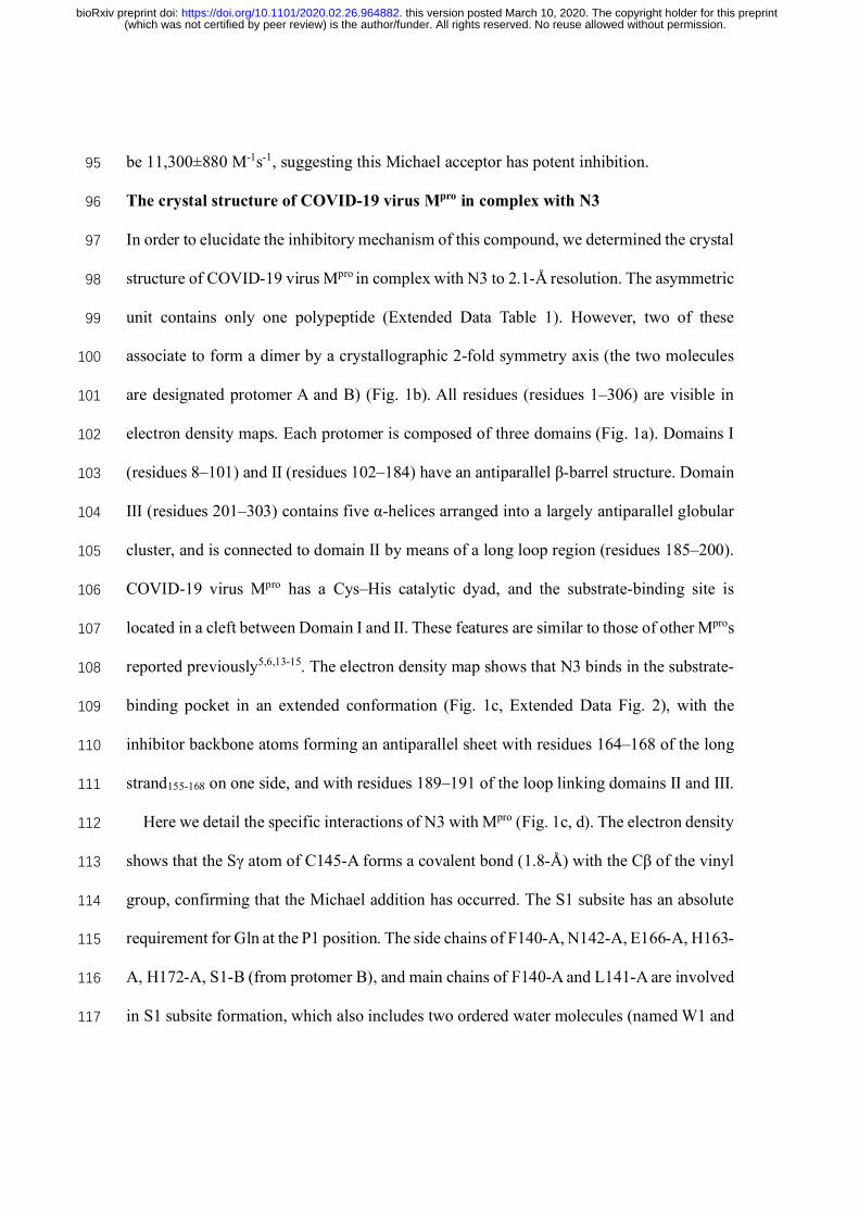
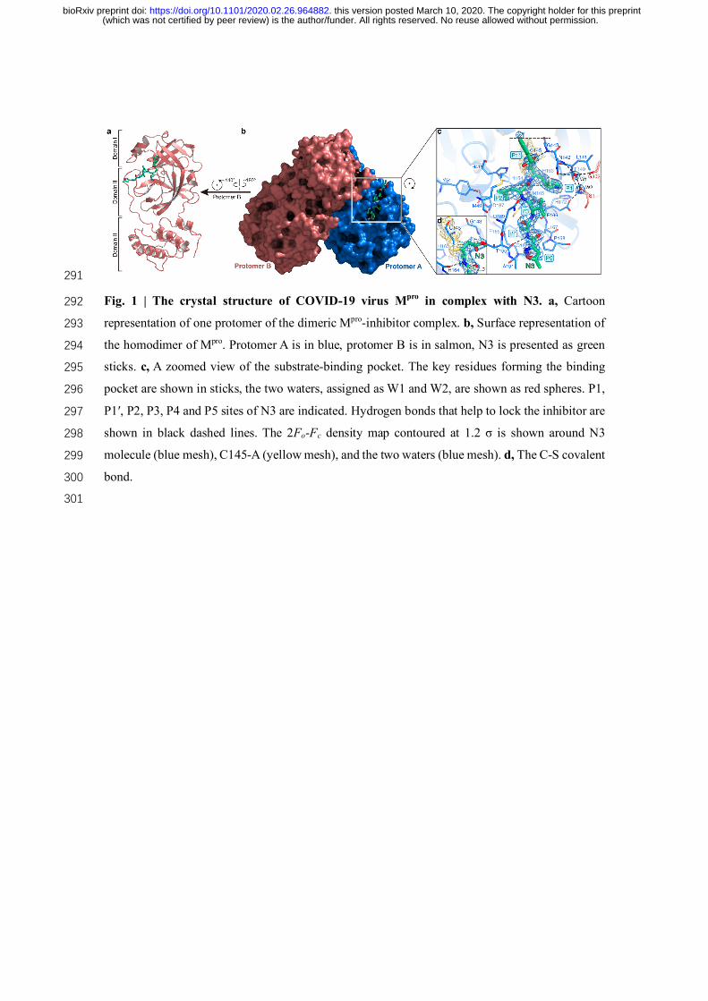
The crystal structure of COVID-19 virus Mpro in complex with N3 96
In order to elucidate the inhibitory mechanism of this compound, we determined the crystal 97
structure of COVID-19 virus Mpro in complex with N3 to 2.1-Å resolution. The asymmetric 98
unit contains only one polypeptide (Extended Data Table 1). However, two of these 99
associate to form a dimer by a crystallographic 2-fold symmetry axis (the two molecules 100
are designated protomer A and B) (Fig. 1b). All residues (residues 1–306) are visible in 101
electron density maps. Each protomer is composed of three domains (Fig. 1a). Domains Ⅰ 102
(residues 8–101) and Ⅱ (residues 102–184) have an antiparallel β-barrel structure. Domain 103
Ⅲ (residues 201–303) contains five α-helices arranged into a largely antiparallel globular 104
cluster, and is connected to domain Ⅱ by means of a long loop region (residues 185–200). 105
COVID-19 virus Mpro has a Cys–His catalytic dyad, and the substrate-binding site is 106
located in a cleft between Domain Ⅰ and Ⅱ. These features are similar to those of other Mpros 107
reported previously5,6,13-15. The electron density map shows that N3 binds in the substrate-108
binding pocket in an extended conformation (Fig. 1c, Extended Data Fig. 2), with the 109
inhibitor backbone atoms forming an antiparallel sheet with residues 164–168 of the long 110
strand155-168 on one side, and with residues 189–191 of the loop linking domains Ⅱ and Ⅲ. 111
Here we detail the specific interactions of N3 with Mpro (Fig. 1c, d). The electron density 112
shows that the Sγ atom of C145-A forms a covalent bond (1.8-Å) with the Cβ of the vinyl 113
group, confirming that the Michael addition has occurred. The S1 subsite has an absolute 114
requirement for Gln at the P1 position. The side chains of F140-A, N142-A, E166-A, H163-115
A, H172-A, S1-B (from protomer B), and main chains of F140-A and L141-A are involved 116
in S1 subsite formation, which also includes two ordered water molecules (named W1 and 117
(which was not certified by peer review) is the author/funder. All rights reserved. No reuse allowed without permission. The copyright holder for this preprintthis version posted March 10, 2020. . https://doi.org/10.1101/2020.02.26.964882doi: bioRxiv preprint
W2). The lactam at P1 inserts into the S1 subsite and forms a hydrogen bond with H163-118
A. The side chain of Leu at P2 site inserts deeply into the hydrophobic S2 subsite, which 119
consists of the side chains of H41-A, M49-A, Y54-A, M165-A, and the alkyl portion of the 120
side chain of D187-A. The side chain of Val at P3 is solvent-exposed, indicating that this 121
site can tolerate a wide range of functional groups. The side chain of Ala at P4 side is 122
surrounded by the side chains of M165-A, L167-A, F185-A, Q192-A and the main chain 123
of Q189-A, all of which form a small hydrophobic pocket. P5 makes van der Waals contacts 124
with P168-A and the backbone of residues 190–191. The bulky benzyl group extends into 125
the S1′ site, forming van der Waals interactions with T24-A and T25-A. In addition, N3 126
forms multiple hydrogen bonds with the main chain of the residues in the substrate-binding 127
pocket, which also helps lock the inhibitor inside the substrate-binding pocket. 128
An overlay of the structures of COVID-19 virus Mpro-N3 and SARS-CoV Mpro-N112 129
shows that N3 and N1 bind to Mpros in a similar mode (Fig. 2a, Extended Data Fig. 3). The 130
major difference lies in the P1´ site. Compared with the benzyl ester portion of N3 in the 131
COVID-19 virus Mpro structure, the ethyl ester portion in N1 adopts a slightly different 132
conformation. This may be attributed to an ordered water (W1) in SARS-CoV Mpro-N1 133
structure, which makes a long distance hydrogen bond to the carboxylate oxygen of the 134
ester and also forms two hydrogen bonds from the backbone NH of G143 and the side 135
chain of N142. In our previous study, we proposed that all the CoV Mpros share a highly 136
conserved substrate-recognition pocket, which could serve as a drug target for the design 137
of broad-spectrum inhibitors12. The recent discovery of new CoVs and accumulation of 138
structural data for CoV Mpros from various species provided the opportunity to further 139
examine this hypothesis. Superposition of the 12 crystal structures of Mpros12-21 have shown 140
(which was not certified by peer review) is the author/funder. All rights reserved. No reuse allowed without permission. The copyright holder for this preprintthis version posted March 10, 2020. . https://doi.org/10.1101/2020.02.26.964882doi: bioRxiv preprint
that the most variable regions were the helical domain Ⅲ and surface loops, but the 141
substrate-binding pockets located in a cleft between domains Ⅰ and Ⅱ are still highly 142
conserved among all CoV Mpros, suggesting the antiviral inhibitors targeting this site should 143
have wide-spectrum anti-CoV activity (Fig. 2b, c). 144
Virtual screening 145
The structure of COVID-19 virus Mpro in complex with N3 provides a model for identifying 146
lead inhibitors to target COVID-19 virus Mpro through in silico screening. To achieve this, 147
an in-house database of potential binding compounds was docked using Glide (v8.2). The 148
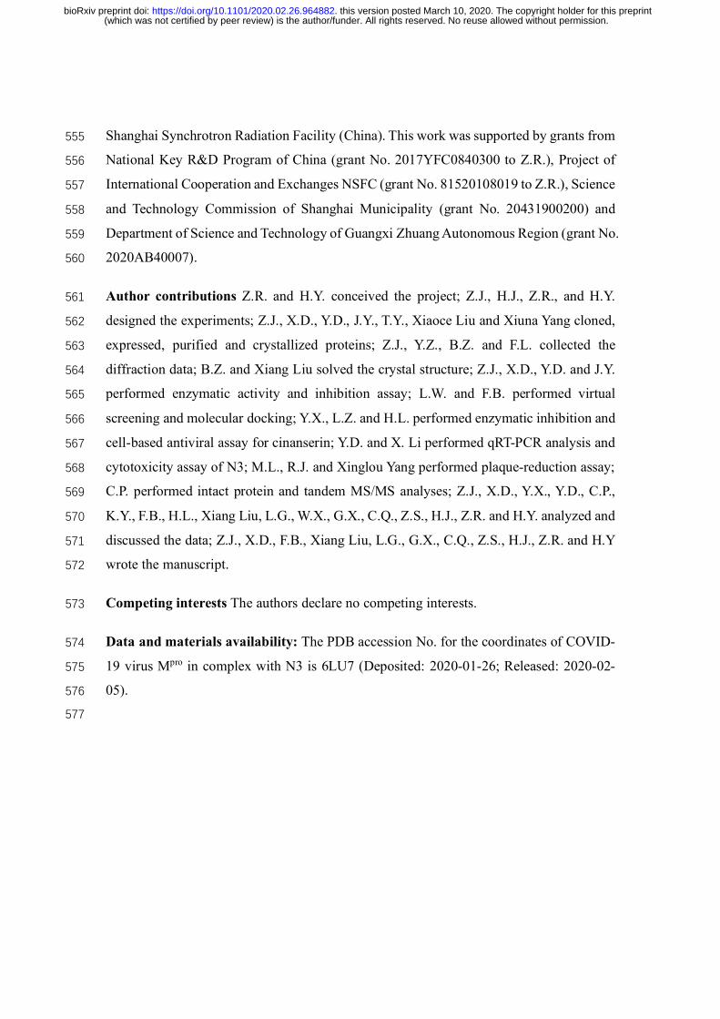
results show that cinanserin fits snugly into the substrate-binding pocket, by interacting 149
with H41 and E166 of Mpro by cation-π. Subsequently we determined this compound has 150
an IC50 value of 125 μM for Mpro. Moreover, cinanserin is a well-characterized serotonin 151
antagonist, which underwent preliminary clinical testing in humans in the 1960s22 and has 152
previously been shown to inhibit SARS-CoV Mpro23. The CC50 of cinanserin is > 200 μM 153
(Extended Data Fig. 4). Thus, it has potential for optimization as an anti-viral drug lead. 154
High-throughput screening 155
Next, we used our FRET assay, to screen a library of ~10,000 compounds consisting of 156
approved drugs, clinical trial drug candidates and natural products. Primary hits included 157
seven compounds that are either FDA-approved drugs or clinical trial/preclinical drug 158
candidates. We then determined their IC50 values, which are in the range from 0.67 to 21.4 159
μM (Fig. 3). Amongst them, disulfiram and carmofur are FDA-approved drugs, whilst 160
ebselen, shikonin, tideglusib, PX-12 and TDZD-8 are currently in clinical trials or 161
preclinical studies. Ebselen has the strongest inhibition of Mpro activity with an IC50 of 0.67 162
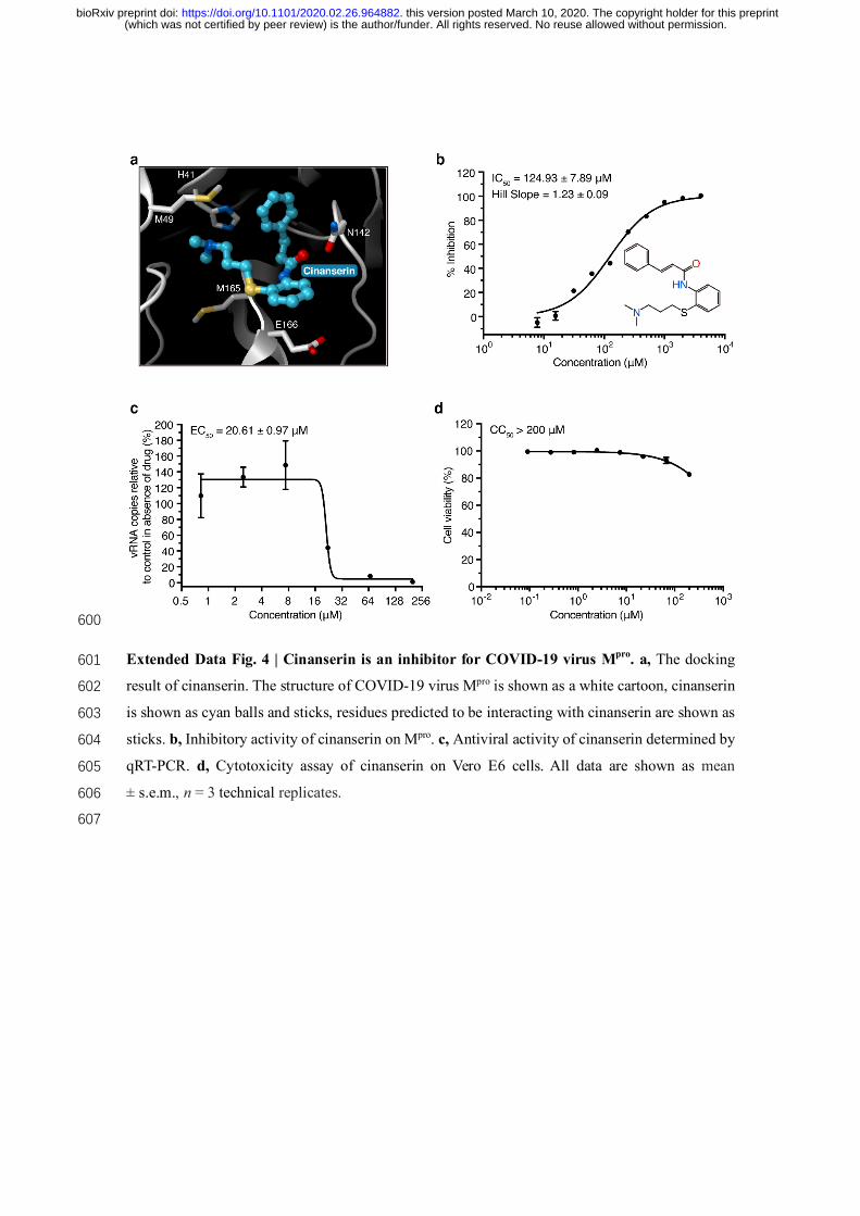
μM. However, in a detergent-based assay24, TDZD-8 was found to be an aggregate-based 163
(which was not certified by peer review) is the author/funder. All rights reserved. No reuse allowed without permission. The copyright holder for this preprintthis version posted March 10, 2020. . https://doi.org/10.1101/2020.02.26.964882doi: bioRxiv preprint
inhibitor, which might non-specifically inhibit Mpro (Extended Data Fig. 5) and was 164
therefore not considered for further investigation. Since our structural data is based on N3, 165
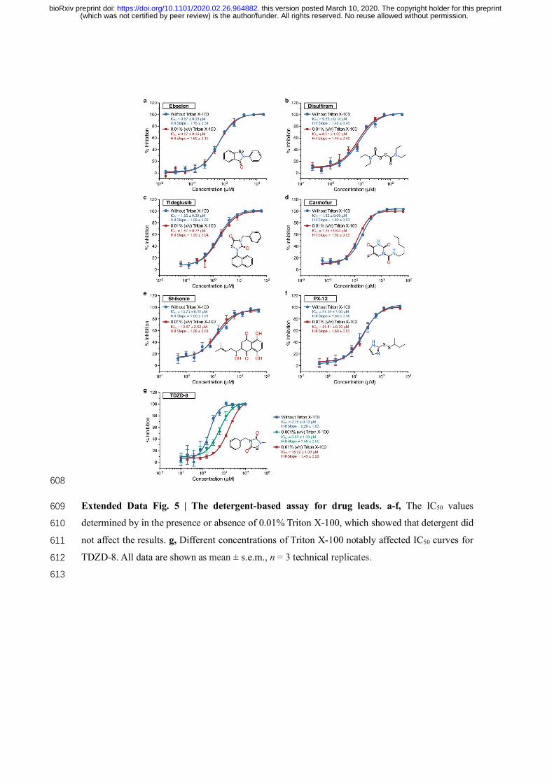
we investigated if molecular docking could predict how other six inhibitors bind to this 166
protein. In all cases, reasonable docking poses were found, demonstrating that they could 167
fit inside the substrate-binding pocket (Extended Data Fig. 6). Next, we set out to identify 168
the potential covalent inhibitors among these compounds through tandem MS/MS analysis. 169
The MS/MS data shows that ebselen, PX-12 and carmofur are all able to covalently bind 170
to C145 of the catalytic dyad in COVID-19 virus Mpro. However, while PX-12 and 171
carmofur completely modified Mpro, ebselen could only partially modify this viral cysteine 172
protease (Extended Data Fig. 7). Since ebselen has even stronger inhibition than the others, 173
there is a possibility that ebselen could also inhibit Mpro through non-covalent binding. 174
Antiviral activity assay 175
To further substantiate the enzymatic inhibition results in vitro, we evaluated whether these 176
compounds could prevent viral replication in cell-based assays. As shown in Fig. 4a, 177
quantitative real-time RT-PCR (qRT-PCR) demonstrated that ebselen and N3 showed the 178
strongest antiviral effects among them at a concentration of 10 μM treatment in COVID-179
19 virus infected Vero cells. A plaque-reduction assay was performed to further assess the 180
efficacy of these two compounds in protecting cells. Ebselen and N3 displayed inhibition 181
against COVID-19 virus with individual EC50 values of 4.67 μM and 16.77 μM, 182
respectively (Fig. 4b, c). The dose-response curves suggest that both of them could be able 183
to penetrate cellular membrane to access their targets. Ebselen is an organoselenium 184
compound with anti-inflammatory, anti-oxidant and cytoprotective properties. This 185
compound has been investigated for the treatment of multiple diseases, such as bipolar 186
(which was not certified by peer review) is the author/funder. All rights reserved. No reuse allowed without permission. The copyright holder for this preprintthis version posted March 10, 2020. . https://doi.org/10.1101/2020.02.26.964882doi: bioRxiv preprint
disorders25 and hearing loss26,27. Ebselen has extremely low cytotoxicity (LD50 in rats > 187
4,600 mg/kg, per os)28 and its safety in humans has been evaluated in a number of clinical 188
trials26,27,29. These data strongly suggest the clinical potential of ebselen for CoV treatment. 189
It is also interesting that cinanserin displayed moderate inhibition against COVID-19 virus 190
with an EC50 value of 20.61 μM from qRT-PCR analysis (Extended Data Fig. 4), which is 191
superior to that in the enzymatic inhibition assay, suggesting that cinanserin might have 192
multi-drug targets in preventing viral infection. In further studies, selection and 193
characterization of drug-resistant mutants will help clarify the mode of cinanserin’s action. 194
Discussion 195
Our crystal structural and docking data have shown that the drug leads identified can bind 196
to the substrate-binding pocket of COVID-19 virus Mpro, which is highly conserved among 197
all CoV Mpros. This strongly supports our hypothesis that development of a single antiviral 198
agent targeting Mpro or in combination with other potential therapies could provide an 199
effective first line of defense against all CoV-associated diseases. 200
In the last twenty years, new infectious agents have emerged to cause epidemics, such 201
as SARS and MERS7. The timely development of effective antiviral agents for clinical use 202
is extremely challenging because conventional drug development approaches normally 203
take years of investigations and cost billions of dollars. The repurposing of approved 204
pharmaceutical drugs and drug candidates provides an alternative approach to rapidly 205
identify potential drug leads to manage rapidly emerging viral infections. Cell-based 206
phenotypic screening has proven to be valuable30, but the complexity of this approach is 207
not readily compatible with high-throughput pipelines, and it cannot identify the molecular 208
target or mechanism of action31. In this study, the convergence of structure-based ab initio 209
(which was not certified by peer review) is the author/funder. All rights reserved. No reuse allowed without permission. The copyright holder for this preprintthis version posted March 10, 2020. . https://doi.org/10.1101/2020.02.26.964882doi: bioRxiv preprint
drug design, virtual screening and high-throughput screening proved to be an efficient 210
strategy to find antiviral leads against COVID-19 virus. The methods presented here can 211
greatly assist in the rapid discovery of drug leads with clinical potential in response to new 212
emerging infectious diseases that currently lack specific drugs and vaccines. 213
References 214 1 Zhu, N. et al. A novel coronavirus from patients with pneumonia in China, 2019. New England 215
Journal of Medicine (2020). 216 2 Qun Li et al. Early Transmission Dynamics in Wuhan, China, of Novel Coronavirus–Infected 217
Pneumonia. New England Journal of Medicine (2020). 218 3 Zhou, P. et al. A pneumonia outbreak associated with a new coronavirus of probable bat origin. 219
Nature, doi:10.1038/s41586-020-2012-7 (2020). 220 4 Wu, F. et al. A new coronavirus associated with human respiratory disease in China. Nature, 221
doi:10.1038/s41586-020-2008-3 (2020). 222 5 Anand, K. et al. Structure of coronavirus main proteinase reveals combination of a chymotrypsin 223
fold with an extra α‐helical domain. The EMBO Journal 21, 3213-3224 (2002). 224 6 Yang, H. T. et al. The crystal structures of severe acute respiratory syndrome virus main protease 225
and its complex with an inhibitor. Proceedings of the National Academy of Sciences of the United 226 States of America 100, 13190-13195, doi:10.1073/pnas.1835675100 (2003). 227
7 de Wit, E., van Doremalen, N., Falzarano, D. & Munster, V. J. SARS and MERS: recent insights 228 into emerging coronaviruses. Nature Reviews Microbiology 14, 523-534, 229 doi:10.1038/nrmicro.2016.81 (2016). 230
8 Hegyi, A. & Ziebuhr, J. Conservation of substrate specificities among coronavirus main proteases. 231 Journal of general virology 83, 595-599 (2002). 232
9 Pillaiyar, T., Manickam, M., Namasivayam, V., Hayashi, Y. & Jung, S. H. An Overview of Severe 233 Acute Respiratory Syndrome-Coronavirus (SARS-CoV) 3CL Protease Inhibitors: Peptidomimetics 234 and Small Molecule Chemotherapy. Journal of Medicinal Chemistry 59, 6595-6628, 235 doi:10.1021/acs.jmedchem.5b01461 (2016). 236
10 Xue, X. Y. et al. Production of authentic SARS-CoV M-pro with enhanced activity: Application as 237 a novel tag-cleavage endopeptidase for protein overproduction. Journal of Molecular Biology 366, 238 965-975, doi:10.1016/j.jmb.2006.11.073 (2007). 239
11 Wang, Q. M., Johnson, R. B., Cox, G. A., Villarreal, E. C. & Loncharich, R. J. A Continuous 240 Colorimetric Assay for Rhinovirus-14 3C Protease Using Peptidep-Nitroanilides as Substrates. 241 Analytical biochemistry 252, 238-245 (1997). 242
12 Yang, H. T. et al. Design of wide-spectrum inhibitors targeting coronavirus main proteases. Plos 243 Biology 3, 2044-2044, doi:10.1371/journal.pbio.0030428 (2005). 244
13 Xue, X. Y. et al. Structures of two coronavirus main proteases: Implications for substrate binding 245 and antiviral drug design. Journal of Virology 82, 2515-2527, doi:10.1128/jvi.02114-07 (2008). 246
14 Ren, Z. L. et al. The newly emerged SARS-Like coronavirus HCoV-EMC also has an "Achilles' 247 heel": current effective inhibitor targeting a 3C-like protease. Protein & Cell 4, 248-250, 248 doi:10.1007/s13238-013-2841-3 (2013). 249
15 Wang, F., Chen, C., Tan, W., Yang, K. & Yang, H. Structure of Main Protease from Human 250 Coronavirus NL63: Insights for Wide Spectrum Anti-Coronavirus Drug Design. Sci Rep 6, 22677-251 22677, doi:10.1038/srep22677 (2016). 252
16 Zhao, Q. et al. Structure of the main protease from a global infectious human coronavirus, HCoV-253 HKU1. Journal of virology 82, 8647-8655, doi:10.1128/JVI.00298-08 (2008). 254
17 Lee, C.-C. et al. Structural basis of inhibition specificities of 3C and 3C-like proteases by zinc-255 coordinating and peptidomimetic compounds. J Biol Chem 284, 7646-7655, 256 doi:10.1074/jbc.M807947200 (2009). 257
18 Ma, Q., Xiao, Y., Hilgenfeld, R. Inhibitor for the Main Protease of Coronavirus Hku4 (2012). 258 19 Wang, F. H. et al. Crystal Structure of Feline Infectious Peritonitis Virus Main Protease in Complex 259
(which was not certified by peer review) is the author/funder. All rights reserved. No reuse allowed without permission. The copyright holder for this preprintthis version posted March 10, 2020. . https://doi.org/10.1101/2020.02.26.964882doi: bioRxiv preprint
with Synergetic Dual Inhibitors. Journal of Virology 90, 1910-1917, doi:10.1128/jvi.02685-15 260 (2016). 261
20 Wang, F. H. et al. Michael Acceptor-Based Peptidomimetic Inhibitor of Main Protease from Porcine 262 Epidemic Diarrhea Virus. Journal of Medicinal Chemistry 60, 3212-3216, 263 doi:10.1021/acs.jmedchem.7b00103 (2017). 264
21 Cui, W. et al. The crystal structure of main protease from mouse hepatitis virus A59 in complex with 265 an inhibitor. Biochemical and Biophysical Research Communications 511, 794-799, 266 doi:10.1016/j.bbrc.2019.02.105 (2019). 267
22 Rubin, B. & Waugh, M. H. Antiphlogistic effects of antiserotonin (SQ 10,643) and aminopyrine in 268 rats versus endotoxin and other agents. Proceedings of the Society for Experimental Biology and 269 Medicine 119, 438-443 (1965). 270
23 Chen, L. et al. Cinanserin is an inhibitor of the 3C-like proteinase of severe acute respiratory 271 syndrome coronavirus and strongly reduces virus replication in vitro. Journal of virology 79, 7095-272 7103 (2005). 273
24 Feng, B. Y. & Shoichet, B. K. A detergent-based assay for the detection of promiscuous inhibitors. 274 Nature protocols 1, 550-553 (2006). 275
25 Singh, N. et al. A safe lithium mimetic for bipolar disorder. Nature communications 4, 1-7 (2013). 276 26 Lynch, E. & Kil, J. Development of ebselen, a glutathione peroxidase mimic, for the prevention and 277
treatment of noise-induced hearing loss. Seminars in Hearing 30, 047-055 (2009). 278 27 Kil, J. et al. Safety and efficacy of ebselen for the prevention of noise-induced hearing loss: a 279
randomised, double-blind, placebo-controlled, phase 2 trial. The Lancet 390, 969-979 (2017). 280 28 Renson, M., Etschenberg, E. & Winkelmann, J. 2-Phenyl-1, 2-benzisoselenazol-3 (2H)-one 281
containing pharmaceutical preparations and process for the treatment of rheumatic diseases. (1982). 282 29 Masaki, C. et al. Effects of the potential lithium-mimetic, ebselen, on impulsivity and emotional 283
processing. Psychopharmacology 233, 2655-2661 (2016). 284 30 Xu, M. et al. Identification of small-molecule inhibitors of Zika virus infection and induced neural 285
cell death via a drug repurposing screen. Nature Medicine 22, 1101-1107, doi:10.1038/nm.4184 286 (2016). 287
31 Aulner, N., Danckaert, A., Ihm, J., Shum, D. & Shorte, S. L. Next-Generation Phenotypic Screening 288 in Early Drug Discovery for Infectious Diseases. Trends in parasitology (2019). 289
290
(which was not certified by peer review) is the author/funder. All rights reserved. No reuse allowed without permission. The copyright holder for this preprintthis version posted March 10, 2020. . https://doi.org/10.1101/2020.02.26.964882doi: bioRxiv preprint
Fig. 1 | The crystal structure of COVID-19 virus Mpro in complex with N3. a, Cartoon 292
representation of one protomer of the dimeric Mpro-inhibitor complex. b, Surface representation of 293
the homodimer of Mpro. Protomer A is in blue, protomer B is in salmon, N3 is presented as green 294
sticks. c, A zoomed view of the substrate-binding pocket. The key residues forming the binding 295
pocket are shown in sticks, the two waters, assigned as W1 and W2, are shown as red spheres. P1, 296
P1′, P2, P3, P4 and P5 sites of N3 are indicated. Hydrogen bonds that help to lock the inhibitor are 297
shown in black dashed lines. The 2Fo-Fc density map contoured at 1.2 σ is shown around N3 298
molecule (blue mesh), C145-A (yellow mesh), and the two waters (blue mesh). d, The C-S covalent 299
bond. 300
301
(which was not certified by peer review) is the author/funder. All rights reserved. No reuse allowed without permission. The copyright holder for this preprintthis version posted March 10, 2020. . https://doi.org/10.1101/2020.02.26.964882doi: bioRxiv preprint
TGEV, HCoV-NL63, HCoV-229E and IBV. The color spectrum represents the root-mean-square 308
deviation (RMSD) of the aligned Cα atoms. c, Surface presentation of conserved substrate-binding 309
pockets of 12 CoV Mpros. Red: residues are entirely identical among all 12 Mpros; violet: conserved 310
substitution in one CoV Mpro; orange: conserved substitution in more than one CoV Mpros. S1, S2, S4, 311
and S1′ subsites are indicated. 312
313
(which was not certified by peer review) is the author/funder. All rights reserved. No reuse allowed without permission. The copyright holder for this preprintthis version posted March 10, 2020. . https://doi.org/10.1101/2020.02.26.964882doi: bioRxiv preprint
Fig. 3 | Drug leads inhibit the activity of COVID-19 virus Mpro. a-f, The hydrolytic activity of 315
COVID-19 virus Mpro was measured in the presence of varying concentrations of the drug 316
candidates. Dose-response curves for half-maximum inhibitory concentration (IC50) values were 317
determined by nonlinear regression. All data are shown as mean ± s.e.m., n = 3 technical replicates. 318
319
(which was not certified by peer review) is the author/funder. All rights reserved. No reuse allowed without permission. The copyright holder for this preprintthis version posted March 10, 2020. . https://doi.org/10.1101/2020.02.26.964882doi: bioRxiv preprint
Fig. 4 | Antiviral activities of the drug leads against COVID-19 virus. a, The quantification of 321
absolute viral RNA copies (per ml) in the supernatant at 72 h post infection (p.i.) determined by 322
qRT-PCR analysis. Data are mean ± s.e.m., n = 3 technical replicates. b, c, Dose-response curves 323
for ebselen and N3 in the plaque-reduction assay, respectively. All data are shown as mean 324
± s.e.m., n = 4 technical replicates. 325
326
(which was not certified by peer review) is the author/funder. All rights reserved. No reuse allowed without permission. The copyright holder for this preprintthis version posted March 10, 2020. . https://doi.org/10.1101/2020.02.26.964882doi: bioRxiv preprint
viral Mpro has been reported previously10. The expression plasmid was transformed into 331
Escherichia coli BL21 (DE3) cells and then cultured in LB medium containing 100 μg/ml 332
ampicillin at 37 °C. When the cells were grown to OD600 of 0.6-0.8, 0.5 mM IPTG was 333
added to the cell culture to induce the expression at 16 °C. After 10 h, the cells were 334
harvested by centrifugation at 3,000g. The cell pellets were resuspended in lysis buffer (20 335
mM Tris-HCl pH 8.0, 300 mM NaCl), lysed by high-pressure homogenization, and then 336
centrifuged at 25,000g for 40 min. The supernatant was loaded onto Ni-NTA affinity 337
column (Qiagen, Germany), and washed by the resuspension buffer containing 20 mM 338
imidazole. The His tagged Mpro was eluted by cleavage buffer (50 mM Tris-HCl pH 7.0, 339
150 mM NaCl) including 300 mM imidazole. Human rhinovirus 3C protease was added to 340
remove the C-terminal His tag. The Mpro was further purified by ion exchange 341
chromatography and size exclusion chromatography. CoV Mpros exist as a mixture of 342
monomers and dimers in solutions32. The purified Mpro was stored in 50 mM Tris-HCl pH 343
7.3, 1 mM EDTA. 344
Crystallization, data collection and structure determination 345
COVID-19 virus Mpro was incubated with 10 mM N3 for 30 min and the complex (5 mg/ml) 346
was crystallized by hanging drop vapor diffusion method at 20 °C. The best crystals were 347
grown with well buffer containing 0.1 M MES pH 6.0, 2% polyethylene glycol (PEG) 6000, 348
3% DMSO, 1 mM DTT. The cryo-protectant solution contained 0.1 M MES pH 6.0, 30% 349
(which was not certified by peer review) is the author/funder. All rights reserved. No reuse allowed without permission. The copyright holder for this preprintthis version posted March 10, 2020. . https://doi.org/10.1101/2020.02.26.964882doi: bioRxiv preprint
X-ray data were collected on beamline BL17U1 at Shanghai Synchrotron Radiation 351
Facility (SSRF) at 100 K and at a wavelength of 1.07180 Å using an Eiger X 16M image 352
plate detector. Data integration and scaling were performed using the program Xia233. The 353
structure was determined by molecular replacement (MR) with the Phaser module34 in 354
CCP435 using the SARS-CoV Mpro (PDB ID: 2H2Z) as a search template. The output model 355
from MR was subsequently subjected to iterative cycles of manual model adjustment with 356
Coot36 and refinement was finished with Phenix37. The inhibitor N3 was built according to 357
the omit map. The phasing and refinement statistics are summarized in Extended Data 358
Table 1. The Rwork/Rfree values are 0.202/0.235, respectively. 97.3% residues are in most 359
favored regions of the Ramachandran plot, and no residues are found in disallowed regions. 360
Coordinates and structure factors for COVID-19 virus Mpro in complex with the inhibitor 361
N3 have been deposited in Protein Data Bank (PDB) with accession number 6LU7. 362
Enzymatic activity and inhibition assays 363
The enzyme activity assays have been described previously10. Briefly, the activity of 364
COVID-19 virus Mpro was measured by a continuous kinetic assay, with the substrate 365
MCA-AVLQSGFR-Lys(Dnp)-Lys-NH2 (GL Biochem, Shanghai), using wavelengths of 366
320 nm and 405 nm for excitation and emission, respectively. The assay started by 367
immediately mixing 0.2 μM COVID-19 virus Mpro with different concentrations of 368
substrate (2.5-100 μM). Fluorescence intensity was monitored with an EnVision multimode 369
plate reader (Perkin Elmer, USA). Initial rates were obtained by fitting the linear portion 370
of the curves to a straight line. The kinetic parameters Km and kcat were calculated from a 371
double-reciprocal plot. As N3 is a mechanism-based irreversible inhibitor for COVID-19 372
(which was not certified by peer review) is the author/funder. All rights reserved. No reuse allowed without permission. The copyright holder for this preprintthis version posted March 10, 2020. . https://doi.org/10.1101/2020.02.26.964882doi: bioRxiv preprint
virus Mpro, kobs/[I] was used as an approximation of the pseudo second-order rate constant 373
to evaluate the inhibition effect of the inhibitor N312. In this case, the measurement was 374
carried out with 0.2 μM of enzyme, 20 μM of substrate and inhibitor at 6 different 375
concentrations (0-1 μM). 376
Virtual screening 377
The virtual screening was performed using our in-house database via a workflow 378
application of Glide (v8.2), Maestro (Schrodinger 2019-1a)38. All compounds in the 379
database were considered to be at pH 7.4 ± 0.2 to estimate their protonation state using the 380
program EpiK39. Their three dimensional conformations were generated by the ligPrep 381
module of Maestro. The structure of COVID-19 virus Mpro (PDB ID: 6LU7) was used to 382
generate receptor grid for docking simulations. The center of active site of the grid was 383
determined according to the position of N3 in the structure. The flexibility of the receptor 384
hydroxyl and thiol groups in side chains of C145, S46 and Y54 were considered. At the 385
very beginning, a relatively fast but raw screening was performed by using the glide 386
standard precision model, and the top 20% of compounds were kept. Then covalent 387
docking simulations were performed by choosing Michael Addition as the reaction type for 388
these top 20% compounds that contained Michael acceptors, and C145 was set as the 389
source of Michael donor. Finally, the candidate molecules were picked by analysing the 390
predicted binding modes and their scores. 391
High-throughput drug screen and IC50 measurement 392
Potential inhibitors against COVID-19 virus Mpro were screened by an enzymatic inhibition 393
assay. When the different compounds were added into the enzymatic reaction mixture, the 394
change of initial rates was calculated to evaluate their inhibitory effect. Five drug libraries, 395
(which was not certified by peer review) is the author/funder. All rights reserved. No reuse allowed without permission. The copyright holder for this preprintthis version posted March 10, 2020. . https://doi.org/10.1101/2020.02.26.964882doi: bioRxiv preprint
FDA-approved Drug Library (Selleck, USA), Natural Product Library (Selleck, USA), and 397
Anti-virus Drug Library (Shanghai Institute for Advanced Immunochemical Studies, 398
SIAIS), which includes ~10,000 compounds, were used. The preliminary screening 399
reaction mixture included 0.2 μM protein, 20 μM substrate and 50 μM compounds. The 400
compounds of interest were defined as those with a percentage of inhibition over 60% 401
compared with the reaction in the absence of inhibitor. IC50 values of seven drug leads were 402
measured using 0.2 μM protein, 20 μM substrate and 11 different inhibitor concentrations. 403
In order to exclude inhibitors possibly acting as aggregators, detergent-based control was 404
performed by adding 0.001% or 0.01% freshly made up Triton X-100 to the reaction at the 405
same time24. All experimental data was analyzed using GraphPad Prism. All experiments 406
were performed in triplicate. 407
Molecular docking 408
To understand the binding interaction of these molecules with COVID-19 virus Mpro, two 409
different molecular docking methods, i.e., Glide (v8.2)38 and iFitDock40 were used to 410
predict their binding poses. Then a 3D molecular similarity calculation method, SHAFTS41, 411
was used for molecular alignment poses enumeration by matching the critical 412
pharmacophore and volumetric overlay between the N3 molecule within the Mpro structure 413
and the six drug candidates. However, the selenium atom of ebselen could not be treated 414
by any of these above methods, so sulfur was used to replace it in the calculations. Then 415
the obtained optimal superposition of these molecules was used to assess the reasonability 416
of the predicted binding poses from the two docking methods, and only the binding 417
orientations which were consistent among different methods were kept for constructing the 418
(which was not certified by peer review) is the author/funder. All rights reserved. No reuse allowed without permission. The copyright holder for this preprintthis version posted March 10, 2020. . https://doi.org/10.1101/2020.02.26.964882doi: bioRxiv preprint
initial complexes. Finally, these complexes were further optimized and re-scored by using 419
MM-GBSA module42 of Schrödinger, and the residues within 5 Å around the ligand were 420
refined. 421
Antiviral and cytotoxicity assays for compounds from high-throughput screening 422
The in vitro antiviral efficacy of the drug candidates on Vero cells were determined by qRT-423
PCR. About 1×10⁴ Vero cells were seeded into a 96-well plate and incubated for 20-24 h 424
at 37 °C. All the infection experiments were performed at biosafety level-3 (BSL-3). Cells 425
were pre-treated with the drug candidates (10 μM) for 1 h, the COVID-19 virus (MOI of 426
0.01) was subsequently added to allow infection for 2 h. Then, the virus-drug mixture was 427
removed and cells were further cultured with fresh drug-containing medium. At 72 h post 428
infection (p.i.), viral RNA (vRNA) was extracted from the culture supernatant using 429
QIAamp viral RNA mini kit (Qiagen, Germany) according to the manufacturer’s 430
recommendation and detected by qRT-PCR assay using the COVID-19 virus-specific 431
primers. Because shikonin showed cellular toxicity at the test concentration, its antiviral 432
activity assay did not further proceed. Viral RNA copies per milliliter were determined 433
using a synthetic RNA fragment to amplify the target region. The linearized plasmid 434
containing S gene of COVID-19 virus was subjected to in vitro transcription. The resulting 435
RNA transcripts were purified and then quantified using spectrophotometry on Nanodrop 436
2000 (Thermo Fisher Scientific, USA). The purified RNA was diluted 10-fold serially 437
using RNase-free water and was detected using qRT-PCR. Threshold cycle (Ct) values for 438
the known concentrations of the RNA were plotted against the log of the number of genome 439
equivalent copies. The resultant standard curve was used to determine the number of 440
genome equivalents of vRNA in the samples. The determination of the detection limit was 441
(which was not certified by peer review) is the author/funder. All rights reserved. No reuse allowed without permission. The copyright holder for this preprintthis version posted March 10, 2020. . https://doi.org/10.1101/2020.02.26.964882doi: bioRxiv preprint
based on the lowest level at which vRNA was detected and remained within the range of 442
linearity of a standard curve (Ct value of 38). TaqMan primers for COVID-19 virus are 5'-443
TCCTGGTGATTCTTCTTCAGG-3' and 5'-TCTGAGAGAGGGTCAAGTGC-3' with 444
COVID-19 virus probe 5'-FAM-AGCTGCAGCACCAGCTGTCCA-BHQ1-3'. The 445
cytotoxicity of the tested drugs on Vero cell were determined by MTS cell proliferation 446
assays (Promega, USA). 1×10⁴ cells were seeded into a 96-well plate and incubated for 20-447
24 h at 37 °C. After that, the medium was removed, and 100 µl of medium containing 448
decreasing concentrations of antiviral compounds were added to the wells. After 4 days 449
incubation at 37 °C, MTS assays were performed according to manufacturer’s protocols. 450
All experiments were performed in triplicate. 451
Antiviral and cytotoxicity assays for cinanserin 452
For the antiviral assay, a clinical isolate COVID-19 virus3 was propagated in Vero E6 cells, 453
and viral titer was determined as described previously43. All the infection experiments were 454
performed at biosafety level-3 (BSL-3). Pre-seeded Vero E6 cells (5×10⁴ cells/well) were 455
pre-treated with the different concentration of cinanserin for 1 h and the virus was 456
subsequently added (MOI of 0.05) to allow infection for 2 h. Then, the virus-drug mixture 457
was removed and cells were further cultured with fresh drug containing medium. At 24 h 458
p.i., the cell supernatant was collected and vRNA in supernatant was subjected to qRT-PCR 459
analysis. For cytotoxicity assays, Vero E6 cells were suspended in growth medium in 96-460
well plates. The next day, appropriate concentrations of cinanserin were added to the 461
medium. After 24 h, the relative numbers of surviving cells were measured by CCK8 462
(Beyotime, China) assay in accordance with the manufacturer’s instructions. All 463
experiments were performed in triplicate. 464
(which was not certified by peer review) is the author/funder. All rights reserved. No reuse allowed without permission. The copyright holder for this preprintthis version posted March 10, 2020. . https://doi.org/10.1101/2020.02.26.964882doi: bioRxiv preprint
1×10⁵ Vero E6 cells were seeded in a 24-well plate and treated with different doses of the 466
inhibitors. All the infection experiments were performed at BSL-3. Inhibitors with different 467
dilution concentrations were mixed with COVID-19 virus (100 PFU), 200 μl mixtures were 468
inoculated onto monolayer Vero E6 cells for 1 h. After removing the supernatant, the plate 469
was washed twice with DMEM medium, cells were incubated with 0.9% agarose 470
containing appropriate concentrations of inhibitors. The overlay was discarded at 4 days 471
p.i. and cells were fixed for 30 min in 4% polyoxymethylene and stained with crystal violet 472
working solution. The plaque forming units were determined. All experiments were 473
performed in four replicates. 474
Intact protein analysis 475
2.5 μl of compounds (10 mM in DMSO) were added into 50 μl of COVID-19 virus Mpro 476
(10 mg ml-1). The mixtures were kept in room temperature for 30 min. Liquid 477
chromatography-mass spectrometry (LC-MS) analyses were performed in positive-ion 478
mode with a quadrupole-time-of-flight (QTOF) mass spectrometer (Agilent 6550, USA) 479
coupled with a high-performance liquid chromatograph (HPLC, Agilent 1260, USA) for 480
detecting the molecular weight of intact proteins. The samples were eluted from a 481
Phenomenex Jupiter C4 300Å LC Column (2×150 mm, 5 μm) over a 15 min gradient from 482
5% to 100% acetonitrile containing 0.1% formic acid at a flow rate of 0.5 ml/min. The 483
acquisition method in positive-ion mode with Dual Agilent Jet Stream electrospray voltage 484
used a capillary temperature of 250 °C, a fragmentor of 175 V, a capillary voltage of 3000 485
V. Mass deconvolution was performed using Agilent MassHunter Qualitative Analysis 486
B.06.00 software with BioConfirm Workflow. 487
(which was not certified by peer review) is the author/funder. All rights reserved. No reuse allowed without permission. The copyright holder for this preprintthis version posted March 10, 2020. . https://doi.org/10.1101/2020.02.26.964882doi: bioRxiv preprint
The samples were precipitated and redissolved by 8 M urea, and then digested for 16 h at 489
25 °C by chymotrypsin at an enzyme-to-substrate ratio of 1:50 (w/w). The digested 490
peptides were desalted and loaded onto a homemade 30 cm-long pulled-tip analytical 491
column (ReproSil-Pur C18 AQ 1.9 μm particle size, Dr. Maisch GmbH, 75 μm ID× 360 492
μm OD) connected to an Easy-nLC1200 UHPLC (Thermo Fisher Scientific, USA) for 493
mass spectrometry analysis. The elution gradient and mobile phase constitution used for 494
peptide separation were as follows: 0-1 min, 4%-8% B; 1-96 min, 8-35% B; 96-104 min, 495
35-60% B; 105-120min, 60-100% B (mobile phase A: 0.1% formic acid in water; mobile 496
phase B: 0.1% formic acid in 80% acetonitrile) at a flow rate of 300 nl /min. Peptides eluted 497
from the LC column were directly electro-sprayed into the mass spectrometer with the 498
application of a distal 1.8-kV spray voltage. Survey full-scan MS spectra (from m/z 300–499
1800) were acquired in the Orbitrap analyzer (Q Exactive, Thermo Fisher Scientific, USA) 500
with resolution r = 70,000 at m/z 400. The top 20 MS/MS events were sequentially 501
generated and selected from the full MS spectrum at a 30% normalized collision energy. 502
The dynamic exclusion time was set at 10 seconds. One acquisition cycle includes one full-503
scan MS spectrum followed by top 20 MS/MS events, sequentially generated on the first 504
to the twentieth most intense ions selected from the full MS spectrum at a 28% normalized 505
collision energy. The acquired MS/MS data were analyzed UniProtKB E.coli database 506
(database released on Nov. 11, 2016) containing nsp5 using Protein Discoverer 2.1. In order 507
to accurately estimate peptide probabilities and false discovery rates (FDR), we used a 508
decoy database containing the reversed sequences of all the proteins appended to the target 509
database. FDR was set at 0.01. Mass tolerance for precursor ions was set at 20 ppm. 510
(which was not certified by peer review) is the author/funder. All rights reserved. No reuse allowed without permission. The copyright holder for this preprintthis version posted March 10, 2020. . https://doi.org/10.1101/2020.02.26.964882doi: bioRxiv preprint
Chymotrypsin was defined as cleavage enzyme and the maximal number of missed 511
cleavage sites was set at 4. Protein N-terminus acetylation, methionine oxidation and 512
compounds covalent bindings were set as variable modifications. The modified peptides 513
were manually checked and labeled. 514
32 Anand, K., Ziebuhr, J., Wadhwani, P., Mesters, J. R. & Hilgenfeld, R. Coronavirus main proteinase 515 (3CL(pro)) structure: Basis for design of anti-SARS drugs. Science 300, 1763-1767, 516 doi:10.1126/science.1085658 (2003). 517
33 Winter, G. xia2: an expert system for macromolecular crystallography data reduction. Journal of 518 applied crystallography 43, 186-190 (2010). 519
34 McCoy, A. J. et al. Phaser crystallographic software. Journal of applied crystallography 40, 658-520 674 (2007). 521
35 Potterton, L. et al. CCP4i2: the new graphical user interface to the CCP4 program suite. Acta 522 Crystallographica Section D-Structural Biology 74, 68-84, doi:10.1107/s2059798317016035 523 (2018). 524
36 Emsley, P., Lohkamp, B., Scott, W. G. & Cowtan, K. Features and development of Coot. Acta 525 Crystallographica Section D: Biological Crystallography 66, 486-501 (2010). 526
37 Afonine, P. V. et al. Towards automated crystallographic structure refinement with phenix.refine. 527 Acta Crystallographica Section D-Structural Biology 68, 352-367, 528 doi:10.1107/s0907444912001308 (2012). 529
38 Friesner, R. A. et al. Glide: A new approach for rapid, accurate docking and scoring. 1. Method and 530 assessment of docking accuracy. Journal of Medicinal Chemistry 47, 1739-1749, 531 doi:10.1021/jm0306430 (2004). 532
39 Greenwood, J. R., Calkins, D., Sullivan, A. P. & Shelley, J. C. Towards the comprehensive, rapid, 533 and accurate prediction of the favorable tautomeric states of drug-like molecules in aqueous solution. 534 Journal of computer-aided molecular design 24, 591-604 (2010). 535
40 Bai, F. et al. Free energy landscape for the binding process of Huperzine A to acetylcholinesterase. 536 Proceedings of the National Academy of Sciences of the United States of America 110, 4273-4278, 537 doi:10.1073/pnas.1301814110 (2013). 538
41 Liu, X. F., Jiang, H. L. & Li, H. L. SHAFTS: A Hybrid Approach for 3D Molecular Similarity 539 Calculation. 1. Method and Assessment of Virtual Screening. Journal of Chemical Information and 540 Modeling 51, 2372-2385, doi:10.1021/ci200060s (2011). 541
42 Guimaraes, C. R. W. & Cardozo, M. MM-GB/SA rescoring of docking poses in structure-based lead 542 optimization. Journal of Chemical Information and Modeling 48, 958-970, doi:10.1021/ci800004w 543 (2008). 544
43 Wang, M. et al. Remdesivir and chloroquine effectively inhibit the recently emerged novel 545 coronavirus (2019-nCoV) in vitro. Cell Research 30, 269-271, doi:10.1038/s41422-020-0282-0 546 (2020). 547
Acknowledgments We would like to thank Ying Lei and Juan Kong from High Throughput 548
Platform, staff from Analytical Chemistry Platform at Shanghai Institute for Advanced 549
Immunochemical Studies, for their technical support. We are grateful to National Centre 550
for Protein Science Shanghai and The Molecular and Cell Biology Core Facility of the 551
School of Life Science and Technology, ShanghaiTech University for use of their 552
instrumentation and technical assistance. We thank Prof. Zhijie Liu, and Haixia Su for 553
discussion. We also thank the staff from beamlines BL17U1, BL18U1 and BL19U1 at 554
(which was not certified by peer review) is the author/funder. All rights reserved. No reuse allowed without permission. The copyright holder for this preprintthis version posted March 10, 2020. . https://doi.org/10.1101/2020.02.26.964882doi: bioRxiv preprint
Shanghai Synchrotron Radiation Facility (China). This work was supported by grants from 555
National Key R&D Program of China (grant No. 2017YFC0840300 to Z.R.), Project of 556
International Cooperation and Exchanges NSFC (grant No. 81520108019 to Z.R.), Science 557
and Technology Commission of Shanghai Municipality (grant No. 20431900200) and 558
Department of Science and Technology of Guangxi Zhuang Autonomous Region (grant No. 559
2020AB40007). 560
Author contributions Z.R. and H.Y. conceived the project; Z.J., H.J., Z.R., and H.Y. 561
designed the experiments; Z.J., X.D., Y.D., J.Y., T.Y., Xiaoce Liu and Xiuna Yang cloned, 562
expressed, purified and crystallized proteins; Z.J., Y.Z., B.Z. and F.L. collected the 563
diffraction data; B.Z. and Xiang Liu solved the crystal structure; Z.J., X.D., Y.D. and J.Y. 564
performed enzymatic activity and inhibition assay; L.W. and F.B. performed virtual 565
screening and molecular docking; Y.X., L.Z. and H.L. performed enzymatic inhibition and 566
cell-based antiviral assay for cinanserin; Y.D. and X. Li performed qRT-PCR analysis and 567
cytotoxicity assay of N3; M.L., R.J. and Xinglou Yang performed plaque-reduction assay; 568
C.P. performed intact protein and tandem MS/MS analyses; Z.J., X.D., Y.X., Y.D., C.P., 569
K.Y., F.B., H.L., Xiang Liu, L.G., W.X., G.X., C.Q., Z.S., H.J., Z.R. and H.Y. analyzed and 570
discussed the data; Z.J., X.D., F.B., Xiang Liu, L.G., G.X., C.Q., Z.S., H.J., Z.R. and H.Y 571
wrote the manuscript. 572
Competing interests The authors declare no competing interests. 573
Data and materials availability: The PDB accession No. for the coordinates of COVID-574
19 virus Mpro in complex with N3 is 6LU7 (Deposited: 2020-01-26; Released: 2020-02-575
05). 576
577
(which was not certified by peer review) is the author/funder. All rights reserved. No reuse allowed without permission. The copyright holder for this preprintthis version posted March 10, 2020. . https://doi.org/10.1101/2020.02.26.964882doi: bioRxiv preprint
Extended Data Fig. 1 | The purification of COVID-19 virus Mpro and the inhibitory assay of 579
N3 compound. a, The SDS-PAGE gel of COVID-19 virus Mpro. The first lane: marker; the second 580
lane: Mpro before treating with rhinovirus 3C protease; third lane: Mpro after the cleavage of C-581
terminal His tag. b, Size-exclusion chromatography profile of Mpro. c, The chemical structure of 582
N3 inhibitor. d, Inhibition mechanism for N3. e, Typical inhibition curves for N3. f, Cytotoxicity 583
assay of N3 on Vero cells, data are shown as mean ± s.e.m., n = 3 technical replicates. 584
585
(which was not certified by peer review) is the author/funder. All rights reserved. No reuse allowed without permission. The copyright holder for this preprintthis version posted March 10, 2020. . https://doi.org/10.1101/2020.02.26.964882doi: bioRxiv preprint
Extended Data Fig. 2 | The interactions between COVID-19 virus Mpro and N3. a, The Fo-Fc 587
omit map (contour level = 3 σ, shown as the blue mesh). b, Detailed view of the interactions 588
between the inhibitor N3 and COVID-19 virus Mpro. Mpro residues are shown in blue (Protomer A) 589
and salmon (Protomer B); N3 is in green, water is in black. The hydrogen bonds are shown as black 590
dashed lines. The covalent bond between N3 and C145-A is in purple. 591
592
(which was not certified by peer review) is the author/funder. All rights reserved. No reuse allowed without permission. The copyright holder for this preprintthis version posted March 10, 2020. . https://doi.org/10.1101/2020.02.26.964882doi: bioRxiv preprint
Extended Data Fig. 3 | Comparison of the binding modes between COVID-19 virus Mpro-N3 594
and SARS-CoV Mpro-N1. a, The chemical structure of N1 inhibitor. b, The binding mode of 595
COVID-19 virus Mpro (blue sticks) with N3 (green sticks). c, The binding mode of SARS-CoV Mpro 596
(grey sticks) with N1 (pink sticks). The hydrogen bonds formed by water (W1) are indicated by the 597
dashed lines. 598
599
(which was not certified by peer review) is the author/funder. All rights reserved. No reuse allowed without permission. The copyright holder for this preprintthis version posted March 10, 2020. . https://doi.org/10.1101/2020.02.26.964882doi: bioRxiv preprint
Extended Data Fig. 4 | Cinanserin is an inhibitor for COVID-19 virus Mpro. a, The docking 601
result of cinanserin. The structure of COVID-19 virus Mpro is shown as a white cartoon, cinanserin 602
is shown as cyan balls and sticks, residues predicted to be interacting with cinanserin are shown as 603
sticks. b, Inhibitory activity of cinanserin on Mpro. c, Antiviral activity of cinanserin determined by 604
qRT-PCR. d, Cytotoxicity assay of cinanserin on Vero E6 cells. All data are shown as mean 605
± s.e.m., n = 3 technical replicates. 606
607
(which was not certified by peer review) is the author/funder. All rights reserved. No reuse allowed without permission. The copyright holder for this preprintthis version posted March 10, 2020. . https://doi.org/10.1101/2020.02.26.964882doi: bioRxiv preprint
Extended Data Fig. 5 | The detergent-based assay for drug leads. a-f, The IC50 values 609
determined by in the presence or absence of 0.01% Triton X-100, which showed that detergent did 610
not affect the results. g, Different concentrations of Triton X-100 notably affected IC50 curves for 611
TDZD-8. All data are shown as mean ± s.e.m., n = 3 technical replicates. 612
613
(which was not certified by peer review) is the author/funder. All rights reserved. No reuse allowed without permission. The copyright holder for this preprintthis version posted March 10, 2020. . https://doi.org/10.1101/2020.02.26.964882doi: bioRxiv preprint
Extended Data Fig. 6 | Docking Poses of different COVID-19 virus Mpro inhibitors. a, The 615
crystal structure of COVID-19 virus Mpro-N3 complex. b-g, The docking results of six drug leads. 616
Mpro is shown as grey background, inhibitors are in different colors. The inhibitors identified 617
through the high-throughput screening are likely to occupy the same pocket as N3. h, Predicted 618
binding affinities for the drug leads to COVID-19 virus Mpro by using MM-GBSA module 619
integrated in Schrödinger. 620
621
(which was not certified by peer review) is the author/funder. All rights reserved. No reuse allowed without permission. The copyright holder for this preprintthis version posted March 10, 2020. . https://doi.org/10.1101/2020.02.26.964882doi: bioRxiv preprint
Extended Data Fig. 7 | Tandem MS/MS analysis reveals that ebselen, PX-12 and carmofur are 623
able to covalently bind to C145 of COVID-19 virus Mpro. a, Molecular weight of apo COVID-624
19 virus Mpro and compounds treated Mpro. The mass shifts (∆m) of the proteins indicate that more 625
than one molecular of the compounds can be covalently bonded to one molecular of Mpro. b-e, A 626
higher-energy collisional dissociation (HCD) MS/MS spectrum recorded on the [M+H]2+ ion b, at 627
m/z 787.3852 of the Mpro unmodified peptide TIKGSFLNGSCGSVGF, c, at m/z 998.4152 of the 628
Mpro modified peptide FTIKGSFLNGSCGSVGF harboring a modification (-C13H9NOSe) induced 629
by ebselen on C145, d, at m/z 831.4080 of the Mpro modified peptide TIKGSFLNGSCGSVGF 630
harboring a modification(-C4H8S) induced by PX-12 on C145, e, at m/z 850.9414 of the Mpro 631
modified peptide TIKGSFLNGSCGSVGF harboring a modification(-C7H13NO) induced by 632
carmofur on C145. Predicted b- and y-type ions (not including all) are listed above and below the 633
peptide sequence, respectively. 634
(which was not certified by peer review) is the author/funder. All rights reserved. No reuse allowed without permission. The copyright holder for this preprintthis version posted March 10, 2020. . https://doi.org/10.1101/2020.02.26.964882doi: bioRxiv preprint
(which was not certified by peer review) is the author/funder. All rights reserved. No reuse allowed without permission. The copyright holder for this preprintthis version posted March 10, 2020. . https://doi.org/10.1101/2020.02.26.964882doi: bioRxiv preprint