Supporting Information Eckhardt et al. 10.1073/pnas.1407385111 SI1: Some Previous “Unique” Hominin Species Although the Flores skeletal remains from Liang Bua Cave, Flores, have some unusual aspects, the gambit of proposing a new species to account for the characteristics of a few unusual bones is a recurrent phenomenon in paleoanthropology. Periodically the discipline is swept by one or another idée fixe stimulated by some specimen or sample. Usually, but not invariably, the “new hominin species” hypothesis results from a fresh discovery, but this is not invariably the case. The proposal first was made in an irregularly issued in-house bulletin for staff (1), that 14–15 Ma “Ramapithecus” was the earliest hominid (in the 1960s, hominid was the quasi-equivalent term for which hominin now is used, the shift being related to common subsequent phylogenetic place- ment of the great apes into the Hominidae). The proposal was based on highly fragmentary gnathic remains that had been known to paleontologists for more than a quarter of a century (2), but “rediscovered” by Elwyn Simons (3) and then promoted by him (4) and his then graduate student, David Pilbeam (5). As with “Homo floresiensis,” within a few years the newest “earliest human” was common textbook fare. Such speculative phylogenetic proposals can be assessed and rejected with assurance by those who have studied the evidence in detail, as in the case of Ramapithecus (5, 6). However, time and again many paleoanthropologists and journalists fail to acquaint themselves with the primary data, instead relying upon and re- peating the opinions of others. In such instances, the general acceptance of data-based refutations can take an extended pe- riod, possibly in part because the “newest new thing” has been disseminated widely through popular articles and textbooks, thereby taking hold on the professional as well as the popular imagination. Later confirmations of the early negations of these popularized concepts often are not acknowledged as such; after all, misdirections, however inevitable in science, do not redound to the credit of the field in which they occur, as recently learned in physics (7), and it sometimes seems that one “newest, new thing” simply is replaced by an alternative focus of enthusiasm, a newer newest, new thing (this is one of the few situations in which a linguistic comparative may trump a superlative). Sometimes novel foci for evolutionary interest may represent genuine advances. Thus, the detailed descriptions and diagnoses of the finds from Hadar and Laetoli (8) attributed to Australopithecus afarensis appropriately shifted professional and popular attention from long ago Asia to comparatively more recent Africa, and thus coincidentally away from Ramapithecus to genuine hominids. More recently, similarly extensive coverage of additional African fossils, denominated as Ardipithecus ramidus (9), is serving to concentrate research into the phases of human evolution earlier than A. afarensis but later than the less-extensively documented earliest stem hominins, such as Orrorin (10). However, unlike A. afarensis and A. ramidus, not all novel taxonomic fabrications prove sustainable; some of them fail to hold up when examined closely and independently by others. The history of paleoan- thropology demonstrates, nonetheless, that rejection of a mis- leading hypothesis, particularly when the hypothesis is supported by established figures and then much more widely by scientific dilettantes, may not take hold for a disturbingly long time. During such interim periods, which can span decades, in- convenient factors and implications often are overlooked. In the case of Ramapithecus, for example, if that hypothetical taxon really had been a hominid, what were its members doing that was sufficiently different to distinguish them as a unique lineage from other dryopithecine apes found with them in the same deposits? And where were its (also presumably hominid) descendant populations in the intervening 10 million y or so before the existence of australopithecines, which on the basis of abundant data had been established as hominids for decades? In the Ramapithecus case the correct answers proved to be “nothing” and “nowhere.” The jaws and teeth allocated to Ramapithecus were a selection of the more gracile fragments from larger samples that included more complete, more apelike specimens; so the more gracile remnants comprised not really a separate taxon on a novel adaptive plateau, and hence they did not have differentiated hominid descendants until many millennia later. However, at the time when Ram- apithecus was promoted as a hominid, not only were there not any good answers to such questions, but—and this is important in the context of H. floresiensis—few paleoanthropologists bothered to raise them at all, and the evolutionary biologists who did paid a price in terms of access to research support and publication. The conventional academic rewards came from staying in the mainstream and generating speculative hypotheses about ramapithecine diet, behavior, and even family composi- tion, among other aspects of postulated “adaptation” (11); that is, publications of speculative epiphenomenological analyses were favored. Drawing attention to the very sparse data was unwelcome and negatively sanctioned professionally, both openly and covertly. Eventually, the Ramapithecus episode ended in con- fused misdirection (12) and more or less faded from journals in any coherent form, although the “existence” of Ramapithecus as a hominid ancestor persisted in textbooks of anthropology and fields peripheral to it for about another decade. Preceding Ramapithecus, the classic case of a mainstream idée fixe, backed by major figures, long accepted, and doing much harm while the illusion lasted, was Piltdown. It is easy, nearly a century after that episode, to excuse the error as coming from a time in which the total fossil record was far more sparse than it is now. Although there is some truth to that rationalization, it should not be forgotten that Piltdown itself was interpolated into a human evolutionary record that then already was nearly a century old, counting, as we well might, from the find in a Welsh cave (Goat Hole) in 1822, of remains from an ana- tomically modern young man whose iron oxide stained bones, accompanied by those of Pleistocene mammals, became ro- mantically but misleadingly dubbed the “Red Lady of Paviland” (13). An excellent overview of the Piltdown diversion exists (14). In the context of the Liang Bua Cave bones, an apposite in- ference is that a plethora of speculative articles published over decades, based on acceptance of some hypothetical phenomenon (a new taxon requiring that all previously accepted patterns of human evolution be discarded) ultimately was worth far less than close examination of the primary specimens from the skeptical standpoint of one human biologist, Joseph Weiner (15). It did not matter that remains of more than one individual had been recovered, or that appropriately primitive stone tools (“eoliths”) and bones of Pleistocene mammals were found in association with the cranial fragments, or that among many supporters of Piltdown as setting a new pattern human evolution were some exceedingly well known anatomists and anthropologists (not only Sir Arthur Keith but also Grafton Elliot Smith and Teilhard de Chardin). Piltdown turned out to be a fraud, whereas Ramapithecus proved merely to be a case of misguided enthusiasm and sus- pension of disbelief, but both cases bear testimony to the ability of paleoanthropologists to create and sustain seemingly plausi- ble delusions for years or even decades, as the primary data are Eckhardt et al. www.pnas.org/cgi/content/short/1407385111 1 of 17
Transcript
Supporting InformationEckhardt et al. 10.1073/pnas.1407385111SI1: Some Previous “Unique” Hominin SpeciesAlthough the Flores skeletal remains from Liang Bua Cave,Flores, have some unusual aspects, the gambit of proposing a newspecies to account for the characteristics of a few unusual bones isa recurrent phenomenon in paleoanthropology. Periodically thediscipline is swept by one or another idée fixe stimulated bysome specimen or sample. Usually, but not invariably, the “newhominin species” hypothesis results from a fresh discovery, butthis is not invariably the case. The proposal first was made in anirregularly issued in-house bulletin for staff (1), that 14–15 Ma“Ramapithecus” was the earliest hominid (in the 1960s, hominidwas the quasi-equivalent term for which hominin now is used, theshift being related to common subsequent phylogenetic place-ment of the great apes into the Hominidae). The proposal wasbased on highly fragmentary gnathic remains that had beenknown to paleontologists for more than a quarter of a century(2), but “rediscovered” by Elwyn Simons (3) and then promotedby him (4) and his then graduate student, David Pilbeam (5). Aswith “Homo floresiensis,” within a few years the newest “earliesthuman” was common textbook fare.Such speculative phylogenetic proposals can be assessed and
rejected with assurance by those who have studied the evidence indetail, as in the case of Ramapithecus (5, 6). However, time andagain many paleoanthropologists and journalists fail to acquaintthemselves with the primary data, instead relying upon and re-peating the opinions of others. In such instances, the generalacceptance of data-based refutations can take an extended pe-riod, possibly in part because the “newest new thing” has beendisseminated widely through popular articles and textbooks,thereby taking hold on the professional as well as the popularimagination. Later confirmations of the early negations of thesepopularized concepts often are not acknowledged as such; afterall, misdirections, however inevitable in science, do not redoundto the credit of the field in which they occur, as recently learnedin physics (7), and it sometimes seems that one “newest, newthing” simply is replaced by an alternative focus of enthusiasm,a newer newest, new thing (this is one of the few situations inwhich a linguistic comparative may trump a superlative).Sometimes novel foci for evolutionary interest may represent
genuine advances. Thus, the detailed descriptions and diagnoses ofthe finds fromHadar and Laetoli (8) attributed toAustralopithecusafarensis appropriately shifted professional and popular attentionfrom long ago Asia to comparatively more recent Africa, and thuscoincidentally away from Ramapithecus to genuine hominids.More recently, similarly extensive coverage of additional Africanfossils, denominated as Ardipithecus ramidus (9), is serving toconcentrate research into the phases of human evolution earlierthan A. afarensis but later than the less-extensively documentedearliest stem hominins, such as Orrorin (10). However, unlike A.afarensis and A. ramidus, not all novel taxonomic fabricationsprove sustainable; some of them fail to hold up when examinedclosely and independently by others. The history of paleoan-thropology demonstrates, nonetheless, that rejection of a mis-leading hypothesis, particularly when the hypothesis is supportedby established figures and then much more widely by scientificdilettantes, may not take hold for a disturbingly long time.During such interim periods, which can span decades, in-
convenient factors and implications often are overlooked. In thecase of Ramapithecus, for example, if that hypothetical taxonreally had been a hominid, what were its members doing that wassufficiently different to distinguish them as a unique lineage fromother dryopithecine apes found with them in the same deposits?
And where were its (also presumably hominid) descendantpopulations in the intervening 10 million y or so before the existenceof australopithecines, which on the basis of abundant data had beenestablished as hominids for decades? In the Ramapithecus case thecorrect answers proved to be “nothing” and “nowhere.” The jawsand teeth allocated to Ramapithecus were a selection of themore gracile fragments from larger samples that included morecomplete, more apelike specimens; so the more gracile remnantscomprised not really a separate taxon on a novel adaptive plateau,and hence they did not have differentiated hominid descendantsuntil many millennia later. However, at the time when Ram-apithecus was promoted as a hominid, not only were there notany good answers to such questions, but—and this is importantin the context of H. floresiensis—few paleoanthropologistsbothered to raise them at all, and the evolutionary biologists whodid paid a price in terms of access to research support andpublication. The conventional academic rewards came fromstaying in the mainstream and generating speculative hypothesesabout ramapithecine diet, behavior, and even family composi-tion, among other aspects of postulated “adaptation” (11); thatis, publications of speculative epiphenomenological analyseswere favored. Drawing attention to the very sparse data wasunwelcome and negatively sanctioned professionally, both openlyand covertly. Eventually, the Ramapithecus episode ended in con-fused misdirection (12) and more or less faded from journals in anycoherent form, although the “existence” of Ramapithecus as ahominid ancestor persisted in textbooks of anthropology andfields peripheral to it for about another decade.Preceding Ramapithecus, the classic case of a mainstream idée
fixe, backed by major figures, long accepted, and doing muchharm while the illusion lasted, was Piltdown. It is easy, nearlya century after that episode, to excuse the error as coming froma time in which the total fossil record was far more sparse than itis now. Although there is some truth to that rationalization, itshould not be forgotten that Piltdown itself was interpolated intoa human evolutionary record that then already was nearly acentury old, counting, as we well might, from the find ina Welsh cave (Goat Hole) in 1822, of remains from an ana-tomically modern young man whose iron oxide stained bones,accompanied by those of Pleistocene mammals, became ro-mantically but misleadingly dubbed the “Red Lady of Paviland”(13). An excellent overview of the Piltdown diversion exists (14).In the context of the Liang Bua Cave bones, an apposite in-ference is that a plethora of speculative articles published overdecades, based on acceptance of some hypothetical phenomenon(a new taxon requiring that all previously accepted patterns ofhuman evolution be discarded) ultimately was worth far less thanclose examination of the primary specimens from the skepticalstandpoint of one human biologist, Joseph Weiner (15). It didnot matter that remains of more than one individual had beenrecovered, or that appropriately primitive stone tools (“eoliths”)and bones of Pleistocene mammals were found in associationwith the cranial fragments, or that among many supporters ofPiltdown as setting a new pattern human evolution were someexceedingly well known anatomists and anthropologists (not onlySir Arthur Keith but also Grafton Elliot Smith and Teilhard deChardin). Piltdown turned out to be a fraud, whereas Ramapithecusproved merely to be a case of misguided enthusiasm and sus-pension of disbelief, but both cases bear testimony to the abilityof paleoanthropologists to create and sustain seemingly plausi-ble delusions for years or even decades, as the primary data are
Eckhardt et al. www.pnas.org/cgi/content/short/1407385111 1 of 17
ignored or misconstrued in support of speculative discussions ofepiphenomena.Ideas have consequences, and bad ideas can have adverse
consequences. Acceptance of the Piltdown bones as tangibleevidence of a new species that supported the incorrect theorythat the brain had led the way in human evolution blocked fordecades general acceptance of small-brained australopithecinesas valid ancestors, despite a growing body of evidence collectedindustriously by Raymond Dart, Robert Broom, and their asso-ciates. In the case of H. floresiensis it seems that scarce scientificresources are being expended in the unproductive search for itas well as a similarly hypothetical, dilettante-imagined “Homosulawesiensis” (16).What do these missteps share in common? The number of
specimens is important. Sample size problems are compounded withother factors, including lack of confirmation by new discoveries andfailure to generate predictions that can be treated as testable hy-potheses. Perhaps most important, purely hypothetical homininstend to persist as long as people focus on the epiphenomena—thepermutations and penumbrae of the explanations—rather thanon the primary data. After all, it seems more sophisticated toconjecture about “big ideas” rather than to look at mundaneevidence, particularly when that evidence is guarded from in-dependent scrutiny. Although Piltdown was accepted as a validphenomenon, there was far too little skepticism about what, inretrospect, seem to be some rather strange elements. Amongthese elements was an implement, carved from the thigh bone oflate Pliocene or early Pleistocene Elephas meridionalis, 400-mmlong, 100-mm wide, and 50-mm thick, shaped rather like theblade of a cricket bat. Where are the comparably needed skep-tical assessments of what should be considered as contradictorybut rationalized explications of the Flores tools?Some doubts are raised about the Liang Bua Cave specimens,
but selectively. One question that our group has faced time andagain from colleagues and journalists is this: How could therehave been a whole population of abnormal (or sick, or diseased,for example) individuals in Liang Bua Cave on Flores?There are several answers to this question, which seems rooted
in misunderstanding, if not a disingenuous façade. Example froma recurrent source: “. . .Jacob et al. (2006) attempted to dismissthese new fossils as pathological, pygmoid, Australomelanesianhumans” (17). Statements of that sort are not isolated, but ratherconstitute a recurrent trope among believers in the reality of thetaxon H. floresiensis. Another recent example is: “The unusualcombination of extremely small brain size, short stature, andother unique physical traits of H. floresiensis have led some toargue that the skeletal remains represent a population of path-ological modern humans” (18). The creation of a straw man toattack and refute the positions of others usually is a sign ofweakness in the position of the fabricators. Our response is that,quite simply, we never have said or written anywhere that all ofthe skeletons from Liang Bua Cave are abnormal; rather, that sofar the evidence indicates abnormality only in LB1. There isanother more complex answer, but that is presented in thecompanion paper to this report (19).
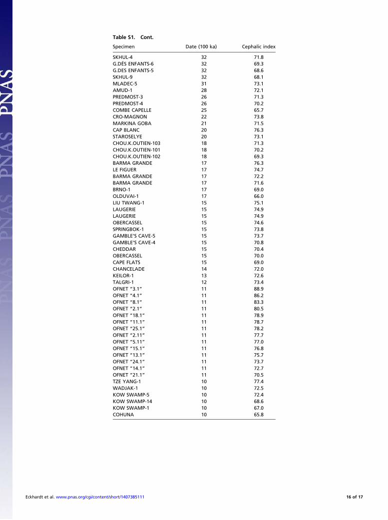
SI2: Cephalic IndexThe cephalic index or cranial index is the ratio of the maximum(biparietal) width of the head of an organism multiplied by 100,divided by its maximum (occipito-frontal) length, with bothmeasurements taken in the transverse plane. The index wasdefined by Anders Retzius in the 19th century and first used tocategorize human remains excavated in Europe. Because softtissues are of relatively uniform thicknesses around the heads ofliving humans in the transverse plane of measurement, figuresfrom past and living people are approximately comparable.By dividing the continuum of ratio values into three discon-
tinuous categories, human populations can be characterized
descriptively as dolichocephalic (long headed), mesocephalic(medium headed), or brachycephalic (short headed).Conventional ranges for these categories are: dolicocephalic
<75, mesocephalic 75.0–79.9, brachycephalic 80.0 (some sourcesuse slightly different dividing lines for females).Measurement of cranial length and breadth is not complex. It
can be accomplished with a pair of spreading calipers on a skull orreasonably accurate cast, and in the case of LB1 approximatedeven in two dimensions with a ruler on widely available publishedphotographs (figure 1 in ref. 20 and figure 1 in ref. 21).The skull shape of LB1, compared with representative Aus-
tralomelanesians, tends toward brachycephaly. The average cra-nial indices of Australomelanesians (22) range from 68.8 to 72.7,with LB1 at 79.0 lying ∼2 SDs above the midpoint average(supplementary table 1 in ref. 20). As noted in the text of ourreport, this index value also was biased downward in the originalpublication (1). Thus, the repeated erroneous descriptive prosereferences to LB1 cranial proportions raise the possibility thatverbal descriptions have been influenced by preconceptions,perhaps unconsciously, thereby tailoring reported results to apriori taxonomic conclusions. Quite simply, the cranial index ofLB1 is borderline brachycephalic, more so than modern humansin its region and more brachycephalic than most fossils (Fig. S1).Numerous previous craniometric analyses have been reviewed
(18); these have produced contradictory results arising fromnumerous shortcomings of experimental design and samplecomposition.
SI3: Skull (Cranium Plus Mandible and Dentition)Vault. Endocranial volume, proportions (length, breadth, andheight), and shape (chiefly left-right asymmetry) all have beendisputed since the earliest published reports on the LB1 skull. Justas the framing hypothesis has shifted from hypothetical islandisolation and dwarfing to (comparably hypothetical) migrationfrom an African origin at a pre-erectus level, it more recently hasbeen conceded that the LB1 cranium is asymmetrical and exhibitsproportions indicative of developmental abnormality, as notedbelow (18, 21).
Endocranial Volume. There have been several measurements ofendocranial volume in LB1: 380 mL (20), 417 mL (23), 430 mL(24), and 426 mL (25). The extremely low value initially re-ported (20) remains below all others published since then by10% or more. The only attempt to explain the endocranialvolume discrepancy between the original report (20) and sub-sequent higher determinations was by Falk et al. (26), who notedthat the “difference is attributable to how cranial holes wereplugged” when Brown measured the endocranial volume withmustard seeds. However, plugging holes (a common practicebefore determining volume with seed filling) is unlikely to leadto a lower volume than the CT scan technique used (23) unlessan excess of the material used to plug holes also somehowprojected into the cranial cavity (and then such excess materialwere removed before the CT scan or not detected by it); sucha result would indicate an improbably low level of technical skillfor Brown et al. (20). Our higher determination of 430 mL[which differs by only 3% from the CT scan made by Falk et al.(26)] also was made by filling with mustard seed (24) after re-moving some matrix inside the vault missed by Brown’s group.A simple inference is that the low endocranial volume, statedexplicitly to be “equal to the minimum estimates for Austral-opithecus” (20), resulted from an apparent technical error thatbiased reported cranial capacity downward toward those ofearlier hominins, coincidentally heightening media attention(27). This impression has been fostered further by visual con-trasts of the LB1 skull with an unusually large Homo sapiensskull of unspecified provenance [see figure 1 in the “FloresHobbit-Like Human Picture Gallery” on the National Geographic
Eckhardt et al. www.pnas.org/cgi/content/short/1407385111 2 of 17
website (http://news.nationalgeographic.com/news/2004/10/photogalleries/homo_floresiensis_1/)]. We provide here a moreappropriate regional and temporal comparison of LB1 withLiang Momer E from Flores, dated to 3,000–5,000 y ago.
Cranial and Endocranial Cast Proportions. Verbal descriptions ofthe LB1 skull have been subjective, and increasingly contradic-tory with time. Reportedly “The cranial vault is long and low”and “. . .indices of cranial shape closely follow the pattern inH. erectus” (20) and “align LB1 with archaic Homo . . . and Homoerectus (s.l.) in particular, such as the long, low cranial profile. . .”(28). In contrast, Falk et al. characterize the skull of LB1 as“extremely brachycephalic” (29) and according to other inves-tigators “The LB1 vault is anteroposteriorly short relative to itsbreadth” (18) , with similar remarks repeated twice elsewhere inthe same text. Our own measurements indicate the index valueof 80.1, whereas breadth and length reported by Kaifu et al. (18)yield an index value 82.0. Brachycephalic deviation from refer-ence population norms is a feature that occurs in numerousdevelopmentally abnormal syndromes, such as trisomy 21.Similar remarks can be made about the several successive
analyses of the LB1 endocast. One report (23) used a sample thatcomprised STS 5, KNM-WT 17000, 5 Homo erectus skulls, 10gorillas, 18 chimpanzees, 10 normal modern humans, and 1 adultfemale pygmy. In that study, one principal components analysisbased on length, breadth, height, and frontal breadth, groupedthe LB1 endocast with H. erectus and separate from H. sapiens;a second principal components analysis that excluded H. erectusendocasts grouped LB1 exclusively with H. sapiens rather thana variety of other hominoid primates. Subsequently, LB1 wascompared with 10 normal modern humans and a somewhatheterogeneous sample of 11 microcephalics (29). In this case,discriminant and canonical analyses were said to group LB1 withthe normal H. sapiens rather than the microcephalics. A still laterpaper by the same set of authors reported essentially the sameresults (26). Within the same research group, the outcomes ofvarious multivariate analyses appear to reflect more the samplecomposition and dimensions selected than the features of LB1itself. Moreover, the overall conclusions about internal endocastdimensions, proportions, and anomalies must remain suspect aslong as repeated denials of external cranial asymmetry remainuncorrected (see below). An independent study derived addi-tional ratios from the same dataset (26, 29), augmented by muchoriginal data (30). This study showed that that several LB1 en-docast ratios not computed by Falk et al. (29) fall largely outsidethe range of H. erectus and normal H. sapiens endocasts butwithin the range of microcephalic endocasts, thus supporting thesuggestion (24, 31) that LB1 represents a pathological microce-phalic H. sapiens rather than a hypothetical new species basedtenuously on a single specimen.
Cranial Base Angle. Purportedly “[T]he cranial base angle (basion-sella-foramen caecum) of 130° is relatively flexed in comparisonwith both Homo sapiens (mean 137°–138°. . . and IndonesianH. erectus (Sambungmacan 4, 141°. . .. Other small brainedhominins, for instance STS5 Australopithecus africanus, have theprimitive less-flexed condition” (20). The source for the 137–138° value (32) does not give sample sizes or measures of dis-persion but much of their data derive from other studies (33, 34),showing that a sample of 99 extant humans yields a cranial baseangle of 134.7° with SD = 6.09° and a range of 116–149°. Con-sequently, LB1 is not unique in this regard, or even unusual.
Cranial Vault Bone Thickness. Reportedly “cranial vault bone isthick and lies within the range of H. erectus and H. sapiens” (20).The deposits from which all of the Liang Bua Cave skeletons arederived span that recorded only for our own species, H. sapiens.Consequently there is no reason to suggest a particular affinity to
H. erectus, unless one accepts that H. erectus should be subsumedinto our own species (35, 36), which is a matter of conceptualimportance but aside from the main point of this paper.Table S1 (20) provides data on cranial vault thicknesses for
LB1 at the following sites: midfrontal, bregma, parietal eminence,lambda, asterion, and e-o-p. For comparison, measurements atthese same points were provided for global pooled-sex H. sapiens,in which sample sizes ranged from 575 to 670, for which sampleswere provided the means and SDs, as well as minimum andmaximum values. In all cases LB1 fell within the minimum/maximum ranges for the recent H. sapiens samples. Conse-quently, in simplest terms there is no objective basis for sayingthat the cranial vault bone of LB1 is thick; the verbal statement(20) is not warranted by the data provided. There is little morethat can be inferred reliably, particularly given the absence of anyinformation on the population and regional composition of therecent H. sapiens samples, which were very heterogeneous. Of thesix cranial thickness measurements given, two (midfrontal andasterion) were above the global population sample midpoint, one(parietal eminence) was the same, and three (bregma, lambda,and e-o-p) were below, with the average of all six thicknessesfalling at only about 55% of the sample maximum.Comparison of LB1 with the global sample data are likely to be
misleading directionally, because the Liang Bua Cave samplefrom Flores lies within the Australomelanesian population re-gion. Australian aboriginal skulls from within that region arethicker than other populations (37) (table 8 in ref. 37 provideslimited data for skull vault thickness); at vertex, Australian ab-original skulls average 26% thicker than North American“Caucasoids” (7.33 mm vs. 5.8 mm). The skull vault of LB1 isnot unusually thick objectively, and in the context of its ap-propriate regional population, can be characterized as thinnerthan average.
Foramen Magnum.Descriptions of fossil hominin skulls commonlyreport the antero-posterior placement and angulation of theforamen magnum, but not its length and breadth. The LB1 fo-ramen magnum is described as narrow (21 mm) relative to itslength (28mm), but it was not stated whether this information wasincluded to suggest that LB1 is unique, primitive, derived, normal,or abnormal (20). The foramen magnum is commonly narrowedin various developmental abnormalities, including achondropla-sia and Down syndrome (38); we deal definitively with this andrelated points in ref. 19.
Other Cranial Base Features. It originally was noted of LB1 that “Incommon with Asian, and some African, H. erectus a deep fissureseparates the mastoid process from the petrous crest of thetympanic. Bilaterally there is a recess between the tympanic plateand the entoglenoid pyramid. These two traits are not seen inmodern humans, and show varied levels of development inAsian and African H. erectus and Pliocene hominins” (20). Thatstatement is incorrect; these features commonly are found inAustralian and Tasmanian crania (39–41) as well as Kow Swamp5 and Keilor (42), a point we made several years ago (24) withoutany rejoinder being published since. These features obviouslyare not unique to LB1 in comparison with present and pastmembers of H. sapiens in the geographic region of which Floresis a part, and logically cannot be a defining feature of the sup-posed new species.
Mandible. “The anterior portion of the corpus is rounded andbulbous and without a chin” (20). Although this absence of anexternal chin has been stressed repeatedly as a “primitive” fea-ture, it is not unusual among populations in the region. Thispoint, made years ago by Jacob et al. (24) has been deniedrepeatedly since, with some of these denials introducingdistortions. One publication (43) provided a radiograph of an
Eckhardt et al. www.pnas.org/cgi/content/short/1407385111 3 of 17
unidentified Australomelanesian whose mandible displays a hardtissue chin, around which a supposedly nonprojecting soft tissuechin was drawn in, with the artificial enhancement made pre-sumably because the image itself was unclear. Regardless ofwhatever information was meant to be conveyed by the alteredimage (43), the anterior region of the mandible is variably de-veloped among Australomelanesian populations including thosenow living on Flores, and nonprojecting bony chins occur widely(Fig. S3).This point has been established definitively for extant Flores
H. sapiens now living on Flores (44). In the sample of 52 Ram-pasasa individuals (22 males, 30 females), 76.9% had neutral ornegative chins as assessed radiographically from hard tissues(Table S2); only one male had a positive hard tissue chin that wasmasked by soft tissue that made it appear negative. Any attemptto demonstrate the range of variation in this phenotypic char-acteristic with a single example is inherently typological.The LB6 mandible is said to confirm key features of LB1
mandibular morphology. This is not the case except in the generalsense of overall size, as can be seen from figures 1–4 in ref. 43.The symphyseal region of LB6 inclines more toward the verticalas in some extant Rampasasa (44) and lacks the supposedly more“primitive” posterior, inferior, curvature of LB1 that is present inother extant Rampasasa (44). This extent of variation in the chinregion even between two LB specimens parallels that docu-mented by Hastuti, et al. (44) in the extant Flores Rampasasapopulation. The variants of the chin in LB1 and LB6 are observedin numerous Australomelanesian populations, past and present.Another mandibular feature of LB1 is described thus: “The
ramus is broadest inferiorly, slopes slightly posteriorly and isthickened medio-laterally, and the coronoid process is higher thanthe condyle” (20). The relative heights of the coronoid process andcondyle have no particular phylogenetic valence that would favorallocation of the Liang Bua Cave bones to H. erectus, to un-specified very early Homo populations, or to australopithecines.Several studies (45, 46) show that relative heights of the condyleand coronoid process vary widely among extant human, UpperPaleolithic, early anatomically modern (i.e., pre-Upper Paleo-lithic), and Neandertal populations. These features and othersfavor the hypothesis that the Liang Bua Cave skeletons rep-resent normally variable H. sapiens rather than some veryprimitive hominin population caught in a time warp.The contrast presented by the LB6 mandible with that of LB1
(43) is even greater for the morphology of the ramus than for thesymphyseal region. The taxonomic irrelevance of a coronoidprocess being higher than the condyle is underscored within theLiang Bua Cave sample itself, because in LB6 the coronoidprocess is not higher than the condyle, but rather is just aboutexactly the same height in the orientation shown.Emphasis on absence of the external bony chin as a purported
taxonomic uniqueness although it is not, and of the coronoidprocess being higher than the condyle, although this also is nota unique feature in the Liang Bua Cave skeletal sample, hasserved to divert attention from a different but genuinely unusualfeature of the LB1 mandible (again, not shared with LB6): itsstrikingly tall ramus.In discussing the possible functional significance of a tall
mandibular ramus, one study (47) presented two alternativemeasurements of mandibular corpus height: from the inferiormargin of the mandible or from the occlusal surface of the teethto the condyle (which was the superior point measured in theirstudy), the latter measurement removing any potentially con-founding effect of mandibular corpus thickness. The resultsshowed that in a sample of 62 extant human mandibles, the meancondylar height above the occlusal plane for both males andfemales is about 36 mm; that is, in their sample there was nodifference attributable to sexual dimorphism. In a sample of 20chimpanzees divided equally between males and females, the
respective measurements were 41 mm and 40 mm, for a differ-ence in ramus height of 2.5%. Ten male and 10 female gorillasshowed mean heights of 72 mm and 60 mm, respectively, theramus in the males thus being 20% taller. In an independentstudy (48) mandibular ramus height in human males was re-ported to be about 10% greater than that in females. Theselimited samples of hominoid primates show the range of sexualdimorphism to be 0–20%, with males on average tending towardtaller rami. We note in passing that LB1 originally was identifiedas female (20), an impression that has been propagated widelyand uncritically. In contrast, our explicit quantitative analysis (5)showed that LB1 was a gracile male, which is reinforced by theresults reported here. The ramus of LB1 is substantially tallerthan that of LB6 (figure 3 in ref. 20). Others have found ramusbreadth to be the best predictor of sex, with males havingbroader rami (49). The ramus of LB1 is broader than that ofLB6, again supporting our diagnosis of LB1 as male and con-tradicting the original impressionistic assessment (20).Our measurements from the paper by Brown and Maeda
(figure 3 in ref. 43) show the height of the mandibular ramus inLB1 to be very substantially greater than that of LB6. From theinferior surface of the mandible to the condyle, the ramus of LB1is 40% taller than that of LB6; measuring to the coronoid tipproduces a moderately higher figure for LB1 of 55%. From theocclusal surface to the occipital condyle, the ramus of LB1 is50% taller than that of LB6; measuring to the slightly highercoronoid tip produces a 67% greater height for LB1.Differences in ramus height between LB1 and LB6 (generally
unremarked until now) are substantially greater than reported inother studies of hominins and, more widely, hominoid primates.These measurements are discrepant enough to direct attention toa situation encountered in robust australopithecines, which alsohave somewhat dished midfacial regions and very tall mandibularrami (50). In robust australopithecines these facial featuresare combined with relatively small braincases; because LB6 isa mandible without a cranium, this comparison cannot be ex-plored except as an intriguing question, but if the australopith-ecine analogy holds, it implies that LB6 had a larger craniumthan that known for LB1; a more extensive comparative study inthat direction is beyond the scope of this paper and the existingdata available to us. In the less hypothetical realm, however,several additional suggestions are possible. First, functionally,the cross-sectional mass of the masseter muscle shows a positivecorrelation with mandibular ramus height; we do not know whatimplications there might be of a relatively larger masseter inLB1. Second, developmentally the human skull achieves its adultsize through a supero-inferior gradient of maturation, with thepotential for bidirectional developmental influences between thelateral cranial floor and the face until about 11–12 y of age, therebeing a structural interface between brain and facial anatomy(51). Third, genetically, there is evidence for an association be-tween mandibular height and the growth hormone receptorgene, as elucidated in a Japanese population (52).One recent computationally ambitious study of mandibular
measurements in the Liang Bua Cave sample (53) puzzlinglyomits inclusion of LB6 entirely. Even so, results of their ca-nonical discriminant analysis group LB1 more closely with the LaQuina and Gibraltar neandertals, and also with Peninj, than withAL 288-1, and as closely with some modern humans as withSk15. Other highly formulaic analyses of landmark data inthe absence of genetic, developmental, and functional consid-erations (54) only reinforce the impression that the morphologyof H. floresiensis really is just that of the individual specimen,LB1. Landmark hyperspace explorations tell us little other thanthat LB1 is dimensionally strange, which from the first has beenevident by inspection.
Eckhardt et al. www.pnas.org/cgi/content/short/1407385111 4 of 17
Dentition. In terms of dimensions and morphology, teeth of LB1and LB6 are not in any way unique compared with those ofanatomically modern H. sapiens. There are some interestingdetails regarding wear and crown morphology alterations of LB1teeth, but these have been described (55) and discussed (56)elsewhere.
SI4: Upper Limb Skeletal ElementsScapula. No scapula has been described for LB1. Three majorportions of this bone are known from specimen LB6/4 (57), anadult. The scapular spine appears horizontally oriented as it is inother H. sapiens. The scapular neck shows a slight dorsal ori-entation to the glenoid fossa, attributed to distortion. The ven-tral bar/glenoid angle is reported as 157°, also in the range ofextant humans. None of the angles identified by Oxnard (58) asdifferentiating scapula form among primate locomotor groupsdistinguishes LB6/4 statistically from those of living humans, andare reported as falling within the corresponding 95% fiduciallimits of modern humans. In the descriptive and comparativecontext provided, the scapula of LB6 accords with our hypothesis(24) that, aside from LB1, the Liang Bua Cave skeletal samplerepresents developmentally normal extant H. sapiens of smallbody mass and stature.
Clavicle. The clavicle is the first long bone to ossify, a process thatbegins in condensed mesenchyme during the fifth and sixthembryonic weeks of embryonic life from two ossification centers,one medial and the other lateral; fusion occurs during fetal de-velopment. Epiphyseal ossification occurs at both the acromialand sternal ends of the bone; the sternal end fuses with the di-aphysis between 18 and 25 y of age, making this the last longbone to fuse (59). Such a developmental pattern would be ex-pected to permit postnatal functional influences on individualdevelopment.There are numerous genetic diseases that alter clavicle mor-
phology. Lenz–Majewski hyperostotic dwarfism causes a broadand thick clavicle, along with progressive skeletal sclerosis, se-vere growth retardation, and mental retardation (60). Melnick–Needles syndrome (Otopalatodigital syndrome spectrum dis-orders), can cause clavicular hypoplasia, curved long bones,and flared metaphyses (61, 62). Otopalatodigital syndrome typeII presents with thin clavicles, microcephaly, cleft palate, andoverlapping fingers (63). Maroteaux-type acromesomelic dys-plasia manifests a curved clavicle with pronounced dispropor-tionate short stature (64). This list is only a partial summary, andthe clavicle remains only minimally described—or undescribed—in many other inherited disorders, including some of the morecommon ones, such as Down syndrome (trisomy 21). Trisomy 13and trisomy 18 exhibit generally thin clavicles, and Turner syn-drome often includes clavicles that are thin laterally (65).An extended description and some dimensions for the right
clavicle, LB1/50, has been previously published (57). It is missingthe sternal end; the lateral end also has been damaged post-mortem. Views include superior, inferior, anterior, and poste-rior. Discussions for most of these views make no claim foruniqueness. An exception is the posterior (i.e., dorsal) view:“Voisin (2006) has reported that modern humans are distinct indisplaying a single inferior curve of the clavicle in posterior view.His preliminary examination of LB1/5 (based on photographsonly) indicates that it retains the primitive double curvature seenin African apes and all hominins except modern humans (Voisin,pers. comm.)” (57).Over more than half a century the clavicle has received a fair
share of attention by physical anthropologists, including an earlypaper on techniques for its measurement (66). More recently,Voisin also has published extensively on this skeletal element(67–73). Although we do not have the text of the personal com-munication from Voisin to Larson et al. (57), two of us (M.H.
and R.B.E.) have examined the original specimen as well asfigure 1 of Larson et al. (57). In the posterior view, both “cur-vatures” of the LB1 clavicle are not pronounced. This findingaccords with the following statement published by Voisin (71):“Sometimes, some individuals [that is, individual extant humans]show two curvatures in dorsal view, but these curvatures areslight in regard to the condition exhibited in the great apes.”The approach of Olivier for the quantifying the degree of
curvature of the clavicle was applied to LB1, with awareness ofstrong methodological limitations. A significant piece of thesternal end of the clavicle is missing. This defect required that thetotal length be extrapolated from the preserved portion, andconsequently that some points of measurement be estimated.Furthermore, the only source available for this study was thepublished composite figure shown in ref. 57; this approach is notas desirable as having a CT scan of the clavicle, which was un-available to us. Three measurements were estimated for eachcurvature, based on the range of extrapolated total clavicle lengthgiven (57). The extrapolated length was assumed to all belong tothe sternal end. This approach probably did not greatly affectmeasurements of the external and inferior curvatures but there isconsiderable uncertainty (Table S3).For the external curvature, LB1 is within 1 SD of the mean of
H. sapiens of 16.1, according to the reference data (71). For theinternal curvature, LB1 is also within 1 SD of the mean ofH. sapiens of 12.6 if the smallest total length is taken. The in-ferior curvature of LB1 is within 1 SD of the mean of H. sapiensof 5.1. LB1 is within 2 SD of the mean for H. sapiens for thesuperior curvature, which has a mean of 2.9.In addition, CT scans of one human subject included in our
pilot study, undertaken to provide comparative developmentalevidence from patients combining short stature and skeletalanomalies, showed clavicles with some modest double curvature(subject’s superior curvature = 4.89 mm vs. human referencesample mean of 2.9 mm, SD = 1.5 mm, n = 33; subject’s inferiorcurvature = 9.45 mm vs. human reference sample mean of5.1 mm, SD = 2.3 mm, n = 33). Against the background of eventhese limited data, use of the term “primitive” for variants thatoccur in some living humans, normal or developmentally ab-normal, is pejorative and misleading.Representation of the short clavicle of LB1 in H. floresiensis
(i.e., members of the Liang Bua Cave skeletal sample) as aprimitive retention, recurs in context of the statement “The re-cent description of comparatively short clavicles from Dmanisi(Lordkipanidze et al. 2007) supports the view that a relativelyshort clavicle is characteristic of early H. erectus and is probablythe primitive condition for hominins” (1). Such a statementimplies that the information contained in human clavicle sizeand shape is exclusively phylogenetic (because no other factorsare discussed) and thus has direct taxonomic valence. Theclavicle and other bones surely reflect evolutionary heritage tosome extent, but there is failure to consider the possibility thatthe morphologies of the clavicle and other bones can containindividual developmental information as well (57). In contrast,there is strong evidence for functional influences on clavicleform and dimensions (74), with recognition that as “. . .an early-ossifying, late-fusing bone . . . [the clavicle] . . . should show clearsigns of lateral loading bias, with the right side being the moreheavily loaded at the population level” (74). Furthermore, basedon a sample of 136 individuals including both sexes and juvenilesthrough adults, “. . .the right clavicle tends to be more robust. Inthe adult males (the group in which the right clavicle is signifi-cantly thicker in both sagittal and vertical dimension at its mid-shaft), lateral curvature is significantly less in the right clavicle—which suggests that the right bone is more adapted to resistlateral buckling stresses in axial loading. The other clear corre-lates of length differences are asymmetrical development ofthe areas of attachment of the trapezoid and costoclavicular
Eckhardt et al. www.pnas.org/cgi/content/short/1407385111 5 of 17
ligaments” (74). Against this broader functional background, aswell as analysis of the limited empirical evidence, the categoricalstatement (57) that the structure of the LB1 clavicle is distinctfrom extant humans by its proportions and supposedly primitivedouble curvature is insupportable from available data.
Humerus. Two initial points should be recalled here: first, at thetime of the initial description and diagnosis of this hypotheticaltaxon (20), the upper limb bones had not yet been recovered, sotheir attributes could not have been part of its supposedly uniquemosaic of features; second, as with the femora, the humerus ofLB1 is the only example of this bone in the entire Liang BuaCave skeletal sample. Thus, as for the femora, any statementabout “the humerus of Homo floresiensis” should be read moreconservatively as “the humerus of LB1.” Pertinent attributes ofthe LB1 humerus include its overall size (principally length),robusticity, and torsion.Length. The LB1 right humerus is complete aside from post-mortem damage to the anterior surface of the head and greatertubercle; its length is given as 243 mm (75). This figure is repeatedby Larson, et al. (57), who note that “The humerus. . . while shortin an absolute sense, can be matched to the lower extremes ofsmall-bodied African pygmies and Andaman Islanders (WLJ,pers. obs.).” Thus, the length of the LB1 humerus simply is notunique among extant humans.Beyond size are bone shape considerations. “When compared
with a modern human humerus scaled to the same length, themost obvious differences are the greater diameter of the LB1shaft and the limited degree of humeral torsion (that is, rotationof the humerus head medially relative to the mediolateral axis ofthe distal end; Fig. 4)” (75).Width. “All the major limb bones of LB1 have shaft and articularsurface dimensions that are robust relative to length” (75). Thisdefinition of robusticity is used repeatedly in numerous sub-sequent papers. “In external dimensions, the humerus of LB1 isindeed robust” (23); similarly, “. . .LB1 has extremely high levelsof robusticity for all limb bones. . .” (76).“The midshaft appears rounded in cross-section with an an-
teroposterior diameter of 17.44 mm and a mediolateral diameterof 16.35 mm” (57). The exclusive use of external bone diameters(in isolation or in proportion to length) to represent robusticity,once standard, now is idiosyncratic and outdated. Among otherinvestigators a broader array of robusticity measurements is used(77). The more common of these measurements involve appli-cation of beam theory, in which the cross-sectional geometryof long bone diaphyses is quantified to assess the mechanicalcompetence of a bone (78). In this approach, calculations ofbiomechanical properties of cross-sections of bone diaphyses aredependent upon accurate determination of periosteal and end-osteal contours of the diaphyses. Methods now available can usesilicone molds and biplanar radiographs to provide accurateestimates of cross-section contours without damaging the speci-men or incurring the costs and other limitations of CT (79, 80).Given the weight placed on the supposed robusticity of LB1 asa species-distinguishing factor, one would expect that the mostfunctionally informative methods would be used.In fact, these methods have been used, although not on LB1.
Jungers previously made a case for use of such methods (81):“Employment of this methodology in the mechanical assessmentof fossil long bones has been limited to date to fortuitous breaksor fragmentary specimens. . ., since postcranial remains of fossilsare usually too rare to be sacrificed by sectioning.” The existenceof LB1 CT data are implied by the statement of Falk, et al. (26),who wrote “. . .CT scans indicate that the cortical bone of theLB1 humerus is not especially thick, but rather is within thehuman range.” It is puzzling that these numerical CT scan datahave not yet been published. They might be redundant, in light ofthe statement by Morwood et al. (75) that in the case of the LB1
humerus “. . .the mid-shaft was broken post-mortem. . .” Thatbeing the case, why were the broken ends of the shaft not pho-tographed or measured directly and the cortical thicknesses re-ported? These data seem to have existed since at least 2005 inthe case of the broken humeral shaft, and since at least 2009 forthe CT scans. Reference to LB1 humeral “robusticity” inferredfrom only the most rudimentary external dimensions is morelimited than necessary if the Liang Bua Cave skeletons are asimportant as represented. In any case, another claim of “primi-tiveness” or “uniqueness” lacks support.Humeral torsion. Measurement of humeral torsion is complex fora number of reasons. The pertinent anatomical and methodo-logical complications are reviewed informatively and indepen-dently of the controversy surrounding interpretation of the LiangBua Cave skeletons (82, 83).Morwood et al. (75) noted “In LB1, humeral torsion is ap-
proximately 110°, which is the norm for Hylobates and quadru-pedal primates such as Macaca, but is significantly less than inlarge-bodied apes, modern humans (141–178°) and other knownhominins, including Australopithecus.” Lack of any qualificationin that report created the impression that the information em-bodied in humeral torsion is only phylogenetic, consequentlypertinent to support for the new species created the year before(20). The reported humeral torsion, being outside the range ofmodern humans, was held to constitute another uniqueness ofH. floresiensis. However, this degree of torsion also is outsidethose of all large-bodied apes and hominins. H. floresiensis, as amember of the genus Homo, should have humeral torsion withinthe hominid (large apes plus humans) range, were it a healthynormal individual, so its reported value was more likely to be di-agnostic of pathology than uniqueness. We responded (24) bynoting that the extent of humeral torsion in any individual isdevelopmentally labile, and that the abnormally low amount ofhumeral torsion in LB1 was consistent with the extremely weakmuscle development indicated by its muscle insertions. Suchweakness also is pertinent to the debate about LB1 humeral“robusticity” (see above). Numerous studies, some cited as ref-erences by Cowgill (82), document developmental influences onhumeral torsion as a result of various pathologies (84), occupa-tions (85), and sports, particularly those involving throwing (85;see also Whiteley et al. 86–88).Larson (89) remarked that “since the most proximal end of the
LB1/50 humerus is damaged, it is possible that the publishedmeasurement of 110° of humeral torsion is inaccurate.” The newmeasurements, using two slightly different indicators of humeralhead position, were 119° and 121°. These measurements, notimplausibly, were averaged to provide a new published estimateof 120°. Then this figure was averaged once again (not so plau-sibly) with the previous estimate—possibly inaccurate by herstatement—to arrive at a new average of averages of 115°. Thispuzzling “best estimate of humeral torsion in LB1/50” was re-peated in Larson et al. (57). Data in figure 4 of Larson (89) showthat the 120° value is within the range of several regional pop-ulations pertinent to Flores inhabitants: Australians (n = 4),Melanesians (n = 14), Senoi (n = 4). Even the dubiously de-termined 115° figure still is higher than the average for EastCentral African Pygmy females (n = 6).Overall, the publication history of LB1 humeral torsion—and
indeed of its humerus length and robusticity as well—is one ofretreat from initial stress on taxonomic uniqueness (75) topresent banality of being somewhat unusual, although not amongextant small-bodied humans (57, 89).
Ulna.Our comments are based on study of the specimens (by twoof us, M.H. and R.B.E.) plus review of two published accounts(1, 23). Discussion is limited here because the publications citedmake no particular claim for taxonomic uniqueness other thanthe cryptic comment in the supporting information published
Eckhardt et al. www.pnas.org/cgi/content/short/1407385111 6 of 17
online by Morwood et al. (75) with reference to the LB1 rightulna: “As LB1 is from a species with different postcranial di-mensions and anatomy to H. sapiens it is not known how accu-rate this estimate is.” Such a statement confuses hypothesis withpostulate in assuming a priori that H. floresiensis is a new species.The material most completely described comprises LB1/51,
a partial left ulna (205 mm in length, lacking proximal and distalportions), and LB1/52, a more nearly complete right ulna (190mm with distal portion of shaft and head missing, reconstructedto ∼205 mm).Also recovered were LB6/3 and LB2/1. From the same spit [51]
as LB1, LB6/3 preserves the proximal portion of a shaft witha length of 137 mm. Unfortunately it is not figured in Morwoodet al. (75) or Larson et al. (57). Alignment of the preservedportions of the LB1 and LB6 ulnae might have been informativewith regard to estimates of overall size (e.g., stature) because theulnae are the only two long bones common to these two in-dividuals, and because statements about the relative and abso-lute statures of various individuals from Liang Bua Cave are veryproblematical and contradictory; see the section on tibia in thispaper (below) and also the monograph by De Klerk (90). Thecorresponding measurements provided in table 3 of ref. 57 forthe proximal ends of the ulnae are consistently smaller in LB6/3than in LB1/52.Regarding the ulna specimen LB2/1, measurements provided
in table 3 of ref. 57 for the proximal ends of its ulna generally, butnot invariably, fall between those of LB6/3 and LB1/52. Giventhe much greater age of this specimen (about 74 ka in compar-ison, with 15.7–17.1 ka for LB6 and 17.1–18.7 ka for LB1), aswell as the allusion by the authors to some unspecified pathol-ogy, further study seems warranted.Morwood et al. (75) conclude with the statement that “Other
H. floresiensis morphological traits, for example in the humeraltorsion and ulna, are not shared with any other known homininspecies. . ..” Previously in this report we found no objectivesupport for that statement with regard to the humerus. Similarly,the ulna manifests no uniqueness at any level, against a back-ground of individual variation that is normal for human pop-ulations, past and present.
Radius. As for the ulna, our comments here derive from the re-stricted study of the specimens (by M.H. and R.B.E. in February2005) plus the published accounts (57, 75) that are overlapping.The length estimated for the LB1 radius is 190 mm with SE =
4.208 mm.Uniqueness in the radius is implied by the statement in the
caption of supporting information figure 5 byMorwood et al. (75),in which LB1 radius length is estimated from ulna and radiuslengths in extant humans: “As LB1 is from a species with dif-ferent postcranial dimensions and anatomy to [sic] H. sapiens it isnot known how accurate this estimate is.” That qualifier standsin contrast to their own statement in the text (75): “The arms ofLB1 share the distinctive elongated distal segment common totropical modern humans, including populations of small averagestature, and have an estimated brachial index ((radius length ×100)/humerus length) of 78, which is close to the African humanmale average.” Synthesizing all this, it seems to be suggested thatthe ulna and radius (as well as humerus) lengths of LB1 may bethose of a (dubiously diagnosed) female of a (hypothetical) dif-ferent species, but they exhibit the proportions that one wouldexpect for extant tropical male H. sapiens.Although no radius appears known certainly for LB1, the
penultimate entry in table 1 of ref. 75 is: “Radius (LB1?) Bothepiphyses missing. 124 × 16.5 mm. (Note: from baulk collapse).”We have not found further references to this possibility. Other
pertinent radius specimens here are LB4/1, a juvenile left radiuswith unfused epiphyses, and adults LB6/2, and LB3. For somereason the juvenile radial fragment LB4/1 is shown in comparison
with (presumably) adult left radius LB3, but is not compareddimensionally or illustratively with any juvenile radii of knownH. sapiens.Adult specimen LB6/2 is a complete right radius with a highly
distorted distal portion, about which Larson et al. (57) write,“which Morwood, et al. (2005) interpreted as the result of anunset, healed fracture with compensatory remodeling and callusdevelopment.” Larson, et al. (57) further comment: “A radio-graph of the radius supports the interpretation of a healedfracture resulting in ulnar displacement of the distal radius(Ortner, pers comm.; Sampson, pers. comm.).” The anatomicallocation of this distal radial irregularity mimics Madelung de-formity, but without our being able to examine the specimen withthis possibility in mind, no definite statement can be made andresemblances probably are coincidental. A healed fracture wouldbe consistent with our overall general position that, aside fromLB1, other individuals from the Liang Bua Cave site appeardevelopmentally normal.Although incomplete, lacking its head and distal portion, the
partial radius LB3 merits discussion. It was recovered from one ofthe oldest sections of the Liang Bua Cave deposits, dated to about74 ka (20) and discussed in table 1 of ref. 57. Although the shaftof this bone is rather straight and its cortical bone is noted to bethin (1.3–1.7 mm) (57), its preserved portion is 164 mm, and itsestimated that total length was ∼210 mm. If that reconstructionis accurate, it would place this bone within the normal modernhuman range. Henneberg and Thorne (31) estimated that thisradius corresponds to a modern human of the stature 1.51–1.62 mtall. Larson et al. (57) comment that “. . .like LB6/2, the radialtuberosity faces more directly medially than in modern humans,judging from the position of the interosseous crest.” However, asno comparative evidence is adduced, this remains a descriptionwithout demonstration of taxonomic uniqueness.
SI5: Carpal BonesComments on Morphology of LB1. The specimens of LB1 describedso far, all from the left side, comprise a scaphoid (LB1/44),capitate (LB1/45), hamate (LB1/46), trapezoid (LB1/47), and apartial lunate.In 2007, left LB1 carpal specimens, comprising a scaphoid
(LB1/44), capitate (LB1/45), and hamate (LB1/46), were de-scribed, illustrated with multicolored cartoon-like drawings (91).Photographs of the actual specimens were not published until 2 ylater (57), at which time the trapezoid (LB1/47) and partial lu-nate also were described and figured. After a gap of another 2 yan additional (right) capitate and two hamates from a differentindividual (most likely LB6) were reported in an abstract (92),followed again after a year by a full-length paper (93).The comparative perspective initially offered (91) is misleading
in a manner that is implicitly typological. That is, publishedfigures of all three bones show several other individual speci-mens in addition to LB1. For the trapezoid, individual specimenscomprise Qafzeh 9 and Kebara 2; for the scaphoid, Qafzeh 9,Regordou 1, and OH7; for the capitate, AL288-1w, AL333-40,and TM1526. The rest of the comparative sample comprises theextant hominoid taxa: Pongo, Gorilla, Pan, and Homo (desig-nated Modern), each of which is represented by a single cartoonimage. None of these illustrations of extant hominoid taxa areidentified as particular specimens (with a collection specimennumber to permit replication), and exactly how composite im-ages might have been derived is unspecified; in some unclear wayeach individual cartoon seems intended to represent an entiregenus or species. The impression thus created by comparison ofsome individuals to entire taxa—implied but not stated—isnecessarily to underrepresent variation (normal and abnormal)in any of the extant taxa that might affect the morphologicalcomparisons. However, even limited visual comparisons amongthe stylized abstractions in figures 1–3 of ref. 92 make it possible
Eckhardt et al. www.pnas.org/cgi/content/short/1407385111 7 of 17
to reject the implied null hypothesis of within-taxon invariance(and consequently the reliability of between-taxon differences).
Trapezoid (figure 1 in ref. 91). In figure 1 of ref. 91, in palmar view(top row) Qafzeh 9 (Upper Paleolithic Homo) resembles Kebara2 (a Neanderthal) more than it resembles the Modern mor-photype; note the extent to which this visual comparison isinfluenced by the nonarticular surface (pink). In proximal view(middle row), Qafzeh 9 resembles the Modern morphotype inoutline, but not in articular surface for the capitate (green). Inulnar view (bottom) Qafzeh 9 resembles generic Modern lessthan it does Kebara 2 (a Neandertal). Among generic ape taxonmorphotypes, in all three views (palmar, proximal, ulnar), Panresembles Pongo strikingly more than either does Gorilla. Thesepatterns of similarity and difference are explicable in functionalterms [indeed, the authors do so to some extent on page S7 oftheir supporting information (91)], but the inescapable criticalinference is that these visual comparisons of overall shape andarticular patterns simply are not reliable as indicators of phylo-genetic relationship, and it is the nature of the phylogeneticrelationship that is the intended focus of that paper. Against thisbackground, any inference about the phyletic position of theindividual LB1 specimen is indeterminate at best and sub-jectively determined a priori at worst. The proximal view (middlerow) in particular emphasizes this point. Although on first glanceLB1 superficially shares an outline more similar to the ape genusmorphotypes, that impression is dominated by its horizontal di-mension in figure 1 of ref. 91; if anything, this is beyond thepongid range in its elongation, which obscures the contradictionposed by the articular surfaces, particularly for the capitate(green), which far more resembles the Modern morphotype.
Scaphoid (figure 2 in ref. 91). In figure 2 of ref. 91, in contrast to thetrapezoid, with Pongo largely eliminated from most comparisonsbecause of its unfused os centrale, other differences in the car-toon images are more minor. In radial and ulnar views, LB1shows comparable extents of resemblance to the Modern humanand Pan morphotypes. The distal views are marginally more in-formative, although unclear in the inferences that can be drawnfrom them. In outline shape of the bone, LB1 resembles Re-gourdou (a Neanderthal) more than any of the others, and itstrapezium-trapezoid articulation (light blue) is closer to OH7(Homo habilis), with neither resembling the Pan morphotype,which in this regard is more like the Gorilla morphotype. Again,these patterns of similarity and difference are conflicting, moreenigmatic than enlightening.The trial study described in Materials and Methods produced
for the first subject we studied a dorsal view of a right scaphoidthat was a nearly perfect (mirror image) match with comparabledorsal view of that shown for H. floresiensis (i.e., LB1; compareour Fig. S4 with figure 7 in ref. 57, both scaphoids being boomerang-shaped in outline rather than resembling a rounded triangleshown for their typical human).Of course far more extensive studies could be pursued, but
there are major cost/benefit ratios that must be considered.Carpal bone variants are extensive, and the causes of thesevariations remain incompletely understood. It is highly probablethat scaling factors and developmental anomalies will influenceheavily the range of carpal variants that will result from fur-ther study.
Capitate (figure 3 in ref. 91). As a general observation applying toall views of this bone shown in figure 3 of ref. 91, there isa contrast between articular facet orientations (broadly similaracross all images) and overall bone shape (more disparate).Particularly in palmar view, the capitate of the Pongo morpho-type is sharply waisted, whereas that of the Modern humanmorphotype is broadly rectangular. Among the early hominin
specimens, AL288-1w (A. afarensis), which is Pan-like, contrastssharply with TM1526 (A. africanus), which appears hyper-Mod-ern in many respects. AL333-40 is intermediate between theother two australopithecines, vitiating formal taxonomic assign-ments. Although LB1 can be said to resemble the Pan morpho-type here in palmar view, in fact there is more apparent visualdifference between AL288-1 and TM1526 than there is betweenLB1 and the Homo morphotype. Similarly, radial views are notparticularly distinguishing, although there is more of an overallpattern contrast between all ape genus morphotypes as a groupwith the group comprising all australopithecines and the Modernmorphotype, particularly with regard to second metacarpal ar-ticulation; in this regard, LB1 groups far more conformably withall of the hominins. In distal view all of the specimens andmorphotypes are complexly disparate. If the Modern morphotypeis representative—which cannot reliably be assumed because itssingle image represents a sample from or composite of at least20 specimens—then it manifests the greatest deviation from theoverall patterns of shape and articulations. Most notably, theModern human morphotype as represented here is matched veryclosely by LB1 in all respects save the portion of the thirdmetacarpal articular facet that extends just below (figure 3 in ref.91) the articulation for the second metacarpal.
Comments on Multivariate Plots of LB1 Carpal Bones. The multi-variate plots convey a similarly confusing impression, with thesame caveat with regard to multidimensional shape complexityand near-impossibility of making any simple inference from themabout the phylogenetic position of the LB1 specimen.
Trapezoid (figure 1 in ref. 91). For specimens from known taxa, theplot of CAN2 vs. CAN1 appears to show a clear separation ofhominins from nonhominins, with LB1 sorting into the latter. Oncloser examination, the placement of LB1 is located centrallywithin the fringes of all of these nonhominin groups, aboutequidistant from the marginal scattering of Pan, Pongo, Gorilla,and Papio. Incidentally, the inclusion of Papio in the comparativesample is puzzling, because to our knowledge, no investigatorwith the exception of Gert van den Berg (94), a member of thegroup that advocates the recognition of H. floresiensis as a sepa-rate species, has postulated a quadrupedal locomotor pattern forLB1; that this (94) astonishingly heterodox (and thoroughlyimplausible) view has gone unremarked is another example ofthe suspension of disbelief about nearly anything said concerningLB1 as long as the taxonomic validity of H. floresiensis remainsunquestioned. Nonetheless, this otherwise inexplicable expan-sion of the comparative sample provides the basis for an un-expectedly telling point: LB1 is morphologically plesiomorphicwith regard not only to hominoids, but with regard to catarrhineprimates more broadly. Because abnormal development verycommonly produces atavistic phenotypes, this is an importantobservation with which we deal elsewhere (19). The plot ofCAN3 vs. CAN2 shows an even more extreme placement of LB1beyond the fringe of all groups represented in this study.
Scaphoid (figure 2 in ref. 91). For various reasons Pongo (unfusedos centrale) and Papio (unspecified) are not included in thecomparative sample for this bone. Based on what evidence isdepicted, LB1 falls among Pan specimens; however, because Panspecimens present such a wide scatter, LB1 is more separatedfrom many Pan than it is from numerous Modern humans.Similarly, LB1 is more distant from OH7 than it is from someModern human specimens.
Capitate (figure 3 in ref. 91).There is broad separation along CAN1of Gorilla and Modern human specimens, with Pan and Pongospecimens scattered between them. All three australopithecines,regardless of taxon, fall more or less between these two groups,
Eckhardt et al. www.pnas.org/cgi/content/short/1407385111 8 of 17
with AL288-1w and AL333-40 closer to Modern humans andTM1526 within Pan and Pongo (which overlap extensively, rais-ing questions about the meaning of these data in terms of wristfunction as well as any inferred phylogenies). Against this back-ground, LB1 falls within both Pan and Pongo. Broadly similarobservations and inferences about patterning of distributionsapply to the plot of CAN3 vs. CAN1. All these carpal bones varyquite complexly in their 3D shapes and articulations; therefore, itis not surprising that the canonical plots below them are atmarked variance with the misleading impression created by themonomorphic cartoon images.Summarizing their findings, Larson et al. (57) commented that
these LB1 carpals lack the suite of derived features that char-acterize modern human and Neandertal carpals, and insteadshow a symplesiomorphic pattern that also characterizes carpalsbelonging to African apes, Homo habilis (OH 7), Austral-opithecus africanus (TM 1526), and Australopithecus afarensis(A.L. 288–1, A.L. 333–40). In contrast, we find the overall mor-phological pattern presented by the LB1 carpals to be far lessclear, with the new species supporters’ phylogenetic inferencescontradicted by their own evidence. In that context, the papersby Tocheri et al. (91) and Larson et al. (57) represent advocacypositions more than hypothesis tests. As supported by our ownobservations above, the results of the analyses by Tocheri et al.(91) and Larson et al. (57), although extensive, do not makea clear or decisive case, especially with respect to the broadlyphylogenetic or narrowly taxonomic inferences that have beendrawn. However, even the limited comparisons that are possibleprovide strong evidence for rejecting the hypothesis that LB1 isespecially similar to, and hence derived in some direct and specialway—that is, separately from the ancestry of other H. sapiens—from Plio-Pleistocene hominin ancestors.On the basis of their results, if anything LB1 appears to most
closely resemble Pongo. In plots of the canonical variables (91) infigures. 1 (trapezoid) and 3 (capitate) of ref. 91, LB1 plots mostsolidly into the point distributions of Pongo specimens; figure 2 ofref. 91 is an exception, but only because it includes no Pongospecimens. If similarity of carpal bones is intended to be the basisfor inferring phylogenetic relationship, and if LB1 is posited to bea normal representative of some hominoid taxon separate fromH.sapiens, then the results presented (91) indicate Pongo as itsclosest congener. This appears to be such a counter-intuitive re-sult that Tocheri et al. (91) and Larson et al. (57) have notmentioned it. We concur that it is not a likely inference, but thisrealization points decisively in another direction. When any par-ticular investigation produces results that are at such variance withmultiple additional lines of evidence, inference from the specificmethodology itself should be called into question.
Comments on Morphology and Multivariate Plots of LB6. Orr et al.(93) add more information on carpal bones but no greaterclarification of the overall problem. This more recent paperpresents description and analysis of carpals attributed to at leastone other individual from Liang Bua Cave represented by a rightcapitate (LB20) that is said to be smaller than LB1-45 and twohamates (LB21 and LB22), with LB20, LB21, and LB22 possiblyall representing the same individual, which may be LB6.In terms of the results presented, several observations are
possible. At the simplest level, in figure 2 of ref. 93 the LB1-45and LB20 capitates do not look “just like” each other, and thatremark applies even more obviously to figure 3 in ref. 93, whichshows the LB1-46 vs. LB21 and LB22 hamates. Furthermore, thevalue of these comparisons is vitiated strongly by the absenceof illustrations of variation among extant humans, develop-mentally abnormal and normal, particularly small-bodied in-dividuals. Much of the needed frame of reference simply isnonexistent in this paper.
Regarding the canonical variates for the capitate metrics,CAN1 vs. CAN2 places LB1 and LB20 into the area of overlapamong gorillas, chimpanzees, and orangutans; CAN3 vs. CAN1 issimilar, with placement if anything more clearly among orang-utans. Canonical variates for the hamatemetrics, CAN2 vs. CAN1place LB1 and LB22 in an area of overlap among normal extanthumans, neandertals, and orangutans, particularly orangutans;CAN3 vs. CAN1 places LB21 and LB22 most clearly amonggorillas. Regarding the many other metrics, ratios, and com-parisons made it would be possible to offer numerous detailedresponses, but the main one is that no clear pattern of similarityemerges, although with the numerous assertions of plesiomorphy,the absence of any discussion of atavism in the context of de-velopmental abnormality is telling. The close similarities of LB1to Pongo in particular, and to the other nonhuman primatesstudied here in general, should at least have led to some con-sideration of the potential presence of atavistic features widelyrecognized by orthopedic surgeons as emblematic of a broadspectrum of developmental abnormalities. As with Tocheri et al.(91), there is some pro forma gesture in the direction of con-sidering developmental abnormalities as influences on the carpalmorphology of LB1, but their analysis in this regard is tangentialat best because it included only two developmentally abnormalmodern human specimens (a pituitary giant and a pituitary dwarf,neither of which is at all pertinent to the Liang Bua Cave problem;it almost seems that for purposes of these authors seeming to haveconsidered developmental abnormality, any abnormality will do).However, there are hundreds of developmental syndromes thatare known to produce the most commented-upon features of theLiang Bua Cave skeletons, unusually short stature and extremelylow endocranial volume (while ignoring its manifest asymmetryand disproportion). Even if carpal bones for all or many of thesesyndromes were not available for morphometric analysis, that isnot a justification for ignoring the extensive orthopedic litera-ture. The case for the LB1 carpals possibly representing ata-visms remains intriguing and worthy of further investigation. Sodoes the possibility that in the case of the carpal bones we arenot dealing with abnormality, but instead with a situation inwhich the features described are influenced heavily by scalingfactors in small-bodied humans. Also seemingly unconsideredare influences of regional human variation. Lost among thetrees of geometric morphometrics is the possibility that impor-tant aspects of developmental forest layout are being over-looked. There is much data here, but one strains to see itspersuasiveness.In their introduction, Orr et al. (93) assert: “. . .two main de-
bates have emerged in relation to H. floresiensis [sic]. The firstinvolves whether these remains represent a hominin speciesdistinct from modern H. sapiens or modern humans with atypicalmorphology due to disease, pathology, or disorder; the secondacknowledges H. floresiensis [sic] as a valid taxon, but addresseswhether or not this taxon evolved from Homo erectus sensu stricto(i.e. Asian H. erectus – the only other fossil hominin currentlyknown from Indonesia and the specific evolutionary processesinvolved. . .).”Taking the second debated point first, in the context of the over-
all problem we find it puzzling that the results of the extensivecarpal bone analyses have caused the advocates of H. floresiensisto abandon the (also flawed) explanatory hypothesis of islandisolation without establishing a credible basis for derivation ofthat hypothetical taxon from any clear Plio-Pleistocene ante-cedent. The papers by Tocheri et al. (91), Larson et al. (57), andOrr et al. (93) fall far short of accomplishing this, insteadshowing only diffuse similarity to various hominoid primates,hominin, and nonhominin (particularly orangutans).On the first point of whether LB1 represents a valid new species
or an abnormal individual, we are skeptical about results of thecarpal analyses and will remain so until there is much more
Eckhardt et al. www.pnas.org/cgi/content/short/1407385111 9 of 17
comparative context in the form of data on carpal morphology indevelopmentally abnormal and normal small-bodied humans. Itshould not be overlooked that the original postulate that LB1was a new species was not based at all on carpal morphology.Symmetrically, our hypothesis that LB1 was developmentallyabnormal was based on developmental signs (chiefly asymmetryand disproportion in many—but not all—cranial and postcranialfeatures) completely without reference to carpal morphologyand metrics. For LB1 to be accepted as developmentally dis-rupted it is not essential that every feature of its skeletal mor-phology must be abnormal, and thus morphologically distinctfrom developmentally normal members of the same population.To assume that this must be the case as implied (4) is to re-inforce the impression, clear from other evidence as well, thatthe authors do not have very much experience in the study ofsyndromes involving disrupted developmental pathways. Inves-tigators familiar with the extensive literature on human de-velopmental genetics know that there are families with bothmultiple affected and multiple unaffected individuals, with theadded complexity that some individuals who are chromosomallyabnormal (as in Down syndrome) can present as phenotypicallynormal in many skeletal features. In such cases, deciding whethersome shared characteristics (as in details of carpal morphology)among individuals (such as LB1 and LB6) are normal or ab-normal is beyond the scope of the data presented so far for theLiang Bua Cave skeletons.Given that the most heavily emphasized features (small brain
and body size) of H. floresiensis are more compatible with de-velopmental abnormality than phylogenetic novelty, and that asshown elsewhere in this paper, all of the original subsidiary“uniquenesses” (low humeral torsion, for example) simply are notunique, special pleading of a totally new sort would be necessaryto establish the hypothetical new species H. floresiensis solely onthe residual basis of carpal morphology. No other hominin spe-cies ever has been defined on the basis of carpal morphologyalone, and H. floresiensis is not supportable on that basis either.
SI6: Lower Limb Skeletal ElementsOssa Coxae. Aside from the degree of iliac flaring, Jungers et al.(17) make a detailed case for correspondence of LB1 withmodern humans: “Total pelvic height. . .is at least 165 mm; this isindeed a small bony pelvis but it can easily be matched in skeletalsamples of African pygmies. . .and in Andaman Islanders. . .. Thebreadth to length ratio of the ilium of LB1/7 is 1.17, and thisshape index also falls well within the range of small-bodied hu-mans. . .. The ratio of ischial length to iliac length is 0.52 in LB1/7,and this proportionality is also well within the range seen inAfrican pygmies and other groups. . ..” Other characteristics fitwith these details: “The anatomy of the acetabulum is decidedlyhuman-like” (17). We concur with this detailed, if inadvertent,support for our long-continued position that the LB1 skeleton isthat of a small anatomically modern human.The degree of iliac flaring is pertinent to discussions of the LB1
individual. We agree that the body proportions and other aspectsof skeletal anatomy make diagnosis of Majewski osteodysplasticprimordial dwarfism type II (MOPD II) questionable, and thatRauch et al. (95) “. . .simply ignored these observations in theirfacile speculation that LB1 suffered from MOPD II” (17).The LB1 specimen is developmentally abnormal, but not all
specific diagnoses are equally well-founded, and our group hadconsidered and rejected MOPD II as an explanation years ago.Jungers et al. (17) critique other diagnoses proposed for LB1 aswell, but their own inference seems to be that because a fewdisorders can be ruled out, therefore LB1 must be a normalrepresentative of a new hominin species. A full assessment of thevery large number of syndromes that combine a strikingly smallbrain with very short stature and documented cranial and postcranial skeletal asymmetry is beyond the scope of this paper, but
the iliac flaring seen in LB1 is consistent with several de-velopmental anomalies, particularly Down syndrome. There isno evidence that any of the supporters of LB1 as the typespecimen of a new human species have carried out in necessarydetail the appropriate differential diagnoses.In one sense, of course, the pelvis of LB1 is unique. That is, it is
the sole example from the Liang Bua Cave skeletal sample that wehave for this portion of the body. However, in such a case, beingthe only one of its kind does not support taxonomic distinc-tiveness, especially against the fact that other descriptive detailsprovided correspond pervasively to small-bodied members ofvarious H. sapiens populations.
Femora and Patella.The only femora that have been recovered fromLiang Bua Cave to date both belong to LB1, so again any dis-cussions of the femora, or limb proportions incorporating thefemur as one element of the hypothetical taxon H. floresiensis, arethose of just this one individual. As we have noted, in describingLB1, Brown et al. (20) inexplicably published the left femur (LB1/9) incorrectly as right (LB1/8), which error they still were ratio-nalizing three years later in a trade book (96), despite our researchgroup having brought this point to their attention early in 2005.Jungers et al. (17) belatedly acknowledged our correction
about the reversal of femur antimeres, but ignore the cautionthat the “uniqueness” of LB1 inheres in there being no otherLiang Bua Cave individuals whose bones duplicate the detailedanatomy of these parts that have been represented as supportiveof a unique species. In this way they have influenced heavily thevarious epiphenomenological aspects of discussions about thehypothetical taxon. The femora of H. floresiensis are the femoraof LB1 alone. Statements such as “It is noteworthy that thesebones bear very little resemblance to modern human femoraexcavated from more recent archeological sites on Flores” (17)can be interpreted alternatively as meaning that the LB1 femoraare either phylogenetically unique, or that they are abnormalmodern human femora. The balance of evidence now favors thelatter hypothesis even more strongly than it did initially, andcarries with it the consequence that, for various reasons, thestature of LB1 is substantially underestimated (24).Overall, the treatment of the femora of LB1 oddly combines
fulsome description with scant dimensional data. There areseveral references to chips and cracks, for example (17): “Thediaphysis of LB1/9 was fractured but repaired just below mid-shaft, and a large anterior bone fragment was glued back intoplace.” However, no measurements are given for the cortical andtrabecular bone thicknesses exposed as a result. Other importantdata are alluded to but not given (17): “. . .CT-based measures ofleft-right asymmetry in cortical area at femoral midshaft are alsowell within the ranges seen in modern humans of both sexes.”Verbal generalizations without dimensions are unexpected.We have documented (24) by CT scans that the cortical bone
thicknesses of the femora are low, and their shaft diameters arelarge in relation to total length, producing an appearance ofshafts of tubular shape enclosing unusually wide marrow cavities.Even if we accept without any data that “the thickness of . . .cortices is well within the ranges seen in healthy modern hu-mans” (17), the fact that this supposedly “normal” corticalthickness occurs in bones that are “robust” in their external di-mensions indicates abnormal shape of shafts: relatively straight,wide, tubular bones with wide marrow cavities. In terms of me-chanics, widening of tubular elements increases their strength towithstand axial pressures without much effect on their cross-sectional strength. In the case of a human body, this means thatwide tubular bones are able to support upper body weight whilenot being much stimulated by actions of muscles providingtransverse force vectors. This shape presents a conundrum in thecontext of very low estimates of LB1 body mass.
Eckhardt et al. www.pnas.org/cgi/content/short/1407385111 10 of 17
The mention that in LB1 the dimensions and “. . .morphologycan be readily matched in the large sample of human patellaefrom the Indian subcontinent. . .” (17) reinforces our point aboutlack of specific uniqueness of the individual in many respectsdespite the anomalies and disproportions already documented.
Tibia. In their description, Jungers et al. (17) reconstruct the lengthof the LB1/13 tibia to be 235–240 mm and comment that “. . .thisis shorter than the tibia of any modern human of which we areaware, including African pygmies and Andaman Islanders.” It isnoted that Brown et al. (20) remarked on the “. . .slight curvaturein the long axis of LB1/13.” Jungers et al. (17) concur that thecurvature of this bone is “. . .very slight indeed. . .especially whenviewed together with an articulated fibula.” In the case of LB1/13,cortical bone thicknesses are given from locations where therewere natural breaks, a salient contrast with the absence of thesedata for the femora, as noted above.Another adult tibia, LB8/1 is also described. In this description
cortical thickness dimensions are alluded to but not given (17): “Itwas possible to examine cortical bone thickness near the mid-shaft when the separate pieces were cleaned prior to being re-glued; the cortical bone of LB8/1 is relatively thick (i.e. thecortical index is in the high normal range).” This statement im-plies the possibility that there is a discrepancy in bone thicknessbetween the two individuals, with LB1 having thinner corticalbone, consistent with our hypothesis that the latter individual isdevelopmentally abnormal. However, in the absence of the ac-tual cortical thickness measurements for LB8/1 this must remaina surmise, one which data that are available but unpublishedmight have dispelled. Views other than anterior for the tibiaealso might have been informative.Overall, the tibiae of the specimens described document rel-
atively short stature, but no morphological uniquenesses. Theyfall within the ranges known for extant H. sapiens.
Fibula. Jungers et al. (17) describe the right fibula LB1/14 and leftfibula LB1/53 as being relatively straight and overall “. . .quitehuman-like in overall morphology.” Again, there is no unique-ness that would warrant invention of a new hominin species.
Foot. Jungers et al. (17) devote more than six pages to describingvarious bones of the foot. There are relatively frequent refer-ences to one or another “primitive” feature, but as a rule thesestatements lack systematic comparative context. Here is oneexample (17): “There is an asymmetrically (proximomedially)placed calcaneal process or ‘beak’ on both cuboids. Althoughthe expression of this process is variable in modern humans, it
usually is regarded as the hallmark of a derived, bipedal calca-neocuboid joint that locks or close-packs in inversion late instance phase, thereby providing a stable longitudinal lateralcolumn.” Referential data are lacking and the comparisons madecreate an aura of oddity rather than convincing genuineuniquenesses. Thus, “Metatarsal I is a robust bone as in hu-mans, and articular and shaft dimensions are large relative tolength. . .. It is clearly an adducted, non-grasping hallux. . .. itappears to be quite broad and relatively flat mediolaterally indistal and dorsal views; in this respect, it is more derived inthe direction of modern humans than is Paranthropus, A.afarensis, and apparently early Homo from Dmanisi. . ..” (17).This presentation does not say that the bone is different fromthe range of variation that might be documented in extanthumans (with no real comparative data provided for these),but through the references to taxa known from genuine fossils,one is left with the impression that, perhaps, there might besomething taxonomically distinctive here from extant humans.There isn’t.Jungers et al. (97) discuss the LB1 foot separately. The authors
state that “LB1’s foot is exceptionally long relative to the femurand tibia, proportions never before documented in hominins. . ..”However, stated later in the same paper is that “the relativelyhigh foot-to-femur ratio, not unlike the high humerofemoralindex, is driven primarily by an exceptionally short hindlimb.” Itis possible to resolve this contradiction by comparing LB1 withnormal and developmentally abnormal members of extant hu-man populations and by matching proportions of its foot withother skeletal elements of the Liang Bua Cave sample. We dealwith the pertinent detailed comparisons of measurements andproportions at length in another paper (19), and here will noteonly that the LB1 foot is not unusually long, but rather the femuris short, and that these statements are not transitive once otherlimb elements are taken into account. Aside from that, the LB1foot exhibits the maladaptive “uniquenesses” of lacking archesthat produce what is known, in simple terms, as a flat foot, andalso has osteophytes that are indicative of high stress. We doconcur with the authors in their conclusion that “The foot of H.floresiensis [sic] was not well-designed for either high-speed orefficient endurance running” (17). However, in contrast we con-clude that flat feet and osteophytes are indicators of func-tional abnormality (and, in the case of flat feet, functionalabnormality that accompanies abnormally short femora indevelopmental abnormalities such as Down syndrome), nottaxonomic distinctiveness.
1. Simons EL (1961) The phyletic position of Ramapithecus. Postilla, Yale PeabodyMuseum, No. 157:1–9.
2. Lewis GE (1934) Preliminary notice of new man-like apes from India. Am J Sci 27(5):161–179.
3. Simons EL (1964) The early relatives of man. Sci Am 211:50–62.4. Simons EL, Pilbeam DR (1965) Preliminary revision of the Dryopithecinae (Pongidae,
(Univ of Michigan, Ann Arbor, MI).6. Eckhardt RB (1972) Population genetics and human origins. Sci Am 226(1):94–103.7. Reich ES (2009) Plastic Fantastic (Palgreave Macmillan, New York).8. Johanson DC, White TD (1979) A systematic assessment of early African hominids.
Science 203(4378):321–330.9. White TD, et al. (2009) Ardipithecus ramidus and the paleobiology of early hominids.
Science 326(5949):75–86.10. Galik K, et al. (2004) External and internal morphology of the BAR 1002’00 Orrorin
tugenensis femur. Science 305(5689):1450–1453.11. Tattersall I (1969) Ecology of North Indian Ramapithecus. Nature 221(5179):451–452.12. Pilbeam D (1982) New hominoid skull material from the Miocene of Pakistan. Nature
295(5846):232–234.13. Eckhardt RB (2000) Human Paleobiology (Cambridge Univ Press, Cambridge, UK).14. Spencer F (1997) Piltdown. History of Physical Anthropology, an Encyclopedia, ed
Spencer F (Garland, New York), pp 821–825.15. Weiner JS, et al. (1953) The solution to the Piltdown problem. Bull Br Mus 2(3):141–146.
16. Zimmer C (2007) On my wish list: Homo sulawesiensis. The Loom. Available at http://scienceblogs.com/loom/2007/01/29/on-my-fossil-wish-list-homo-su. Accessed January24, 2005.
17. Jungers WL, et al. (2009a) Descriptions of the lower limb skeleton of Homofloresiensis. J Hum Evol 57(5):538–554.
18. Kaifu Y, et al. (2011) Craniofacial morphology of Homo floresiensis: Description,taxonomic affinities, and evolutionary implication. J Hum Evol 61(6):644–682.
19. Henneberg M, Eckhardt RB, Chavanaves S, Hsü KJ (2014) Evolved developmentalhomeostasis disturbed: LB1 from Flores, Indonesia, manifests Down syndrome, notdiagnostic traits of new species. Proc Natl Acad Sci USA, 10.1073/pnas.1407382111.
20. Brown P, et al. (2004) A new small-bodied hominin from the Late Pleistocene ofFlores, Indonesia. Nature 431(7012):1055–1061.
21. Kaifu Y, et al. (2009) Brief communication: “Pathological” deformation in the skull ofLB1, the type specimen of Homo floresiensis. Am J Phys Anthropol 140(1):177–185.
22. Milicerowa AG (1955) Crania Australica (Materialy i Prace Antropologiczne vol 6.Polska Akademia Nauk, Wroclaw).
23. Falk D, et al. (2005) The brain of LB1, Homo floresiensis. Science 308(5719):242–245.24. Jacob T, et al. (2006) Pygmoid Australomelanesian Homo sapiens skeletal remains
from Liang Bua, Flores: Population affinities and pathological abnormalities. ProcNatl Acad Sci USA 103(36):13421–13426.
25. Kubo D, Kono RT, Kaifu Y (2013) Brain size of Homo floresiensis and its evolutionaryimplications. Proc Biol Sci 280(1760):20130338.
26. Falk D, et al. (2009) The type specimen (LB1) of Homo floresiensis did not have Laronsyndrome. Am J Phys Anthropol 140(1):52–63.
Eckhardt et al. www.pnas.org/cgi/content/short/1407385111 11 of 17
27. Schlanger SO (1991) Foreword. Controversies in Modern Geology, eds Müller DW,McKenzie JA, Weissert H (Academic, London), p xi.
28. Baab KL, McNulty KP (2009) Size, shape, and asymmetry in fossil hominins: The statusof the LB1 cranium based on 3D morphometric analyses. J Hum Evol 57(5):608–622.
29. Falk D, et al. (2007) Brain shape in human microcephalics and Homo floresiensis. ProcNatl Acad Sci USA 104(7):2513–2518.
30. Vannucci RC, Barron TF, Holloway RL (2011) Craniometric ratios of microcephaly and LB1,Homo floresiensis, using MRI and endocasts. Proc Natl Acad Sci USA 108(34):14043–14048.
31. Henneberg M, Thorne A (2004) Flores human may be pathological Homo sapiens.Before Farming: The Archaeology and Anthropology of Hunter-Gatherers, Available atwww.waspress.co.uk/journals/beforefarming/journal_20044/news/index.php. Accessed ▪▪▪.
32. Lieberman DE, Ross CF, Ravosa MJ (2000) The primate cranial base: Ontogeny,function, and integration. Am J Phys Anthropol 43(Suppl 31):117–169.
33. Ross C, Henneberg M (1995) Basicranial flexion, relative brain size, and facial kyphosisin Homo sapiens and some fossil hominids. Am J Phys Anthropol 98(4):575–593.
34. Ross CF, Ravosa MJ (1993) Basicranial flexion, relative brain size, and facial kyphosis innonhuman primates. Am J Phys Anthropol 91(3):305–324.
35. Wolpoff M, Thorne AG, Jelinek J, Yinyun Z (1994) The case for sinking Homo erectus:100 years of Pithecanthropus is enough! Courier Forschungsinstitut Senckenberg 171:341–361.
36. Henneberg M (1990) Brain size/body weight variability in Homo sapiens: Consequencesfor interpreting hominid evolution. Homo 39:121–130.
37. Brown T, Pinkerton SK, Lambert W (1976) Thickness of the cranial vault in Australianaboriginals. Archaeology & Physical Anthropology in Oceania XIV:54–71.
38. Peyton W, Peterson H (1942) Congenital deformities in the region of the foramenmagnum: Basilar impression. Radiology 38(2):131–144.
39. Larnach SL, Macintosh NWG (1966) The Craniology of the Aborigines of Coastal NewSouth Wales, Oceania Monographs (Oceania, Sydney), Vol 13.
40. Larnach SL, Macintosh NWG (1970) The Craniology of the Aborigines of Queensland,Oceania Monographs (Oceania, Sydney), Vol 15.
41. Larnach SL, Macintosh NWG (1971) The Mandible in Eastern Australian Aborigines,Oceania Monographs (Oceania, Sydney), Vol 17.
42. Curnoe D, Thorne A (2006) Human origins in Australia: The skeletal evidence. BeforeFarming 2006/1, article 5.
43. Brown P, Maeda T (2009) Liang Bua Homo floresiensis mandibles and mandibularteeth: A contribution to the comparative morphology of a new hominin species. JHum Evol 57(5):571–596.
44. Hastuti J, Rahmawati NT, Suriyanto RA, Jacob T (2007) The chin in Rampasasapygmies, West Flores. International Seminar on Southeast Asian PaleoanthropologyProgram Guidebook. (Gadjah Mada Univ, Yogyakarta, Indonesia), p. 84.
45. Wolpoff MH, Frayer DW (2005) Unique ramus anatomy for Neandertals? Am J PhysAnthropol 128(2):245–251.
46. Nicholson E, Harvati K (2006) Quantitative analysis of human mandibular shape usingthree-dimensional geometric morphometrics. Am J Phys Anthropol 131(3):368–383.
47. Rak Y, Hylander WL (2008) What else is the tall mandibular ramus of the robustaustralopiths good for? Primate Craniofacial Function and Biology, eds Vinyard C,et al. (Springer, New York), pp 431–442.
48. Mangla R, Singh N, Dua V, Padmanabhan P, Khanna M (2011) Evaluation of mandibularmorphology in different facial types. Contemp Clin Dent 2(3):200–206.
49. Indira AP, Markande A, David MP (2012) Mandibular ramus: An indicator for sexdetermination—A digital radiographic study. J Forensic Dent Sci 4(2):58–62.
50. Rak Y (1983) The Australopithecine Face (Acaemic, New York).51. Bastir M, Rosas A, O’Higgins P (2006) Craniofacial levels and the morphological
maturation of the human skull. J Anat 209(5):637–654.52. Tomoyasu Y, et al. (2009) Further evidence for an association between mandibular
height and the growth hormone receptor gene in a Japanese population. Am JOrthod Dentofacial Orthop 136(4):536–541.
53. Viterbo KMA, Jungers W, Sutikna T, Saptomo EW, Morwood M (2012) 3D geometricmorphometrics of the LB1 mandible support the new species diagnosis (Homofloresiensis). Am J Phys Anthropol 54(Supplement):294.
54. Baab KL, McNulty KP, Harvati K (2013) Homo floresiensis contextualized: A geometricmorphometric comparative analysis of fossil and pathological human samples. PLoSONE 8(7):e69119.
55. Henneberg M, Eckhardt RE, Schofield J (2010) The Hobbit Trap: How New Species areInvented (Left Coast Press, Walnut Creek, CA).
56. Jungers WL, Kaifu Y (2011) On dental wear, dental work, and oral health in the typespecimen (LB1) of Homo floresiensis. Am J Phys Anthropol 145(2):282–289.
57. Larson SG, et al. (2009) Descriptions of the upper limb skeleton of Homo floresiensis. JHum Evol 57(5):555–570.
58. Oxnard CE (1963) Locomotor adaptations of the primate forelimb. Symp Zool SocLond 10:165–182.
59. Moore K, Dalley A (2010) Clinically Oriented Anatomy (Lippincott, Williams andWilkins, Hagerstown, MD).
60. Robinow M, Johanson AJ, Smith TH (1977) The Lenz-Majewski hyperostotic dwarfism.A syndrome of multiple congenital anomalies, mental retardation, and progressiveskeletal sclerosis. J Pediatr 91(3):417–421.
62. Stevenson RE, Hall JG (2006) Human Malformations and Related Anomalies (OxfordUniv Press, Oxford).
63. Preis S, Kemperdick H, Majewski F (1994) Oto-palato-digital syndrome type II in twounrelated boys. Clin Genet 45(3):154–161.
64. Bartels CF, et al. (2004) Mutations in the transmembrane natriuretic peptide receptorNPR-B impair skeletal growth and cause acromesomelic dysplasia, type Maroteaux.Am J Hum Genet 75(1):27–34.
65. Freyschmidt J, Köhler A, Zimmer EA, Schmid H (2003) Freyschmidt’s “Koehler/Zimmer”Borderlands of Normal and Early Pathologic Findings in Skeletal Radiography(ParisThieme, Stuttgart).
66. Olivier G (1951) Technique de mesure des courbures de la clavicule. Comptes rendusde l’Asociation des anatomistes 69:753–764.
67. Voisin J-L (2000a) La clavicule humaine: Adaptation à la station érigée? Biom Hum etAnthropol 18(1-2):15–22.
68. Voisin J-L (2000b) L’épaule des hominids: Aspects architecturaux et functionnels,references particuliéres à la clavicule. PhD thesis. (Thése duMuséumNational d’HistoireNaturelle, Paris).
69. Voisin J-L (2001) Evolucion de la morphologie claviculaire au seine du genre Homo:Consequences architecturales et functionelles sur la ceinture scapulaire. Anthropologie05:449–468.
70. Voisin J-L (2004) Clavicule: Approche architecturale de l’epaule et réflexion sur lestatut systématique des neanderthaliens. Comp Rendus Acad Sér Palevol 3:133–142.
71. Voison J-L (2006) Clavicle, a neglected bone: Morphology and relation to armmovements and shouldera Architecture in primates. The Anatomical Record Part A288A(9):944–953.
72. Voisin J-L (2008) The Omo I hominin clavicle: Archaic or modern? J Hum Evol 55(3):438–443.
73. Vosisin J-L, Balzeau A (2004) Internal structures of the clavicle. Method andpreliminary results on Pan, Gorilla and Homo. Bull Mém Soc Anthropol 16:5–16.
74. Mays S, Steele J, Ford M (1999) Directional asymmetry in the human clavicle. Int JOsteoarchaeol 9(1):18–28.
75. Morwood MJ, et al. (2005) Further evidence for small-bodied hominins from the LatePleistocene of Flores, Indonesia. Nature 437(7061):1012–1017.
76. Brown P (2012) LB1 and LB6 Homo floresiensis are not modern human (Homo sapiens)cretins. J Human Evolut 62(2):2012–224.
77. Stock JT, Shaw CN (2007) Which measures of diaphyseal robusticity are robust? Acomparison of external methods of quantifying the strength of long bone diaphysesto cross-sectional geometric properties. Am J Phys Anthropol 134(3):412–423.
78. Kuperavage AJ, Eckhardt RB (2009) Biomechanical inferences about the origins ofbipedal locomotion from Ancient African femora. J Eng Mech 135(5):479–484.
79. Stock J (2002) A test of two methods of radiographically deriving long bone cross-sectional properties compared to direct sectioning of the diaphysis. Int J Archaeol12(5):335–342.
80. O’Neill MC, Ruff CB (2004) Estimating human long bone cross-sectional geometricproperties: a comparison of noninvasive methods. J Hum Evol 47(4):221–235.
81. Jungers WL, Minns RJ (1979) Computed tomography and biomechanical analysis offossil long bones. Am J Phys Anthropol 50(2):285–290.
82. Cowgill LW (2007) Humeral torsion revisited: A functional and ontogenetic model forpopulational variation. Am J Phys Anthropol 134(4):472–480.
83. Waters PM, Smith GR, Jaramillo D (1998) Glenohumeral deformity secondary tobrachial plexus birth palsy. J Bone Joint Surg Am 80(5):668–677.
84. Rhodes JA (2006) Adaptations to humeral torsion in medieval Britain. Am J PhysAnthropol 130(2):160–166.
85. Reagan KM, et al. (2002) Humeral retroversion and its relationship to glenohumeralrotation in the shoulder of college baseball players. Am J Sports Med 30(3):354–360.
86. Whiteley R, Ginn K, Nicholson L, Adams R (2006) Indirect ultrasound measurement ofhumeral torsion in adolescent baseball players and non-athletic adults: Reliability andsignificance. J Sci Med Sport 9(4):310–318.
87. Whiteley RJ, Ginn KA, Nicholson LL, Adams RD (2009) Sports participation andhumeral torsion. J Orthop Sports Phys Ther 39(4):256–263.
88. Whiteley RJ, Adams RD, Nicholson LL, Ginn KA (2010) Reduced humeral torsion predictsthrowing-related injury in adolescent baseballers. J Sci Med Sport 13(4):392–396.
89. Larson SG (2007) Evolutionary transformation of the hominin shoulder. Evol Anthropol16(5):182–187.
90. De Klerk B (2012) Size variation and body proportions in an isolated Holocene-agedpopulation of hominids from Palau, Micronesia and its impact on our understanding ofvariation in extinct hominids. PhD dissertation (Univ of Witwatersrand, Johannesburg).
91. Tocheri MW, et al. (2007) The primitive wrist of Homo floresiensis and its implicationsfor hominin evolution. Science 317(5845):1743–1745.
92. Orr CM, et al. (2011) New wrist bones from Homo floresiensis. Amer J Phys AnthropolSuppl S 52:230–231.
93. Orr CM, et al. (2013) New wrist bones of Homo floresiensis from Liang Bua (Flores,Indonesia). J Hum Evol 64(2):109–129.
94. van den Berg G (2005) Floresmens was mogelijk een viervoeter. Natuurwetenschapund Techniek. 11 November 2005.
95. Rauch A, et al. (2008) Mutations in the pericentrin (PCNT) gene cause primordialdwarfism. Science 319(5864):816–819.
96. Morwood M, van Oosterzee P (2007) A New Human (Harper Collins, New York).97. Jungers W, et al. (2009b) The foot of Homo floresiensis. Nature 459(7243):81–84.
Eckhardt et al. www.pnas.org/cgi/content/short/1407385111 12 of 17
Fig. S1. LB1 skull in dorsal view showing brachycephalic proportions, in contrast to verbal descriptions of its proportions being long and low or resemblingHomo erectus.
Fig. S2. LB1 skull (Left) compared with Liang Momer E skull (Right). Photograph of Liang Momer E skull taken at Naturalis Biodiversity Center (Leiden, TheNetherlands).
Fig. S3. Mandible of Australian Aboriginal woman buried at Roonka Flat on the Murray River in South Australia. The site is dated at the Holocene (10,000–200 y B.P.).
Eckhardt et al. www.pnas.org/cgi/content/short/1407385111 13 of 17