+ All Categories
Home > Documents > Surface characterization of nickel titanium orthodontic arch wires

Surface characterization of nickel titanium orthodontic arch wires

Date post: 30-Dec-2016
Category:
Upload: vimal
View: 217 times
Download: 1 times
Share this document with a friend
11
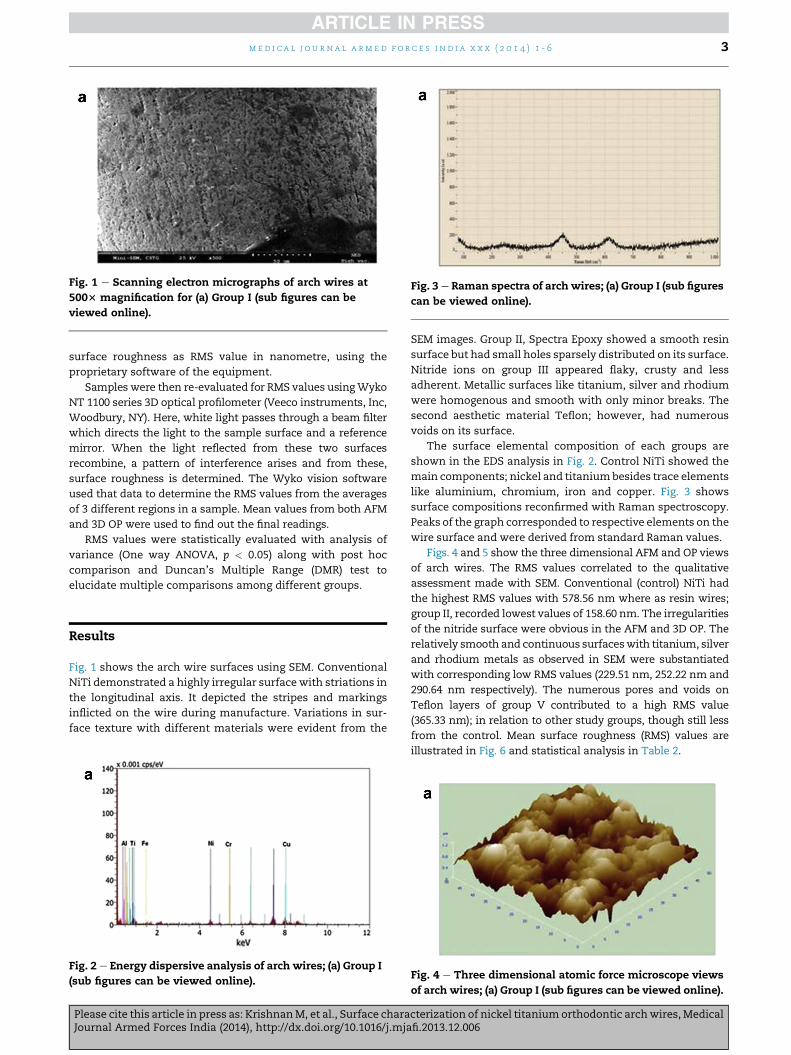
Original Article Surface characterization of nickel titanium orthodontic arch wires Lt Col Manu Krishnan a, *, Saraswathy Seema b , Brijesh Tiwari c , Col Himanshu S. Sharma d , Maj Gen Sanjay Londhe e , Lt Gen Vimal Arora, AVSM,VSM**, PHDS f a Classified Specialist (Orthodontics), Dept of Dental Research & Implantology, Institute of Nuclear Medicine and Allied Sciences (INMAS), Defence Research and Development Organization (DRDO), Timarpur, Delhi 1100054, India b Research Scholar, School of Medicine and Paramedical Health Sciences, Guru Gobind Singh Indraprastha University, Delhi Cantt, India c Senior Research Fellow (Project), Dept of Dental Research & Implantology, Institute of Nuclear Medicine and Allied Sciences (INMAS), Defence Research and Development Organization (DRDO), Timarpur, Delhi, India d Commanding Officer, Military Dental Centre, Delhi Cantt, India e Addl Director General Dental Services, IHQ of MoD (Army), New Delhi 110001, India f Director General Dental Services & Colonel Commandant, O/o DGDS, Adjutant General’s Branch, IHQ of MoD, L Block, New Delhi 110001, India article info Article history: Received 13 September 2013 Accepted 11 December 2013 Available online xxx Keywords: Nickel titanium arch wire Root mean square roughness Scanning electron microscopy Raman spectroscopy Atomic force microscopy 3D profilometry abstract Background: Surface roughness of nickel titanium orthodontic arch wires poses several clinical challenges. Surface modification with aesthetic/metallic/non metallic materials is therefore a recent innovation, with clinical efficacy yet to be comprehensively evaluated. Methods: One conventional and five types of surface modified nickel titanium arch wires were surface characterized with scanning electron microscopy, energy dispersive analysis, Raman spectroscopy, Atomic force microscopy and 3D profilometry. Root mean square roughness values were analyzed by one way analysis of variance and post hoc Duncan’s multiple range tests. Results: Study groups demonstrated considerable reduction in roughness values from conventional in a material specific pattern: Group I; conventional (578.56 nm) > Group V; Teflon (365.33 nm) > Group III; nitride (301.51 nm) > Group VI (i); rhodium (290.64 nm) > Group VI (ii); silver (252.22 nm) > Group IV; titanium (229.51 nm) > Group II; resin (158.60 nm). It also showed the defects with aesthetic (resin/Teflon) and nitride surfaces and smooth topography achieved with metals; titanium/silver/rhodium. Conclusions: Resin, Teflon, titanium, silver, rhodium and nitrides were effective in decreasing surface roughness of nickel titanium arch wires albeit; certain flaws. Findings have clinical implications, considering their potential in lessening biofilm adhesion, reducing friction, improving corrosion resistance and preventing nickel leach and allergic reactions. ª 2014, Armed Forces Medical Services (AFMS). All rights reserved. * Corresponding author. Tel.: þ91 (0) 11 23939588, þ91 8860821484. E-mail addresses: [email protected], [email protected] (M. Krishnan). Available online at www.sciencedirect.com ScienceDirect journal homepage: www.elsevier.com/locate/mjafi medical journal armed forces india xxx (2014) 1-6 Please cite this article in press as: Krishnan M, et al., Surface characterization of nickel titanium orthodontic arch wires, Medical Journal Armed Forces India (2014), http://dx.doi.org/10.1016/j.mjafi.2013.12.006 0377-1237/$ e see front matter ª 2014, Armed Forces Medical Services (AFMS). All rights reserved. http://dx.doi.org/10.1016/j.mjafi.2013.12.006
Transcript

ww.sciencedirect.com

med i c a l j o u r n a l a rm e d f o r c e s i n d i a x x x ( 2 0 1 4 ) 1 - 6

Available online at w

ScienceDirect

journal homepage: www.elsevier .com/locate/mjafi

Original Article

Surface characterization of nickel titaniumorthodontic arch wires

Lt Col Manu Krishnan a,*, Saraswathy Seema b, Brijesh Tiwari c,Col Himanshu S. Sharma d, Maj Gen Sanjay Londhe e,Lt Gen Vimal Arora, AVSM,VSM**, PHDS

f

aClassified Specialist (Orthodontics), Dept of Dental Research & Implantology, Institute of Nuclear Medicine and

Allied Sciences (INMAS), Defence Research and Development Organization (DRDO), Timarpur, Delhi 1100054, IndiabResearch Scholar, School of Medicine and Paramedical Health Sciences, Guru Gobind Singh Indraprastha University,

Delhi Cantt, IndiacSenior Research Fellow (Project), Dept of Dental Research & Implantology, Institute of Nuclear Medicine and Allied

Sciences (INMAS), Defence Research and Development Organization (DRDO), Timarpur, Delhi, IndiadCommanding Officer, Military Dental Centre, Delhi Cantt, IndiaeAddl Director General Dental Services, IHQ of MoD (Army), New Delhi 110001, IndiafDirector General Dental Services & Colonel Commandant, O/o DGDS, Adjutant General’s Branch, IHQ of MoD,

L Block, New Delhi 110001, India

a r t i c l e i n f o

Article history:

Received 13 September 2013

Accepted 11 December 2013

Available online xxx

Keywords:

Nickel titanium arch wire

Root mean square roughness

Scanning electron microscopy

Raman spectroscopy

Atomic force microscopy

3D profilometry

* Corresponding author. Tel.: þ91 (0) 11 2393E-mail addresses: manukrishnanin@yaho

Please cite this article in press as: KrishnaJournal Armed Forces India (2014), http:/

0377-1237/$ e see front matter ª 2014, Armhttp://dx.doi.org/10.1016/j.mjafi.2013.12.006

a b s t r a c t

Background: Surface roughness of nickel titanium orthodontic arch wires poses several

clinical challenges. Surface modification with aesthetic/metallic/non metallic materials is

therefore a recent innovation, with clinical efficacy yet to be comprehensively evaluated.

Methods: One conventional and five types of surface modified nickel titanium arch wires

were surface characterized with scanning electron microscopy, energy dispersive analysis,

Raman spectroscopy, Atomic force microscopy and 3D profilometry. Root mean square

roughness values were analyzed by one way analysis of variance and post hoc Duncan’s

multiple range tests.

Results: Study groups demonstrated considerable reduction in roughness values from

conventional in a material specific pattern: Group I; conventional (578.56 nm) > Group V;

Teflon (365.33 nm) > Group III; nitride (301.51 nm) > Group VI (i); rhodium (290.64 nm) >

Group VI (ii); silver (252.22 nm) > Group IV; titanium (229.51 nm) > Group II; resin

(158.60 nm). It also showed the defects with aesthetic (resin/Teflon) and nitride surfaces

and smooth topography achieved with metals; titanium/silver/rhodium.

Conclusions: Resin, Teflon, titanium, silver, rhodium and nitrides were effective in decreasing

surface roughness of nickel titanium arch wires albeit; certain flaws. Findings have clinical

implications, considering their potential in lessening biofilm adhesion, reducing friction,

improving corrosion resistance and preventing nickel leach and allergic reactions.

ª 2014, Armed Forces Medical Services (AFMS). All rights reserved.

9588, þ91 8860821484.o.co.in, [email protected] (M. Krishnan).

nM, et al., Surface characterization of nickel titaniumorthodontic archwires,Medical/dx.doi.org/10.1016/j.mjafi.2013.12.006

ed Forces Medical Services (AFMS). All rights reserved.

Table 1 e Study design (n [ 6 per group) of arch wires in0.016 inch (0.406 mm) round dimensions.

Group Product Manufacturer

I Conventional NiTi Ortho Organizers, San Marcos, CA

II Spectra Epoxy GAC International, Bohemia, NY

III Neo Sentalloy GAC International, Bohemia, NY

IV Black Titanium Class One Orthodontics, St. Lubbock

V Teflon d-Tech Asia Ltd, Pune

VI SilvereRhodium d-Tech Asia Ltd, Pune

me d i c a l j o u r n a l a rm e d f o r c e s i n d i a x x x ( 2 0 1 4 ) 1 - 62

Introduction

Ever since the report of ‘shape memory effect’ in nickel tita-

nium (NiTi) alloy in 1962, several applications of the material

in medical and dental disciplines have been identified till

now.1 Nickel titanium (NiTi) arch wires with its unique shape

memory and super elasticity properties are integral compo-

nents of contemporary orthodontic practice.2 However, the

high content of nickel (Ni: 47e50%) in NiTi alloys and its

extremely rough surface topography are confronting issues in

orthodontics.3 The increased propensity of plaque accumu-

lation, frictional forces at wire-bracket interface, nickel leach

and wire fracture ensuing intra oral corrosion are consequent

to it.4 Nickel release, in turn is known to initiate several

adverse responses, ranging from allergic hypersensitivity re-

actions to extremes of carcinogenic changes.5 As for any other

metallic alloy, NiTi also has oxide layers on its surface (TiO2,

TiO, Ti2O5 and NiO), which renders it the natural protection.6

These oxides are formed on the wire surface during its ‘wire

drawing procedures’ from large ‘ingot’ blocks.1 Yet, these are

removed during clinical use and electrochemical potential

differences are generated which initiates pitting and crevice

corrosion.7,8 To a large extent, all these have been attributed to

the high surface roughness of NiTi wires.9e11

In this context, there are some attempts to modify NiTi

arch wire surface with metals, non-metals and aesthetic

materials with the objective of reducing surface roughness so

as to enhance esthetics and to lessen friction, corrosion and

nickel leach.12 Surface engineering as a distinct discipline has

made remarkable strides in the field of material technology

during the last two decades and its medical and dental ap-

plications are manifold.12e14 Configuring a surface barrier

layer on biomaterials like pacemakers, stents, implants and

other devices with an ‘environment-friendly material’ is an

innovative step for improving biocompatibility.15 Surface

modification of dental materials are done either through

plasma spraying or physical/chemical vapour deposition;

where atoms, ions or molecules activated by plasma, laser or

high energy beams are condensed on the substrates.16,17 The

methods specifically used for orthodontic arch wires are

electron beam deposition, magnetron sputtering, cathodic arc

deposition or pulsed laser deposition.18

Surface roughness of materials is measured by profilo-

metric or optical methods and is generally expressed as root

mean square (RMS) values.19 Earlier, invasive profilometric

procedures were used to determine surface roughness of NiTi

wires.20 At present, there are many non invasive options for

assessing the exteriors ofmaterials used in industry,medicine

and dentistry. These include qualitative and quantitative

means like scanning electron microscopy (SEM), energy

dispersive analysis (EDS), spectroscopic techniques like

Raman spectroscopy, atomic force microscopy (AFM) and of

late, the advanced three dimensional optical profilometry (3D

OP).21 Still, these have not so far been comprehensively used

to evaluate the topography of surface modified NiTi wires. In

this study, prototypes of all currently available versions of

these wires were included to assess the surface features,

which have a close bearing on their clinical performance.

Additionally, none of these products are indigenously

Please cite this article in press as: KrishnanM, et al., Surface charaJournal Armed Forces India (2014), http://dx.doi.org/10.1016/j.mja

manufactured and there is an influx of these imported prod-

ucts into Indian dental market at a high cost but with fewer

evidences in favour of them.

The aim of the current study was therefore to characterize

the topographic features of five newly introduced surface

modified NiTi wires along with a conventional type, using

advanced optical methods.

Material and methods

The study groups included 5 types of surface modified nickel

titanium wires and one group of conventional NiTi in 0.016

inch (0.406 mm) round dimension. Group 1: Conventional

NiTi; (Ortho Organizers, San Marcos, CA), Group II: Spectra

Epoxy (GAC International, Bohemia, NY), Group III: Neo Sen-

talloy (GAC International, Bohemia, NY), Group IV: Black Ti-

tanium (Class One Orthodontics, St. Lubbock), Group V: Teflon

(d-TechAsia Ltd, Pune) and GroupVI: SilvereRhodium (d-Tech

Asia Ltd, Pune). Since group VI had a dual covering of silver

and rhodium, they are represented as groupVI (i) for silver and

group VI (ii) for rhodium. The study design is shown in Table 1.

Preliminary surface analysis of the arch wires were done

with SEM (SNE-3000M model, SEC, Korea) at 500� magnifica-

tion. Elemental mapping was carried out with EDS (SNE-

3000M model, SEC, Korea) and Raman Spectroscopy (HR 800,

Jobin Yvon, Spectrometer, Horiba Ltd, Minami-Ku, Kyoto)

equipped with 1800 grooves/mm holographic grating. Heli-

umeNeon laser of 633 nm was used as the excitation source.

The laser spot size of 3 mm diameter was focused on the

sample surface using a diffraction limited 10� objective. The

laser power at the sample was z20 mW and slit width of the

monochromator was 400 mm. The back scattered Raman

spectra were recorded using super cooled (<�110 �C)1024 � 256 pixels charge coupled device (CCD) detector with

range from 80 cm�1 to 2000 cm�1 with 5 s exposure time and

20 CCD accumulations. All the spectra were then baseline

corrected. Three different areas of the wire were checked for

each sample.

Surface roughness was initially evaluated with Solver Pro

EC atomic force microscope (NT-MDT, Zelenograd, Moscow).

All measurements were carried out in contact mode using a

standard conical silicon tip attached to a cantilever having a

force constant of 5 nNm�1 with a frequency limit from 50 to

150 Hz. The radius of curvature of the tip was 10 nm and the

cone angle was <22�. The scan area was 50 � 50 mm of each

sample, at three different locations. Averages of these from

six (n ¼ 6/group) wire samples were taken to express the

cterization of nickel titaniumorthodontic archwires,Medicalfi.2013.12.006

Fig. 1 e Scanning electron micrographs of arch wires at

5003 magnification for (a) Group I (sub figures can be

viewed online).

Fig. 3 e Raman spectra of arch wires; (a) Group I (sub figures

can be viewed online).

med i c a l j o u r n a l a rm e d f o r c e s i n d i a x x x ( 2 0 1 4 ) 1 - 6 3

surface roughness as RMS value in nanometre, using the

proprietary software of the equipment.

Samples were then re-evaluated for RMS values usingWyko

NT 1100 series 3D optical profilometer (Veeco instruments, Inc,

Woodbury, NY). Here, white light passes through a beam filter

which directs the light to the sample surface and a reference

mirror. When the light reflected from these two surfaces

recombine, a pattern of interference arises and from these,

surface roughness is determined. The Wyko vision software

used that data to determine the RMS values from the averages

of 3 different regions in a sample. Mean values from both AFM

and 3D OP were used to find out the final readings.

RMS values were statistically evaluated with analysis of

variance (One way ANOVA, p < 0.05) along with post hoc

comparison and Duncan’s Multiple Range (DMR) test to

elucidate multiple comparisons among different groups.

Results

Fig. 1 shows the arch wire surfaces using SEM. Conventional

NiTi demonstrated a highly irregular surface with striations in

the longitudinal axis. It depicted the stripes and markings

inflicted on the wire during manufacture. Variations in sur-

face texture with different materials were evident from the

Fig. 2 e Energy dispersive analysis of arch wires; (a) Group I

(sub figures can be viewed online).

Please cite this article in press as: KrishnanM, et al., Surface charaJournal Armed Forces India (2014), http://dx.doi.org/10.1016/j.mja

SEM images. Group II, Spectra Epoxy showed a smooth resin

surface but had small holes sparsely distributed on its surface.

Nitride ions on group III appeared flaky, crusty and less

adherent. Metallic surfaces like titanium, silver and rhodium

were homogenous and smooth with only minor breaks. The

second aesthetic material Teflon; however, had numerous

voids on its surface.

The surface elemental composition of each groups are

shown in the EDS analysis in Fig. 2. Control NiTi showed the

main components; nickel and titaniumbesides trace elements

like aluminium, chromium, iron and copper. Fig. 3 shows

surface compositions reconfirmed with Raman spectroscopy.

Peaks of the graph corresponded to respective elements on the

wire surface and were derived from standard Raman values.

Figs. 4 and 5 show the three dimensional AFM and OP views

of arch wires. The RMS values correlated to the qualitative

assessment made with SEM. Conventional (control) NiTi had

the highest RMS values with 578.56 nm where as resin wires;

group II, recorded lowest values of 158.60 nm. The irregularities

of the nitride surface were obvious in the AFM and 3D OP. The

relatively smooth and continuous surfaceswith titanium, silver

and rhodium metals as observed in SEM were substantiated

with corresponding low RMS values (229.51 nm, 252.22 nm and

290.64 nm respectively). The numerous pores and voids on

Teflon layers of group V contributed to a high RMS value

(365.33 nm); in relation to other study groups, though still less

from the control. Mean surface roughness (RMS) values are

illustrated in Fig. 6 and statistical analysis in Table 2.

Fig. 4 e Three dimensional atomic force microscope views

of arch wires; (a) Group I (sub figures can be viewed online).

cterization of nickel titaniumorthodontic archwires,Medicalfi.2013.12.006

Fig. 5 e Three dimensional profilometry views of arch

wires; (a) Group I (sub figures can be viewed online).

Table 2 e One way analysis of variance of mean rootmean square (RMS) values for different groups (n [ 6 pergroup and P < 0.001).

Parameter Group Mean RMSvalue in nm

SD

Surface

Roughness

I; Conventional NiTi 578.56a 48.68

II; Spectra Epoxy 158.60b 28.49

III; Neo Sentalloy 301.51c 19.95

IV; Black Titanium 229.51d 11.11

V; Teflon 365.33e 12.65

VI (i) Silver 252.22f 17.34

VI (ii) Rhodium 290.64g 10.26

Different superscripts; a, b, c, d, e, f and g indicate that mean values

differed significantly from each other for all the groups (Duncan’s

multiple range test).

me d i c a l j o u r n a l a rm e d f o r c e s i n d i a x x x ( 2 0 1 4 ) 1 - 64

Discussion

Surface roughness; usually measured by RMS values, is a

fundamental property of an arch wire. AFM and 3D OP, which

offer good accuracy in measuring surface roughness, were

used in the present investigation. Conventional/control NiTi

topped among the study groups in terms of RMS values at

578.56 nm; well within the range (100e1300 nm) reported

previously for NiTi.21,22 This high roughness is mainly

ascribed to the grain re-crystallizations that occur when NiTi

wires are pulled through diamond moulds during its fabrica-

tion.5 The SEM images also depicted an exceedingly rough

surface, with areas of ‘pickling/pores/white inclusion spots’

that are characteristically described for NiTi wires.21

EDS determines the elemental composition of a material

on interaction with X-rays, depending on the energy differ-

ences that occur during excitation and down fall of its elec-

trons.23 Raman spectroscopy, on the other hand is based on

the in-elastic scattering of a monochromatic laser with a

material. Frequency of the re-emitted photons from the

Fig. 6 e Mean root mean square (RMS)

Please cite this article in press as: KrishnanM, et al., Surface charaJournal Armed Forces India (2014), http://dx.doi.org/10.1016/j.mja

material shows a characteristic ‘up’ or ‘down shift’ with

respect to the original, known as ‘Raman Effect’. Raman

spectroscopy thereby gives information on the low frequency

transitions in molecules and delineates its material

composition.24

Surface modification using titanium nitride (TiN) over NiTi

alloys have been used in industry for different purposes. But

with straight grain boundaries and open porosities, they did

not form a homogenous surface; instead, rendered open

percolation of reactive agents.16 At present, finer titanium

aluminium nitride (TiAlN) is used for making impervious

layers over NiTi alloys. Similarmethods are being used for NiTi

arch wires also, but exact parameters of temperature and

pressure used for surface modification are not known.17

Nitride ion implanted wires were among the first to be mar-

keted in the surfacemodifiedNiTi series.14 In this study, nitride

ion deposited (Group III) showed a low RMS value (301.51 nm),

with respect to the control (578.56 nm). However, the surface

appeared loose and grossly incongruous in the SEM.

Demand for aesthetic orthodontic appliances ismostlymet

by transparent ceramic or composite brackets. Tomatch these

values of arch wires in nanometre.

cterization of nickel titaniumorthodontic archwires,Medicalfi.2013.12.006

med i c a l j o u r n a l a rm e d f o r c e s i n d i a x x x ( 2 0 1 4 ) 1 - 6 5

brackets, resin/Teflon modified NiTi wires are attempted by

atomization procedures. Teflon wires demonstrated rough-

ness values (365.33 nm) less than the control, but were higher

than other study groups. It correlated well with the large

number of elliptical voids observed in SEM, AFM and 3D OP

images, indicative of inadequate surface modification with

Teflon. Characteristic to Teflon or poly tetraflouro ethylene

(PTFE) is a fluoridated chain which is responsible for its

aesthetic and non adherent nature. Confirming this, EDS and

Raman spectra showed predominance of fluoride ions on its

surface. Since frictional coefficient of Teflon is low, arch wires

with Teflon surface are cited to have reduced resistance to

sliding mechanics.25 Another aesthetic material; resin wires

(Group II) showed carbon, hydrogen and oxygen peaks in the

EDS and Raman spectroscopy. Carbon on NiTi surface is

known to form ‘nickel-titanium-carbide’ or ‘titanium carbide’

hard layers capable of preventing nickel leach.4 Contrary to

Teflon; resin NiTi had an extremely smooth topography with

the lowest RMS value (158.60 nm). Though less in number than

Teflon, resin group also showed few voids on its surface in the

SEM and AFM images, suggestive of defects in arch wire

modification with aesthetic materials.

Biocompatibility of titanium can be the persuading factor

for using it for surface modification over NiTi arch wire. Same

is the case with other biocompatible metals like silver and

rhodium. However, colour of these metals may not fetch

necessary appeal among patients and clinicians. Comparable

RMS values, were seen for group IV (titanium; 229.51 nm) and

VI which had dual surfaces with rhodium (290.64 nm) on the

anterior and silver (252.22 nm) on the posterior spans. This

implied that modifying NiTi arch wire surface with metals

offer a promising option for reducing roughness. SEM, AFM

and 3D OP images proved the uniform topography accom-

plished with metals.

Irrespective of the metallic structure, orthodontic alloys

undergo corrosion of varying degrees due to the effects of pH,

temperature, microbes and enzymes. Corrosion causes

disintegration of orthodontic appliances, release of constitu-

ent elements and deterioration of their mechanical and clin-

ically desirable properties. Orthodontic wires are constantly

engaged with brackets using ligatures or modules and there-

fore make conducive sites for corrosion. Arch wire surface

being themain interacting area in this, altering its topography

can bring out favourable changes in corrosion features. It is

based on the conviction that the high surface roughness of

NiTi is a chief causative factor for corrosion.11 Among the

several natural oxides on its surface; TiO2 is the predominant

one, which gives inherent protection to NiTi against corrosion.

Even this protective layer is disrupted by mechanical, biolog-

ical and chemical actions in the oral cavity and cause

‘hydrogen ion entrapment,’ which makes the alloy brittle and

susceptible to fracture.10,26 Tan and co-workers12 found that

for the NiTi alloy; the ‘breakdown potential;’ an index of the

corrosion resistance, can be increased by ion implantation

with oxygen. Similar results were reported by Sawase et al as

well giving credence to the view point of modifying NiTi sur-

face for evading corrosion.27

Orthodontic wires containing nickel have been implicated

to cause a Type IV delayed hypersensitivity immune response,

mediated by the release of nickel ions into the oral cavity. Use

Please cite this article in press as: KrishnanM, et al., Surface charaJournal Armed Forces India (2014), http://dx.doi.org/10.1016/j.mja

of NiTi archwires can convert 20% of non sensitive Ni subjects

into Ni sensitive subjects where the allergy is manifested as

burning papular erythema or papulovesicular dermatitis.14

Nickel also affects polymorphonuclear leukocytes, mono-

cytes and endothelial cells, causing inflammatory responses.

Nickel complexes in the form of arsenides and sulfides are

known carcinogens andmutagens effecting DNAdamages.5 In

view of the probable health hazards of nickel leach from bio-

materials, its topographic correction has tremendous impor-

tance for safe orthodontic practice.

Investigations into the shape memory and super elasticity

properties of surfacemodifiedNiTi wires are so far, very rare. In

an exclusive study, using differential scanning calorimetry and

X-ray diffraction on a surfacemodifiedNiTi with certain oxides,

no differences in these features were observed.28 This does not

mean that all the surface modified NiTi products currently

available have their requisite physical properties. It follows that

experimental procedures for the correction of surface rough-

ness of NiTi wires should not merely focus on reducing RMS

values or surface roughness but should give due recognition for

its physical properties like shape memory and super elasticity.

The current study proved that all surface modified NiTi

groups showed considerable reduction in surface roughness

compared with control. RMS values showed the following

order: Group I; conventional NiTi (578.56 nm) > Group V;

Teflon (365.33 nm) > Group III; nitride (301.51 nm) > Group VI

(i); rhodium (290.64 nm) > Group VI (ii); silver (252.22 nm) >

Group IV; titanium (229.51 nm) > Group II; resin (158.60 nm).

Mean values of study groups differed significantly from the

control in the analysis of variance (One-way ANOVA). The

groups differed significantly from each other too, in the post

hoc Duncan’s multiple range (DMR) test.

From the foregoing, it is evident that surface roughness is a

key property of NiTi arch wires, though certain aspects of re-

lations between RMS values versus friction and corrosion still

await clarifications.19,21,22 Further, role of the rough surface of

NiTi in attracting oral plaque, biofilm organization and nickel

leach have important clinical implications.5 The study

explicitly brought out the defects of esthetic; resin/Teflon and

nitride surfaces over NiTi. At the same time, it showed the

correction of surface roughness achieved with metals. It thus

stressed the need for having appropriate quality control in

manufacturing surface modified NiTi wires. Since these pro-

cedures most likely entail high temperature and pressure

applications on NiTi wires, its effects on shape memory and

super elasticity are also to be closely scrutinized for ensuring

optimum clinical results. For all these, surface roughness and

RMS values would be a reckonable entity and a crucial

assessment factor in NiTi arch wire research.

Inferences in the current study were based on the char-

acterization tests done on ‘as- received’ samples. The efficacy

of coatings and alterations in surface roughness values can be

better understood, if the samples are retrieved after clinical

use and subjected to a similar set of scrutiny. Future in-

vestigations based on clinically used samples would therefore

be appropriate. Notwithstanding that, the results give clini-

cians firsthand information on topographic features of NiTi

wires with surface coatings. It certainly adds on to the clinical

knowhow and offers an opportunity to practice evidence

based orthodontics with these new wires.

cterization of nickel titaniumorthodontic archwires,Medicalfi.2013.12.006

me d i c a l j o u r n a l a rm e d f o r c e s i n d i a x x x ( 2 0 1 4 ) 1 - 66

Conflicts of interest

This study has been financed by the research grants from the

O/o DGAFMS, New Delhi.

Intellectual contribution of authors

Studyconcept: LtColManuKrishnan,ColHimanshuSSharma,

Maj Gen Sanjay Londhe, Lt Gen Vimal Arora, AVSM, VSM**, PHDS.

Drafting $ Manuscript revision: Lt Col Manu Krishnan,

Brijesh Tiwari, Lt Gen Vimal Arora, AVSM, VSM**, PHDS.

Statistical analysis: Saraswathy Seema.

Study supervision: Col Himanshu S Sharma, Maj Gen Sanjay

Londhe.

Acknowledgement

Dr Parvatha Varthini, Dr Sole and Dr Vanitha Kumari, Scien-

tists at Indira Gandhi Centre for Atomic Research (IGCAR),

Department of Atomic Energy (DAE), Kalpakkam, Tamil Nadu.

r e f e r e n c e s

1. Brantley WA. Orthodontic wires. In: Brantley WA, Eliadas T,eds. Orthodontic Materials: Scientific and Clinical Aspects.Stuttgart: Thieme; 2001:77e103.

2. Kusy RP. A review of contemporary arch wires: theirproperties and characteristics. Angle Orthod. 1997;67:197e207.

3. Locci P, Lilli C, Marinucci L, et al. In vitro cytotoxic effects oforthodontic appliances. J Biomed Mater Res. 2000;53:560e567.

4. von Fraunhofer JA. Corrosion of orthodontic devices. SeminOrthod. 1997;3:198e205.

5. Eliades T, Athanasiou AE. In vivo aging of orthodontic alloys:implications for corrosion potential, nickel release, andbiocompatibility. Angle Orthod. 2002;72:222e237.

6. Iijima M, Endo K, Ohno H, Yonekura Y, Mizoguchi I. Corrosionbehavior and surface structure of orthodontic NiTi alloywires. Dent Mater J. 2001;20:103e113.

7. Rondelli G, Vicentini B. Evaluation by electrochemical tests ofthe passive film stability of equiatomic NiTi alloy also inpresence of stress induced martensite. J Biomed Mater Res.2000;51:47e54.

8. Huang HH, Chiu YH, Lee TH, et al. Ion release from NiTiorthodontic wires in artificial saliva with various acidities.Biomaterials. 2003;24:3585e3592.

9. Hunt NP, Cunningham SJ, Golden CG, Sheriff M. Aninvestigation into the effects of polishing on surface hardnessand corrosion of orthodontic archwires. Angle Orthod.1999;69:433e440.

Please cite this article in press as: KrishnanM, et al., Surface charaJournal Armed Forces India (2014), http://dx.doi.org/10.1016/j.mja

10. Oshida Y, Sachdeva RC, Miyazaki S. Microanalyticalcharacterization and surface modification of TiNi orthodonticarch wires. Biomed Mater Eng. 1992;2:51e69.

11. Widu F, Drescher D, Junker R, Bourauel C. Corrosion andbiocompatibility of orthodontic wires. J Mater Sci Mater Med.1999;10:275e281.

12. Tan L, Dodd RA, Crone WC. Corrosion and wear-corrosionbehavior of NiTi modified by plasma source ion implantation.Biomaterials. 2003;24:3931e3939.

13. Sarholt-Kristensen L. Effect of Nþ implantation on the shapememory behaviour and corrosion resistance of an equiatomicNiTi alloy. J Mater Sci Lett. 1993;12:618e619.

14. House K, Sernetz F, Dymock D, Sandy JR, Ireland AJ. Corrosionof orthodontic appliances-should we care? Am J OrthodDentofacial Orthop. 2008;133:584e592.

15. Black J. Biological Performance of Materials: Fundamentals ofBiocompatibility. New York, NY: Marcel Decker; 1999:28e44.

16. Wu SK, Chu CL, Lin HC. Ion nitriding of equiatomic TiNi shapememory alloys II: corrosion properties and wearcharacteristics. Surf Coat Technol. 1997;92:206e211.

17. Husmann P, Bourauel C, Wessinger M, Jager A. The frictionalbehavior of coated guiding arch wires. J Orofac Orthop.2000;63:199e211.

18. Harlin P, Bexell U, Olsson M. Influence of surface topographyof arc-deposited TiN and sputter-deposited WC/C coatings onthe initial material transfer tendency and frictioncharacteristics under dry sliding contact conditions. Surf CoatTechnol. 2009;203:1748e1755.

19. Kusy RP, Whitley JQ, Mayhew MJ, Buckthal JE. Surfaceroughness of orthodontic arch wires via laser spectroscopy.Angle Orthod. 1988;58:33e45.

20. Porosoki RR, Bagby MD, Erickson LC. Static frictional force andsurface roughness of nickel titanium arch wires. Am J OrthodDentofacial Orthop. 1991;100:341e348.

21. Bourauel C, Fries T, DrescherD, Plietsch R. Surface roughness oforthodontic wires via atomic force microscopy, laser specularreflectance, and profilometry. Eur J Orthod. 1998;20:79e92.

22. Huang HH. Variation in corrosion resistance of nickeltitanium wires from different manufacturers. Angle Orthod.2005;75:661e665.

23. Eliades T. Research Methods in Orthodontics. A Guide toUnderstanding Orthodontic Research. 1st ed. Springer;2013:1e24.

24. van Meerbeek B, Mohrbacher H, Celis JP, et al. Chemicalcharacterization of the resin dentin interface by micro RamanSpectroscopy. J Dent Res. 1993;72:1423e1428.

25. Farronato GP, Casiraghi G, Giannı̀ AB, Salvato A. The use ofPTFE in orthodontics. Mondo Ortod. 1988;13:83e89.

26. Yokoyama K, Hamada K, Moriyama K, Asaoka K. Degradationand fracture of NiTi superelastic wire in an oral cavity.Biomaterials. 2001;22:2257e2262.

27. Sawase T, Wennerberg A, Baba K, et al. Application of oxygenion implantation to titanium surfaces: effects on surfacecharacteristics, corrosion resistance, and bone response. ClinImplant Dent Relat Res. 2001;3:221e229.

28. Krishnan M, Seema S, Sukumaran K, Pawar V. Phasetransitions in coated nickel titanium arch wires: a differentialscanning calorimetric and X-ray diffraction analysis. BullMater Sci. 2012;35:905e911.

cterization of nickel titaniumorthodontic archwires,Medicalfi.2013.12.006

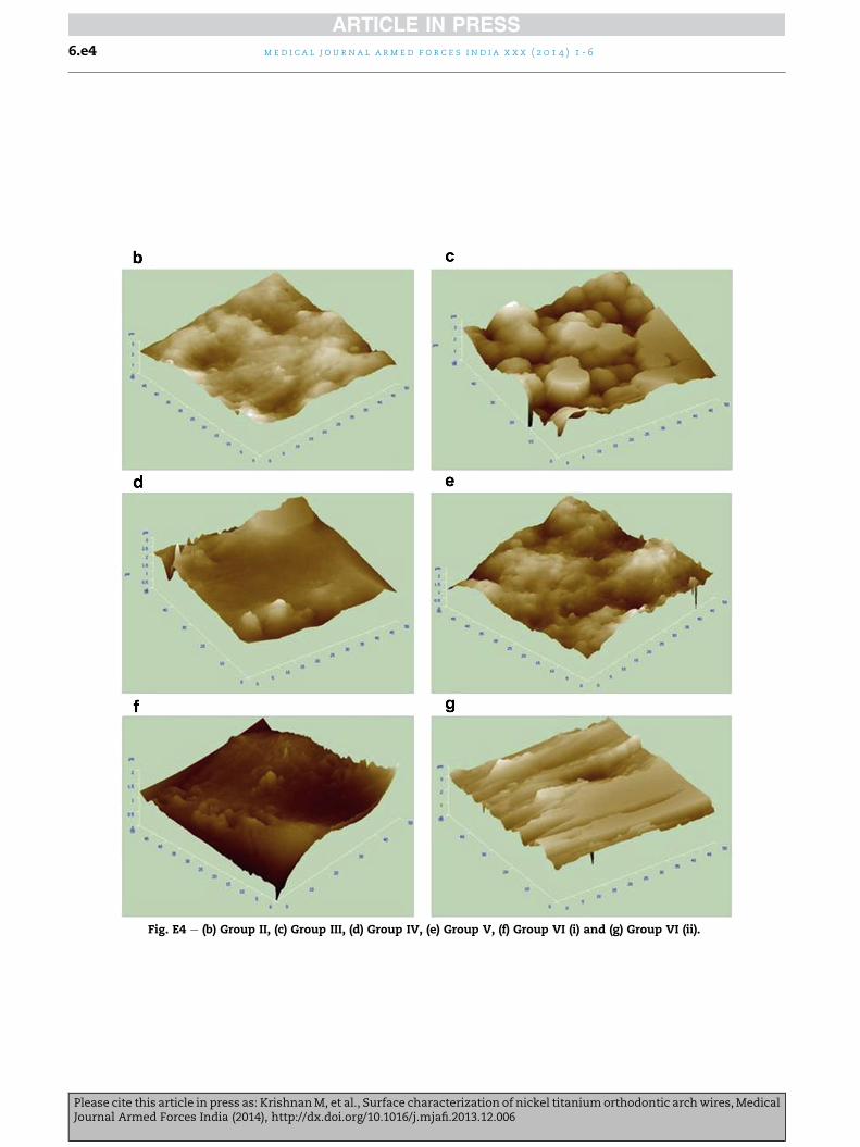
Fig. E1 e (b) Group II, (c) Group III, (d) Group IV, (e) Group V, (f) Group VI (i) and (g) Group VI (ii).

med i c a l j o u r n a l a rm e d f o r c e s i n d i a x x x ( 2 0 1 4 ) 1 - 6 6.e1

Please cite this article in press as: KrishnanM, et al., Surface characterization of nickel titaniumorthodontic archwires,MedicalJournal Armed Forces India (2014), http://dx.doi.org/10.1016/j.mjafi.2013.12.006

Fig. E2 e (b) Group II, (c) Group III, (d) Group IV, (e) Group V, (f) Group VI (i) and (g) Group VI (ii).

me d i c a l j o u r n a l a rm e d f o r c e s i n d i a x x x ( 2 0 1 4 ) 1 - 66.e2

Please cite this article in press as: KrishnanM, et al., Surface characterization of nickel titaniumorthodontic archwires,MedicalJournal Armed Forces India (2014), http://dx.doi.org/10.1016/j.mjafi.2013.12.006

Fig. E3 e (b) Group II, (c) Group III, (d) Group IV, (e) Group V, (f) Group VI (i) and (g) Group VI (ii).

med i c a l j o u r n a l a rm e d f o r c e s i n d i a x x x ( 2 0 1 4 ) 1 - 6 6.e3

Please cite this article in press as: KrishnanM, et al., Surface characterization of nickel titaniumorthodontic archwires,MedicalJournal Armed Forces India (2014), http://dx.doi.org/10.1016/j.mjafi.2013.12.006

Fig. E4 e (b) Group II, (c) Group III, (d) Group IV, (e) Group V, (f) Group VI (i) and (g) Group VI (ii).

me d i c a l j o u r n a l a rm e d f o r c e s i n d i a x x x ( 2 0 1 4 ) 1 - 66.e4

Please cite this article in press as: KrishnanM, et al., Surface characterization of nickel titaniumorthodontic archwires,MedicalJournal Armed Forces India (2014), http://dx.doi.org/10.1016/j.mjafi.2013.12.006

Fig. E5 e (b) Group II, (c) Group III, (d) Group IV, (e) Group V, (f) Group VI (i) and (g) Group VI (ii).

med i c a l j o u r n a l a rm e d f o r c e s i n d i a x x x ( 2 0 1 4 ) 1 - 6 6.e5

Please cite this article in press as: KrishnanM, et al., Surface characterization of nickel titaniumorthodontic archwires,MedicalJournal Armed Forces India (2014), http://dx.doi.org/10.1016/j.mjafi.2013.12.006


Recommended